F
ACULTE DES SCIENCES
–
D
EPARTEMENT DE
C
HIMIE
C
ENTRE DE
R
ECHERCHES DU
C
YCLOTRON
Synthesis of stable and radioactive
isotope-containing peptidoglycan fragments
Directeur: Professeur André L
UXENDissertation présentée par
Nicolas Lamborelle
Pour l’obtention du grade de
Docteur en Sciences
Committee
Promoter: Professor André Luxen (Université de Liège) President: Professor Christian Damblon (Université de Liège) Members: Doctor Christophe Dugave (Institut Frédéric-Joliot)
Doctor Simon Lacroix (Université Libre de Bruxelles) Professor Lionel Delaude (Université de Liège) Professor Bernard Joris (Université de Liège) Doctor Christian Lemaire (Université de Liège)
Abstract
In this work, a method for the total synthesis of different isotope-containing meso-diaminopimelic acids (m-A2pm or (2S,6R)-A2pm) is presented. This compound,
specific of bacterial cell wall, allows for the reticulation of peptidoglycan in most Gram-negative and some Gram-positive bacteria. A preparation of new isotope-containing compounds (1-13C)(2S,6R)-A2pm and (4,5-2H2)(2S,6R)-A2pm is hereby
presented. These compounds can be used as chromatography and quantitative mass spectrometry references for the study of peptidoglycan dynamics. The first total stereoselective synthesis of high specific activity (30 Ci/mmol) tritiated [4,5-3H](2S,6R)-A
2pm is also described. Furthermore, the synthesis of differentially
protected (2S,6R)-A2pm and (4,5-2H2)(2S,6R)-A2pm allowed the preparation of
peptidoglycan peptide fragments, which broaden the scope of applications to studies of b-lactamase induction phenomenon in penicillin resistant bacteria as well as to studies in the field of human innate immune system.
Résumé
Ce travail décrit une méthode de synthèse totale d’acides méso-diaminopimélique (m-A2pm ou (2S,6R)-A2pm) marqués. Ce composé, spécifique de la paroi bactérienne,
permet la réticulation du peptidoglycane de la plupart des bactéries à Gram-négatif et de quelques bactéries à Gram-positif. La synthèse de nouveaux composés marqués, (1-13C)(2S,6R)-A2pm et (4,5-2H2)(2S,6R)-A2pm, pouvant servir de références
en chromatographie et en spectrométrie de masse quantitative pour l’étude du métabolisme du peptidoglycane est décrite. La première synthèse totale stéréoselective d’acide méso-diaminopimélique tritié [4,5-3H](2S,6R)-A
2pm à haute
activité spécifique (30 Ci/mmol) est également décrite. Enfin, la préparation de (2S,6R)-A2pm et (4,5-2H2)(2S,6R)-A2pm différemment protégés est présentée. Ces
composés permettent la synthèse de fragments peptidiques de peptidoglycane, ce qui élargit le champ des applications aux études du phénomène d’induction de b-lactamases chez les bactéries résistantes aux pénicillines ainsi qu’à des études dans le domaine du système immunitaire inné chez l’humain.
Acknowledgements
First and foremost, I would like to express my gratitude to my promoter Professor André Luxen for giving me the opportunity to be a part of his laboratory, the Cyclotron Research Center, during these years. I’m thankful for his guidance and support all along my work.
I also thank the jury members, Doctor Christophe Dugave, Doctor Simon Lacroix, Professor Lionel Delaude, Professor Bernard Joris, Doctor Christian Lemaire and the president Professor Christian Damblon for the approval to examine this work.
I thank the Chemistry Department at the University of Liège for giving me the privilege to be a teaching assistant and for the funding of my PhD research.
I thank the members of the Laboratoire de Marquage par le Tritium of the Commissariat à l’Énergie Atomique of Paris-Saclay for welcoming me in their laboratory. I would like to specially thank Doctor Sophie Feuillastre, Olivia Carvalho and Sebastien Garcia-Argote who performed the synthesis of tritiated meso-diaminopimelic acid. I also thank Doctor Bernard Rousseau for saving me from the snow storm which paralyzed Paris during my time there.
I thank Doctor Jean-Christophe Monbaliu for his precious collaboration for the development of the preparation of vinylglycine derivatives by flow chemistry and for what he brings to the Chemistry Department. I thank Professor Christian Damblon for our collaboration in the structural analyses classes, for the liberty he offered me in this context and for all our passionate discussions around and away from the NMR instruments. I thank Professor Lionel Delaude for his help during the first cross-metathesis test. I thank Doctor Astrid Zervosen for her support and advice in the laboratory and on the agrégation benches. I thank Doctor Justine Simon for all
our debates over synthesis strategies and her support during these years, sharing laboratories, ideas, laughter and doubts. I also thank the former +4 team and all the CRC staff members for their help, kindness and welcome.
I thank my friends and my family for their love and support. And finally, I thank my lovely wife Aurélie for her love, her everlasting support and for all the adventures awaiting us.
List of abbreviations
AG allylglycine
Ala alanine
APT attached proton test
ATP adenosine triphosphate
B. Bacillus
BAIB bis(acetoxy)iodobenzene
BPR back pressure regulator
Boc tert-butoxycarbonyl
br broad
Cbz carboxybenzyl
CEA Commissariat à l’énergie atomique et aux énergies
alternatives
CiTOS Center for Integrated Technology and Organic Synthesis
CRC Cycloton Research Center
COD cycloocta-1,5-diene
COSY correlation spectroscopy
d doublet
DBU diazabicycloundecene
DCC dicyclohexylcarbodiimide
DCU dicyclohexylurea
DET diethyl tartrate
DHB dehydrobutyrine
DIBAL diisobutylaluminium hydride
DMA N,N-dimethylacetamide
DMAC dimethyl acetylenedicarboxylate
DMAP 4-dimethylaminopyridine
DMF N,N-dimethylformamide
DNA deoxyribonucleic acid
DOPA 3,4-dihydroxyphenylalanine dr diastereomeric ratio E. Escherichia EDCI 1-ethyl-3-(3-dimethylaminopropyl)carbodiimide ee enantiomeric excess equiv. equivalent
FADH flavin adenine dinucleotide
Fmoc fluorenylmethyloxycarbonyl
GlcNAc N-acetylglucosamine
Glu glutamic acid
HBTU hexafluorophosphate benzotriazole tetramethyl uronium
HMBC heteronuclear multiple bond correlation
HMDS bis(trimethylsilyl)amide
HMM high molecular mass
HPLC high performance liquid chromatography
HRMS high resolution mass spectrometry
HSQC heteronuclear single quantum coherence
J coupling constant
LMT Laboratoire de Marquage par le Tritium
LMM low molecular mass
Lys lysine
m multiplet
m-A2pm meso-diaminopimelic acid
mp melting point
mRNA messenger RNA
MurNAc N-acetylmuramic acid
NAC N-acetyl-L-cysteine
NADP nicotinamide adenine dinucleotide phosphate
NBD nobornadiene
NMM N-methylmorpholine
NMR nuclear magnetic resonance
NOD nucleotide-binding oligomerization domain
OP operator OPA o-phthaldehyde PBP penicillin-binding protein PCC pyridinium chlorochromate PDC pyridinium dichromate PHA polyhydroxyalkanoate PPTS pyridinium p-toluenesulfonate PTC phase-transfer catalysis PyBOP benzotriazol-1-yl-oxytripyrrolidinophosphonium hexafluorophosphate q quadruplet
RNA ribonucleic acid
rt room temperature
s singlet
SCBM Service de Chimie Bio-organique et de Marquage
SPPS solid-phase peptide synthesis
t triplet
TBHP tert-butyl hydroperoxide
TEMPO 2,2,6,6-tetramethylpiperidine-1-oxyl
TFA trifluoroacetic acid
THF tetrahydrofuran
TMS trimethylsilyl
tRNA transfer RNA
UDP uridine diphosphate
UMP uridine monophosphate
Contents
CHAPTER I INTRODUCTION 1
I.1.GENERAL INTRODUCTION 3
I.2.THE BACTERIAL CELL 5
I.3.THE BACTERIAL CELL WALL 6
I.4.PEPTIDOGLYCAN 7
I.5.PEPTIDOGLYCAN METABOLISM 9
I.5.A) PEPTIDOGLYCAN BIOSYNTHESIS 9
I.5.B) PEPTIDOGLYCAN TURNOVER AND RECYCLING 11
I.5.C) PENICILLIN-BINDING PROTEINS 13
I.6.ANTIBIOTICS 15
I.7.RESISTANCE 18
I.7.A) THE PHENOMENON 18
I.7.B) INDUCTION OF A b-LACTAMASE IN B. LICHENIFORMIS 20
I.8.OBJECTIVES 22
CHAPTER II BIBLIOGRAPHY 25
II.1.JURGENS AND CHEN SYNTHESES 29
II.2.HOLCOMB AND WANG SYNTHESES 31
II.3.HERNANDEZ SYNTHESIS 32
II.4.KAWASAKI SYNTHESIS 33
II.5.DEL VALLE,CHOWDHURY AND SAITO SYNTHESES 34
II.6.PEPTIDES 36
CHAPTER III RESULTS AND DISCUSSION 39
III.1.STRATEGY 41
III.2.ALLYLGLYCINE AND (13C)ALLYLGLYCINE PREPARATION 43
III.2.A) STRATEGY 43
III.2.B) PREPARATION OF SCHIFF BASES 43
III.2.C) CHIRAL ALKYLATIONS AND PROTECTIONS 46
III.3.VINYLGLYCINE PREPARATION 50
III.4. M-A2PM SYNTHESES 53
III.4.A) DIFFERENTIALLY PROTECTED M-A2PM SYNTHESES 53
III.4.B) FREE M-A2PM AND (2H2)M-A2PM SYNTHESES 56
III.4.C) [3H]M-A2PM SYNTHESIS 60
III.4.D) FREE (13C)M-A
2PM SYNTHESIS 64
III.5.PEPTIDE PREPARATION 67
III.5.A) SOLID-PHASE PEPTIDE SYNTHESIS 67
III.5.B) PROTECTING GROUP CHOICES 68
CHAPTER IV CONCLUSION 75
CHAPTER V REFERENCES 83
CHAPTER VI SUPPLEMENTARY DATA 93
Chapter I
Introduction
I.1. General Introduction
Humans have been intrigued by the sky and the far infinite since the dawn of times, but a whole other infinite became accessible when microscopes came up in the late 16th – early 17th centuries. Antoine van Leeuwenhoek brought some improvements
to these early models and was the first to observe microorganisms using hand-crafted microscopes of his own in 1674. He called these microorganisms animalcula. Most of these tiny animals are now called bacteriaa as a result from Ehrenberg’s
studies in 1828. This new world has been intensively studied since and results from these works changed our world. From Louis Pasteur (1822-1894) and his understanding of the fermentation process and his development of the pasteurization process to Robert Koch (1843-1910, Nobel Prize in medicine in 1905) who postulated that microorganisms are the cause of infectious diseases, the discoveries of these times still have a tremendous impact on our current way of life. Bacteria appeared on Earth about 3.5 billion years ago during the Archean Eon. They were among the first living organisms on this planet. Bacteria are prokaryotic forms of life, most of the time unicellular, and display various shapes and sizes (0.5-5 µm). They can be found everywhere, even in the harshest conditions, where no other form of life can survive. Some can live in hot springs and around deep sea hydrothermal vents (thermophilic bacteria), some bacteria are found in extreme cold environment such as Antarctica (psychrophilic bacteria). Others are capable to cope with very acidic conditions (acidophilic bacteria) and some can even survive in highly radioactive environments.
Though many bacteria are known because of the illnesses they cause, the vast majority of them are harmless and many are even beneficial to humans. As an example, Escherichia Coli, a symbiont in the human intestine, fulfills an important role in the digestion and in the secretion of vitamins. Bacteria are also essential to
the cheese production, turning sugars found in milk into lactic acid. Some bacteria are used as depolluting agents to convert organic matter found in sewage into fertilizer. Some biodegradable plastics such as PHA (polyhydroxyalkanoate), are produced by bacteria from sugars derived from corn1.
But it is obvious why bacteria got their negative reputation. It is estimated that they cause about half of all the human diseases. Tuberculosis, caused by Mycobacterium
tuberculosis, kills 2 million people a year and about 2 more million people die each
year from diarrheal diseases caused by various bacteria1. Among the most common
bacterial infections, we may also cite the Lyme disease caused by bacterium Borrelia
burgdorferi carried by ticks, tetanus caused by Clostridium tetani found in soil,
cholera, botulism and typhoid fever.
Bacterial diseases were very often lethal before the development of antibiotics. In the late 19th century, the German chemical industry had provided dyes able to
selectively stain tissues or pathogens. This permitted Paul Ehrlich to propose his concept of selective chemotherapy. He reasoned that if it is possible to selectively stain a pathogen, it should also be possible to kill it using what he called a “magic bullet” without harming the surrounding cells2. However, it is Alexander Fleming
who made the famous, though fortuitous, discovery of penicillin in 1929 as the first antibiotic3. Many more antibiotics are now available but their overuse and misuse
I.2. The bacterial cell
Selective chemotherapy can only be achieved because the bacterial cell is different from ours. Bacteria are prokaryotes
in opposition to the
eukaryotes cells we are made of1. The most noticeable
difference lies in the absence of a membrane-bound nucleus in prokaryotic cellsb (Figure 1).
The prokaryotic genome,
usually a single circular chromosome, is therefore floating in the cytoplasm and folded into a compact structure called the nucleoid. A typical prokaryotic cell also has smaller circular DNA molecules called plasmids independent from the single chromosome. The cytoplasm contains ribosomes (slightly smaller than their eukaryotic counterparts and different in their protein and RNA content), which read mRNA and use tRNA to synthesize proteins. The slight differences between prokaryotic and eukaryotic ribosomes can be exploited to design antibiotics that do not harm prokaryotic cells such as ours1. Some vesicles or inclusion bodies may be
observed in the bacterial cytoplasm as nutrients stocks.
The outer structure, called bacterial cell wall, contains a layer of peptidoglycan, a rigid macromolecule. This component is essential to the bacteria to withstand its internal osmotic pressure as its cytoplasm is highly hypertonic5. Therefore,
peptidoglycan metabolism represents a perfect target for a “magic bullet”.
b from Greek pro "before" and karyon "nut, kernel" here referring to the nucleus. Figure 1. A typical rod-shaped bacterium4
I.3. The bacterial cell wall
Bacterial cell wall represents a very active research topic. This barrier allows bacteria to withstand their high internal osmotic pressure due to the highly concentrated cytosol compared to their outer environment1,5.It is also the theater of exchanges
with the exterior and the target of many antibiotics. Bacteria can be categorized in two groups: Gram-positive and Gram-negative, depending on their ability to retain Gram stainc. This is due to a structural difference between their walls. Both have a
layer of peptidoglycan but the one found in Gram-positive bacteria is much thicker (20-80 nm vs 1-3 nm). Additionally, Gram-negative bacteria peptidoglycan is surrounded by an external asymmetric membrane made of phospholipids and lipopolysaccharides5 (Figure 2).
Figure 2. Gram stain and structures of bacterial cell walls5.
I.4. Peptidoglycan
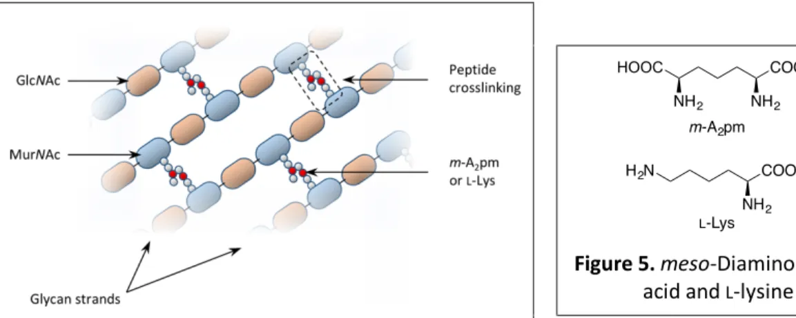
Peptidoglycan is a giant macromolecule made of glycan strands reticulated by short peptides, that envelops the plasma membrane as a mesh net called sacculus6,7 (Figure 3). This impressive
structure is responsible for stabilizing and shape-maintaining the cell wall. Glycan strands are made of alternating units of
N-acetylglucosamine (GlcNAc) and N-acetylmuramic acid (MurNAc) linked together
by b-(1®4) glycosidic bonds (Figure 4). Each MurNAc unit is substituted by a short peptide; most of them with the following structure: L-Ala-g-D-Glu-L-diamino acid-D-Ala-OH. These tetrapeptides are at the origin of the peptidoglycan reticulation. The nature of the diamino acid varies from species to species. In most Gram-negative bacteria and in Bacilli (a genus of Gram-positive, rod shaped bacteria), it is
meso-diaminopimelic acid (m-A2pm, an amino acid specific to bacterial
peptidoglycan, Figure 5) and for the majority of Gram-positive bacteria, m-A2pm is
replaced by L-Lysine7.
Figure 4. General structure of peptidoglycan.
Figure 5. meso-Diaminopimelic acid and L-lysine NH2 COOH HOOC NH2 H2N COOH NH2 m-A2pm L-Lys Figure 3. Model of a sacculus6.
Lines represent glycan strands and arrows represent peptide reticulations
The reticulation of peptidoglycan is generally due to a link between the free amino group of the amino acid in position-3 of a tetrapeptide (m-A2pm or L-Lys) of one
glycan strand and the carboxylic moiety of D-Ala in position-4 of another tetrapeptide attached to a second glycan strand7. The detailed structure of mature peptidoglycan
of E. coli is given in Figure 6 as an example.
Figure 6. Molecular structure of mature peptidoglycan of E. coli8.
Repeated GlucNAc-MurNac-tetrapeptide unit is highlighted in yellow. The crosslinking between two glycan strands is colored in blue.
I.5. Peptidoglycan metabolism
I.5.a) Peptidoglycan biosynthesis
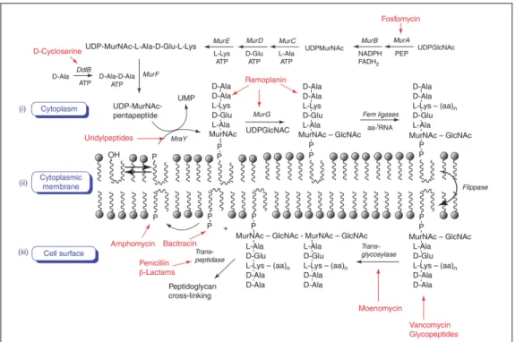
The peptidoglycan biosynthesis is a complex multistep process occurring in three different bacterial compartments (Figure 7):
1. In the cytoplasm;
2. In the cytoplasmic membrane;
3. Outside the cell (in the periplasm in case of Gram-negative bacteria).
1. The cytoplasmic phase10,11 starts with the addition of enolpyruvate to uridine
diphosphate-N-glucosamine (UDP-GlcNAc) by enolpyruvyl transferase MurA. Reductase MurB then catalyzes the reduction of the previously introduced alkene to form UDP-MurNAc. Then MurC, MurD, MurE and MurF ligases, grow a L-Ala-g-D-Glu-m-A2pm-D-Ala-D-Ala-OH fragment to afford an
UDP-MurNAc-pentapeptide often referred to as the Park nucleotide9.
2. The phospho-MurNAc-pentapeptide fragment of this molecule is then transferred to an undecaprenyl-phosphate anchored in the membrane under the action of MraY12, a membrane enzyme to yield Lipid I. The membrane
phase continues as MurG glycosyltransferase adds a GlcNAc moiety to form Lipid II. This building bloc of the peptidoglycan needs to be translocated by the flippase MurJ13,14 to allow next steps.
3. Once flipped, a glycosyltransferase outside the membrane assembles lipid II blocs through polymerization to form glycan strands. Reticulation of peptidoglycan is finally performed by the action of a DD-transpeptidase15.
During the crosslinking process, the active serine of this enzyme is acylated by the penultimate D-alanine of a pentapeptide from a first glycan strand (the donor strand). This binding provokes the concomitant release of the terminal D-alanine. The resulting DD-transpeptidase-tetrapeptide adduct then undergoes a nucleophilic attack from the D-side amino group of a m-A2pm of
second pentapeptide from a second glycan strand (the acceptor strand)11,16,17.
Maturation of peptidoglycan consists in the cleavage of ultimate D-Ala from the remaining D-Ala-D-Ala residues thanks to a DD-carboxypeptidase18–20.
All these enzymes (MurA, MurB, MurC, MurD, MurE, MurF, MraY, MurG, MurJ, glycosyltransferase, DD-transpeptidase, DD-carboxypeptidase) have important roles in the peptidoglycan biosynthesis. Therefore, if any of these were to be inhibited, growth and life of a bacteria population could be seriously compromised.
I.5.b) Peptidoglycan turnover and recycling
Peptidoglycan being a crucial element of the bacterial structure, its metabolism is intimately bound to bacterial growth and division. In view of its function of shape-maintaining the cell wall, one might think that peptidoglycan is a static structure. But it is actually highly dynamic in the sense that it is constantly renewed21.
It is estimated that in some Gram-negative and Gram-positive species, up to 50% of the preexisting peptidoglycan is degraded and replaced during a single cellular cycle22. Some enzymes keep on breaking down the peptidoglycan so that others can
insert new fragments during cell growth. This process is called peptidoglycan turnover.
Because such a loss of material would be unbearable for these organisms, biochemists hypothesized that fragments resulting from the peptidoglycan turnover are taken up and recovered by bacteria23. This was actually observed in
Gram-negative bacteria such as E. coli, in which peptidoglycan fragments are reimported from the periplasmic region where they accumulate. The current model explaining peptidoglycan recycling shows a complex pathway catalyzed by a dozen of dedicated enzymes breaking down bigger fragments into smaller ones ready to be incorporated in the peptidoglycan biosynthesis21,24.
Peptidoglycan recycling would be even more beneficial to Gram-positive bacteria given their much larger peptidoglycan layer. Indeed, it has recently been observed that Gram-positive model bacteria Staphylococcus aureus, Bacillus subtilis, and
Streptomyces coelicolor all recycle the sugar N-acetylmuramic acid (MurNAc)25.
Whether or not Gram-positive organisms recycle other peptidoglycan fragments remains unclear. One might say that peptidoglycan metabolism is a clever dynamic equilibrium of biosynthesis, catabolism and recycling (Figure 8).
Figure 8. Cell wall recycling in Gram-negative bacteria23.
Several models6,17 describe peptidoglycan turnover in different organisms but some
similarities appear:
- Sacculus meshes have to be cleaved to insert new peptidoglycan fragments - Catabolism and anabolism of peptidoglycan have to be concerted to avoid
the bacteria lysis.
Models can be categorized among two different insertion strategies. In the first one, hydrolases act first to cut the mesh and then, synthases come into play to polymerize glycan strands. The second main strategy is known as “make-before-break”: first, synthases polymerize glycan strands and then, hydrolases make incision to blend new strands in the existing sacculus. The key point of these mechanisms is that the cell wall must never be weakened to avoid the bacteria lysis. This is especially true in Gram-negative bacteria since their peptidoglycan layer is thinner6.
Peptidoglycan fragments not reintroduced into the bacterial cell through recycling can trigger a response from the human innate immune system in case of an infection (Figure 8). These danger-associated molecular patterns are recognized by pattern recognition receptors NOD1 and NOD2 (for nucleotide-binding oligomerization domain)26. Once activated NOD1 and NOD2 containing proteins (or peptidoglycan
recognition proteins PGRP) trigger an inflammatory response trough signaling cascades27. These cytosolic proteins are associated with several inflammatory
diseases such as asthma28 or Crohn’s disease29,30 upon mutation of NOD1 and NOD2
genes.
I.5.c) Penicillin-Binding Proteins
Many essential enzymatic activities related to peptidoglycan metabolism are assured by “Penicillin-Binding Proteins” or PBPs17. As suggested by their name, PBPs are able
to covalently bond penicillin, which affects their activities. PBPs have several activities:
- Transglycosylation (polymerization of glycan strands) - DD-transpeptidation (peptidoglycan cross-linking)
- DD-carboxypeptidation (cleavage of the last D-alanine of pentapeptides) - DD-Endopeptidation (reticulation hydrolysis)
PBPs can be categorized in two main groups: high molecular mass (HMM) PBPs and low molecular mass (LMM) PBPs. HMM PBPs are multimodular enzymes. Their C-terminal penicillin-binding domain has a DD-transpeptidase activity. HMM PBPs can be divided in class A PBPs and class B PBPs depending on their N-terminal domain activity. Class A PBPs N-terminal domains are responsible for glycan strand elongation (glycosyltransferase activity) and class B PBPs N-terminal domains are believed to be active in cell morphogenesis.
LMM PBPs are sometimes referred to as class C PBPs. They can be divided in four subcategories depending on their primary structure homology with E. coli Class C main PBPs (PBP4, PBP5, PBP7, AmpH), which are involved in cell separation, peptidoglycan maturation and recycling.
All PBPs display some kind of peptidase activity (transpeptidase, DD-carboxypeptidase or DD-endopeptidase). The terminal D-Ala-D-Ala bond is necessary for these enzymes to form a Henri-Michaelis complex with their substrate.
While the L-Ala-g-D-Glu-m-A2pm (or L-Lys)-D-Ala-D-Ala-OH pentapeptide stem forms
a short-lived complex with PBPs before hydrolysis of the D-Ala-D-Ala peptide bond (DD-carboxypeptidase activity) or before substitution of the last D-Ala by the amino group of m-A2pm or L-Lys of another peptide (DD-transpeptidase activity), the
structurally similar penicillin (Figure 9) durably binds with PBPs and therefore disables their activities17. Bacteria cannot survive as PBP-penicillin complex half-life
exceeds bacteria generation time.
Figure 9. Penicillin structure (a) compared to
D-Ala-D-Ala pentapeptide moiety (b). N O NH R O S OH O H N O NH R O OH O a) b)
I.6. Antibiotics
Many antibiotics have been discovered and developed since Alexander Fleming published his work on penicillin in 19293. Some of them induce cell death
(bactericidal drugs) and others only inhibit cell growth (bacteriostatic drugs)31. Most
of antibiotics used nowadays are natural or semi-synthetic products but some fully synthetic molecules are also available. These drugs can be classified following their bacterial target (Table 1).
Table 1. Main classes of antibiotics and their targets31.
DNA Synthesis
inhibitors RNA synthesis inhibitors Protein synthesis inhibitors Cell wall synthesis inhibitors
• Fluoroquinolones • Trimethoprim – sulfamethoxazole • Rifamycins • Tetracyclines • Aminoglycosides • Macrolides • Streptogramins • Phenicols • b-lactams • Glycopeptides • Glycolipopeptides • Lipopeptides
Figure 10. Structures of selected antibiotics. N O NH O S OH O N COOH O F N HN Ciprofloxacine (a fluoroquinolone) NH OH OH O O OH N N N O HO O H3CO HO AcO Rifampicin (a rifamycin) N OH H O O NH2 O HO O OH H OHH Doxicycline
Some antibiotics such as fluoroquinolones inhibit bacterial DNA replication by interfering with the maintenance of chromosomal topology. This class of antibiotics targets topoisomerases II and IV at the DNA cleavage stage preventing them from joining DNA strands back. Others like rifamycins inhibit RNA synthesis by stopping DNA transcription. These drugs prevent RNA strand from emerging out of RNA polymerases by blocking their b-subunit. Tetracyclines inhibit proteins synthesis by targeting ribosomes. These compounds block the access of aminoacyl tRNAs to the 30S ribosomal subunit. b-lactams such as penicillin interfere with PBPs and inhibit the cell wall synthesis leading to the lysis of the bacteria31. Notable drugs from these
classes of antibiotics are depicted in Figure 10.
As previously mentioned, the bacterial cell wall metabolism is a key target for antibiotics. A few of these and their mode of actions over the peptidoglycan synthesis are shown in Figure 11.
The Golden Years of antibiotic discovery and development have passed. Arrivals of these “wonder drugs” permitted the rise of the pharmaceutical industry and the antibiotic market is still a very lucrative business today, but as years went by, drug resistant bacteria appeared and the number of treatment options went down. Paradoxically, there is less effort today to find new drugs when the medical need keeps on increasing.
I.7. Resistance
I.7.a) The phenomenon
Bacteria may be seen as primitive or inferior organisms to a human eye but they are actually highly evolved species. They responded to 3.5 billion years of environmental challenges. Bacteria have short generation times and reproduce by binary fission. This explains why they evolve so quickly. Each division may induce mutations in genes of the newly formed cells. This allows large amounts of genetic variation in a population. An example with an average population of E. coli in a human intestine might help the reader to have a better grasp on this concept. The probability to have a mutation of a given gene during a cell division is about one in 10 million (10-7).
About 2 ´ 1010 new E. coli cells arise each day in a person’s intestine. This translate
to approximately 2000 bacteria with a mutation in that gene per day (10-7 ´ 2 ´ 1010
= 2000). If one take into account that E. coli has around 4300 genes, it is estimated that the total number of mutations among the E. coli population in a human host is roughly 9 million per day1.
In addition to these random mutations, genotype and possibly phenotypes of a bacteria can be altered by different mechanisms1:
- Transformation: exchange of homologous DNA segments with the surroundings (they might be from broken-open cells)
- Transduction: exchange of bacterial DNA with bacteriophage homologous DNA segments
- Conjugation: transfer of DNA material between two different cells that are temporarily joined (they may be from different species).
Antibiotic drugs are just another challenge to bacteria: each time antibiotics are used, a selective pressure is applied and some survivors among the targeted pathogens might engender new populations of resistant bacteria. This became a serious issue since antibiotics are everywhere, especially in farming and healthcare
facilities. And now, resistant bacteria are escaping the hospitals. Some even say that we are now in a post-antibiotic era33–35. It is estimated that antibiotic resistance
causes over 25 000 deaths each year in the European Union, with an estimated cost of 1.5 billion € per year36.
Figure 12. Bacterial resistance mechanisms37.
A variety of resistance mechanisms have been reported so far (Figure 12): efflux of antibiotics by efflux pumps; decreased influx of antibiotics; sequestration of antibiotics; target modification to avoid binding with antibiotics; target protection to avoid binding with antibiotics; target bypass (new metabolic pathways not requiring the target intermediary); target amplification (synthesis of more targets than usual so the bacteria can survive even if a portion of these is affected); antibiotic inactivation by specific enzymes.
The next section will detail one of these resistance mechanisms: the penicillin inactivation by a b-lactamase in Bacillus licheniformis (a Gram-positive bacteria).
I.7.b)
Induction of a b-lactamase in B. licheniformis
b-lactamases are enzymes capable of deactivating b-lactam antibiotics such as penicillin by hydrolyzing their b-lactam ring. The first of this kind to be identified was penicillinase in 1940 (isolated from E. coli), even before penicillin entered clinical use33,38.
Bacillus licheniformis strain 749/I is also able to produce a b-lactamase (BlaP) coded
by blaP gene. When there is no penicillin, BlaP is only produced at a very low level. The current model39 shows that in the absence of penicillin the bla divergon coding
for BlaP, BlaR1 and BlaI is expressed at a low level (Figure 13a). BlaR1 is a membrane-bound penicillin-binding receptor and BlaI acts as a cytoplasmic repressor of bla divergon when dimerized. In this case, PBP1 (a membrane bound PBP with a transpeptidase activity) is active and peptidoglycan reticulation occurs normally.
When a sub-lethal dose of penicillin is added into the medium, PBP1 is partially inactivated by penicillin and reticulation of peptidoglycan is compromised. Because of this, the anabolism-catabolism equilibrium of peptidoglycan gets dubious and quantities of cell wall turnover products is on the rise (a penicillin stress is generated by the partial deactivation of PBP1, Figure 13b).
Penicillin also binds to receptor BlaR1. This opens a new path for cell wall turnover products: H2N-L-Ala-g-D-Glu-m-A2pm tripeptide resulting from the activity of YkfA is
hydrolyzed into g-D-Glu-m-A2pm dipeptide by activated BlaR1. This dipeptide
inactivates BlaI dimer repressor so the divergon bla is expressed and production of b-lactamase BlaP is triggered.
I.8. Objectives
As pharmaceutical companies neglect the development of new antibiotics in the era of resistance, fundamental research in this area becomes increasingly essential. Biochemists face huge challenges when they strive to study such complex biochemical mechanisms. These studies require chemical tools. Some can be prepared by enzymatic synthesis, but others are more challenging. The goal of this work is to broaden the biochemist toolbox to study peptidoglycan metabolism and antibiotics resistance mechanisms.
Enzymatic in vitro studies require substrates. NMR experiments designed to study the tridimensional structure of peptidoglycan could benefit from 13C or 15N labelled
compounds. Quantitative analyses to study peptidoglycan dynamics require chromatography references as well as isotopologues for mass spectrometry. Non-radioactive and radioactive probes allow for peptidoglycan recycling analyses. Peptide fragments of peptidoglycan are also useful in order to study the innate immune system as they are sensed as danger associated patterns by receptors like NOD containing proteins. Due to its specificity to the bacterial cell wall,
meso-diaminopimelic acid (Figure 14) seems to be a perfect probe to study
peptidoglycan metabolism. This particular amino acid is also implicated in each of the peptides intervening in the current model for the induction of b-lactamase BlaP in B. licheniformis39.
Figure 14. meso-diaminopimelic acid and foreseen substitution and labelling positions.
NH2 COOH HOOC NH2 m-A2pm L(S) D(R) (2H)/[3H] (13C)
Chemistry is the only viable way to prepare these tools as enzymatic syntheses produce very low amounts of products and require multiple purification steps. In addition, these methods do not allow for selective labelling. Therefore, we will in this work synthesize different radioactive and non-radioactive isotope-containing
m-A2pm and m-A2pm containing peptides.
Although m-A2pm appears as a very simple compound at a first glance, the organic
chemist will face some challenges in the syntheses of its isotope-containing analogues and its incorporation in peptides. Indeed, we will have to find a way to prepare a symmetric compound with two chiral centers of opposite configuration. Moreover, several differentially protected m-A2pm will have to be prepared in
anticipation of their insertion in peptide synthesis (Figure 15). Indeed, the biochemist toolbox remains incomplete as enzymatic digestions cannot provide every possible fragment of the peptidoglycan cross-link. PEP2 and a deuterated analogue of it will be useful as chromatography and mass spectrometry references for the study of dynamics of the induction of b-lactamase in B. licheniformis as it appears in the current model. PEP1 and PEP3 contain the peptide bond responsible of the cross-linking between glycan strands in peptidoglycan and therefore will be useful to study the enzymatic activity of DD-transpeptidases. All these compounds could also be used in studies of the human innate immune system. The next chapter will focus on the different strategies reported so far for the preparation of such compounds.
Figure 15. target peptides NH2 CO-D-Ala-OH HOOC CO-D-Ala-H2N NH-γ-D-Glu NH2 HOOC NH-γ-D-Glu-L-Ala-NH2 CO-D-Ala-OH HOOC H2N NH-γ-D-Glu-L-Ala-NH2 CO-D-Ala-OH HOOC H2N-D-Ala-H2N PEP1 PEP2 PEP3
Chapter II
Bibliography
It is not surprising that several groups investigated the preparation of
meso-diaminopimelic acid, this compound being such an important link of
peptidoglycan, both figuratively and literally. Efforts toward the chemical synthesis of isotope-containing m-A2pm (2H, 3H, 14C) were reported from as early as the
1960’s40–45. The first total syntheses did not display any selectivity toward the meso
diastereoisomer. However, a notable preparation of tritiated m-A2pm was reported
in 1985 by Schott et al.46 (Scheme 1). It begins with a photochemically induced radical
chlorination of an existing m-A2pm, probably isolated from peptidoglycan.
Chloro-m-A2pm derivatives were then converted into [3H]m-A2pm by catalytic
dehydrohalogenation and reduction using tritium gas.
Scheme 1. Tritiation of an existing m-A2pm (Schott et al.)46.
(a) conc. HCl, Cl2, hn, 70-80 °C; (b) T2 (1 atm), Pd/C (10 % wt).
Alongside, some efforts have also been made to isolate m-A2pm from the
peptidoglycan of bacteria grown in 14C or 3H enriched media47. But these methods
did not offer the aimed flexibility required for the variety of target molecules of this work. NH2 NH2 HOOC COOH NH2 NH2 HOOC COOH Cl NH2 NH2 HOOC COOH NH2 NH2 HOOC COOH Cl Cl H2N NH2 HOOC COOH [3H] [3H] [3H] a b
Many groups have investigated the chemical synthesis of peptidoglycan fragments during the last 25 years and several stereoselective syntheses of m-A2pm and
m-A2pm containing peptides were reported. A few notable syntheses will be
discussed in the following paragraphs and ultimately, we will choose one to base our strategy on. A few factors helped us to decide which strategy to opt for:
- Stereoselectivity;
- Carbon-13 compatibility;
- Deuterium and tritium compatibility; - Peptide synthesis possibility.
As its name suggest, m-A2pm or (2S,6R)-A2pm includes two stereogenic carbons of
opposite configurations. This represents one of the challenges that we will face in this work, and of course high optical purity is a decisive factor in the strategic choices we will have to make.
Since we want to obtain a carbon-13 substituted m-A2pm, the synthetic pathway that
we will choose has to start with a carbon-13 substituted synthon. The commercial availability of a such starting material is therefore a necessity. This compound should not be used in excess at any point during the synthesis as for obvisou cost reasons. Only syntheses respecting this last criterion will be mentioned.
We also intend to prepare deuterium-substituted and tritium-labelled m-A2pm, so
an easy to label intermediate is needed (e.g. by alkene reduction). If we want to perform a tritium labelling, this should be done during the last chemical step. Finally, we would like to prepare some peptides using solid-phase peptide synthesis and depending on the target molecule, there should be a high flexibility over the protecting groups we may have to use.
II.1. Jurgens and Chen syntheses
Jurgens was the first to publish a total enantioselective synthesis of a differentially protected meso-diaminopimelic acid in 199248. His starting material was Garner’s
aldehyde, already containing the first chiral center of m-A2pm (Scheme 2). The key
step of this strategy is the introduction of the second chiral center by alkylation of a Schollkopf reagent, a chiral auxiliary (step d). The alkene function in this product is later reduced by hydrogen making it a good substrate for an eventual deuterium labelling. However, a tritium reduction is not conceivable since a few more steps are required to produce free m-A2pm.
It should also be noted that no carbon-13 substituted Garner’s aldehyde is commercially available, although, such compounds could be prepared from (13C)serine49. All carbon-13 substituted analogues of serine are available in
L-configuration but none in D-configuration, meaning that a (R)-configured Schollkopf reagent would be necessary to prepare m-A2pm.
Chen et al. followed a very similar approach in 201450. Yet, they used a different
starting material and their meso-diaminopimelic acid skeleton did not feature an alkene anymore (Scheme 2). In this case, the oxazolidine precursor could be prepared from L-glutamic acid (available in a variety of stable isotopomers)50.
These two syntheses will be disregarded as the deuterium/tritium introduction opportunity happens at a too early stage of the strategy.
Scheme 2. Jurgens (1992)48 and Chen (2014)50.
(a) Ph3P=CHCHO, toluene,
D
, 80%; (b) DIBAL, CH2Cl2, 0°C, 60%; (c) Ph3P, CBr4, CH2Cl2, 0°C, 95%; (d) n-BuLi, THF, -78°C,95%; (e) 0.1M HCl, THF, H2O, 48%; (f) H2, Pd/C, EtOAc, quant.; (g) Cbz-Cl, Et2O, NaHCO3, 84%; (h) p-TsOH, H2O, CH3OH,
91%; (i) PDC, DMF, 75%. (j) n-BuLi, THF, -78°C, 3h, 65%; (k) 0.5M HCl, THF, 0°C, 2h; (l) CbzCl, Et3N, CH2Cl2, rt, 2h, 70% (2
steps); (m) p-TsOH , H2O, MeOH, rt, 36h, 80%; (n) TEMPO, BAIB, CH2Cl2, H2O, rt, 1h, 72%.
(R) (R) (S)(S) N BocN O N (S) (S) OMe N N (S) (S) OMe OMe (S) (S) BocN O R (R) (R) BocN O O R = CHO R = CH2OH R = CH2Br b c (R) (R) (S)(S) NH2 BocN O MeOOC (R) (R) (S)(S) NHCbz NHBoc MeOOC OH (R) (R) (S)(S) NHCbz NHBoc MeOOC COOH R = H R = Cbz g (R) (R) (S)(S) NHR BocN O MeOOC (S) (S) BocN O Br N N (S) (S) OEt OEt OMe (R) (R) (S)(S) N BocN O N (S) (S) OEt OEt (R) (R) (S)(S) NH2 BocN O EtOOC (R) (R) (S)(S) CbzHN BocN O EtOOC (R) (R) (S)(S) NHCbz NHBoc EtOOC OH (R) (R) (S)(S) NHCbz NHBoc EtOOC COOH
D-serine L-glutamic acid
a d e f h i n m l k j
II.2. Holcomb and Wang syntheses
Holcomb et al. (1994) started their chemical pathway from L-glutamic acid (readily available in any stable isotopomer)51. The second chiral center is introduced via an
asymmetrical hydrogenation of an alkene obtained by a Wittig reaction (Scheme 3). Wang et al. adopted an identical strategy in 2001, which differed only in the choice of protecting groups52. It should be noted that the chiral selectivity of the Wang
synthesis is vastly superior to the one reported by Holcomb, which requires a chromatography separation of the diastereoisomers. The reduction step represents an easy way to introduce deuterium atoms but the protecting groups would have to be changed to afford free tritiated m-A2pm in a single reduction step. As mentioned
in the previous section, L-glutamic acid is available as a variety of stable isotopomers.
Scheme 3. Holcomb (1994)51 and Wang (2001)52.
(b) (1) BH3.SMe2, 0
®
25°C (2) PCC, 25°C, 51% (2 steps); (c) KHMDS -78®
25°C, 75%; (d) H2 (40 psi),[Rh(I)(NBD)2]ClO4.(S,S)-chiraphos, 25°C, 80% (3:1 R:S); (e) TMSCH2CH2OH, LiHMDS, 0°C, 53%. (x) DIBAL,
Et2O, -78°C, 91%; (y) DBU, CH2Cl2, rt, 5.5h, 91%; (z) H2 (70 psi), [Rh(I)(COD)(R,R)-Et-DuPHOS]OTf, MeOH, 24h, 88%.
(R) (R) (S)(S) COOCH 2CH2TMS NHCbz NHBoc MeOOC (R) (R) (S)(S) CbzN BocHN MeOOC O O (S) (S) CbzN MeOOC O BocHN R (S)(S) CbzN O R = COOH R = CHO b (R) (R) (S)(S)COOt-Bu NBoc2 NHCbz MeOOC (S) (S)COOt-Bu NBoc2 MeOOC NHCbz R (S)(S)COOt-Bu NBoc2 MeOOC BocHN P O OMe OMe MeOOC CbzHN P O OMe OMe
L-glutamic acid L-glutamic acid
R = COOMe R = CHO x O O c d e y z
II.3. Hernandez synthesis
Hernandez and Martin reported a new preparation of m-A2pm from L-aspartic acid
in 200153. The key step of this synthesis is the introduction of the second asymmetric
carbon thanks to an Katsuki-Sharpless asymmetric epoxidation54 of an alkene
obtained by a Wittig reaction (Scheme 4). The final differentially protected molecule is obtained after the epoxide opening, several protection/deprotection and reduction/oxidation steps. Although carbon-13 labelled L-aspartic acid are available, there is no satisfying opportunity for a deuterium or tritium introduction.
Scheme 4. Hernandez (2001)53.
(a) benzene, 0°C, 89%; (b) DIBAL, -78°C, 85%; (c) Ti(i-PrO)4, (R,R)-(+)-DET, TBHP, CH2Cl2, -20°C, 82%; (d) NaN3,
NH4Cl, MeOH/H2O (8:1), reflux, 83%; (e) H2C=C(OMe)CH3, PPTS (cat.), CH2Cl2, rt, 85%; (f) (1) H2, Pd/C, EtOAc, rt,
(2) Cbz2O, CH2Cl2, rt, (3) MeOH, p-TsOH (cat.), (4) NaIO4, Na2CO3, KMnO4, dioxane/H2O (3:1), rt, 60% (4 steps).
(R) (R) (R) (R) (S)(S)COOMe N3 NHBoc OH HO (S) (S) (S) (S) (S) (S) COOMe NHBoc HO O (S) (S) COOMe NHBoc R (R) (R)(R)(R) (S)(S) COOMe N3 NHBoc O O R = COOMe R = CH2OH b O (S)(S) COOMe NHBoc P MeOOC HOOC(R)(R) (S)(S)COOMe NHCbz NHBoc L-aspartic acid a c d e f
II.4. Kawasaki synthesis
The synthesis reported by Kawasaki et al.55 in 2008 enforces a Julia-Kocienski
olefination56 reaction as the key step (step a, Scheme 5). The two chiral centers are
already present in the aldehyde and sulfone precursors. Both of these are derived from D-serine, which is available as 13C-substituted isotopomers. This chemical
pathway features an alkene reduction making it suitable for a deuterium labelling. However, this reduction step occurs at an early stage of the synthesis compromising the possibility of a tritium labelling.
Scheme 5. Kawasaki (2008)55.
(a) NaHMDS, THF, -70°C, 71%; (b) HCl 1M in MeOH; (c) triphosgene, Et3N, CH2Cl2, 22% (2 steps); (d) (1) H2, Pd/C,
MeOH, (2) CbzCl, NaHCO3, 1,4-dioxane/H2O (1:1), 82% (2 steps); (e) RuCl3.nH2O, NaIO4, acetone/H2O (1:1), 66%;
(f) AcCl, MeOH, 89%; (g) Boc2O, DMAP, Et3N, THF, 86%; (h) LiOH, THF/H2O (3:1); (i) (1) Cs2CO3, MeOH, (2) BnBr,
DMF, 83% (3 steps); (j) PDC, DMF, 86%. BnOOC(R)(R) (S)(S) COOH NHCbz NHBoc BnOOC(R)(R) (S)(S) NHCbz NHBoc OH HOOC (R) (R) (S)(S) NHCbz NHBoc OH MeOOC(R)(R) (S)(S) CbzHN BocN O O MeOOC(R)(R) (S)(S) CbzHN HN O O HOOC(R)(R) (S)(S) CbzHN HN O O HOH2C(R)(R) (S)(S) CbzHN HN O O HOH2C(R)(R) (S)(S) CbzHN HN O O (R) (R) (S)(S) NCbz BocN O O (S) (S) BocN O (R) (R) (S)(S) NHCbz NH2 OH HO O (S) (S) N O Cbz S O O N N N N Ph D-serine a b c d e f g h i j
II.5. Del Valle, Chowdhury and Saito syntheses
These three syntheses have a common key step: an olefin metathesis (steps c, g and I, Scheme 6). All three groups opted for olefinic synthons already containing the chiral centers present in m-A2pm.
Del Valle and Goodman chose a ring closing metathesis (RCM) ensuring almost quantitative yields in their 2004 publication57 (Scheme 6, left). However, this strategy
requires more steps to prepare the diene substrate needed for RCM and to open the resulting ring. Alkene is then reduced and alcohol oxidized to afford the target molecule.
Saito et al. performed a cross-metathesis between a protected allylglycine and an oxazolidine derived from D-serine58 (Scheme 6, right). An excess of the oxazolidine
synthon is necessary to limit the formation of homodimers of allylglycine. As for Del Valle synthesis, three more steps are required to reach the target molecule: alkene reduction, oxazolidine opening and alcohol oxidation.
Chowdhury and Boons presented a very straightforward approach in 200559 (Scheme
6, middle). The m-A2pm framework is formed in a single step from protected
(R)-allylglycine and (S)-vinylglycine. The remaining step being a reduction of the alkene function. As for Saito’s synthesis, an excess of vinylglycine synthon is necessary to limit the formation of homodimers of allylglycine. Vinylglycine does not react with itself probably due to steric reasons.
These three strategies use precursors that can be prepared from amino acids available in a variety of 13C-substitued isotopomers. But only Chowdhury’s synthesis
is worth considering since the reduction could be applied as a final step. This strategy is very appealing to us because, with the right choices of protecting groups, a variety may of m-A2pm derivatives may be produced with different goals in mind (peptides,
Scheme 6. Del Valle (2004)57, Chowdhury (2005)59 and Saito (2013)58 strategies.
(a) (1) PyBOP, Et3N, CH2Cl2, (2) Ac2O, pyridine, CH2Cl2, 83% (2 steps); (b) Boc2O, DMAP, THF, 77%; (c) 2 mol% Grubbs 2nd
gen. cat., CH2Cl2, reflux, 2h, 99%; (d) (1) LiOH 2M, THF, (2) MeI, K2CO3, DMF; (e) H2, 2mol% [Ir(COD)PyPCy3]PF6, CH2Cl2,
56% (3 steps); (f) NaClO, NaClO2, TEMPO, MeCN, H2O (92%). (g) Grubbs 2nd gen. cat., 64%; (h) H2, 3% Pt/C, EtOH, CH2Cl2,
H2O, 98%. (i) 5mol% Grubbs 2nd gen. cat., 73%; (j) H2, PtO2, EtOAc, rt, 14h, 97%; (k) p-TsOH, H2O, MeOH, 81%; (l) NaClO,
NaClO2, TEMPO, MeCN, H2O.
BnOOC(R)(R) (S)(S) CbzHN BocN O (S) (S) BocN O BnOOC(R)(R) NHCbz glycine D-serine BnOOC(R)(R) (S)(S) CbzHN BocN O BnOOC(R)(R) (S)(S) NHCbz NHBoc OH BnOOC(R)(R) (S)(S) NHCbz NHBoc COOH t-BuOOC(R)(R) NHBoc (S) (S) NHFmoc COOBn t-BuOOC(R)(R) NHBoc (S) (S) NHFmoc COOBn t-BuOOC(R)(R) NHBoc (S) (S) NHFmoc COOH L-methionine glycine (R) (R) NHBoc (S) (S) NCbzBn COOMe HO HOOC(R)(R) NHBoc (S) (S) NCbzBn COOMe (S) (S) Boc N (R) (R) NCbzBn O (R) (R) (S) (S) COOMe NHBoc NCbzBn HO AcO (S) (S)NCbzBn O N Boc (R) (R) AcO (S) (S)NCbzBn O N H (R) (R) AcO (S) (S) NCbzBn O HO NH2 (R) (R) HO glycine L-methionine a b c d e f g h i j k l
II.6. Peptides
Several groups have reported syntheses of m-A2pm containing
peptides and muropeptides. Some prepared short peptides in solution50,51,55,57–61 while others
inserted differentially or orthogonally protected m-A2pm in
solid-phase peptide synthesis59,62–64
(SPPS, concept illustration in Scheme 7). The former strategies will be dismissed as they require extensive purification after each coupling whereas the latter only require a single purification step after cleaving the peptide from resin.
Recently, Dr J. Simon et al. proposed a new methodology for the synthesis of m-A2pm containing
peptides inspired by Chowdhury cross-metathesis65. Rather than
inserting an adequately protected
m-A2pm directly into SPPS, they
grew a peptide containing an allylglycine and then, performed an on-resin cross-metathesis with a vinylglycine synthon to turn the
Scheme 7. SPPS of a m-A2pm containing
peptide (classical approach). allylglycine into an m-A2pm (concept
illustration in Scheme 8). NHFmoc NHFmoc HOOC FmocHN NHR2 COOR1 NHR2 COOR1 HN O N H O NH2 COOH HN O N H HO O NHR2 COOR1 NHFmoc N H O NHFmoc NH2
Scheme 8. SPPS of a m-A2pm containing peptide
via on-resin cross-metathesis.
NHFmoc NHFmoc HOOC NHFmoc HN O N H O NHFmoc N H O HN O N H O COOR1 NHR2 NHFmoc O NH2 COOH HN O N H HO NH2 NH2 NHR2 COOR1
Chapter III
Results and discussion
Sc he m e 1. Ge ne ral st rat eg y fo r t he p re par at io n of fr ee m -A2 pm , ( 2 H 2 )m -A2 pm , [ 3 H] m -A2 pm a nd pe pt ide fr ag m ent s o f pe pt ido gl yc an. H2 N-D -Al a -m -A 2 pm -D -Al a -O H H2 N-L -Al a -D -G lu γ R 1 OOC NHR 2 NHR 4 COOR 3 HOOC H2 N NH 2 COOH H/ D / T H/ D / T R1 OOC NHR 2 NHR 4 COOR 3 + fre e m -A 2 pm d e h yd ro -m -A 2 pm a llyl g lyci n e vi n yl g lyci n e g lyci n e D -me th io n in e HN HOOC NH O NH COOH O N H COOH O H2 N H/ D H/ D O H2 N L D D D D L HN HOOC H2 N O NH COOH O N H COOH O H2 N H/ D H/ D L D D D L HN HOOC H2 N O NH O NH O H2 N COOH D D D L NH 2 HOOC O N H COOH D D L m -A 2 pm -D -Al a -O H H2 N-L -Al a -D -G lu γ m -A 2 p m-D -Al a -m -A 2 p m-D -Al a -O H D -G lu γ
III.1. Strategy
As a reminder, the first goal of this work is the preparation of non-radioactive and radioactive isotope-containing meso-diaminopimelic acid: m-A2pm, (2H2)m-A2pm,
[3H]-m-A2pm and (13C)m-A2pm.
The second goal is the synthesis of m-A2pm and (2H2)m-A2pm containing peptides.
Because of the variety of target molecules (Scheme 1), The most versatile and straightforward strategy found so far will be enforced. Chowdhury’s approach allows to mix and match couples of differently protected allylglycine (AG) and vinylglycine (VG) synthons to afford m-A2pm with several sets of protecting groups.
Protecting groups and absolute configurations of AG and VG synthons must be chosen wisely depending on the anticipated fate of the dehydro-m-A2pm derivatives
obtained by cross metathesis, especially for peptide synthesis purposes. Depending on the targeted peptide, either orthogonally or differentially protected m-A2pm will
be needed. To avoid any information overdose to the reader at this early stage, actual protecting groups choices are argued in the following subchapters depending on the target molecule discussed (see paragraph III.5.b protecting groups choice). Syntheses of free m-A2pm, (2H2)m-A2pm and [3H]m-A2pm will only differ in the
catalytic reduction conditions of a common cross-metathesis dehydro-m-A2pm key
intermediate, but in contrast to deuterium and tritium, carbon-13 has to be introduced at the beginning of the synthetic pathway (Scheme 2). Although D-methionine and glycine are commercially available as several carbon-13 isotopomers, (1-13C)glycine is the cheapest option available (172 €/g). In addition, in
order to avoid large amounts of homodimers of allylglycine synthons during the cross metathesis key step, Chowdhury uses an excess of vinylglycine synthon, making it a poor choice as a carbon-13 substituted synthon for cost reasons. Consequently, it
was decided to produce (13C)allylglycine from (1-13C)glycine. We opted for (R)-VG and
(13C)(S)-AG synthons.
Scheme 2. General strategy for the preparation of (13C)m-A 2pm.
The first part of this chapter will focus on the preparation of AG and (13C)AG
synthons. The second part will describe the synthesis of the VG synthons. Then, syntheses of free and protected m-A2pm will be discussed. The last subchapter will
discuss peptides syntheses.
HOOC H2N NH2 13COOH (13C)m-A 2pm R1OOC NHR2 NHR4 13COOR 3 (13C)dehydro-m-A 2pm R1OOC NHR2 NHR4 13COOR 3 + (13C)allylglycine vinylglycine (1-13C)glycine D-methionine
III.2. Allylglycine and (
13C)allylglycine preparation
III.2.a) Strategy
Allylglycine derivatives can be enantioselectively produced by phase-transfer catalysis (PTC) of a Schiff base using a chiral quaternary ammonium salt as a catalyst66
(see retrosynthetic path described in Scheme 3). Such Schiff bases can be prepared from glycine. A benzophenone imine Schiff base was chosen since it is known to ensure monoalkylation in opposition to benzaldimine Schiff bases which are prone to dialkylation67,68. Due to the relatively high price tag of (1-13C)glycine (172 €/g), all
chemical steps were optimized on the cheap natural isotopic abundance glycine (0.1 €/g).
Scheme 3. Retrosynthetic path for the preparation of allylglycine derivatives.
III.2.b) Preparation of Schiff bases
In order to prepare the desired natural isotopic abundance and carbon-13 substituted Schiff bases 5 and (13C)5, glycine tert-butyl esters 4 and (13C)4 (Scheme
4) had to be prepared first. This particular type of ester can be tricky to prepare and requires a protection of glycine’s amino group. So, glycine 1 was first converted into a carboxybenzyl carbamate 2. This protecting group was chosen because its catalytic reduction removal conditions are compatible with a tert-butyl ester. The protection went smoothly on glycine 1 and was repeated on (1-13C)glycine (13C)1 with similar
yields (96%).
NHPG 13COOPG
NH2
13COOH
Schiff base (1-13C)glycine
(1-13C)allylglycine
derivative
N 13COOPG
Scheme 4. Preparation of Schiff bases 5 and (13C)5.
(a) Cbz-Cl, NaOH, toluene, 0 °C to rt, 96%; (b) t-BuOH, DCC, DMAP, CH2Cl2, 93%;
(c) H2 (5 bar), Pd/C, HCl, MeOH; (d) Ph2C=NH, CH2Cl2, anh. MgSO4, 94% (2 steps).
This first step was the occasion to witness analytical particularities of carbon-13 substituted compounds in mass spectrometry and NMR spectroscopy. Obviously, mass spectra were shifted one unit to the right but more interestingly, some carbon-hydrogen couplings were observed on 1H NMR as well as carbon-carbon couplings
on 13C NMR (in addition to a hundred times more intense than usual peak for the
carbon-13 substituted atom). NMR couplings observed on (13C)2 are displayed in
Figure 1. Similar behaviors were observed in every carbon-13 substituted compounds synthesized from this point (see Chapter VI: Supplementary Data).
Figure 1. Comparison of NMR signals of CH2a in 2 and (13C)2.
Ha of (13C)2 appears as a triplet due to similar JHH and JHC in 1H NMR. Ca appears as a doublet in 13C NMR.
Spectra taken in DMSO-d6 on a 400MHz Bruker instrument.
NHR2 *COOR1 N *COOt-Bu 1 R1 = H R2 = H 2 R1 = H R2 = Cbz 3 R1 = t-Bu R2 = Cbz 4 R1 = t-Bu R2 = H 5 a b c d N H C 13C O OH O O H H JHC = 5.8 Hz JHH = 5.8 Hz JCC = 58.8 Hz N H C O OH O O H H JHH = 5.8 Hz 2 (13C)2
Tert-butyl protection of the carboxylic group was tested using Steglich’s
esterification69. Two carbodiimides were considered for this reaction: EDCI and DCC.
The former has the advantage of being converted into a water soluble, easy to extract, corresponding urea. However, DCC gave better yields (93% vs 72%) and we had to cope with DCU elimination by multiple precipitations in ether. (13C)3 was
obtained using these last conditions with a 93% yield.
Removal of the amino protecting group through catalytic hydrogenation surprisingly proved to be more difficult than expected (Table 1). Two different catalysts (Pd/C 5% wet and Pd/C 10% dry) were tested but none of them afforded satisfying results using THF as the solvent (entries 1, 2). Quantities of crude product 4 obtained after work-ups were very low. Our first hypothesis was a poor washing of the filtered catalyst. So, methanol was used and higher amounts of 4 although still lower than expected were obtained (entry 3). Using methanol as the reaction solvent improved a bit but still not in a satisfactory manner (entry 4). Our hypothesis then evolved: the free amino group might act as a poison to the catalyst. To avoid this issue, it was decided to add some acid to the medium. The acid choice and its quantity are crucial in order to avoid a cleavage of the tert-butyl ester. We first settled on using 1 equivalent of acetic acid (entry 5). Conversion improved a lot, comforting our hypothesis. Finally, using 0.48 equivalent of sulfuric acid or 0.95 equivalent of hydrochloric acid led to yields around 95% (entries 6, 7). These last conditions were then repeated on (13C)3.
Table 1. Optimization of Cbz carbamate cleavage (step c, Scheme 4).
# P(H2) Solvent Washing solvent Catalyst (quantity) Duration Yield
1 1-3 bar THF THF Pd/C 5% wet (1%) 5 h <1%
2 3 bar THF THF Pd/C 10% dry (1%) 18 h 4%
3 5 bar THF MeOH Pd/C 10% dry (1%) 120 h 23%
4 5 bar MeOH MeOH Pd/C 10% dry (1%) 120 h 37%
5 5 bar MeOH + 1.0 equiv. HOAc MeOH Pd/C 10% dry (1%) 120 h 56% 6 5 bar MeOH + 0.48 equiv. H2SO4 MeOH Pd/C 10% dry (2%) 2h 95%
The last step toward the formation of the desired Schiff base can be accomplished using different reactants: dichlorodiphenylmethane, benzophenone or benzophenone imine70–72. The best results were obtained using the latter. The
driving force of this reaction is the precipitation of by-product NH4Cl. Addition of
anhydrous MgSO4 to the medium ensured precipitation of the salt and allowed total
conversion of glycine tert-butyl ester 4 into Schiff base 5. These optimized conditions were repeated on crude (13C)4 and allowed the formation of (13C)5 with a satisfying
94% yield over two steps. Carbon-13 substituted Schiff base (13C)5 was prepared on
an over 10 g scale from (1-13C)glycine (13C)1 with an overall yield of 84% over four
steps.
III.2.c) Chiral alkylations and protections
The Cyclotron Research Center (CRC) is familiar with chiral phase transfer catalysis, for instance in the synthesis of a-benzylated lanthionine (an analogue of m-A2pm)73
or [18F]fluoro-L-DOPA74–76, 6-[18F]fluoro-L-tyrosine75 and 6-[18F]fluoro-L-m-tyrosine76,
for PET applications. Among the chiral quaternary ammonium salts already tested at the CRC, we may cite cinchonine and cinchonidine derived quaternary ammoniums and Maruoka’s catalysts66 (Figure 2).
Figure 2. Selected chiral phase transfer catalysts.
Maruoka’s phase transfer catalysts 6a and 6b are exceptionally active (only 0.5 mol% is usually needed) and stereoselective (enantiomeric excesses over 95%). They even
N H O N Cl H N H H O N Cl 7b 7a N F F F F F F Br N F F F F F F Br 6a 6b
can afford high enantioselectivity at high temperatures. They are now commercially available (50 mg = 261 €), but it was not the case at the beginning of this work. The very long multistep preparation of these catalysts led us to consider cinchonine and cinchonidine derivatives 7a and 7b. These compounds, although less efficient than 6a and 6b, stand out as easy to prepare catalysts from cheap cinchonine (100 g = 110 €) and cinchonidine (100 g = 167 €) while remaining sufficiently active and selective. They were synthesized following Lygo’s procedure77 depicted in Scheme 5.
Cinchonidine derivative 7b was used to prepare (S)-AG derivatives and cinchonine derivative 7a for the preparation of (R)-AG as chiral HPLC references.
Scheme 5. Preparation of catalyst 7b.
(a) H2 (5 bar), Pd/C (10%, dry), rt, 97%; (b) 9-chloromethylanthracene, toluene, N2, 95 °C, 72%; (c) BnBr, NaOH
50%, CH2Cl2, 0 °C, 72%.
Scheme 6. Preparation of AG derivatives 10, (13C)10 and 11.
(a) CH2=CH-CH2-Br, 7b, KOH 50%, toluene/CH2Cl2 10:1, -20°C, 95%; (b) citric acid, H2O/THF; (c) Fmoc-OSu,
THF/H2O 65% (2 steps); (d) Cbz-Cl, Na2CO3, toluene, 0°C to rt, % (55%, 2 steps).
PTC alkylations of Schiff bases 5 and (13C)5 were performed using a mixture of
toluene and dichloromethane (10:1) as the organic phase and 50% aqueous KOH as a base (step a, Scheme 6). Toluene usually helps achieving better enantioselectivity but a fraction of dichloromethane was necessary to solubilize the catalyst. As with
N OH N H Cl N OHH N H Cl N OHH N H N OHH N H a b c 7b cinchonidine N *COOt-Bu N *COOt-Bu NHR *COOt-Bu 5 8 9 (R = H) 10 (R = Fmoc) 11 (R = Cbz) c d a b