Edited by JE Thomas
the safe movement of
planet.
We deliver scientific evidence, management practices and policy options to use and safeguard agricultural and tree biodiversity to attain sustainable global food and nutrition security. We work with partners in low-income countries in different regions where agricultural and tree biodiversity can contribute to improved nutrition, resilience, productivity and climate change adaptation. Bioversity International is a member of the CGIAR Consortium – a global research partnership for a food-secure future.
www.bioversityinternational.org
The Global Musa Genetic Resources Network (MusaNet) is a global collaborative framework for Musa genetic resources and a partnership of all key stakeholders, aiming at ensuring the long-term conservation on a cooperative basis, and facilitating the increased utilization of Musa genetic resources globally.
Our vision is a world in which Musa genetic resources diversity is valued, secured and supporting all life.
To create this world our mission is: “To build upon existing strengths in the global, regional and national collections by bringing people to optimize the effort to conserve, add value and promote the use and safe distribution of a wide range of Musa genetic diversity as a foundation for further breeding or direct use by farmers.”
www.musanet.org
Citation:
Thomas, JE. 2015. MusaNet Technical Guidelines for the Safe Movement of Musa Germplasm. 3rd Edition. Bioversity International, Rome.
ISBN 978-92-9255-034-9 © Bioversity International 2015 Cover photos:
of Musa germplasm
1. Introduction ... 1
1.1. Guidelines structure ... 2
1.2. Guidelines update ... 2
1.3. Definitions ... 2
2. Contributors and acknowledgements ... 3
3. Transit centre and virus indexing centres ... 5
3.1. International Transit Centre ... 5
3.2. Virus indexing centres ... 5
4. General recommendations and technical protocols ... 6
4.1. General recommendations ... 6
4.2. Technical protocols ... 7
5. Virus diseases of Musa ... 9
5.1. Banana bunchy top ... 9
5.2. Abaca bunchy top... 16
5.3. Banana bract mosaic ... 18
5.4. Abaca mosaic ... 21
5.5. Banana mosaic ... 24
5.6. Banana streak ... 27
5.7. Banana mild mosaic ... 35
5.8. Banana virus X ... 39
6. References ... 40
Appendix 1. Diagnostic protocols ... 54
1
1. Introduction
1These guidelines describe technical procedures that minimize the risk of pest introductions due to the movement of germplasm for research, crop improvement, plant breeding, exploration or conservation. It is important to emphasize that these guidelines are not meant for trade and commercial consignments concerning export and import of germplasm.
Collection, conservation and utilization of plant genetic resources and their global distribution are essential components of international crop improvement programmes.
Inevitably, the movement of germplasm involves a risk of accidentally introducing plant pestsalong with the host plant. In particular, pathogens that are often symptomless, such as viruses, pose a special risk. To minimize such risks, preventive measures and effective testing procedures are required to ensure that distributed material is free of pests of potential phytosanitary importance. Eradication of pathogens from a region or country is extremely difficult, and even low levels of infection or infestation may result in the establishment of pathogens in new areas.
The international, and inter-regional, movement of plant germplasm for research, conservation and basic plant breeding purposes (including plant biotechnology) requires complete and up-to-date information concerning the phytosanitary status of the plant germplasm. In addition, the relevant and current national regulatory information governing the export and importation of plant germplasm in the respective countries is essential.
The recommendations made in these guidelines are intended for small, specialized consignments used in research programmes, e.g. for collection, conservation and utilization for breeding of plant genetic resources and crop improvement by conventional and modern technologies. When collecting and transporting germplasm, International Standards for Phytosanitary Measures (ISPMs) established by the Food and Agriculture Organization of the UN (FAO)’s International Plant Protection Convention (IPPC; https://www.ippc.int/core-activities/standards-setting/ispms) should be considered.
This revision of the technical guidelines for banana has been produced by the Conservation Thematic Group of MusaNet, an international network for Musa genetic resources coordinated by Bioversity International (www.musanet.org). These guidelines supersede those published in 1989 (Frison and Putter 1989) and 1996 (Diekmann and Putter 1996). The experts who contributed to the
2
production of these technical guidelines are listed in Section 2 of this publication. They have contributed to the preparation of the technical guidelines in their personal capacity and do not represent or commit the organizations for which they work. The guidelines are intended to provide the best possible phytosanitary information to institutions involved in small-scale plant germplasm exchange for research purposes. Bioversity International and the contributing experts cannot be held responsible for any problems resulting from the use of the information contained in these guidelines.
1.1. Guidelines structure
The guidelines cover virus diseases only, as they are the major pathogen contaminants found in in vitro Musa germplasm. Section 3 lists the Inter-national Transfer Centre and indexing laboratories, while Section 4 makes general and technical recommendations on safe procedures to move banana germplasm. The largest section, Section 5, covers pests of phytosanitary concern for the international or regional movement of Musa. The information given on a particular pest is not exhaustive but rather concentrates on those aspects that are most relevant to the safe movement of germplasm. No specific information on treatment is given in the pest descriptions. A pest risk analysis (PRA) will produce information on which management options are appropriate for the case in question. General precautions are given in the technical recommendations.
1.2. Guidelines update
These guidelines reflect the consensus and knowledge of the specialists who have contributed to this revision, but it is anticipated that the information will need to be regularly updated as new information becomes available. We ask our readers to kindly bring to our attention any developments that may require a review of the guidelines, such as new records, detection methods or control methods.
Correspondence regarding this publication should be addressed to Bioversity International, Parc Scientifique Agropolis II, 34397 Montpellier Cedex 5, France.
1.3. Definitions
Germplasm Germplasm is living tissue from which new plants can be grown. It can be a seed or pollen or vegetative material (e.g. a sucker, leaf, piece of stem, corm, meristem or regenerable cells).
Pest Any species, strain or biotype of plant, animal, or pathogenic agent, injurious to plants or plant products (FAO 1996).
3
2. Contributors and acknowledgements
Contributor Address Email John Thomas
(Editor)
The University of Queensland,
Ecosciences Precinct, Level 2C West,
GPO Box 267, Brisbane, Queensland 4001, Australia
Murray Sharman Queensland Dept of Agriculture and Fisheries, Ecosciences Precinct, Level 2C West,
GPO Box 267, Brisbane, Queensland 4001, Australia
Ludivine Lassois University of Liege, Gembloux Agro-Biotech, Passage des Déportés, 2, B-5030 Gembloux, Belgium [email protected] Sebastien Massart [email protected] Caroline De Clerck [email protected] Marie-Line Caruana CIRAD, UMR-BGPI TA A-54/K, Campus International de Baillarguet, 34398 Montpellier Cedex 5, France [email protected] Matthieu Chabannes [email protected] Pierre-Yves Teycheney CIRAD, Station de Neufchâteau, Sainte-Marie, 97130 Capesterre-Belle-Eau, Guadeloupe [email protected]
P. Lava Kumar IITA-Nigeria Ibadan, PMB 5320,
Ibadan, Oyo State, Nigeria
4
Contributor Address Email Ines Van den
houwe Bioversity International Transit Centre, c/o KU Leuven, Division of Crop Biotechnics - Laboratory of Tropical Crop Improvement,
Willem de Croylaan 42 box 2455 , BE3001 Heverlee, Belgium [email protected] Nicolas Roux (MusaNet Coordinator) Bioversity International, 1990 Boulevard de la Lironde,
Parc Scientifique Agropolis II,
34357 Montpellier, France
These guidelines were developed and published through funds from the CGIAR’s Roots Tubers and Bananas (RTB) Research Program and Managing and Sustaining Crop Collections Research Program (Genebanks).
The editorial assistance of Rachel Chase, Bioversity International, Montpellier, France is gratefully acknowledged.
5
3. Transit centre and virus indexing centres
3.1. International Transit Centre
The International Transit Centre (ITC) is operated by Bioversity International under the auspices of FAO, and is hosted by the Katholieke Universiteit Leuven, Belgium. Its role is to facilitate safe exchange of Musa germplasm. With over 1,500 accessions, the ITC contains the largest banana in vitro collection in the world.
3.2. Virus indexing centres
Virology research units having expertise with Musa viruses, and collaborating with Bioversity International for the virus indexing of banana, currently include the Centre de coopération internationale en recherche agronomique pour le développement (CIRAD), Montpellier, France (Officer-in-Charge: Dr Marie-Line Iskra-Caruana); University of Queensland, Brisbane, Australia (Officer-in-Charge: Dr John Thomas); University of Liege (ULg), Gembloux Agro-Biotech, Gembloux, Belgium (Officer-in-Charge: Dr. Sébastien Massart) (currently Bioversity’s Virus Indexing and Sanitation Centre (VISC)); and the International Institute of Tropical Agriculture (IITA), Ibadan, Nigeria (Officer-in-Charge: Dr Lava Kumar).
6
4. General recommendations and technical protocols
4.1. General recommendations
The following general recommendations apply for all international Musa germplasm exchanges.
Germplasm should be obtained from the safest source possible. There is for example a pathogen-tested in vitro Musa germplasm collection available at the ITC, from which safe germplasm can be ordered online (http://www.crop-diversity.org/banana). All germplasm moving from one continent to another should transit through the ITC or, if possible, be obtained from the ITC. Other sources of safe germplasm may be available where indexing laboratories have the capacity and expertise to test for the complete range of viruses.
All germplasm should ideally be moved in the form of tissue culture. If this is not possible, full quarantine measures, including containment in secure post-entry quarantine facilities, must be taken until the vegetative material or seed is cultured in vitro.
Germplasm should be tested for all viruses known to affect Musa according to the protocols described in these guidelines. However, in some instances tests may be omitted if there is reliable evidence that particular viruses are not present in the country of origin of the germplasm.
Indexing procedures and results should be documented, e.g. in a germplasm health statement. A sample copy is included at the end of this publication (Appendix 2).
The movement of germplasm should be carefully planned in consultation with quarantine authorities and the relevant indexing laboratory, and should comply with the regulatory requirements of the importing country. The ISPM measures as published by the Secretariat of the International Plant Protection Convention (IPPC) should be followed during movement of germplasm. In accordance with IPPC regulations, any material being transferred internationally must be accompanied by a phytosanitary certificate.
7
4.2. Technical protocols
Vegetative material
The following protocol should be followed to prepare vegetative material for virus indexing and exchange (distribution) of germplasm.
Select a sucker from a plant without symptoms of systemic pathogen infection.
Trim the sucker to remove soil, roots and any other extraneous material, leaving part of the central corm containing the meristem and about 10 cm of the pseudostem tissue above it. The overall dimensions of the block of tissue will be about 20 cm high and 10−15 cm in diameter.
Air-dry the block for 24 h and wrap in newspaper. Label the material and dispatch in a cardboard box using the fastest transportation method. Plastic should not be used for wrapping.
Send the material to the nearest appropriate tissue culture laboratory in the country of origin, or if this is not possible, to a tissue culture laboratory preferably in a non-banana-growing area.
The meristem tip should be excised, surface disinfected (Hamill et al. 1993) and cultured aseptically (Strosse et al. 2004; Van den Houwe and Swennen 2000; Vuylsteke 1989; Wong 1986).
A single meristem-tip-derived subculture should be further cloned to seven plantlets, of which five should be sent to an indexing facility and two should remain in culture for future multiplication.
At the indexing facility, four plants should be established in a potting mix in a vector-free, insect-proof greenhouse under conditions conducive to vigorous plant growth. The fifth plant serves as a back-up. The plants to be indexed should be physically isolated from other bananas so as to minimize the possibility of cross-contamination.
After three and six months of growth, tissue samples should be taken from the three youngest expanded leaves and indexed for viruses as described in Appendix 1.
In addition, electron microscopic observations should be undertaken at both three and six months to look for the presence of any undescribed viruses.
8
If all tests are negative, the four indexed plants may be released and the cultures derived from the two remaining plants in vitro may be further propagated and distributed in vitro.
If found to be infected, the material should be subjected to a virus eradication treatment if available. The plant material must not be released until its virus-free status has been confirmed using the testing procedures described in Appendix 1.
For the movement of in vitro material, neither charcoal nor antibiotics should be added to the culture medium, as these may mask contamination in the culture.
In vitro cultures should be shipped in transparent tubes, preferably plastic,
and visually inspected for bacteria, fungi and arthropods. Contaminated germplasm should be destroyed. Special care should be taken to protect the material from extremes of temperatures (not below 15°C and not above 35°C) during shipment.
Seeds
Some viruses may be transmitted through seeds. The following protocol should be followed before seeds are moved internationally.
Collect seeds from plants that are free from pests and disease symptoms. Make sure seeds are free of pulp, and there is no visual evidence of
insects, mites or nematodes. Air-dry and, if necessary, fumigate. Banana seeds are sensitive to desiccation and therefore should be moved immediately to an appropriate tissue culture laboratory in the country of origin, or if not possible, to a tissue culture laboratory preferably in a non-banana-growing area.
Seed should be surface-disinfected. The embryos should be isolated and germinated in vitro to establish sterile cultures (Bakry 2008; Pancholi et al. 1995; Uma et al. 2011).
Materials derived from embryo culture should be indexed and moved in the same way as material derived from meristem tip culture, as described above.
9
5. Virus diseases of Musa
5.1. Banana bunchy top
Cause
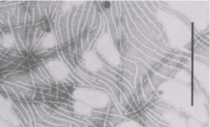
Banana bunchy top virus (BBTV) has been consistently associated with the
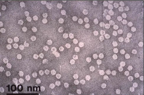
disease. This persistently aphid-transmitted virus is restricted to the phloem tissue, and has 18−20 nm isometric virions (Figure 1) with a coat protein subunit of 20.1 kDa and a multicomponent single-stranded DNA genome. It is a member of the genus Babuvirus (family Nanoviridae). Two geographical groups are described: the South Pacific group (isolates from Australia, Africa, the Pacific islands and the Indian sub-continent) and the Asian group (South-east Asia and China).
Figure 1. Purified preparation of BBTV particles (JE Thomas).
Significance
The most devastating effects of the virus in recent years have been reported in Pakistan (Soomro et al. 1992), Hawaii (Ferreira et al. 1989) and sub-Saharan Africa (Adegbola et al. 2013; Kumar et al. 2011; Lokossou et al. 2012).
Several countries (e.g. Australia, Taiwan and the Philippines) have implement-ed comprehensive roguing/sanitation programmes.
10
Symptoms
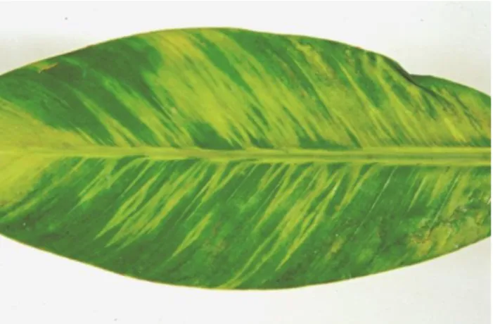
Typical severe symptoms include dark green streaks of variable length in the leaf veins, midribs and petioles. These streaks may, however, be rare or absent in some banana cultivars, abaca (Musa textilis) and Ensete spp. Leaves become progressively shorter and develop marginal chlorosis (Figure 2). As the disease progresses, leaves become more upright or ‘bunched’ at the apex of the plant (Figures 3 and 4).
Figure 2. A healthy banana leaf (left) and a BBTV-infected banana leaf (right) (C De Clerck).
11
Plants can be infected at every development stage, but symptoms do not appear until at least two new leaves are produced (Thomas 2008).
Depending on when the plant becomes infected, it may produce no fruit or the bunch may not emerge from the pseudostem. When infection takes place very late in the season, there may be no leaf symptoms but dark green streaks may be seen on the tips of the bracts.
Plants coming from infected planting material show strong symptoms on all leaves, with leaves remaining bunched at the top of the pseudostem and showing chlorotic edges (Figure 4). Aphid-inoculated plants show symptoms only on leaves formed after infection. All the suckers from an infected plant will be infected.
Mild symptoms of vein clearing as well as symptomless infections have been reported from Taiwan (Su et al. 2003) and Vietnam. Attenuation of initial severe symptoms has been reported in the cv. Veimama from Fiji (Magee 1953).
Figure 4. Severe BBTV symptoms on plants grown from infected planting material; Burundi, near Bujumbura (C De Clerck).
12
Hosts
All Musa species and cultivars are potential hosts for BBTV. To date, no resistant cultivar has been identified but varietal differences in susceptibility have been reported (Espino et al. 1993; Hooks et al. 2009; Stover 1972; ADW Geering and JE Thomas, unpublished). Cultivars in the Cavendish subgroup are severely affected. Ensete ventricosum has been experimentally infected.
Canna indica (Pinili et al. 2013; Su et al. 1993), Hedychium coronarium (Su et al.
1993), Alpinia zerumbat and one selection of Colocasia esculenta (Pinili et al. 2013) have been reported as experimental hosts of BBTV using Asian subgroup isolates of the virus. Attempts to confirm these results have been unsuccessful with South Pacific subgroup isolates (Geering and Thomas 1997; Hu et al. 1996; Manickam et al. 2002). There are no reports of natural infections of hosts outside the Musaceae.
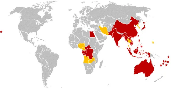
Geographical distribution
The map in Figure 5 highlights the countries affected by BBTV. The affected countries are also listed below.
Figure 5. Distribution of BBTV. Countries in red were identified in the 1996 Technical Guidelines (Diekmann and Putter 1996) while those in orange (and the countries in bold in the list below) have been identified since 1996.
13 Countries affected by BBTV (by region)
Note: new records which were not in the 1996 Technical Guidelines (Diekmann and Putter 1996) are printed in bold; * unconfirmed (this status conferred by the quoted author).
Africa
Angola (Kumar et al. 2008) Benin (Lokossou et al. 2012)
Burundi (Sebasigari and Stover 1988) Cameroon (Oben et al. 2009)
Central African Republic (Foure and Lassoudiere, unpublished) Congo Republic (Wardlaw 1961)
Democratic Republic of Congo (formerly Zaire) (Manser 1982) Egypt (Magee 1953)
Equatorial Guinea (Manser 1982) Gabon (Manser 1982)
Malawi (Kenyon et al. 1997) Nigeria (Adegbola et al. 2013) Rwanda (Sebasigari and Stover 1988) Zambia (Gondwe et al. 2007)
Asia
Bangladesh* (Fouré and Manser 1982) China (Thomas and Dietzgen 1991) Hong Kong* (Buddenhagen 1968) Indonesia (Sulyo and Muharam 1985) India (Magee 1953)
Iran (Bananej et al. 2007)
Japan (Ogasawara-gunto, formerly Bonin Island: Gadd 1926; Okinawa: Kawano and Su 1993)
14 Korea* (Kiritani 1992)
Laos (Chittarhat et al., unpublished, 2015) Malaysia (Su et al. 1993)
Myanmar (Furuya and Natsuaki 2006) Pakistan (Soomro et al. 1992)
Philippines (Castillo and Martinez 1961) Sri Lanka (Magee 1953)
Taiwan (Sun 1961)
Thailand (Wongsuwan and Chawpongpang 2012) Vietnam (Vakili 1969)
Oceania
Australia (Magee 1927) Fiji (Magee 1927)
Kiribati (formerly Gilbert Islands; Shanmuganathan 1980)
Marianas Islands (Guam: Beaver 1982; Saipan, Tinian and Rota: Miller et al. 2011)
New Caledonia (Kagy et al. 2001) Tonga (Magee 1927)
Tuvalu (formerly Ellice Islands: Campbell 1926)
USA (American Samoa: Magee 1927; Hawaii: Dietzgen and Thomas 1991) Wallis Island (Simmonds 1933)
Western Samoa (Magee 1927)
Transmission
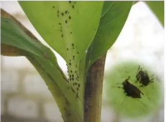
The virus is transmitted vegetatively, through tissue culture and conventional planting material, and by the aphid vector Pentalonia nigronervosa (banana aphid; Figure 6). The virus does not replicate in the aphid and is not transmitted from an infective adult to the newly hatched nymphs. The virus does not replicate in leaves developed prior to the infection. Aphids are thus only able to acquire the virus from symptomatic leaves formed after infection. Recently, P. caladii was also shown to be a vector (Watanabe et al. 2013). No mechanical transmission has been reported.
15
Figure 6. Banana bunchy top aphid vector Pentalonia nigronervosa (C De Clerck).
Detection
The virus can be detected by ELISA (enzyme-linked immunosorbent assay) on samples from field plants, tissue culture plants and infective aphids (Thomas and Dietzgen 1991; Thomas et al. 1995). Monoclonal and polyclonal antibodies are commercially available (Dietzgen and Thomas 1991; Wu and Su 1990). Polymerase chain reaction (PCR) assays are a sensitive alternative for detection (Hu et al. 1996; Mansoor et al. 2005; Sharman et al. 2000b) and DNA probes and primers are available for BBTV DNA components 1 to 6 (Burns et al. 1995; Stainton et al. 2012). Recently real time PCR (Chen and Hu 2013), loop-mediated isothermal amplification (LAMP; Peng et al. 2012b), electro-chemiluminescence PCR (Tang et al. 2007) and impedance spectroscopy (Majumder et al. 2013) based methods have also been described for the detection of BBTV. However, these latter methods have not yet been tested against a genetically diverse range of BBTV isolates to test their wider applicability. Multiplex PCR assays have also been reported (Sharman et al. 2000b; Liu et al. 2012).
The optimal tissue for indexing is the midrib of the youngest leaves, though the virus can also be detected in the leaf lamina, pseudostem, dormant meristems, the bunch stalk, bracts and fruit peel from infected plants. The virus can sometimes be detected in the leaf immediately preceding the youngest symptomatic leaf.
16
Therapy
Meristem or shoot tip culture (<1 to 2 mm), possibly combined with heat therapy (Helliot et al. 2001; Lassois et al. 2013; Ramos and Zamora 1990; Thomas et al. 1995), has been successful in achieving a proportion of virus-free plantlets. Testing after treatment (ideally after an 8−12 month period in the greenhouse) is essential in case the virus is reduced to undetectable levels, but not eliminated, during tissue culture.
5.2. Abaca bunchy top
Cause
Abaca bunchy top virus (ABTV) is a member of the genus Babuvirus (family Nanoviridae) and has a multicomponent single-stranded DNA genome, a
predicted coat protein size of ca. 19.5 Mr, and is presumed to have ca. 20 nm isometric virions similar to BBTV.
Significance
There are no reports on the economic effects of abaca bunchy top disease, though the virus is distantly related to and causes similar symptoms to BBTV, and is expected to have a similar impact on production.
Symptoms
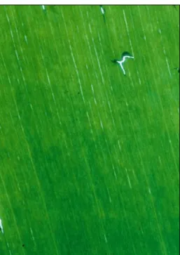
The symptoms caused by ABTV are indistinguishable from those caused by BBTV. Banana cv. Cavendish displays vein clearing and dark green streaking on the leaf lamina, reduced leaf size, bunching of young leaves and plant stunting (Su et al. 2003). Leaf samples from the only abaca plants confirmed to be infected with ABTV displayed vein clearing flecks (Figure 7) and narrow, brittle leaves with chlorotic, upturned margins consistent with descriptions of abaca bunchy top disease in the early literature (Ocfemia 1930).
17
Figure 7. Vein clearing flecks in a banana leaf infected with ABTV (JE Thomas).
Hosts
Abaca (Musa textilis) and banana cv. Cavendish are the only reported hosts.
Geographical distribution
The virus has been reported only from the Philippines and Malaysia (Sarawak). However, some diagnostic ELISA and PCR tests for BBTV will cross-react with ABTV, therefore it is possible that some detections of ABTV may have been mistakenly identified as BBTV.
Transmission
The virus is transmitted vegetatively, through tissue culture and conventional planting material, and by the aphid vector Pentalonia nigronervosa. By analogy to the related BBTV, ABTV is likely to be transmitted in a persistent circulative manner by its aphid vector.
Detection
The virus can be detected by ELISA in field plants. Some commercially available BBTV monoclonal and polyclonal antibodies (from Agdia, Inc., USA) are known to cross-react with ABTV, albeit with a reduced sensitivity.
18
Specific PCR assays are a sensitive and reliable alternative for detection (Sharman et al. 2008). Some PCR primers for BBTV are known to cross-react with ABTV (Su et al. 2003).
ABTV is likely to be phloem limited, and the optimal tissue for indexing is the midrib of the youngest leaves.
Therapy
There are no reports on attempts to eliminate ABTV from infected germplasm, but it is likely that meristem tip culture will be effective, as it is for BBTV (Ramos and Zamora 1990; Thomas et al. 1995; Wu and Su 1991).
5.3. Banana bract mosaic
Cause
The disease is caused by the potyvirus Banana bract mosaic virus (BBrMV). The virus has flexuous filamentous particles about 700−750 nm in length (Figure 8), with a coat protein of ca 39.3 kDa, and a single stranded RNA genome.
Significance
Up to 40% yield loss is reported in the Philippines, where comprehensive roguing/sanitation programmes are implemented (Magnaye 1994). Fruits fail to fill on infected plants in India (DR Jones, unpublished). On export bananas, deformations and streaks on the fruit are a cause for rejection.
Symptoms
Chlorotic spindle-shaped lesions parallel to the veins may occur on the leaf lamina, especially on younger leaves (Figure 9). Dark coloured broad streaks and mosaic patterns on the bracts of the inflorescence are characteristic of the disease (Figure 10). Chlorotic streaks may occur on the peduncles, and a shortening of bunch internodes is also characteristic. After removal of outer leaf sheaths, the presence of irregular coloured stripes or mosaic patterns is diagnostic of the disease (Figure 11). These markings can be chlorotic, yellow or reddish, depending on the pseudostem colour. Greenish to brownish broad, irregularly scattered spindle streaks develop along the petioles (Figure 12).
19
Figure 8. Purified virions of BBrMV; bar represents 400 nm (JE Thomas).
Figure 9 (left). Chlorotic spindle-shaped lesions parallel to the veins on the leaf lamina of a BBrMV-infected banana (JE Thomas).
Figure 10 (right). Dark coloured broad streaks and mosaic patterns on the bracts of the inflorescence of a BBrMV-infected banana (JE Thomas).
20
Figure 11 (left). Irregular stripes and mosaic patterns on the pseudostem of a BBrMV-infected banana, after removal of the outer leaf sheaths (JE Thomas). Figure 12 (right). Irregular streaks and mosaic patterns on the leaf midrib of a BBrMV-infected banana (JE Thomas).
BBrMV symptoms may be masked by other virus infections, and the virus has been recovered from plants with a mild Cucumber mosaic virus (CMV)-like mosaic symptom, often co-infected with CMV, and from banana streak virus (BSV)-infected plants with typical BSV symptoms (Rodoni et al. 1997; 1999). Symptomless infections have also been reported (Feng 2006).
Hosts
Hosts include Musa species and cultivars (Diekmann and Putter 1996), Alpinia
purpurata (Wang et al. 2010) and Elettaria cardamomum (Siljo et al. 2012).
BBrMV infects abaca (Musa textilis) causing symptoms similar to those caused by the abaca strain of Sugarcane mosaic virus (Sharman et al. 2000a; see Section 5.4 on abaca mosaic disease).
Geographical distribution
BBrMV has been reported in the Philippines (Magnaye and Espino 1990), India (where the disease is called ‘Kokkan’) and Sri Lanka (Thomas et al. 1997), Thailand, Western Samoa, Vietnam (Rodoni et al. 1999), Taiwan (Feng 2006)
21
and Hawaii (Wang et al. 2010). Reports from Colombia and Ecuador (Quito-Avila et al. 2013) are yet to be confirmed.
Transmission
No mechanical transmission has been reported in Musa. In addition to transmission through vegetative propagation and tissue culture, transmission by the aphid species Rhopalosiphum maidis, Aphis gossypii and Pentalonia
nigronervosa has been reported (Thomas et al. 2000a).
Detection
Leaf symptoms can be erratic and bract symptoms are evident only during flowering. Dead leaf sheaths must be removed to reveal mosaic and streaking on the pseudostem.
The virus can be detected in extracts of leaf lamina, midribs and flower bracts by ELISA using BBrMV-specific polyclonal and/or monoclonal antibodies. The virus can also be detected by RT-PCR (Iskra-Caruana et al. 2008), immunocapture RT-PCR (Sharman et al. 2000b) or LAMP assays (Siljo and Bhat 2014). Multiplex (IC)-PCR assays are available (Sharman et al. 2000b; Liu et al. 2012).
Therapy
Both meristem culture and heat treatment of shoot cultures have resulted in elimination of BBrMV from infected banana cv. Señorita (Ramos and Zamora 1999).
5.4. Abaca mosaic
Cause
Abaca mosaic is caused by a distinct strain of the potyvirus Sugarcane mosaic
virus (SCMV-Aba; Eloja and Tinsley 1963; Gambley et al. 2004). The flexuous
filamentous particles measure about 680 nm (Figure 13), have a ca. 35 kDa coat protein, and contain a single-stranded RNA genome.
22
Figure 13. Virions of SCMV-Aba; bar represents 100 nm (JE Thomas).
Significance
The disease has been a significant constraint to abaca production in the Philippines. The disease affects fibre yield as well as fibre quality.
Symptoms
Leaves can show yellowish or light green spindle-shaped streaks (Figures 14 and 17) or a mosaic pattern (Figures 15 and 16). Petioles and midribs are mottled with dark green and yellowish streaks, even when no symptoms appear on the leaves (Figure 16) (Thomas and Magnaye 2000).
Figure 14. Spindle-shaped chlorotic lesions caused by SCMV-Aba on banana cv. Lakatan (JE Thomas).
23
Figure 15. Mosaic symptoms caused by SCMV-Aba on abaca (JE Thomas).
Figure 16 (left). Mosaic pattern caused by SCMV-Aba in banana cv. Buhutan (JE Thomas).
Figure 17 (right). Symptoms caused by a mixed infection of SCMV-Aba and BBrMV in abaca (JE Thomas).
Hosts
Natural hosts include Musa textilis (abaca, Manila hemp), Marantha
arundinacea and Canna indica.
There are several experimental hosts, including banana and some species in the Poaceae (Eloja and Tinsley 1963).
24
Geographical distribution
Abaca mosaic disease has been reported only in the Philippines (Eloja and Tinsley 1963).
Transmission
The virus is transmitted by vegetative propagation and tissue culture, as well as by aphids (mainly Rhopalosiphum maidis and Aphis gossypii) in a non-persistent manner (Gavarra and Eloja 1969; Ocfemia and Celino 1938). It is mechanically transmitted with difficulty (Dante A Benigno and Del Rosario 1965; Eloja et al. 1962).
Detection
The virus can be detected by ELISA using antibodies prepared against the abaca strain of SCMV (Gambley et al. 2004), or by RT-PCR (ADW Geering et al., unpublished) – see Appendix 1.
Therapy
No information reported.
5.5. Banana mosaic
Cause
The disease is caused by Cucumber mosaic virus (CMV) belonging to the genus
Cucumovirus, family Bromoviridae. CMV has icosahedral particles 28−30 nm in
diameter (Figure 18), separately encapsidating the tripartite single-stranded RNA genome components. Subgroup I and II isolates of CMV have both been recorded from banana.
Figure 18. Virions of CMV; bar represents 200 nm (Rothamsted Research, UK).
25
Significance
Most strains of the virus are so-called ‘common’ strains, which do not produce severe symptoms or cause significant crop damage (Lockhart and Jones 2000a), though sometimes bunches may be distorted or bear smaller or fewer fruit. However, some severe strains are more damaging, with infection resulting in internal pseudostem necrosis (Figure 19) and even plant death (Niblett et al. 1994). The heart-rot strain found in Morocco is particularly destructive (Bouhida and Lockhart 1990). Plantlets derived from tissues culture are more prone to infection.
Hosts
CMV is known to infect over 800 different plant species, including cultivated crops (e.g. tomato, pepper and cucurbits) and weeds (e.g. Commelina), which serve as reservoirs for infection of banana.
Geographical distribution
CMV has a cosmopolitan distribution, i.e. it is found on all continents and in many countries, but some strains causing severe symptoms, e.g. heart-rot strain, are limited in distribution.
Symptoms
Symptoms are very variable, and include foliar mosaic (Figure 20), chlorotic streaking or flecking (Figure 21), and occasional leaf deformation, especially in young suckers developing from infected mother plants (Lockhart 2002). Uneven ripening has been also associated with the virus. Infected plants may show no symptoms, particularly suckers. In some varieties, high temperature may suppress symptoms. Symptoms have often been confused with those of BSV.
Figure 19. Internal necrosis (heart rot) symptoms in a CMV-infected banana plant (BEL Lockhart).
26
Figure 20. (left) Foliar mosaic streaking in CMV-infected leaves (JE Thomas). Figure 21. (right) Chlorotic streaking in CMV-infected leaves (JE Thomas).
Transmission
CMV is transmitted from plant to plant in a non-persistent manner by many aphid species including Aphis gossypi, Myzus persicae, Rhopalosiphum maidis and R. prunifoliae. These aphids colonize a wide range of plant species and visit but do not colonize banana. As a result, CMV infection of banana occurs almost exclusively by aphid transmission from other plant species rather than from banana to banana (Lockhart 2002). The virus can also be spread over long distances during traditional vegetative propagation using suckers or during in
vitro multiplication. Seed transmission has also been reported (Gold 1972).
Detection
A range of techniques can be used to detect CMV, including observation of symptoms, electron microscopy, the use of indicator plants, nucleic acid hybridization, serology and PCR. The latter two are recommended for reliable and sensitive routine detection of CMV. A number of ELISA kits are commercially available and several PCR assays have been described (e.g. Hu et al. 1995; Sharman et al. 2000b; Wylie et al. 1993), including multiplex assays (Sharman et al. 2000b; Liu et al 2012).
Therapy
Different methods have been developed and tested for CMV eradication, such as thermotherapy, chemotherapy, meristem culture and cryotherapy. A combination of these techniques can improve the eradication rate (Gupta 1986; Helliot et al. 2002; 2004). The efficiency of the eradication technique depends on (i) the virus strain characteristics, (ii) the type of tissue treated and (iii) the banana genotype. However, meristem culture from heat-treated in
27
vitro plantlet is recommended for best results. Protocols are available in
Lassois et al. (2013).
5.6. Banana streak
Cause
Banana streak disease is caused by a group of related viruses (banana streak viruses, BSV) which are plant pararetroviruses belonging to the genus
Badnavirus of the family Caulimoviridae (King et al. 2012). The viruses have
non-enveloped bacilliform particles measuring 120−150 30 nm (Figure 22). Particles contain a non-covalently closed circular double-stranded DNA genome of approximately 7.4 kb made of three different open reading frames (King et al. 2012). BSV display important serological and molecular variability with sometimes more than 20% nucleotide differences in the most conserved RT-RNase H region of the genome. They are considered different badnavirus species, all causing the same disease. BSV exist in two states: as an episomal form, producing infection in the plant cells, or as viral DNA integrated within the B genome of banana. Both forms can be infectious in banana plants.
Figure 22. Electron micrograph of Banana streak Mysore virus particles (J Vo).
Significance
Few quantitative studies are known. Disease incidence varies between countries and this may be related to species differences, banana genotypes and/or vector activity. There is the potential for serious yield losses with some isolates (Lassoudière 1974). Plant death has been reported in Africa. Studies in
28
Australia with Banana streak Cavendish virus have demonstrated yield losses of 6−11% (Daniells et al. 2001).
Symptoms
Symptoms vary with isolates and cultivars. Most isolates produce broken or continuous chlorotic streaks or spindle-shaped patterns which are first chlorotic, then become increasingly dark in colour, and finally result in necrotic streaking in older leaves (Figure 23B and C). A range of other symptoms have been observed including failure of bunch emergence, splitting of the pseudostem (Figure 23A), and a ‘traveller’s palm’ leaf arrangement (Daniells et al. 2001; Lockhart and Jones 2000b). Some isolates of BSV occurring in Africa produce severe necrosis, which begins with the cigar leaf and results in internal pseudostem necrosis and plant death. Other isolates produce very fine, indistinct broken brown interveinal streaks or pinpoints. Bunches may be reduced in size. Symptomless infection occurs frequently. Symptoms can appear sporadically, and may be absent for many months before reappearing. Symptom appearance and severity are associated with temperature changes and physiological age, but the precise correlation has not been experimentally determined. Symptoms are often confused with those caused by CMV.
Hosts
Musa species and cultivars are the natural hosts.
29
Figure 23. (A) Splitting of the pseudostem associated with BSV infection on plantain. (B) Discrete leaf symptoms associated with BSV on young leaves of a plantain (AAB). (C) Chlorotic and necrotic symptoms of BSV on an older leaf of the same plantain. (Photographs taken at the Centre Africain de Recherches sur Bananiers et Plantains (CARBAP) in Cameroon by M Chabannes.)
A
B
30
Geographical distribution
Figure 24 shows the distribution of BSV. The affected countries are also listed below (*unconfirmed – this status conferred by the quoted author).
Figure 24. Distribution of BSV (affected countries shown in red).
Africa
Benin* (Pasberg-Gauhl et al. 1996)
Cameroon* (Gauhl et al. 1997; Lockhart 1995) Cape Verde * (Lockhart 1995)
Côte d’Ivoire* (Lassoudière 1974, M-L Caruana, unpublished) Ghana* (Pasberg-Gauhl et al. 1996)
Guinea Bissau* (Lockhart 1995) Kenya* (Lockhart 1995)
Madagascar* (Jones and Lockhart 1993) Malawi* (Vuylsteke et al. 1996)
Mauritius* (Jaufeerally-Fakim et al. 2006) Morocco* (Lockhart 1986)
31
Nigeria* (Gauhl et al. 1999; Pasberg-Gauhl et al. 1996) Rwanda* (Sebasigari and Stover 1988)
Sierra Leone (Lockhart 1995)
South Africa* (Jones and Lockhart 1993) Tanzania* (Sebasigari and Stover 1988) Togo (Lockhart 1995)
Uganda* (Dabek and Waller 1990; Geering et al. 2005; Harper et al. 2005) Zanzibar* (Vuylsteke et al. 1998)
Asia
China, Peoples’ Republic* (Zhuang et al. 2011) India* (Cherian et al. 2004)
Indonesia (New Guinea)* (Davis et al. 2000; Lockhart 1995) Jordan (Lockhart 1995)
Malaysia (Ang and Ong 2000) Philippines* (Lockhart 1995) Sri Lanka* (Lockhart 1995) Taiwan* (Su et al. 1997)
Thailand* (Aung et al. 2012; Lockhart 1995) Vietnam* (Lheureux et al. 2007; Lockhart 1995) Europe
Spain (Canary Islands) (M-L Caruana, unpublished) Portugal (Madeira) (Jones and Lockhart 1993) Oceania
Australia* (Thomas et al. 1994) New Caledonia* (Lockhart 1995) Papua New Guinea (Davis et al. 2000) Tonga* (Thomas et al. 1994)
32 South and Central America
Brazil* (Carnelossi et al. 2014) Colombia* (Reichel et al. 2003)
Costa Rica* (Pasberg-Gauhl et al. 2000)
Cuba* (Javer et al. 2009; Jones and Lockhart 1993) Dominican Republic* (P-Y Teycheney et al., unpublished) Ecuador* (Jones and Lockhart 1993)
Grenada (Jones and Lockhart 1993) Guadeloupe* (Jones and Lockhart 1993) Honduras* (Jones and Lockhart 1993) Jamaica (Jones and Lockhart 1993) Martinique* (Jones and Lockhart 1993) Nicaragua (Lockhart 1995)
Peru* (Pasberg-Gauhl et al. 2007) Trinidad* (Jones and Lockhart 1993)
USA (Florida, Puerto Rico, Virgin Islands)* (Lockhart 1995) Venezuela* (Garrido et al. 2005)
Transmission
Two means of transmission have been observed: horizontal and vertical transmission.
Horizontal transmission
BSV cannot be transmitted to Musa by mechanical inoculation. Field spread is mainly due to the citrus mealybug Planococcus citri. Sugarcane bacilliform virus (ScBV), which is closely related serologically to BSV, can be transmitted from infected sugarcane to banana by P. citri and the pink sugarcane mealybug (Saccharicoccus sacchari) in laboratory conditions, and produces typical streak symptoms (Lockhart and Autrey 1988). Such transmission has never been reported in field conditions. The mealybugs Dysmicoccus brevipes and
33 Vertical transmission
The virus is transmitted by vegetative propagation to 100% of progeny plants and the use of infected suckers contributes to the spread of the disease. Since the late 1990s, numerous spontaneous outbreaks of banana streak disease have been observed in many banana-producing areas in banana breeding lines and micropropagated interspecific Musa hybrids originating from virus-free parents. The origin of these outbreaks was correlated with the presence of endogenous sequences of banana streak viruses (eBSV) integrated into the Musa balbisiana genome of the hybrids and cultivars. Infectious eBSV contain the complete functional viral genome. Three natural widespread BSV species have so far been identified as infectious integrants arising from the M.
balbisiana genome: Banana streak OL virus (BSOLV), Banana streak IM virus (BSIMV) and Banana streak GF virus (BSGFV)(Chabannes et al. 2013; Gayral et al. 2008; Iskra-Caruana et al. 2010). In banana, abiotic stresses such as micropropagation by in vitro culture processes (Côte et al. 2010; Dallot et al. 2001), temperature differences or water stresses and genetic crosses (Lheureux et al. 2003) are known to contribute to triggering the activation of eBSV to form infectious, episomal BSV.
The presence of infective eBSV species in most B genomes poses a unique problem for the distribution of germplasm. No methods for inactivating these eBSVs are currently available. Therefore, a strategy has been proposed and accepted by the research community for the distribution of this germplasm from the ITC, which minimizes the risks associated with the distribution of BSV, especially non-eBSVs, to the recipient country. Details of this strategy can be found at www.musanet.org.
There is evidence that BSV is seed-transmitted in Musa (Daniells et al. 1995), though this work was conducted before the phenomenon of integration was discovered. This work involved the use of embryo-rescued hybrids of cv. Mysore (AAB) × I.C.1 (AAAA), and the infection of progeny through activation of integrants, rather than through seed transmission, cannot be discounted.
Detection
The main difficulty in diagnosing BSV is to distinguish active infections (episomal) from integrated viral sequences. Although infectious eBSV are only present in the M. balbisiana genome, all Musa spp. also carry partial or ‘dead’ integrated BSV sequences in their genomes. Thus PCR tests on any banana DNA can result in BSV-positive reactions. Diagnostic PCRs must be designed to accommodate this problem.
34
Serological detection of BSV is complicated by the wide serological diversity among virus isolates, some of which are serologically unrelated (Lockhart and Olszewski 1993). A polyvalent polyclonal antiserum raised against BSV species and Sugarcane bacilliform virus species (Ndowora 1998) is capable of detecting all known isolates by immunosorbent electron microscopy (ISEM) in partially purified extracts, even in asymptomatic leaf tissue (BEL Lockhart, unpublished). Plants derived from in vitro material should be tested at least 3 months after plantlets have been acclimatised and planted out.
Detection of BSV by ELISA
ELISA test kits for BSV are commercially available from Agdia and DSMZ companies. Samples of laminar and midrib tissue from the three youngest expanded leaves, or symptomatic tissue if present, should be tested by ELISA. Detection of BSV by ISEM
A partial purified extract is used to observe virus particles under the electron microscope (Geering et al. 2000).
Detection of BSV by IC-PCR
Immunocapture PCR (IC-PCR) to detect specific BSV species has been described (Geering et al. 2000; ADW Geering unpublished – see Appendix 1; Le Provost et al. 2006). Primers for the detection of various BSV species are shown in Appendix 1. Immunocapture conditions were optimized to minimize non-specific binding of plant genomic DNA to the immunocapture tubes. Alternatively, to avoid contamination by plant genomic DNA, a DNAse I treatment is performed (Appendix 1).
Detection of BSV by RCA
Rolling circle amplification (RCA) is a technique that selectively amplifies circular DNA (such as encapsidated BSV genomic DNA) resulting in a specific amplification of DNA from episomal forms of BSV only. James et al. (2011) have developed and used RCA with the addition of BSV-specific primers for the detection of episomal BSV, and demonstrated the ability of this method to differentiate between episomal and integrated viral genomic sequences. This method avoids the need for antiserum, but does require knowledge of the virus sequence in the selection of restriction enzymes required for visualization of the virus-specific products. Identification of a particular BSV also requires subsequent PCR or sequencing.
35 Detection of BSV by LAMP
Loop-mediated isothermal amplification (LAMP) assay is a DNA amplification technique that does not require thermal cycling (Notomi et al. 2000). LAMP products can be detected using conventional agarose gel electrophoresis or using visual observation to estimate turbidity or colour changes (Mori et al. 2001). Peng et al. (2012a) have used the LAMP assay to detect BSV particles in infected banana plants. However this technique cannot differentiate between integrated and episomal forms so false positives could be very frequent. Also, primer design is complicated and is likely to be a constraint with the genetic diversity found in germplasm indexing.
Detection of eBSV
Based on the sequences and the structure of the different eBSV described in the seeded diploid (BB) M. balbisiana Pisang Klutuk Wulung (PKW), several PCR markers have been developed in order to genotype those integrations in different banana genotypes (Chabannes et al. 2013; Gayral et al. 2008; 2010), including (i) integration markers targeting eBSV insertion sites, (ii) structure markers targeting internally reorganized eBSV structures and (iii) allelic markers which allow the distinction between alleles of the same eBSV species. The targeted eBSV, the type of marker developed, the name and the sequence of the primer as well as the size of the PCR product are described in Chabannes et al. (2013).
Therapy
Cryopreservation followed by apical meristem culture has a high rate of success in eliminating episomal BSV infections in cv. Williams, a banana genotype free of eBSV (Helliot et al. 2002). Two retroviral and anti-hepadnavirus molecules have also been effective in eradicating the episomal form of BSV from banana plants (Helliot et al. 2003). Methods are described in Lassois et al. (2013).
5.7. Banana mild mosaic
Cause
The disease is caused by Banana mild mosaic virus (BanMMV), an unassigned virus in the Betaflexiviridae. The virus has flexuous filamentous particles about 580 nm in length (Figure 25), with a coat protein of ca 26.8 kDa, and a single stranded RNA genome (Gambley and Thomas 2001; Thomas et al. 2000b).
36 Figure 25. Virions of BanMMV (JE Thomas).
Significance
The economic impact of this virus has not been quantified, but observations suggest that yield may be affected in both banana and plantain in Latin America (Thomas et al. 2000b).
Symptoms
In many cases infected plants are symptomless, and in other cases symptoms are transitory. In cv. Ducasse (syn. Pisang Awak), chlorotic streaks and mosaic (Figure 26) may be present, especially on younger leaves, but these symptoms often disappear as the plant matures. Other cultivars, including Pisang Seribu, show fine silvery streaks on the leaves (Figure 27). Mixed infections with BSV are very common, and in these cases symptoms are dominated by those typical of BSV (JE Thomas and KS Parmenter, unpublished). There was a high correlation between leaf necrosis symptoms and mixed infections of BanMMV and Cucumber mosaic virus (CMV) in Guadeloupe, compared to mosaic symptoms with CMV infection alone (Caruana and Galzi 1998). BBrMV-infected samples from the Philippines were frequently found to be mixed infections with BanMMV (M-L Caruana, unpublished).
Figure 26. Mosaic symptoms on a young leaf of banana cv. Ducasse (syn. Pisang Awak) (JE Thomas).
37
Figure 27. Chlorotic and silvery streaks on the leaf of a BanMMV-infected banana cv. Rose (JE Thomas).
Hosts
Hosts of BanMMV include Musa species and cultivars (Thomas et al. 2000b).
Geographical distribution
BanMMV is widespread and probably present in most banana-producing countries.
Transmission
BanMMV is transmitted through vegetative propagation and tissue culture. No mechanical transmission has been reported in Musa and no natural vectors have been identified. Attempts to transmit the virus from single infections in cv. Ducasse were unsuccessful with mealybugs (Planococcus citri), aphids (Pentalonia nigronervosa), via soil collected from around infected plants, or by root-to-root contact (M Sharman and JE Thomas, unpublished). However, circumstantial evidence for plant-to-plant spread by an unknown mechanism has been noted in Guadeloupe. Initially virus-free planting material was shown after field planting to be subsequently infected with CMV and BanMMV, implying co-transmission of the viruses in the field (Caruana and Galzi 1998). Also, analysis of 154 partial RdRp sequences of BanMMV from 68 banana accessions supported the possibility of horizontal transfer of the virus between plants. In general the sequences of different isolates displayed a high level of diversity (mean 20.4%), whereas the variability within individual isolates seemed to be only about 2%. Identical sequences were obtained from two independent pairs of plants, and there was a very steep gradient separating plants sharing identical or near identical sequences. About three-quarters of these plants were located 1−5 metres apart (Teycheney et al. 2005a).
38
Detection
Polyclonal antisera to BanMMV have been developed (Teycheney et al. 2007; JE Thomas, unpublished), though they are not commercially available. These antisera can be used in immunocapture RT PCR (see below) or in ISEM.
BanMMV displays a high degree of genetic diversity making detection by PCR particularly challenging. A set of 154 partial RdRp sequences showed a mean pairwise nucleotide sequence divergence of 20.4% (Teycheney et al. 2005a) and similarly, a set of 14 complete coat protein sequences showed a mean of 20.6% divergence (JE Thomas, unpublished). A group of partial CP sequences, primarily from the Ivory Coast, was particularly variable both compared with all other isolates (mean 35%) and within the group itself (mean 35%).
Teycheney et al. (2007) have described a nested immunocapture RT PCR assay for the detection of BanMMV, targeting the RdRp region of the flexiviridae genome and based on the primers of Foissac et al. (2005). This assay is not specific for BanMMV, but does not detect Banana virus X (Teycheney et al. 2007).
Alternative primers, targeting the coat protein gene (upstream primer) and the poly-A tail (downstream primer), have been developed (Gambley et al. 1999; Teycheney et al. 2005a; M Sharman and JE Thomas, unpublished). The published upstream primer sequence of BanCP2 (Teycheney et al. 2005a) contains a probable typographical error and should read 5´-TATGCNGCNTTYGAYTTCTTRGAYG-3´ at positions 7042−7066 on the BanMMV genome. However this primer has from one to three mismatches within the first eight 3´ bases in 23 of 54 available partial CP sequences on GenBank. Upstream primer BanMMV CP2 (3´-TGCCAACTGAYGARGAGCTRAATGC-5´; M Sharman, unpublished; see protocol in Appendix 1) similarly has mismatches in the first seven bases against 12 of 54 sequences. However, there is good correlation between ISEM results and IC RT PCR results when the latter primer has been used for extensive testing of banana germplasm (KS Crew and JE Thomas, unpublished). The latter primer has been modified to eliminate the majority of mismatches in the 3´ end (BanMMV CP8; 5´-TGCCAACTGAYGARGARYTRAAHGC-3´; JE Thomas, unpublished).
Therapy
BanMMV has been eliminated from cvs. Pisang Awak and Pisang Seribu using meristem culture and chemotherapy with a number of chemicals (of which ribavirin was clearly the most effective; Busogoro et al. 2006). Meristem culture alone has inadvertently eliminated BanMMV from selections of cv. Ducasse (syn. Pisang Awak) (SD Hamill, personal communication).
39
5.8. Banana virus X
Cause
The disease is caused by Banana virus X (BVX), an unassigned virus in the
Betaflexiviridae (Teycheney et al. 2005b).
Significance
The economic significance is not known.
Hosts
The only known hosts are Musa spp. (Teycheney et al. 2005b).
Geographical distribution
BVX has been recorded only in Guadeloupe. However, the virus was relatively common in randomly sampled symptomless plants, and was associated with a
Musa germplasm collection (Teycheney et al. 2005b). Although no surveys
have been reported, it is likely to occur more widely in banana-growing countries.
Symptoms
All detections of the virus to date have been in symptomless plants.
Transmission
BVX is not transmitted by sap inoculation to a number of herbaceous test plant species (Teycheney et al. 2005b). No vector has been reported, but the virus is presumably transmitted through vegetative propagation, including tissue culture.
Detection
A nested RT PCR has been reported for the detection of BVX (Teycheney et al. 2007).
Therapy
40
6. References
Adegbola RO, Ayodeji O, Awosusi OO, Atiri GI, Kumar PL (2013) First report of Banana bunchy top virus in banana and plantain (Musa spp.) in Nigeria. Plant Disease 97, 290.
Ang O, Ong M (2000) Virus and virus-like diseases of banana and citrus in Malaysia: status and control strategies. In: Workshop on Disease Management of Banana and Citrus Through the Use of Disease-free Planting Materials, 14−16 October 1998, Davao City, Philippines. Bioversity International.
Aung B, Swangpol S, Somana J (2012) Record on the occurrence of Banana Streak Virus in Thailand during the survey from 2007 to 2011. In: 2012 CU-MUSC Graduate Forum in Plant Biotechnology, Faculty of Science, Mahidol University, Bangkok, Thailand.
Bakry F (2008) Zygotic embryo rescue in bananas. Fruits 63, 111−115.
Bananej K, Ghotbi T, Vahdat A (2007) First report of Banana bunchy top virus infecting banana in Iran. Plant Pathology 56, 719.
Beaver RG (1982) Use of picloram for eradication of banana diseased with bunchy top. Plant Disease 66, 906−907.
Bouhida M, Lockhart BE (1990) Increase in importance of cucumber mosaic virus infection in greenhouse-grown bananas in Morocco. Phytopathology 80, 981.
Buddenhagen IW (1968) Banana diseases in the Pacific area. FAO Plant Protection Bulletin 16, 17−31.
Burns TM, Harding RM, Dale JL (1995) The genome organization of banana bunchy top virus: analysis of six ssDNA components. Journal of General Virology 76, 1471−1482.
Busogoro J-P, Vandermolen M, Masquelier L, Jijakli H (2006) Development of a chemotherapy protocol to sanitise banana genotypes infected by banana mild mosaic virus (BanMMV). In: XVII Reuniao Internacional da Associação para a Cooperação nas Pesquisas sobre Banana no Caribe e na América Tropical (ACORBAT), pp. 752−756. Joinville -Sanla Catarina, Brazil.
Campbell JG (1926) Annual Report of the Fiji Department of Agriculture (1925). Carnelossi PR, Bijora T, Facco CU, Silva JM, Picoli MHS, Souto ER, Oliveira FTd (2014) Episomal detection of Banana streak OL virus in single and mixed infection with Cucumber mosaic virus in banana ‘Nanicão Jangada’. Tropical Plant Pathology 39, 342−346.
41
Caruana ML, Galzi S (1998) Identification of uncharacterised filamentous viral particles on banana plants. Acta Horticulturae 490, 323−335.
Castillo BS, Martinez AL (1961) Occurrence of bunchy top disease of banana in the Philippines. FAO Plant Protection Bulletin 9, 74−75.
Chabannes M, Baurens F-C, Duroy P-O, Bocs S, Vernerey M-S, Rodier-Goud M, Barbe V, Gayral P, Iskra-Caruana M-L (2013) Three infectious viral species lying in wait in the banana genome. Journal of Virology 87, 8624−8637.
Chen Y, Hu XP (2013) High-throughput detection of banana bunchy top virus in banana plants and aphids using real-time TaqMan (R) PCR. Journal of Virological Methods 193, 177−183.
Cherian A, Baranwal V, Malathi V, Pant R, Ahlawat Y (2004) Banana streak virus from India and its detection by polymerase chain reaction. Indian Journal of Biotechnology 3.
Côte FX, Galzi S, Folliot M, Lamagnere Y, Teycheney PY, Iskra-Caruana ML (2010) Micropropagation by tissue culture triggers differential expression of infectious endogenous Banana streak virus sequences (eBSV) present in the B genome of natural and synthetic interspecific banana plantains. Molecular Plant Pathology 11, 137−144.
Dabek AJ, Waller JM (1990) Black leaf streak and viral leaf streak: new banana diseases in East Africa. Tropical Pest Management 36, 157−158.
Dallot S, Acuña P, Rivera C, Ramírez P, Côte F, Lockhart BEL, Caruana ML (2001) Evidence that the proliferation stage of micropropagation procedure is determinant in the expression of Banana streak virus integrated into the genome of the FHIA 21 hybrid (Musa AAAB). Archives of Virology 146, 2179−2190.
Daniells J, Thomas JE, Smith M (1995) Seed transmission of Banana streak virus confirmed. Infomusa 4(1), 7.
Daniells JW, Geering ADW, Bryde NJ, Thomas JE (2001) The effect of Banana
streak virus on the growth and yield of dessert bananas in tropical Australia.
Annals of Applied Biology 139, 51−60.
Dante A Benigno DA, Del Rosario MSE (1965) Mechanical transmission of the abaca mosaic virus and some of its physical properties. The Philippine Agriculturist 49, 197−210.
Davis RI, Geering ADW, Thomas JE, Gunua TG, S. Rahamma (2000) First records of Banana streak virus on the island of New Guinea. Australasian Plant Pathology 29, 281.
42
Diekmann M, Putter CAJ (1996) FAO/IPGRI Technical Guidelines for the Safe Movement of Germplasm. Musa spp. (2nd Ed). Food and Agriculture Organization of the United Nations/International Plant Genetic Resources Institute, Rome.
Dietzgen RG, Thomas JE (1991) Properties of virus-like particles associated with banana bunchy top disease in Hawaii, Indonesia and Tonga. Australasian Plant Pathology 20, 161−165.
Eloja AL, Tinsley TW (1963) Abaca mosaic virus and its relationship to sugarcane mosaic. Annals of Applied Biology 51, 253−258.
Eloja AL, Velasco LG, Agati JA (1962) Studies on the abaca mosaic disease 1. Sap transmission of the abaca mosaic virus. The Philippine Journal of Agriculture 27, 75−84.
End MJ, Daymond AJ, Hadley P (Eds) (2010) Technical Guidelines for the Safe Movement of Cacao Germplasm (Revised from FAO/IPGRI Technical Guidelines No. 20). Global Cacao Genetic Resources Network (CacaoNet)/Bioversity International, Montpellier, France.
Espino RC, Magnaye LV, Johns AP, Juanillo C (1993) Evaluation of Philippine banana cultivars for resistance to bunchy-top and fusarium wilt. In: RV Valmayor, SC Hwang, R Ploetz, SW Lee and VN Roa (Eds), Proceedings of an International Symposium on Recent Developments in Banana Cultivation Technology, 14−18 December 1992, Chiuju, Pingtung, Taiwan, pp. 89−102. INIBAP/ASPNET, Los Ba¤os, Laguna, Philippines.
FAO (1996) International Standards for Phytosanitary Measures No. 5. Glossary of Phytosanitary Terms. FAO, Rome, 23 pp.
Feng Y-C (2006) Identification and survey of Banana bract mosaic potyvirus (BBrMV), a new banana virus in Taiwan. Masters Thesis, National Taiwan University, 88pp.
Ferreira SA, Trujillo EE, Ogata DY (1989) Bunchy top disease of bananas. Commodity Fact Sheet BAN-4(A), FRUIT, Hawaii Cooperative Extension Service, Hawaii Institute of Tropical Agriculture and Human Resources, University of Hawaii at Manoa.
Foissac X, Svanella-Dumas L, Gentit P, Dulucq MJ, Marais A, Candresse T (2005) Polyvalent degenerate oligonucleotides reverse transcription-polymerase chain reaction: A polyvalent detection and characterization tool for trichoviruses, capilloviruses, and foveaviruses. Phytopathology 95, 617−625.
Fouré E, Manser PD (1982) Note sur l'apparition au Gabon d'une grave maladie virale des bananiers et plantains: le Bunchy top. Fruits 37, 409−414.