UNIVERSITÉ DE MONTRÉAL
BLADDER VOLUME DECODING FROM AFFERENT NEURAL
ACTIVITY
ARNALDO MENDEZ
INSTITUT DE GÉNIE BIOMÉDICAL ÉCOLE POLYTECHNIQUE DE MONTRÉAL
THÈSE PRÉSENTÉE EN VUE DE L’OBTENTION DU DIPLÔME DE PHILOSOPHIAE DOCTOR
(GÉNIE BIOMÉDICAL) AOÛT 2013
UNIVERSITÉ DE MONTRÉAL
ÉCOLE POLYTECHNIQUE DE MONTRÉAL
Cette thèse intitulée :
BLADDER VOLUME DECODING FROM AFFERENT NEURAL
ACTIVITY
présentée par : MENDEZ Arnaldo
en vue de l’obtention du diplôme de : Philosophiae Doctor a été dûment acceptée par le jury d’examen constitué de :
M. SAVARD Pierre, Ph.D., président
M. SAWAN Mohamad, Ph.D., membre et directeur de recherche M. MATHIEU Pierre-A., D.Sc.A, membre
DÉDICACE
ACKNOWLEDGEMENTS
I would like to express my immense gratitude to my advisor Professor Mohamad Sawan, first for believing in me since the very beginning, for his support, encouragement, generosity and advice during my Ph.D. studies in École Polytechnique de Montréal. I also thank Prof. Sawan for giving me the opportunity to work on such a difficult but fascinating subject in which we strongly believe.
I would also like to specially say thank you to Dr. Tomonori Minagawa, from Shinshu University in Japan, and Dr. Jean-Jacques Wyndaele, from University of Antwerp in Belgium, for the realization of the very difficult animal experimentation conducted at the Urology Department in University of Antwerp that provided us with neural data from the bladder. I also express my gratitude to Abrar Belghith from Enseirb-Matmeca University in France for her support in the VHDL-programming of the DSP during her internship at Polystim Neurotechnologies Laboratory. I would also like to thank my professors from École Polytechnique and Université de Montréal for allowing me to update and enhance my knowledge and evolve in a context of high-level research. I also thank sincerely Fayçal Mounaim, Marie-Yannick Laplante and Laurent Mouden for their valuable support through these years at Polytechnique. I am also grateful for support from the Canada Research Chair in Smart Medical Devices and le Fonds Québécois de la Recherche sur la Nature et les Technologies (FQRNT).
Finally, but not the least, I infinitely thank my family for their unconditional love and support, especially my wife and my three children who have waited with patience and love for Daddy during these years I have worked hard to achieve the results shown in this thesis.
RÉSUMÉ
Lorsque les fonctions de stockage et de miction de la vessie échouent à la suite de traumatismes médullaires, ou en raison d'autres maladies neurologiques, de conditions de santé ou au vieillissement, des complications graves pour la santé du patient se produisent. Actuellement, il est possible de restaurer partiellement les fonctions de la vessie chez les patients réfractaires aux médicaments à l'aide des neurostimulateurs implantables. Pour améliorer l'efficacité et la sécurité de ces neuroprothèses, il faut un capteur de la vessie capable de détecter l’urine stockée afin de mettre en place un système en boucle fermée qui applique la stimulation électrique uniquement lorsque nécessaire. Le capteur peut également servir à aviser les patients ayant des sensations affaiblies pour les aviser en temps opportun le moment où la vessie doit être vidée ou quand un volume résiduel postmictionnel anormalement élevé reste après une miction incomplète. Dans cette thèse, on présente de nouvelles méthodes de mesure, ainsi qu’un processeur de signal numérique dédié pour décoder en temps réel le volume de la vessie à partir des enregistrements neuronaux afférents provenant des récepteurs naturels présents dans la paroi de la vessie. Nos principales contributions sont rapportées dans trois articles de journaux avec comité de lecture. On présente d'abord une revue exhaustive de la littérature comprenant des articles de journaux, des brevets et les livres les plus réputés portant sur l'anatomie, la physiologie et la physiopathologie du tractus urinaire inférieur ainsi que sur la mesure du volume ou la pression de la vessie. Cette étude nous a permis d'identifier les besoins qu'un capteur de la vessie doit satisfaire pour être utilisé dans des applications chroniques telles que celles proposées dans cette thèse. On présente aussi le résultat d’une analyse exhaustive des caractéristiques anatomiques et physiologiques de la vessie que nous avons identifiées d’avoir exercé une influence, ou même d’avoir empêché, la réalisation d'un tel capteur dans des études faites au cours des dernières années. Sur la base de cette étude et de l'évaluation systématique des méthodes de mesure pour la vessie, on a conclu que le principe de mesure le mieux adapté pour la surveillance chronique du volume de la vessie était la détection, la discrimination et le décodage de l'activité neuronale afférente découlant des récepteurs spécialisés du volume (mécanorécepteurs), au sujet desquels certains auteurs ont émis l'hypothèse de leur existence dans la muqueuse interne de la vessie. Ensuite, on présente la méthode de mesure qui permet d'estimer en temps réel le volume de la vessie à partir de l'activité afférente des mécanorécepteurs. Notre méthode a été validée avec les
données acquises à partir de rats anesthésiés dans des expériences aiguës. Il a été possible d'estimer qualitativement trois états de remplissage de la vessie dans 100 % des essais où l'activité afférente enregistrée présentait un coefficient de corrélation de Spearman supérieur ou égal à 0,6. Par ailleurs, on a pu estimer quantitativement le volume de la vessie, et aussi sa pression, en utilisant des fenêtres de temps convenablement choisies. L'erreur moyenne d'estimation du volume fut de 5,8 ± 3,1 %. Nos résultats nous ont également permis de faire la lumière sur un sujet controversé concernant le type de réponses détectables à partir d'enregistrements afférents de la vessie. Nous avons démontré qu'il était possible de quantifier autant les réponses phasiques que les réponses toniques de la vessie lors de son remplissage lent et lors des mesures isovolumétriques, respectivement.
Enfin, on présente un processeur de signal numérique dédié (DSP, sigle en anglais) capable de surveiller le volume de la vessie en exécutant les méthodes de mesure qualitative et quantitative proposées. Le DSP exécute en temps réel la détection et la discrimination (classification) des potentiels d'action extracellulaires (PAEs) suivies par le décodage neuronal pour estimer soit trois niveaux qualitatifs de remplissage ou la valeur du volume de la vessie, en fonction du mode de sortie sélectionné. Le DSP proposé a été testé en utilisant des signaux synthétiques réalistes et des signaux réels de nerfs afférents de la vessie enregistrés au cours des expériences aiguës avec des modèles animaux. Le circuit de traitement pour faire la détection et discrimination des PAEs a donné une exactitude moyenne de 92% en utilisant des signaux contenant des PAEs avec formes d'onde fortement corrélées et avec un faible rapport signal sur bruit. Les circuits d'estimation du volume, qui ont été testés avec des signaux réels, ont reproduit les valeurs d’exactitude obtenues lors des simulations faites hors ligne en utilisant Matlab, c’est-à-dire, 94 % et 97 % pour les estimations quantitatives et qualitatives, respectivement. Pour évaluer la faisabilité, le DSP a été déployé dans le FPGA Actel Igloo AGL1000V2, qui a montré une consommation de puissance de 0,5 mW et une latence de 2,1 ms à une fréquence d’opération de 333 kHz. Ces performances démontrent qu'un capteur de la vessie implantable qui réalise la détection, la discrimination et le décodage de l'activité neuronale afférente est faisable.
ABSTRACT
Failure of the storage and voiding functions of the urinary bladder due to spinal cord injury (SCI), neural diseases, health conditions, or aging, causes serious complications in a patient's health. Currently, it is possible to partially restore bladder functions in drug-refractory patients using implantable neurostimulators. Improving the efficacy and safety of these neuroprostheses used for bladder functions restoration requires a bladder sensor (BS) capable of detecting urine volume in real-time to implement a closed-loop system that applies electrical stimulation only when required. The BS can also trigger an early warning to advise patients with impaired sensations when the bladder should be voided or when an abnormally high post-voiding residual volume remains after an incomplete voiding. In this thesis, we present new measurement methods and a dedicated digital signal processor for real-time decoding of the bladder volume through afferent neural signals arising from natural receptors present in the bladder wall. The main contributions of this thesis have been reported in three peer-reviewed journal papers. We first present a comprehensive literature review, including papers, patents and mainstay books of bladder anatomy, physiology, and pathophysiology. This review allowed us to identify the requirements (user needs) that a BS must meet for chronic applications, such as those proposed in this thesis. An exhaustive analysis of the particular anatomical and physiological characteristics of the bladder, which we realized had influenced or prevented the achievement of a BS for monitoring the bladder volume or pressure in past studies, are also presented. Based on this study and on a systematic assessment of the measurement methods published in past years, we determined the best measurement principle for chronic bladder volume monitoring: the detection, discrimination and decoding of the afferent neural activity stemming from specialized volume receptors (mechanoreceptors), on which some authors had hypothesized about its existence in the bladder inner mucosa.
Next, we present methods that allows for a real-time estimation of bladder volume through the afferent activity of the bladder mechanoreceptors. Our method was validated with data acquired from anesthetized rats in acute experiments. It was possible to qualitatively estimate three states of bladder fullness in 100% of trials when the recorded afferent activity exhibited a Spearman’s correlation coefficient of 0.6 or better. Furthermore, we could quantitatively estimate the bladder volume, and also its pressure, using time-windows of properly chosen duration. The mean
volume estimation error was 5.8 ± 3.1%. Our results also allowed us to shed light on the controversial subject of the type of responses that are detectable from bladder afferent recordings. We demonstrated that it is possible to quantify not only phasic but also tonic bladder responses during slow filling and isovolumetric measurements, respectively.
Finally, we present a dedicated digital signal processor (DSP) capable of monitoring the bladder volume running the proposed qualitative and quantitative measurement methods. The DSP performs real-time detection and discrimination of extracellular action potentials (on-the-fly spike sorting) followed by neural decoding to estimate either three qualitative levels of fullness or the bladder volume value, depending on the selected output mode. The proposed DSP was tested using both realistic synthetic signals with a known ground-truth and real signals from bladder afferent nerves recorded during acute experiments with animal models. The spike-sorting processing circuit yielded an average accuracy of 92% using signals with highly correlated spike waveforms and low signal-to-noise ratios. The volume estimation circuits, which were tested with real signals, reproduced the accuracies achieved by offline simulations in Matlab, i.e., 94% and 97% for quantitative and qualitative estimations, respectively. To assess feasibility, the DSP was deployed in the Actel FPGA Igloo AGL1000V2, which showed a power consumption of 0.5 mW and a latency of 2.1 ms at a 333 kHz core frequency. These performance results demonstrate that an implantable bladder sensor that detects, discriminates and decodes afferent neural activity is feasible.
TABLE OF CONTENTS
DÉDICACE ... III ACKNOWLEDGEMENTS ... IV RÉSUMÉ ... V ABSTRACT ... VII TABLE OF CONTENTS... IX LIST OF TABLES ...XIII LIST OF FIGURES ... XIV LIST OF ACRONYMS AND ABBREVIATIONS ... XIX LIST OF APPENDICES ...XXIVINTRODUCTION ... 1
Motivation ... 3
Objectives... 4
Thesis works ... 4
Contributions ... 9
Social and economic impact... 11
Thesis organization ... 13
CHAPTER 1 OVERVIEW OF THE URINARY BLADDER... 15
1.1 Bladder anatomy and physiology... 15
1.2 Bladder biomechanics ... 17
1.3 Bladder innervation ... 19
1.4 Bladder dysfunction ... 22
CHAPTER 2 CHRONIC MONITORING OF BLADDER VOLUME: A CRITICAL
REVIEW AND ASSESSMENT OF MEASUREMENT METHODS ... 25
2.1 Background ... 29
2.2 The Anatomical and Physiological Characteristics of the Bladder that Challenge Chronic Monitoring ... 30
2.3 An Exploration of Methods Used for Bladder Volume Monitoring ... 31
2.3.1 Intravesical Pressure Measurement ... 33
2.3.2 Electrical Impedance Plethysmography ... 34
2.3.3 Strain-gauge plethysmography ... 38
2.3.4 Wearable ultrasonography ... 39
2.3.5 Electroneurographic signal recording and processing ... 41
2.3.6 Electromagnetic Plethysmography... 45
2.4 Assessment of the Measurement Methods ... 47
2.5 Discussion ... 50
2.6 Conclusion ... 51
2.7 Acknowledgement ... 52
CHAPTER 3 ESTIMATION OF BLADDER VOLUME FROM AFFERENT NEURAL ACTIVITY ... 53
3.1 Introduction ... 57
3.2 Experimental Methods ... 59
3.2.1 Experimental setup ... 59
3.2.2 Acute experiments ... 61
3.3 Bladder volume estimation method ... 63
3.3.1 The training phase ... 63
3.4 Results ... 69
3.4.1 Bladder afferent activity detection and classification ... 69
3.4.2 Qualitative bladder volume estimation... 69
3.4.3 Quantitative bladder volume estimation ... 73
3.4.4 Results from other test runs ... 79
3.4.5 Bladder pressure estimation ... 81
3.5 Discussion ... 82
3.6 Conclusion ... 85
3.7 Acknowledgment ... 85
CHAPTER 4 DEDICATED ON-CHIP PROCESSOR FOR SENSING THE BLADDER VOLUME THROUGH AFFERENT NEURAL PATHWAYS ... 86
4.1 Introduction ... 89
4.2 Bladder sensor deployment ... 91
4.2.1 Overall system description ... 92
4.2.2 Spike Detector Block... 96
4.2.3 Spike classification block ... 99
4.2.4 The Spike Rate Integrator Block ... 102
4.2.5 Bladder Volume Decoder Block ... 104
4.3 Results of the validation tests ... 107
4.3.1 Spike Detector Block results... 108
4.3.2 Spike Classifier Block results ... 111
4.3.3 Spike Rate Integrator results ... 114
4.3.4 Bladder Volume Decoder results ... 114
4.3.5 FPGA resources, power consumption and latency results ... 116
4.5 Conclusion ... 119
4.6 Acknowledgment ... 120
CHAPTER 5 GENERAL DISCUSSION ... 121
CONCLUSION ... 126
Recommendations for future work ... 127
BIBLIOGRAPHY ... 128
APPENDICES... 146
LIST OF TABLES
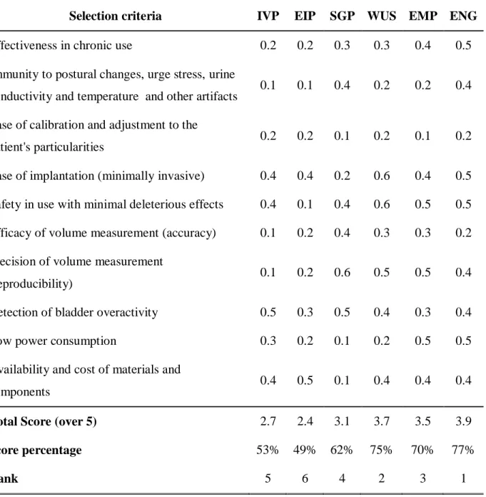
Table 2-1 Methods Assessment results ... 49
Table 3-1: RMS Errors (%) from experiments Type A with model order N of 6... 76
Table 3-2: RMS Errors (%) for bladder Pressure estimation by the best BW ... 82
Table 4-1: Classification Accuracy comparison (%) ... 112
Table 4-2: FPGA Resources usage and Power consumption... 117
Table 4-3: Performance comparison of the on-the-fly spike sorting process ... 117
Table A1-1: Scale and criteria to evaluate system needs ... 148
Table A1-2: Assessment of system needs... 149
Table A2-1: BS target technical specifications ... 152
Table A3-1: Set of weights used for the selection criteria used to assess the measurement methods. ... 154
LIST OF FIGURES
Figure 1-1 Components of the urinary system, from [48]. ... 16 Figure 1-2 Anatomy of the urinary bladder, from [48] ... 17 Figure 1-3: Neural circuits controlling the storage and voiding reflexes. A) Storage reflexes
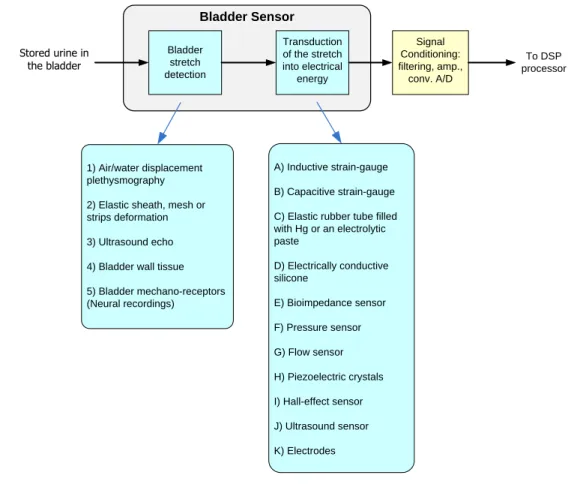
pathways. B) Voiding reflexes pathways. PAG: Periaqueductal grey, R: receptors on afferent nerve terminals [50]. ... 20 Figure 2-1: Primary and secondary transduction stages of the bladder sensor. A summary of the
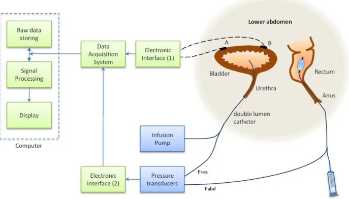
potential measurement principles that may be used in implementing each stage is also shown. ... 27 Figure 2-2: Typical setup used for studies of bladder activity. A and B are generic transducer or
electrodes located on different parts of the lower abdomen depending on experiment goals. ... 34 Figure 2-3: Neural pathways of the pelvis showing the afferent nerves commonly used the ENG
recordings (see text). SPLN, splanchnic nerves; GR, gray rami; WR, white rami; BC, bulbocavernosus muscle; IC, ischiocavernosus muscle; SPH, sphincter. From [49], reproduced with permission from John Wiley and Sons. ... 42 Figure 3-1: Experimental setup used for identification of bladder afferent nerves projecting into
the L6 root and for recording the afferent activity during a filling cystometry. (MPG: Major Pelvic Ganglion). ... 60 Figure 3-2 : Filling profiles used in three types of experiments. A) Standard filling-voiding
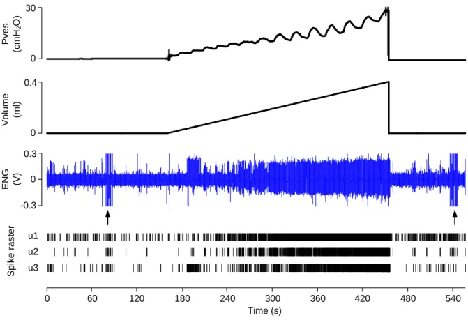
cycle. B) Standard filling and passive withdrawing. C) Filling to different levels followed by an isovolumetric phase. ... 62 Figure 3-3: Bladder afferent activity recordings (ENG) using filling profile A (from animal No.
15, A-fiber). The arrows point to the artifacts elicited by electrical stimulation. The spike raster of the three units identified is shown... 64
Figure 3-4 : Qualitative estimation of bladder volume (Ex. from animal No. 21, C-fiber). A) Pressure, volume and ENG recorded during a filling cystometry. The effect of the optimal BW selection is shown by a comparison of the estimation errors achieved in B) 13.7% for 1 s and C) 0% for 30 s. (Volume*: quantized volume; L: Low, M: Medium, H: High, represent the bladder fullness states, see text). ... 71 Figure 3-5: Confusion matrices with average results in percentage from all trials of the
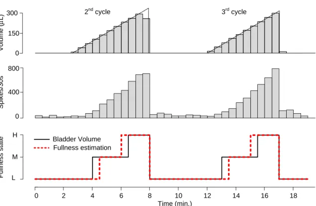
experiments type A during the training phase (A) and during the real-time-like monitoring phase (B). ... 72 Figure 3-6: Qualitative volume estimation for simulated real-time data processing (Ex. from
animal No. 25, C-fiber). The second and third measurement cycles are depicted. The first cycle was used in the learning phase. The qualitative estimation error for depicted cycles was 10.3%. ... 72 Figure 3-7: Quantitative volume estimation by the proposed method (Ex. from animal No. 18,
A-fiber). Comparative results for BW selection are shown. A) Estimation using a BW of 1 s yields an RMSEall of 50.2 L (13%) and an RMSEFill (t > tthr) of 58 L (15%). B)
Estimation using the optimal BW of 35 s produced an RMSEall of 16.2 L (4.2%) and an
RMSEFill (t > tthr) of 5.2 µL (1.4%)... 74 Figure 3-8: Quantitative volume estimation in a simulated real-time data processing experiments
(Ex. form animal No. 31, A fiber). The first cycle was used as a training period to estimate the bladder volume in two consecutives filling and voiding cycles. The resting activity threshold, the optimal BW (47 s) and a polynomial order (N = 6) were determined. The
RMSEall was 2%, 3.9% and 4.1% for each of the three cycles, respectively... 76
Figure 3-9: Assessment of the model goodness of fit. A) Average coefficient of determination (R2), and B) Average Akaike’s Information Criterion (AIC) for all fibers, both computed using the best BW for each model order. ... 77 Figure 3-10: Effect of bin-width and polynomial order on the values of RMSEall. Both
parameters were swept from 1 s to 60 s and from N = 2 to 6, respectively. The averaged values (solid lines) ± standard deviation (dashed lines) computed from all fibers are shown. ... 77
Figure 3-11 Results for volume estimation from all ENG recordings using profile A depicted in Figure 3-2 (nA = 25, 107 trials). A) Estimation RMSEs for different model order. B) The mean and standard deviation of the best BW showing a steep linear drop with increasing polynomial order. ... 78 Figure 3-12: Volume estimation during passive saline withdrawing using profile B shown in
Figure 3-2 (Ex. from animal No. 35, C-fiber). The resting and filling phase were used as learning periods to estimate volume in the withdrawing phase. The RMSEs were 2.1%, 4.1% and 3.1% for the filling, holding and withdrawing phases, respectively. ... 80 Figure 3-13: Volume estimation during the tonic response of the bladder afferent activity
performed during five filling–holding–voiding cycles using profile C (Ex. from animal No. 40, C-fiber). A) The first cycle was used to compute the optimal bin-width (53 s), the polynomial order (N = 6) and the resting activity threshold. B) The isovolumetric measurements performed during the 4 holding phases yielded RMSEiso values of 12.8%, 4.0%, 0.1% and 3.1%, respectively. ... 80 Figure 3-14: Intravesical pressure (Pves) estimation during passive saline filling using profile (A)
(Ex. from animal No. 31, A- fiber). This estimation tracks small spontaneous bladder contractions with low errors; RMSEFill = 7%, RMSEAll = 6.9% and RMSE for Pves>10cmH2O of 5% (1.7cmH20). ... 82 Figure 4-1: Schematic of the general architecture of the Polystim neuroprosthetic device intended
to restore the storing and voiding functions of the urinary bladder in paraplegic patients. ... 87 Figure 4-2: Bladder volume sensor. A) DSP system architecture. B) Flowchart of the DSP main
functions. ... 93 Figure 4-3: Spike detector block (SDB). A) Circuit architecture. B) Flowchart of the SDB main
functions. ... 99 Figure 4-4: Spike classification block (SCB). A) Circuit architecture. B) Flowchart of the SCB
main functions. ... 103 Figure 4-5: Bladder volume decoding (BVD). A) Qualitative estimation circuit. B) Quantitative
Figure 4-6: Spike detection performance comparison. A) Input neural signal (synthetic) with SNR of 1dB; B) kNEO preprocessor output; C) and D) Zoomed-in signal from A) and B), respectively. ... 110 Figure 4-7: Results of the spike detection performed using different methods. A) Detection
accuracy (F-score) achieved while the scaling factor C was swept; B) Detection accuracy achieved for signal with different SNR; and C) ROC curves for each method. ... 111 Figure 4-8: Results of the spike classification using the WED method. A) and E) Templates with
low and high degree of resemblance, respectively, along with the curve of weights to compute the WED. B) to D) and F) to H) The probability density functions (PDF) corresponding to the WED of all the classified spikes computed with each template class of low and high resemblance, respectively. ... 113 Figure 4-9: Processing stage outputs for the quantitative and qualitative volume estimation. A)
Input neural signal previously amplified, filtered, and digitized. B) Spike raster obtained after signal processing performed by the SDB and SCB circuits. C) Output of the SRI circuit using a tbw of 39 s. D) The quantitative volume estimation is compared with the expected
output (volume discretized). E) The qualitative degree of fullness computed is compared with the expected output. ... 115 Figure A2-1: House of Quality (HoQ).………....153 Figure A4-1: Multiple Comparison test among the groups of coefficients using a one-way
ANOVA with a bin-width of 1 s. Pearson’s, Spearman’s and Kendall’s correlation coefficient means showed a statistically significant difference among them (p < 0.05) ………..157 Figure A4-2: Multiple Comparison test among the groups of coefficients using a one-way
ANOVA with a bin-width of 10 s. Pearson’s and Spearman’s correlation coefficient means showed a statistically significant difference with Kendall’s (p < 0.05) ………..157 Figure A4-3: Multiple Comparison test among the groups of coefficients using a one-way
ANOVA with a bin-width of 30 s. Spearman’s and Pearson’s correlation coefficient means were statistically significant different (p < 0.05)………..158
Figure A4-4: Multiple Comparison test among the groups of coefficients using a one-way ANOVA with a bin-width of 60 s. Spearman’s and Kendall’s correlation coefficient means showed a statistically significant difference with Pearson’s coefficient (p < 0.05) ………..………158
LIST OF ACRONYMS AND ABBREVIATIONS
ATD Absolute Thresholding DetectionAIC Akaike’s Information Criterion ANOVA Analysis of variance
ASIC Application Specific Integrated Circuit BIR Bin-integrated-rate
BW Bin-width
BS Bladder sensor
BVD Bladder Volume Decoder BCI Brain-Computer Interface BC Bulbocavernosus muscle CNS Central Neural System CVI Cerebral Vascular Incident CAPs compounded action potentials CV Conduction velocity
CUSUM Cumulative sums
DSD Detrusor-sphincter dyssynergia DSP Digital Signal Processor
DRG Dorsal root ganglia
EIP Electrical Impedance Plethysmography EIT Electrical Impedance Tomography EEG Electroencephalogram
EMP Electromagnetic plethysmography
EMG Electromyogram
ENG Electroneurogram ED Euclidian Distance
EUS External urethral sphincter FPGA Field-programable gate array FIR Finite input response filter FSM Finite State Machine FR Firing rate
FIFO First In First Out
FES Functional electrical stimulation FES Functional Electrical Stimulation
GR Gray rami
HoQ House of Quality
ICS International Continence Society IP Intraperitoneal injection
IVP Intravesical Pressure IC Ischiocavernosus muscle LFP Local field potentials LUT Lower urinary tract
MRI Magnetic Resonance Imaging MCU Microcontroller
MEA Multi-Electrodes Array
NBO Neurogenic bladder overactivity NCU Neurostimulator Control Unit NEO Nonlinear Energy Operator OAB Overactive Bladder
OSR Overall Success Rate PAG Periaqueductal gray matter PC Personal Computer
PMC Pontine micturition center PVR Post void residual urine
PDF Probability Density Function QFD Quality Function Deployment RAM Random Access Memory
ROC Receiver Operating Characteristic RBI Rectifying and bin integration RMS Root Mean Square
RMSE Root Mean Square Error SNR Signal to noise ratio SAR Specific Absorption Rate SPH Sphincter
SCB Spike Classifier Block SDB Spike Detector Block SRI Spike Rate Integrator SCI Spinal cord injury SPLN Splanchnic nerves
SGP Strain-gauge plethysmography SPC Superparamagnetic clustering TEO Teager Energy Operator
UI Urinary incontinency UTI Urinary tract infection
USEA Utah Slanted Electrode Array VHSIC Very-high-speed Integrated Circuits VHDL VHSIC Hardware Description Language WUS Wearable ultrasonography
WED Weighted Euclidian Distance
LIST OF APPENDICES
APPENDIX 1 – BLADDER SENSOR: USER NEEDS…..………146 APPENDIX 2 – TARGET TECHNICAL SPECIFICATIONS………..……….151 APPENDIX 3 – ADDITIONAL INFORMATION ON THE ASSESSMENT OF THE MEASUREMENT METHODS………..………...154 APPENDIX 4 – CORRELATION COEFFICIENT COMPARISON………..………...156
INTRODUCTION
Nowadays there is an ongoing revolution in the field of neuroprosthetics devices that extend to different applications, allowing a total or partial rehabilitation of patients suffering from different neural diseases as consequence of spinal cord injury, stroke, traumatic brain injury, cerebral palsy, deafness, blindness, paralysis, movement disorders, and some mental illness and seizure disorders, among other diseases or non pathologic conditions as aging.
As consequence of the vertiginous development over the last decades, a new field of engineering known as neuroengineering or neural engineering emerged. This new field make use of all knowledge and cumulated experience of different fields of engineering and medicine with the aim of learn, understand, restore or augment functions and treat neural system diseases which includes both motor and sensory prostheses.
The introduction of new neuroprosthetic devices to the current clinical practice has been slower than other biomedical technology because of the high degree of complexity of the neural system and current technology limitations. One of the reasons of the restricted extension of recent neuroprosthetics devices is the limited efficacy controlling muscles, or organs like the urinary bladder, through open-loop systems due to the lack of a signal to feed back the neuroprosthetic device with the ongoing sensory information about the state, position or speed during muscles contraction and relaxation. Therefore, it can easily be deduced that the sensory feedback is crucial to effectively mimic the natural control of the targeted muscles or organs.
It has been show that the extraction of useful information from muscles (Electromyogram - EMG), nerves (Electroneurogram - ENG) and brain (Electroencephalogram - EEG) to feed back or command neurostimulator devices is an effective way to improve the Functional Electrical Stimulation (FES) efficacy restoring the lost or reduced capacity in handicapped patients [1]. These signals coming from natural receptors found in the skin, muscles, tendons, and joints are still present in many patients suffering from the diseases or conditions mentioned above. Capturing and interpreting reliably these signals by means of implanted interfaces in chronic applications is a great challenge due to the limited knowledge of the neural system and technological issues not solved yet.
The EEG, EMG and ENG signal can be used as source to command or feedback implanted neurostimulators depending on where the signal extraction is made and which the intended use of the FES device is. In clinical experiences with neuroprosthesis the EEG and EMG have been commonly considered as command signal, whereas ENG have been mainly used as feedback [1]. To achieve this goal real-time signal recording and sensory decoding is required.
The intelligent or smart neuroprosthesis with feedback capabilities are composed by two basic parts: the implanted unit (IU) and the external unit (EU). Usually both units are connected through a wireless link. The UI comprises mainly the recording subsystem that performs the front-end signal processing of the signals conveying sensory information, the neurostimulation subsystem implementing the FES approach, and a control logic driving both subsystems and the communication circuits with the EU. The IU sends the full recorded signal, or properly selected snips, to the EU unit that performs the back-end processing of the recorded signals and sends back the commands. The EU may also implement the user interface and the implant-computer interface. The amount of information to exchange between the implanted and the external unit is limited by the wireless-link bandwidth and the maximum dissipated power allowable for a safe implant operation. The required power to the implanted device is typically supplied by the external unit by employing the same wireless-link with the data encoded properly.
The research conducted in this thesis is part of a larger project of the Polystim Neurotechnologies research group at the École Polytechnique de Montreal that for several years have been working under the direction of Prof. Mohamad Sawan in the search for solutions to restore urinary functions of paraplegic patients who underwent an spinal cord injury. Particularly, this thesis was focused on the research of an effective sensory feedback from the urinary bladder able to be used either as an embedded bladder sensor in a closed-loop system neuroprosthesis or as a standalone sensor able to advise patients with impaired sensations when the bladder need to be emptied. It is worth noting that in spite of the attempts done in past years, chronic bladder monitoring by an implantable device was not achieved due to limited knowledge and technological challenges that were undertaken in this work.
Motivation
FES has been studied over the last two decades to treat neurogenic bladder dysfunction [2-16]. Implantable FES devices were able to partially restore the bladder functions in patients who were considered refractory to more conservative treatments or those who could not tolerate the side-effects of prescription drugs [17],[18].
FES therapy can improve the patients’ quality of life by preventing overactive bladder symptoms, non-obstructive urinary retention and bladder-sphincter dyssynergia [10],[17],[19]. This therapy can also reduce the frequency of urethral catheterization performed several times a day to empty the bladder, which causes much suffering to patients, recurrent urinary tract infections, and economical and physiological troubles. However, important side-effects of bladder FES therapy have been reported, such as an irreversible sacral deafferentation (dorsal rhizotomy), nerve-tissue injury produced by the continuous electrical stimulation, uncomfortable or painful sensations, infections, and changes in bowel function, among others [17],[19].
The restoring electrical neurostimulation performed by devices researched in past and those currently used in clinical practice is applied continuously without any sensory feedback (‘blind stimulation’). Nevertheless, sensory feedback can improve the neuroprostheses effectiveness with the implementation of a conditional neurostimulation approach based on the ongoing bladder volume or pressure. This approach in turns provides the intrinsic self-regulation advantages of the closed-loop control systems [1],[8]. An FES device implementing a conditional neurostimulation approach applies the electrical stimuli as and when needed, thus favoring energy saving and minimizing the deleterious effects on the neural tissue caused by continuous electrical stimulation [20]. Thereby, chronic monitoring of the bladder volume is required to improve the effectiveness, tolerability and safety of the neuroprosthetic devices used for restoring bladder functions. Attempts over the past few years, which are comprehensively reviewed in Chapter 2, have been made to find a suitable method for continuous bladder monitoring. However, a reliable, precise and robust device for providing feedback with sensory information from the bladder had not yet been identified.
Moreover, a sensor for chronic monitoring of bladder volume can also be useful as a standalone device able to provide early warnings to patients with impaired sensations when the bladder is approaching its capacity or when abnormally high post-voiding residual volume remains after an
incomplete voiding. Such a sensor may also be helpful in bladder differential diagnosis, bladder disease research and in the choice of best suited clinical therapy approaches.
Objectives
Our main objective was to find an effective method for chronic bladder volume monitoring with the aim of supplying an FES device with the feedback information required to restore bladder functions in a safe and effective manner. To accomplish the general objective, the following specific objectives were developed:
1. Identify user needs that a sensor for chronic bladder volume monitoring must fulfill for the intended application.
2. Determine whether a measurement principle based on artificial sensors or on natural receptors is best suited to satisfy the user needs.
3. Propose a bladder volume measurement method based on the selected measurement principle.
4. Validate the proposed measurement method for bladder volume monitoring using in vivo recordings from rats.
5. Assess the feasibility of an electronic device deploying the proposed bladder measurement method.
Thesis works
To clarify the problem, it was necessary to identify the main issues of chronic bladder volume monitoring while considering the anatomical and physiological characteristics of the lower urinary tract (LUT) and its innervation. Thus, an exhaustive review was conducted of updated literature from the different sources indexed in the PubMed, Compendex, Inspec and Derwent Innovation Index on-line databases and the recent editions of well-known reference books on clinical urology practice and neurophysiology [21-26]. The most relevant achievements and pitfalls in the bladder monitoring studies performed in the past years were identified from this analysis.
Once the problem was clarified and the challenges were recognized, the user needs were identified. These are the medical, technical, and ergonomics needs that should be satisfied by a
bladder volume monitoring device to provide the information about bladder volume or pressure. This identification was a crucial step toward the proposition of the measurement method to be used in the implementation of such a device.
The identified needs were translated into technical specifications, also known as target specifications, using the quality function deployment (QFD) methodology [27]. Some published studies and the standards defined by the International Continence Society (ICS) [28],[29], were used as references during this process.
Next, a functional decomposition of the measurement system was performed to identify the critical sub-functions and to focus the search for their solutions. A comprehensive literature review was performed with this goal, and all measurement principles and methods for volume measurement in medical and non-medical applications, particularly the methods used in past research for monitoring bladder volume or pressure, were compiled.
Subsequently, the compiled volume measurements methods were evaluated to identify those that better satisfied the needs and specifications defined in the preceding analyses. The evaluation criteria were obtained from the most important user needs and carefully matched with relative importance weights. An initial screening was performed to keep the most promising methods for the next analysis. These methods were chosen based on the expected feasibility, the technological availability and the go/no-go test that used the most relevant user needs as a threshold. A much more refined selection process was completed using evaluation matrices with the selection criteria and the corresponding weights. To ensure that the method selection was robust enough, i.e., less dependent on the variation of the assigned weights, various sets of weights were used, and the final score was calculated by averaging the resultant score for each set, while keeping the rating given to each method. To rate each measurement method, a qualitative scale was employed. This methodology allowed best methods to be chosen more objectively, overcoming the most evident bias that is often present in any researcher analysis.
From the results of the comprehensive literature review, user needs identification and an exhaustive analysis of all possible measurement methods, we concluded that the bladder mechanoreceptors were the best choice for the primary transducer for chronic monitoring.
The hypothesis and research questions were defined based on the published experimental results about the afferent activity of bladder mechanoreceptors in validated animal models (rats, cats,
dogs, and pigs). These types of studies have not been done in humans due to the highly invasive nature of the experiments needed to draw conclusions about the functioning of the mechanoreceptors. The conclusions from some of these studies suggested that volume-specific mechanoreceptors should exist in the bladder wall for properly controlling the bladder [30] and that the activity of these receptors increases with different response patterns during the bladder filling [31-34].
The initial evidence for the power feasibility of implantable circuits for real-time neural source discrimination was obtained from a theoretical study based on simulations [35]. Nevertheless, we did not find any study demonstrating the feasibility of real-time neural source discrimination together with sensory information decoding, both of which are required for the standalone (autonomous) operation of implantable neural sensors.
Consequently, we defined the two main thesis hypotheses as follows:
H1. When the local nervous system is intact, the bladder volume monitoring can be performed by decoding the afferent mechanoreceptors activity using an implantable device.
H2. The measurement method is deployed in a standalone, implantable system that estimates the ongoing bladder fullness state with an accuracy of 75% or better.
We chose 75% as an acceptable value of accuracy for a difficult measurement to be performed by an implantable device monitoring a physiologic variable in real-time.
The respective research questions underlying these hypotheses were as follows:
1. Can the afferent activity of bladder mechanoreceptors be detected and decoded in real-time to monitor the volume or pressure?
2. Is the measurement method electronically feasible and effective for a standalone implantable device considering power and real-time constraints?
As is customary in the research process, we performed an iterative search of the proper method for sensory decoding of bladder volume or pressure from neural afferent activity. We started with a preliminary method proposition, and we performed experiments using animal models.
Subsequently, the estimation methods were improved gradually to achieve the targeted objectives. Finally, the measurement methods and the thesis hypothesis H1 were validated. Acute experiments with rats were carried out to record bladder mechanoreceptor afferent activity along with the pressure and the known volume of the infused saline. Three types of experiments were performed with particular objectives. The first type of experiment allowed us not only validation of the proposed measurement methods but also to research the afferent activity during the resting, filling and voiding phase using a saline infusion pump with a physiologic filling rate and the instruments required for pressure and neural signal recordings. The second type of experiments was performed to research the reversibility of the mechanoreceptor afferent response by passively withdrawing the infused saline using a second infusion pump. The last experiment type allowed us to research whether the bladder showed phasic and tonic responses during the slow filling and during the isovolumetric measurement phase when the bladder was filled to different level of fullness. Important conclusions were drawn from the experimental results, which demonstrated the feasibility of bladder volume and pressure estimation from the mechanoreceptor afferent activity, the hysteretic mechanoreceptor response and the presence of both phasic and tonic responses. The latter response is a controversial subject we noticed during the literature review.
Two decoding methods were proposed to estimate the bladder volume in real-time. The first method allowed qualitative volume estimations, i.e., three levels of fullness defined as low-volume, comfortable level; medium level, the need-to-void within a proper timeframe; and high level, the urge-to-void because there is a risk of an imminent leaking. The second, a quantitative method, allowed the real-time estimation of the bladder volume or pressure value.
The qualitative method required low-computational cost and only a few hardware resources for its electronic implementation and yielded a high accuracy estimation of the bladder volume. The three qualitative levels defined appear to be sufficient for proper feedback of an FES device for restoring bladder functions and for advising patients with impaired sensations. On the other hand, the quantitative method can accurately compute the bladder volume for future applications, such as patient warning, differential diagnosis, and clinical research. The quantitative method was implemented by a regression model optimized to run in real-time with much lower computational
cost than those found in published works for decoding sensory information from neural recordings.
Both methods required neural source detection and discrimination in real-time, also known as on-the-fly spike sorting. This is a high demanding task even for a powerful personal computer and commonly requires human supervision, which are huge limitations that prevented the use of such algorithms in our intended application. Therefore, new algorithms were proposed and optimized to run unsupervised in real-time with low-computational cost to detect extracellular action potentials (spikes) immersed in noisy signals and to perform the on-the-fly spike sorting process. The estimation methods were designed to run in two phases to reduce the system complexity and the amount of hardware resources required by the implantable device for real-time bladder volume monitoring. The first is a training phase that is executed offline, and the second is the monitoring phase in which the unsupervised real-time volume estimation is performed. The training phase is used to determine the parameters required for the real-time monitoring phase. During the training phase, the most suitable algorithms were executed, regardless of their complexity, to compute the optimal parameters; whereas the monitoring phase, which is to be implemented in an electronic system, used lower complexity and much faster algorithms. Using this approach, some of the complexity from the monitoring phase could be transferred to the training phase without affecting the estimation accuracy.
The simulation and validation tests of the methods were realized in Matlab/Simulink with a real-time-like signal processing approach using realistic synthetic signals with known ground-truth to test the spike sorting process and real recordings from the in vivo experiments to test and validate the measurement methods.
To deploy the method in a standalone electronic system that could be implanted in the lower abdomen or near the targeted spinal root, several algorithms for the detection, classification and decoding of neural signals were designed and optimized to run unsupervised in real-time using few hardware resources and low-power consumption. An improved digital circuit was designed for detecting the spikes in signals with high background noise by estimating the instantaneous energy and comparing it with a self-tuned detection threshold. Furthermore, a digital circuit was designed for the spike classification using the template matching technique with a new metric
that allowed unsupervised on-the-fly spike sorting with high classification accuracies, even for highly correlated spike waveforms.
The new algorithms implemented by the real-time signal processing circuits were previously tested in Matlab/Simulink using custom-written software. Fixed-point arithmetic was chosen to reduce the signal processing retrieval times (latency) as much as possible and also the hardware burden compared with those required for floating-point arithmetic implementation. The number of bits for number representation was carefully determined using simulations performed with the Fixed-point toolbox of Matlab/Simulink to account for the trade-off between the amount of hardware required and the computation accuracy.
The electronic system was implemented in a dedicated digital signal processor (DSP) using a low-power field-programmable gate array (FPGA) to account for the trade-off between a fast and cost-effective R&D process for the bladder sensor and the overall performance required for future tests in chronic experiments. Finally, the results of the validation tests, which were also performed using the realistic synthetic signals and the signals from animal recordings, allowed us to validate the research hypothesis H2 by showing that an implantable low-power bladder sensor is feasible and accurate for the intended application.
Note: Throughout the document, the International System of Units was used for all measurements except for pressure. We used centimeters of water (cmH20) instead of Pascal (Pa) because cmH20 is the standard unit for research on this subject and for urodynamic clinical diagnostic tests. For references purposes, 1 cmH20 (4°C) = 98.0638 Pa.
Contributions
The thesis contributions were reported in three peer-reviewed journal papers. The two major contributions of our research works were as follows:
1. New measurement methods for decoding the bladder volume and pressure from the neural afferent activity arising from the bladder mechanoreceptors. The achievement of this contribution required original research based on the data gathered from experimentation
with animal models. No previous study has demonstrated the feasibility of volume estimation using the afferent activity of the bladder mechanoreceptors.
2. On-chip implementation of a dedicated DSP capable of performing in real-time the spike sorting process and the neural decoding with low-power consumption. To the best of our knowledge, this is the first time that such a DSP performing all these complex tasks, usually run on a personal computer, is reported.
Other important contributions that allowed the achievement of these two major contributions are summarized below:
Identification of the user needs that a bladder sensor must meet to be effective, safe and tolerable. This comprehensive list of needs was obtained from a thorough study of the specialized medical literature and was crucial for achieving our objectives. This user needs list may also help in future related research.
Selection of the bladder volume measurement method best suited for neuroprosthetic implants. This selection was not evident from the commonly used pros and cons analyses found in past studies focused on this subject. Some of these studies showed some issues in their methodological approaches and misunderstanding of the bladder neurophysiology. The review, assessment and selection of the measuring method for bladder monitoring were reported in [36].
Based on the results of specially designed in vivo experiments and the subsequent signal processing and statistical analyses, we showed that it is possible to measure accurately the tonic and phasic responses of the bladder detrusor muscle using the proposed method. The experiments with animal models and the neural signal processing used for the real-time decoding of the sensory information arising from the bladder were reported in [37].
To demonstrate the feasibility of an implantable bladder sensor that deploys the proposed decoding methods, several interesting solutions were found to meet the real-time and low-power consumption constraints imposed on the implantable sensors. The main contributions reported in [38] on this subject were as follows:
o Accurate detection of action potentials (spikes) immersed in signals with a low signal-to-noise ratio using a self-adjusted threshold circuit.
o Accurate spike discrimination using a method that does not require the customary processing sequence comprising spike feature extraction, dimensional reduction, and clustering, thus allowing for a frugal deployment of a digital circuit with low-power consumption.
o Digital implementation of the decoding method with fixed-point arithmetic that was accurate enough using a readily accessible input parameter, such as the spike-rate count, within a time-window of optimally chosen duration.
Social and economic impact
Patients suffering from urinary dysfunction are a topic of high interest because it affects millions of people all over the world [39]. The huge number of patients living with this problem and the economic impact is overwhelming. In just the United States, an estimated 34 million community-dwelling men and women have an overactive bladder. Managing urinary incontinence and overactive bladder costs $19.5 billion a year [40],[41].
The urinary incontinency (UI) prevalence in the Canadian population is estimated to be 3.3 million (10%) [42]. UI is a common condition in the elderly, affects 30% to 60% of patients over 65 years of age, and increases exponentially with age [43]. The projections of Statistics Canada estimate that the number of seniors will double by 2031. There are nearly 4 million of people aged 65 or older in Canada today, but in 30 years there will be 8.7 million, with 2.3 million who are at least of 80 years old. Age is strongly associated with the onset of chronic bladder conditions, which can lead to activity limitations, disabilities, and institutionalization. Therefore, it is expected that this growth in the elderly population will exert increasing pressure on the health care system [42]. Furthermore, there are 85,556 patients in Canada living with spinal cord injury (SCI) [44], which is a major cause of bladder dysfunction [40],[41].
These are, indeed, staggering economical and epidemiological statistics, but it also important to consider the social impact of disease and the patients’ impairment. Stigmatization, isolation, loss of self esteem, depression, and risk of institutionalization are present in many patients suffering
from bladder dysfunction. A study with data from the Canadian Community Health Survey shows that urinary incontinence is a major cause of depression in Canadian women with an average prevalence of 15.5%, which increases to 30% in women with ages between 18 and 44 years [45]. Moreover, a recent study demonstrated that the UI has a negative impact on the psychological burden of family caregivers [46].
With this thesis research, we aimed to improve the quality of life and life expectancy and to reduce the high cost of patient care for those suffering from urinary dysfunction due to different diseases, neurological disorders, and other non-pathologic conditions such as aging. We intended to achieve these goals by providing new knowledge and solutions for an unsolved problem, such as the chronic monitoring of bladder volume and pressure. We hope that the result presented in this thesis can help in bladder dysfunction treatment and diagnosis.
Thesis organization
The thesis comprises this Introduction, four chapters, and a final chapter with a general discussion and conclusions. We present the following work: a careful study of the targeted biological system (lower urinary tract), a literature review, an assessment of the measurement methods used in past studies, the proposal of new measurement methods based on recorded afferent neural activity, and the deployment of the proposed dedicated electronic system deploying the measurement method researched.
The first chapter of the thesis introduces the reader to the fundamentals of the anatomy, physiology and physiopathology of the urinary bladder that are necessary for a better understanding of the thesis work, the scope, objectives, impact and contributions. Particular emphasis is given to the neural control of the urinary bladder due to its relevance for the approach adopted in our research.
The second chapter presents three important subjects related to bladder volume monitoring: 1) a comprehensive review of the previous measurement method for monitoring the bladder volume and pressure; 2) the anatomical and physiological characteristics that we found to have the greatest impact in past studies that failed to find a solution for the bladder monitoring problem; and 3) an assessment of all of the measurement methods used in past studies, which allowed us to determine the method that was most suitable for our research objectives.
The third chapter first describes the acute experiments we performed in anesthetized rats and then proposes two measurement methods. These methods, one qualitative and another quantitative, estimated the bladder volume or pressure from the afferent neural activity arising from bladder mechanoreceptors. Finally, the result of the validation tests of these estimation methods and other test runs are presented.
The fourth chapter explains the design of the electronic system for the proposed measurement method to estimate in real-time the bladder volume or pressure from the recorded neural activity using an on-chip dedicated digital signal processor implemented in a low-power FPGA. Each processing block and the whole system is described and validated using both realistic synthetic signal and real signal recordings, thus showing the feasibility of such a system intended to be used in intelligent neuroprosthetic devices.
The fifth and last chapter discusses the thesis results. Finally, conclusions and recommendations for future work are presented.
Four Appendices are presented at the end of this thesis. Appendix 1 presents the list and the weightings of all of the identified user needs; Appendix 2 lists of all of the targets specification obtained using the QFD method; Appendix 3 presents the values of the weights and rates used in the assessment of the measurement methods; and finally, Appendix 4 shows the result of the analyses performed to choose the correlation coefficient better suited to identify the best unit for bladder volume decoding.
CHAPTER 1 OVERVIEW OF THE URINARY BLADDER
The urinary bladder presents challenging characteristics that have greatly hampered the achievement of a sensory device capable of monitoring urine volume and vesical pressure. Understanding the anatomy, physiology and physiopathology of the urinary bladder is crucial for accomplishing any successful work concerning sensory recovery and restoration of the storing and voiding functions.
An overview of these subjects, with a particular emphasis on the neural control involved in bladder functions, is provided to facilitate the understanding of the thesis works. Finally, the clinical practice standards for evaluating the bladder urine volume are introduced to show their particularities and limitations for chronic bladder monitoring.
1.1 Bladder anatomy and physiology
The urinary bladder is part of the urinary system, which is also composed of the kidneys, the ureters, and the urethra (Figure 1-1). The function of urinary systems is to regulate the water and ionic composition of the body, to excrete waste and potentially toxic products of metabolism, to remove foreign chemicals and to produce several hormones.
The bladder functions are the storing and voiding of urine in a coordinated and controlled manner. The bladder must store a socially adequate volume of urine until voiding is voluntarily elicited. To store urine, the bladder smooth muscle (detrusor) must be relaxed, and the sphincter must be contracted (closed). In contrast, emptying the bladder requires synchronous activation of all detrusor muscles because contracting only one part would stretch the uncontracted compliant (flexible) areas, thus preventing the increase in pressure necessary for urine to be expelled through the urethra. At the same time, these contractions must be followed by sphincter relaxation and opening to discharge the urine.
The bladder detrusor is a smooth muscle that is more adaptable than skeletal muscle and is able to adjust its length over a much wider range. For instance, if the bladder is filled to 400 mL with a diameter of 30 cm and is then emptied to a residual volume of 10 mL with a diameter of 8 cm,
the diameter would have decreased ~75%; thus, the detrusor would have a change in muscle length of same magnitude [47].
Figure 1-1 Components of the urinary system, from [48].
The bladder can be divided into two parts: a body lying above the ureteral openings and a base consisting of the trigone and bladder neck. The bladder outlet is composed of the bladder base, the urethra, and the internal and external urethral sphincter (rhabdosphincter), as shown in Figure 1-2.
The urethra and the sphincter system (Figure 1-2) play an important role in the two principal bladder functions of storing and voiding. These components not only provide controlled urine conduction way but also play an important role in the guarding and micturition reflexes.
There are anatomical and functional differences between the male and female urethral sphincter systems. Males have an internal sphincter composed of smooth muscle at the level of the bladder neck that is innervated by the sympathetic nervous system to prevent retrograde ejaculation. The female urethra is much shorter than that of the male. This is one factor which accounts for the higher prevalence of incontinence in females.
Figure 1-2 Anatomy of the urinary bladder, from [48]
Both the external urethral sphincter (EUS) and the internal urethral sphincter (IUS) allow the control of the opening for urine discharge. The EUS is composed of striated muscles that allow voluntary control of micturition by somatic innervation. In contrast, the IUS, which is composed of smooth muscle, is controlled by autonomic innervation.
The bladder has a storage capacity of approximately 400 – 600 mL, which can accommodate the production of 1 – 2 L of urine per day, in an average adult. The bladder is typically emptied 5 – 7 times a day, often at much smaller volumes. The average urine flow time is less than 30 seconds; thus, the bladder is actively emptying less than 1% of the time. Therefore, the predominant function of the bladder is storing urine [49].
1.2 Bladder biomechanics
The fundamental mechanical properties of the bladder include the stress-strain relationship, viscoelasticity, and deformation of bladder tissue. Whole-bladder properties include bladder shape, mass, and distention.
Basic bladder hydrodynamics and biomechanical properties depend on the relationship between bladder shape, size, pressure, and tension, as expressed by Laplace's law, which is shown in equation (1-1). There is a direct relationship between the wall tension and the intravesical pressure and bladder size. In this equation, T is the bladder wall tension, Pves is the intravesical
pressure, R is the bladder radius, and d is the wall thickness. During normal bladder filling, Pves
increases very slowly. When the bladder is completely distended, the wall thickness d decreases significantly relative to the other parameters unless a hypertrophied wall exists. At this point, a further increase in Pves will produce high wall tension and strong afferent activity.
(1-1)
The bladder compliance (C) is defined as the ratio between the variations in the bladder volume and detrusor pressure (Pdet) as shown in equation (1-2).
(1-2)
The viscoelastic behavior of the bladder and urethra depends on both neuromuscular and mechanical properties. The mechanical properties rely on the magnitude of the wall stretch (distention) and the tissue structure and composition. The bladder is mainly composed of smooth muscle, 50% collagen and 2% elastin [47]. The collagen content increases with injury, obstruction, and denervation, leading to decreased bladder compliance. Conversely, the compliance will be higher when the elastin content exceeds the collagen content. A decrease in compliance or in the efferent neural input modifies the wall tension, causing an abnormal increase in the afferent activity, which may disturb normal bladder sensations and the threshold volume for micturition.
The bladder is typically modeled as a spheroid; however, this organ exhibits one of the most irregular anatomic shapes, especially when it is full, due to the contact with surrounding pelvic structures. Estimating the volume by instrumentation principles based on bladder geometry becomes more difficult because of this shape irregularity.
The bladder intravesical pressure depends on both the abdominal (Pabd) and detrusor (Pdet)
pressures, as shown in equation 1-3. Bladder emptying depends on the detrusor contraction for increasing Pdet without a significant increase in Pabd.
P
ves= P
det+ P
abd(1-3)
Pves measurement during bladder filling shows low and relatively constant bladder pressures (6 to
10 cm H2O in humans) when the bladder volume is below the threshold for inducing voiding. The accommodation of the bladder to increasing volumes of urine is primarily a passive phenomenon that depends on the viscoelastic intrinsic properties of the vesical smooth muscle and the quietness of the parasympathetic efferent pathway.
1.3 Bladder innervation
Bladder innervation involves both the autonomic (involuntary) and the somatic (voluntary) nervous system. Pelvic parasympathetic nerves, which are located at the sacral level of the spinal cord, drive micturition reflexes by contracting the detrusor muscle and relaxing the urethra. The sympathetic nerves, which are located in the lumbar zone, have an opposite action: they promote the storing reflex by relaxing the detrusor muscle and contracting the bladder base and the urethra. The pudendal somatic nerve, which contains afferent sensory axons and efferent motor axons, excites the EUS.
In humans, the afferent axons in the pelvic, hypogastric, and pudendal nerves transmit information from the lower urinary tract to the lumbosacral spinal cord from L1 to S4 roots. The location of these nerves varies from one experimental model to another. The protocols for studying bladder innervation are consequently adjusted.
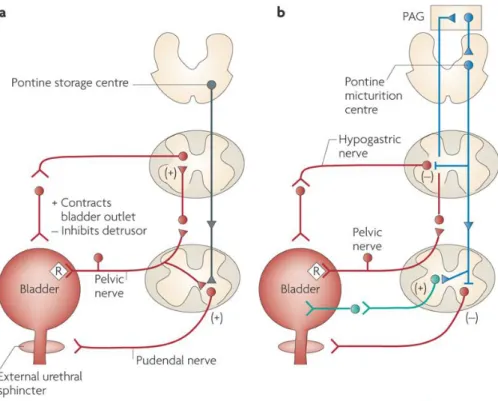
The bladder wall contains sensory receptors that work as physiological transducers, transforming natural energy into a train of all similar action potentials where sensory information is encoded in the time interval between them. Most of these receptors respond only to one type of energy (mechanical, thermal, or chemical), but others can respond to a combination of different types of energy. Mechanoreceptors respond to bladder wall distension and contraction by increasing or decreasing their firing frequency. Trough the afferents pelvic, hypogastric and pudendal nerves,
these action potentials are reaching the spinal cord at L1 to S4 levels and are relayed to the pontine storage and micturition centers, as shown in Figure 1-3.
Figure 1-3: Neural circuits controlling the storage and voiding reflexes. A) Storage reflexes pathways. B) Voiding reflexes pathways. PAG: Periaqueductal grey, R: receptors on afferent nerve terminals [50].
The mechanoreceptors can be slow- or fast-adapting receptors. The slow-adapting mechanoreceptors detect changes in pressure, while the fast-adapting mechanoreceptors respond to rapid changes and vibration. The frequency of action potential discharge is proportional to the intensity of the stimulus. The sensitiveness threshold (minimum pressure required to excite the mechanoreceptor) of the physiological pressure in humans is between 5 and 40 cmH2O. This pressure is in the range of the compliant part of the pressure-volume curve (25 - 75%). This threshold is well correlated with the point where the first sensation of filling is normally detected, although the threshold can be higher, for example, 95 cmH2O after a spinal cord injury [22].
![Figure 1-1 Components of the urinary system, from [48].](https://thumb-eu.123doks.com/thumbv2/123doknet/2339212.33539/40.918.314.642.183.519/figure-components-urinary.webp)
![Figure 1-2 Anatomy of the urinary bladder, from [48]](https://thumb-eu.123doks.com/thumbv2/123doknet/2339212.33539/41.918.152.771.106.433/figure-anatomy-urinary-bladder.webp)