Université de Montréal
Intraoperative hemodynamic instability during and after separation from cardiopulmonary bypass: importance, mechanism and prevention
par
André-Yvan Denault MD PhD FRCPC ABIM CCM FASE
Département d’anesthésiologie Faculté de médecine
Thèse présentée à la Faculté des études supérieures en vue de l’obtention du grade de PhD en Sciences Biomédicales (3-484-1-0)
Septembre 2009
Université de Montréal Faculté des études supérieures
Cette thèse intitulée:
Difficult separation from cardiopulmonary bypass: Importance, mechanism and prevention
Présentée par : André-Yvan Denault
a été évaluée et défendue le 17 décembre 2009 Pour voir la vidéo de la présentation :
www.mediaenligne.umontreal.ca/medclin/anesth/denault_phd.wmv
par un jury composé des personnes suivantes :
Pierre Beaulieu PhD, président-rapporteur Jean Lambert PhD, directeur de recherche
Jean-Claude Tardif, co-directeur Patrick Mathieu, membre du jury Jean-Yves Dupuis, examinateur externe François Reeves, représentant du doyen de la FES
Résumé
Chaque année, environ 1 à 1,25 million d’individus subiront une chirurgie cardiaque. [1] Environ 36 000 chirurgies cardiaques sont effectuées au Canada et 8000 procédures au Québec (http://www.ccs.ca). Le vieillissement de la population aura pour conséquence que la chirurgie cardiaque sera offerte à des patients de plus en plus à risque de complications, principalement en raison d’une co-morbidité plus importante, d’un risque de maladie coronarienne plus élevée, [2] d’une réserve physiologique réduite et par conséquent un risque plus élevé de mortalité à la suite d’une chirurgie cardiaque. L’une des complications significatives à la suite d’une chirurgie cardiaque est le sevrage difficile de la circulation extracorporelle. Ce dernier inclut la période au début du sevrage de la circulation extracorporelle et s’étend jusqu’au départ du patient de la salle d’opération. Lorsque le sevrage de la circulation extracorporelle est associé à une défaillance ventriculaire droite, la mortalité sera de 44 % à 86 %. [3-7] Par conséquent le diagnostic, l’identification des facteurs de risque, la compréhension du mécanisme, la prévention et le traitement du sevrage difficile de la circulation extracorporelle seront d’une importance majeure dans la sélection et la prise en charge des patients devant subir une chirurgie cardiaque. Les hypothèses de cette thèse sont les suivantes : 1) le sevrage difficile de la circulation extracorporelle est un facteur indépendant de mortalité et de morbidité, 2) le mécanisme du sevrage difficile de la circulation extracorporelle peut être approché d’une façon systématique, 3) la milrinone administrée par inhalation représente une alternative préventive et thérapeutique chez le patient à risque d’un sevrage difficile de la circulation extracorporelle après la chirurgie cardiaque.
Mots-clés : ventricule droit, circulation extracorporelle, chirurgie cardiaque, instabilité hémodynamique, échocardiographie transoesophagienne, hypertension pulmonaire
Abstract
Every year, 1 million to 1.25 million patients worldwide undergo cardiac surgery. [1] Up to 36,000 cardiac surgeries are performed each year in Canada and close to 8000 in Quebec (http://www.ccs.ca). Because of the aging of the population, cardiac surgery will increasingly be offered to patients at a higher risk of complications. Indeed, elderly patients have increased co-morbidities, and aging is also a significant risk factor in the prevalence of coronary artery disease. [2] The consequence is a reduced physiologic reserve, hence an increased risk of mortality. These issues will have a significant impact on future healthcare costs, because our population undergoing cardiac surgery will be older and more likely to develop postoperative complications. One of the most dreaded complications in cardiac surgery is difficult separation from cardiopulmonary bypass (CPB). The definition of difficult separation from CPB includes the time period from when CPB is initiated and until the patient leaves the operating room. When separation from CPB is associated with right ventricular failure, the mortality rate will range from 44% to 86%. [3-7] Therefore the diagnosis, the preoperative prediction, the mechanism, prevention and treatment of difficult separation from CPB will be crucial in order to improve the selection and care of patients and to prevent complications for this high-risk patient population. The hypotheses of this thesis are the following: 1) difficult separation from CPB is an independent factor of morbidity and mortality, 2) the mechanism of difficult separation from CPB can be understood through a systematic approach, 3) inhaled milrinone is a preventive and therapeutic approach in the patient at risk for difficult weaning from CPB after cardiac surgery.
Keywords : Right ventricle; Cardiopulmonary bypass; Cardiac surgery; Hemodynamic instability; Transesophageal echocardiography; Pulmonary hypertension.
Table of contents
Table of contents ... v
List of tables ... viii
List of figures ... x
List of appendices ... xiii
Abbreviations ... xiv
Remerciements ... xxiii
Foreword ... 25
Introduction ... 27
Chapter 1 Definition and importance of difficult separation from CPB ... 28
1.1 Definition of difficult separation from CPB ... 29
1.2 Predictors of difficult separation from CPB... 41
1.2.1 Demographic and surgical variables ... 41
1.2.2 Biochemical variables ... 43
1.2.3 Hemodynamic and echocardiographic variables ... 46
1.2.4 Patient-prosthesis mismatch ... 47
1.2.5 Other factors involved in the risk of difficult separation from CPB ... 49
1.3 The significance and consequence of difficult separation from CPB ... 55
1.4 Research and development since the beginning of the PhD in 2006 at the MHI... 57
1.4.1 Studies on arterial pressure and separation from CPB ... 57
1.4.2 Studies on diastolic function and separation from CPB ... 57
1.4.3 Studies on predictors of difficult separation from CPB ... 62
1.4.4 Studies on the outcome of difficult separation from CPB ... 62
Chapter 2 Manuscript #1 ... 70
Foreword to Manuscript #1 ... 71
3.1 Mechanism of hemodynamic instability ... 98
3.1.1 Reduction in mean systemic pressure ... 104
3.1.2 Increased right atrial pressure ... 109
3.1.3 Increased resistance to venous return ... 146
3.1.4 Combined mechanism ... 159
3.2 Research and development since the beginning of the PhD in 2006 at the MHI... 168
3.2.1 Studies on alternative measurement of venous return and cardiac output ... 168
3.2.2 Studies on causes of increased Pra ... 171
Chapitre 4 Manuscript #2 ... 178
Foreword to Manuscript #2 ... 179
Chapitre 5 Manuscript #3 ... 203
Foreword to Manuscript #3 ... 204
Chapter 6 Inhaled milrinone ... 229
6.1. Definition of pulmonary hypertension ... 230
6.2. Pulmonary hypertension in cardiac surgery: mechanism and etiology ... 236
6.2.1 Factors involved in pulmonary hypertension in cardiac surgery ... 237
6.3 Importance and impact of pulmonary hypertension in cardiac surgery ... 242
6.4 Right ventricular dysfunction ... 243
6.5 Treatment and prevention of pulmonary hypertension in cardiac surgery ... 244
6.4.1 Treatment of pulmonary hypertension ... 248
6.4.2 Treatment of right ventricular failure ... 252
6.4.3 Milrinone ... 256
6.4.4 Inhaled milrinone ... 262
6.5. Prevention of pulmonary hypertension ... 264
6.5.1 Pharmacological approach ... 264
6.5.2 Protamine ... 264
6.5.3 Non-pharmacological approach ... 265
6.6 Research and development since the beginning of the PhD in 2006 at the MHI... 266
6.6.2 Intravenous therapy ... 267
6.6.3 Inhalation therapy: inhaled milrinone ... 272
Chapitre 7 Manuscript #4 ... 277
Foreword to Manuscript #4 ... 278
Chapitre 8: Discussion ... 305
8.1 Summary and originality of the thesis ... 305
8.2 Limitations and future projects ... 314
Conclusion ... 318
Appendices ... 321
Bibliography ... 347
Curriculum vitae ... 385
Coauthor consent form ... 386
List of tables
Table 1 Various definitions of difficult separation from CPB proposed in the literature .... 34
Table 2 Studies on difficult separation from CPB and postoperative inotropes ... 53
Table 3 Risk factor for difficult separation from CPB and postoperative inotropes ... 54
Table 4 Left and right ventricular diastolic function ... 61
Table 5 Univariate and multivariate analysis for hemodynamic complications ... 64
Table 6 Outcome and degree of separation from CPB at the Montreal Heart Institute ... 67
Table 7 Outcome and degree of separation from CPB in the BART study ... 86
Table 8 Predictors of the degree of separation from CPB in the BART study ... 91
Table 9 Predictors of mortality in the BART study ... 92
Table 10 Postoperative outcome in the BART study ... 93
Table 11 Mechanism of hemodynamic instability in cardiac surgery ... 103
Table 12 Prognostic value of right ventricular function in cardiac surgery ... 124
Table 13 Abdominal compartment syndrome ... 154
Table 14 Mechanisms of hemodynamic instability and therapeutic implication ... 161
Table 15 Summary of the hemodynamic and echocardiographic measurements ... 164
Table 16 Multivariate analysis for mortality ... 173
Table 17 Mortality in patients undergoing coronary artery bypass grafting ... 173
Table 18 Mortality in patients undergoing non-coronary artery bypass grafting ... 174
Table 19 Characteristics of the amiodarone versus placebo group ... 195
Table 20 Biochemical, hemodynamic and Doppler variables ... 197
Table 21 Outcome data ... 199
Table 22 Characteristics of patients with and without inotropes ... 221
Table 23 Biochemical, hemodynamic and Doppler variables ... 223
Table 24 Outcome data ... 225
Table 25 Definitions of pulmonary hypertension used in clinical research ... 231
Table 27 Clinical studies on the use of milrinone in cardiac surgery ... 257
Table 28 Hemodynamic values during surgery ... 269
Table 29 Right ventricular echocardiographic data ... 271
Table 30 Baseline Characteristics of the Study Population ... 293
Table 31 Hemodynamic variables: one-way repeated ANOVA ... 295
Table 32 Echocardiographic variables: one-way repeated ANOVA ... 297
Table 33 One-way ANCOVA adjusted for T1 at separate time interval ... 299
List of figures
Figure 1 Time sequence of a cardiac surgical procedure ... 30
Figure 2 Radial to femoral artery pressure gradient during cardiac surgery ... 38
Figure 3 Mortality and morbidity in relation with lactate level during CPB ... 44
Figure 4 Intramyocardial acidosis and inotropic requirement ... 45
Figure 5 Patient–prosthesis mismatch ... 48
Figure 6 Retrograde cardioplegia cannula ... 49
Figure 7 Calcium emboli in valvular surgery ... 50
Figure 8 Dysfunctional AoV bioprosthesis after AVR ... 51
Figure 9 Hemodynamic instability and brain desaturation ... 56
Figure 10 Biventricular cardiac dimensions and Doppler during CABG ... 58
Figure 11 Interactions between risk factors ... 94
Figure 12 Mechanism of difficult separation from CPB ... 95
Figure 13 Venous return and cardiac output ... 99
Figure 14 Pressure and volume during a cardiac cycle ... 100
Figure 15 Venous return and pressure-volume loop concept... 101
Figure 16 Reduction in mean systemic venous pressure ... 105
Figure 17 Bilateral pleural effusions ... 106
Figure 18 Abdominal examination using transesophageal echocardiography ... 107
Figure 19 Biventricular systolic dysfunction ... 110
Figure 20 Simpson’s method of discs ... 111
Figure 21 Left ventricular fractional area change ... 112
Figure 22 Echocardiographic classification of diastolic dysfunction ... 114
Figure 23 Biventricular diastolic dysfunction ... 116
Figure 24 Right ventricular systolic and diastolic function ... 119
Figure 26 Tricuspid annular plane systolic excursion (TAPSE) ... 121
Figure 27 Isolated right ventricular systolic dysfunction ... 123
Figure 28 Dynamic left ventricular outflow tract (LVOT) obstruction ... 130
Figure 29 Ventricular outflow tract obstruction... 131
Figure 30 Left ventricular outflow tract obstruction (LVOTO)... 132
Figure 31 Risk factors of systolic anterior motion (SAM) ... 134
Figure 32 Pulmonary embolism immediately after coronary revascularization ... 137
Figure 33 Air embolism ... 138
Figure 34 Air embolism ... 139
Figure 35 Carbon dioxide (CO2) embolism ... 140
Figure 36 Hemodynamic effect of hypoxemia... 141
Figure 37 Patent foramen ovale (PFO) ... 142
Figure 38 Paradoxical pulmonary embolism ... 143
Figure 39 Hypercapnia and cardiac function ... 144
Figure 40 Hypercapnia and right atrial dimension and pressure... 145
Figure 41 Mechanism of increased resistance to venous return during tamponade ... 147
Figure 42 Classical tamponade. ... 149
Figure 43 Regional tamponade. ... 150
Figure 44 Hemodynamic consequence of a pneumothorax ... 151
Figure 45 Mediastinal tamponade ... 152
Figure 46 Acute abdominal compartment syndrome after induction of anesthesia. ... 155
Figure 47 Partially occluded inferior vena cava (IVC) ... 156
Figure 48 Inferior vena cava (IVC) occlusion during Fontan procedure ... 157
Figure 49 Intra-aortic balloon pump (IABP) catheter in the inferior vena cava (IVC). .... 158
Figure 50 Brain desaturation during cardiac transplantation. ... 159
Figure 51 Mechanism of hemodynamic instability in cardiac surgery ... 163
Figure 52 Brain-heart interaction ... 170
Figure 53 Dynamic right ventricular outflow tract (RVOT) obstruction ... 175
Figure 55 Left ventricular diastolic dysfunction (DD) algorithm ... 200
Figure 56 Right ventricular diastolic dysfunction (DD) algorithm ... 201
Figure 57 Hemodynamic and echocardiographic summary... 202
Figure 58 Hemodynamic and echocardiographic summary... 226
Figure 59 Probability of survival at 6 years. ... 228
Figure 60 Doppler estimation of the severity of pulmonary hypertension ... 232
Figure 61 The MAP/MPAP ratio ... 233
Figure 62 Pulmonary hypertension and outcome... 234
Figure 63 Doppler and hemodynamic signs of right ventricular dysfunction ... 235
Figure 64 Mechanisms that could induce pulmonary hypertension in cardiac surgery ... 237
Figure 65 Pulmonary reperfusion syndrome after cardiopulmonary bypass ... 239
Figure 66 Effect of lung volume on pulmonary vascular resistance ... 241
Figure 67 Vent complication ... 251
Figure 68 Proposed approach in the treatment of right ventricular dysfunction ... 253
Figure 69 Right ventricular pressure waveform... 255
Figure 70 Right ventricular pressure waveform with hemodynamic instability ... 255
Figure 71 Right ventricular pressure waveform after inhaled milrinone ... 256
Figure 72 Milrinone plasma concentration–time profile ... 273
Figure 73 Milrinone plasma concentration during inhalation ... 274
Figure 74 Pharmacokinetic/pharmacodynamic analysis of inhaled milrinone ... 275
Figure 75 Hemodynamic and echocardiographic changes... 302
Figure 76 Inhaled milrinone in two patients ... 303
Figure 77 Hemodynamic and echocardiographic summary... 304
List of appendices
Appendix 1 Definitions of variables in the BART study ... 322
Appendix 2 Protocol for vasoactive management during CPB ... 324
Appendix 3 Protocol for vasoactive management during weaning from CPB ... 325
Appendix 4 Arterial blood gases and biochemistry variables ... 326
Appendix 5 Hemodynamic variables ... 328
Appendix 6 Two-dimensional echocardiographic variables ... 330
Appendix 7 Doppler echocardiographic variables ... 332
Appendix 8 Diastolic function evaluation ... 335
Appendix 9 Arterial blood gases and biochemistry variables ... 336
Appendix 10 Hemodynamic variables ... 338
Appendix 11 Two-dimensional echocardiographic variables ... 340
Appendix 12 Doppler echocardiographic variables ... 342
Abbreviations
2D two-dimensional
A dur duration of mitral inflow A-wave
A peak late or atrial diastolic flow velocity ABC Airway-Breathing-Circulation
AC aortic occlusion
ACC/AHA American College of Cardiology and American Heart Association ACE angiotensin converting enzyme
ACS abdominal compartment syndrome Am atrial mitral annular velocity AML anterior mitral leaflet length AMP adenosine monophosphate ANCOVA analysis of covariance ANOVA analysis of variance
Ao aorta
AoV aortic valve AP arterial pressure
APP abdominal perfusion pressure AR aortic regurgitation
AR atrial reversal ASD atrial septal defect
At atrial tricuspid annular velocity AVR aortic valve replacement
BART Blood Conservation Using Antifibrinolytics in a Randomized Trial BMI body mass index
BP blood pressure BSA body surface area
CABG coronary artery bypass grafting CAD coronary artery disease
CARE Cardiac Anesthesia Risk Evaluation CASS Coronary Artery Surgery Study CBF cerebral blood flow
CHF congestive heart failure CI cardiac index
CI confidence interval CK creatinine kinase CO cardiac output
COPD chronic obstructive pulmonary disease CPB cardiopulmonary bypass
CTICU cardiothoracic intensive care unit CVD cerebrovascular disease
CVP central venous pressure
D diastolic
DAP diastolic arterial pressure
DPAP diastolic pulmonary arterial pressure DSB difficult separation from bypass DT deceleration time
E early
ECMO extra-corporeal membrane oxygenator EDA end-diastolic area
EDV end-diastolic volume EF ejection fraction EKG electrocardiogram
Em early mitral annular velocity EOA effective orifice area
ESV end-systolic volume
Et early tricuspid annular velocity ET ejection time
FAC fractional area change
Fem Femoral
FRC functional residual capacity
Gd gradient
GEE generalized estimating equation
HR heart rate
HVF hepatic venous flow IABP intra-aortic balloon pump IAH intra-abdominal hypertension IAP intra-abdominal pressure ICU intensive care unit
iEOA indexed effective orifice area
IL interleukin
iMil Inhaled milrinone iNO inhaled nitric oxide iPGI2 inhaled prostacyclin IU international unit;
IV intravenous
IVC interior vena cava
IVCT isovolumic contraction time IVRT isovolumic relaxation time
LA left atrium
LAA left atrial appendage
LADt left atrial transverse dimension LAP left atrial pressure
LHV left hepatic vein
LIJV left internal jugular vein LIMA left internal mammary artery LOF low output failure
LUPV left upper pulmonary vein LV left ventricle or left ventricular LVAD left ventricular assist device
LVDD left ventricular diastolic dysfunction LVEDA left ventricular end-diastolic area LVEDP left ventricular end-diastolic pressure LVEF left ventricular ejection fraction LVESA left ventricular end-systolic area LVFAC left ventricular fractional area change LVOT left ventricular outflow tract
LVOTO left ventricular outflow tract obstruction LVWMSI left ventricular wall motion score index MAP mean arterial pressure
MAV mitral annular velocity MHI Montreal Heart Institute MI myocardial infarction
MPAP mean pulmonary artery pressure MPI myocardial performance index MR mitral regurgitation
MV mitral valve
MVO2 mixed venous oxygen MVR mitral valve replacement NIH National Institute of Health NIRS near-infrared spectroscopy NO nitric oxide
NTG nitroglycerin NTP nitroprusside
NYHA New York Heart Association OM obtuse marginal
OR odds ratio
OR operating room Pa arterial pressure PA pulmonary artery
PAC pulmonary artery catheter Paf femoral arterial pressure PAF platelet activating factor PAP pulmonary artery pressure Par radial arterial pressure
PCWP pulmonary capillary wedge pressure PEEP positive end-expiratory pressure PFO patent foramen ovale
PGE1 prostaglandin E1 PGI2 prostacyclin
PH pulmonary hypertension PML posterior mitral leaflet length Pms mean systemic pressure PMV prosthetic mitral valve
PN pseudonormal
Ppa pulmonary artery pressure PPM patient-prosthesis mismatch Pra right atrial pressure
Prv right ventricular pressure PVF pulmonary venous flow PVR pulmonary vascular resistance
PVRI indexed pulmonary vascular resistance
PW pulsed-wave
QHLI Quebec Heart and Lung Institute Ra arterial resistance
RA relaxation abnormality RA right atrium
Rad Radial
RADt right atrial transverse diameter RBC red blood cell;
RCA right coronary artery RCT randomized controlled trial ROC receiver operating characteristics RPA right pulmonary artery
Rrv resistance to venous return RV residual volume
RV right ventricle or right ventricular RVAD right ventricular assist device
RVDD right ventricular diastolic dysfunction RVED right ventricular end-diastolic volume RVEDA right ventricular end-diastolic area RVEF right ventricular ejection fraction RVES right ventricular end-systolic volume RVESA right ventricular end-systolic area RVFAC right ventricular fractional area change,
RVMPI right ventricular myocardial performance index RVOT right ventricular outflow tract
RVOTO right ventricular outflow tract obstruction Rvr resistance to venous return
RWMSI regional wall motion score index
S systolic
SAM systolic anterior motion SAP systemic arterial pressure ScO2 cerebral oxygen saturation SCV subclavian vein
SD standard deviation; SE standard error
Sec seconds
SLCL septal to leaflet coaptation length Sm systolic mitral annular velocity SPAP systolic pulmonary artery pressure St systolic tricuspid annular velocity STS Society of Thoracic Surgeons
SV stroke volume
SVC superior vena cava
SVR systemic vascular resistance
SVRI indexed systemic vascular resistance TAPSE tricuspid annular plane systolic excursion TAV tricuspid annular velocity
TD thermodilution
TDI tissue Doppler imaging
TEE transesophageal echocardiography TLC total lung capacity
TMF transmitral flow TNF tumor necrosis factor TO2 oxygen transport TTF transtricuspid flow TV tricuspid valve
UK United Kingdom
USA United States of America VAD ventricular assist device Vp velocity of propagation
À la mémoire de Raymond Martineau dont le dévouement à la recherche au département d’anesthésiologie de l’Institut de Cardiologie de Montréal ne sera pas oublié
Remerciements
Depuis 2006, Jean Lambert m’a appuyé dans cette thèse. Sa disponibilité, son enthousiasme exceptionnel et son enseignement de qualité ont été et demeureront déterminants et hautement « significatifs » dans ma carrière. Sa direction dans l’élaboration de cette thèse fut un privilège inestimable. Je souligne le soutien du docteur Jean-Claude Tardif qui m’a appuyé depuis mon arrivée à l’Institut de Cardiologie en 1999. Sa vision de la recherche, sa générosité et ses commentaires pertinents ont joué un rôle majeur dans ma carrière et dans l’essor de la recherche au département d’anesthésiologie de l’Institut de Cardiologie. Mes collègues anesthésiologistes, chirurgiens, cardiologues m’ont épaulé au cours des dix dernières années et continuent à participer aux projets de recherche et d’éducation. Je les remercie sincèrement. Ces activités de recherche ne seraient possibles sans le soutien de nombreuses autres personnes qui font partie de l’Institut de Cardiologie. Ceux-ci incluent les membres du centre de recherche, l’équipe de recherche, notre assistante administrative, Pierrette Thivierge, l’équipe en salle d’opération (inhalothérapeutes, infirmières, perfusionnistes), l’équipe du génie biomédical, particulièrement Alain Girard qui m’a toujours aidé dans mes demandes technologiques, le service audiovisuel, l’administration hospitalière dont le directeur général M. Busilacchi a tellement à cœur l’épanouissement et le rayonnement de l’Institut de Cardiologie, finalement à Jennifer Stroude et Sylvie Lévesque pour les conseils de rédaction et de statistique. Merci à toute cette équipe et tout spécialement à Denis Babin, mon assistant de recherche depuis 1998 dont la disponibilité, la vision et les talents en informatique ne cessent de me surprendre. Les travaux présentés dans cette thèse ont été soutenus financièrement par la Fondation de la recherche en santé du Québec, la Fondation des maladies du cœur, la Fondation de l'Institut de Cardiologie de Montréal, la Bourse Herbert Black 2007, la Société canadienne d’anesthésiologie via la bourse Earl Wynands et les Instituts de recherche en santé du Canada.
Finalement, tout ce travail ne serait possible sans le support et la compréhension de ma conjointe le Dr Denise Fréchette et de mes trois enfants, Jean-Simon, Gabrielle et Julien. Puisse ce travail être utile à tous les professionnels de la santé qui ont à cœur la réanimation en chirurgie cardiaque du patient instable hémodynamiquement.
Foreword
Every year, 1 million to 1.25 million patients worldwide undergo cardiac surgery. [1] Up to 36,000 cardiac surgeries are performed each year in Canada and close to 8000 in Quebec (www.ccs.ca). With the aging of the population, cardiac surgery will increasingly be offered to patients at a higher risk of complications. Elderly patients have increased co-morbidities and aging is also a significant risk factor in the prevalence of coronary artery disease. [2] The consequence is a reduced physiologic reserve. These issues will have a significant impact on future healthcare costs, because our population undergoing cardiac surgery will be older and more likely to develop postoperative complications. The relation between these postoperative complications and the impact of cardiac surgery has been the object of intensive research performed by several pioneers at the Montreal Heart Institute (MHI) [8] at large, and in the anesthesia department in particular by Dr. Raymond Martineau.
Dr. Raymond Martineau, anesthesiologist at the MHI, played a pivotal role in the creation of a database compiling the data of all patients operated in this institution between 1995 and 1999. Unfortunately, Dr. Martineau was unable to sustain that very important activity and the Department of Anesthesia was saddened when he passed away in 2005. I had the privilege to work with Dr. Martineau and, in collaboration with the Department of Cardiac Surgery, we published several reports using this database. [9-11] When I was recruited by the Department of Anesthesiology of the MHI in 1999, my duty was to develop the use of transesophageal echocardiography (TEE) in the operating room. To do so, Dr. Pierre Couture and I trained our colleagues, developed a database, published in collaboration with Drs. Jean Buithieu and Jean-Claude Tardif a textbook on TEE, [12] the second edition with the collaboration of Dr. Annette Vegas, [13] and performed several research projects regarding the use of TEE in the operating room and in the intensive care unit. [10-12;14-50] In 2006, I enrolled in a PhD program at the University of Montreal. My objective was to improve my research skills in order to perform clinical studies tackling a
problem I felt was important in clinical medicine, namely the issue of hemodynamic instability. When this phenomenon occurs in the setting of cardiac surgery, namely between the initiation of the weaning process of cardiopulmonary bypass (CPB) and the moment when the patient leaves the operating room, we have been calling it difficult separation from CPB. Since the beginning of my PhD in 2006, in collaboration with other investigators and as the supervisor of residents as well as fellow, master and PhD students, we conducted several investigations regarding the issue of difficult weaning from CPB. [10;11;38-44;46;47;51-53] The results of four of these investigations will be presented in this work. Firstly, we will define difficult weaning from CPB and explore its consequences. Secondly, the mechanism of difficult weaning from CPB will be presented based on the physiological concept of venous return described by Guyton [54] and our experience using TEE since 1992 in more than 15,000 patients. Finally, the rationale and the preliminary studies of a novel approach using inhaled milrinone will be presented.
Introduction
One of the dreaded complications in cardiac surgery is difficult separation from CPB. In the setting of cardiac surgery, we define difficult separation from CPB as the process that may take place between the beginning of the weaning process of CPB and the moment the patient leaves the operating room. When difficult separation from CPB is associated with right ventricular (RV) failure, the mortality rate will range from 44% to 86%. [3-7] For this reason the diagnosis, the preoperative prediction, the mechanism, prevention and treatment of difficult separation from CPB will be crucial in order to improve the selection and care of patients and to prevent complications for the cardiac surgical population.
The hypotheses of this thesis are the following: 1) difficult separation from CPB is independently associated with an increased risk of morbidity and mortality, 2) the mechanism of difficult separation from CPB can be understood through a systematic approach based on the concept of venous return, 3) inhaled milrinone is a preventive and therapeutic approach in the patient at risk of difficult weaning from CPB after cardiac surgery.
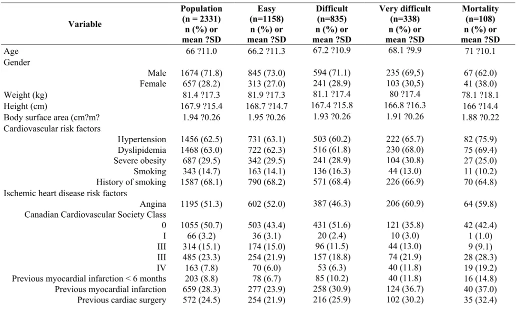
The thesis will include four key studies. The first study will demonstrate the prognostic importance of difficult weaning from CPB in a multicentered Canadian study in 2331 patients [55] in which the MHI participated. The second and third studies are part of a single-centered randomized controlled trial [56] in which we explore the natural hemodynamic and echocardiographic evolution of 120 patients undergoing valvular surgery and describe the characteristics of patients randomized to amiodarone and those requiring inotropes to be weaned from CPB. Finally, the fourth study is the first randomized controlled trial on the intraoperative use of inhaled milrinone for the prevention of difficult separation from CPB.
Chapter 1 Definition and importance of difficult
separation from CPB
In this first chapter, we will define difficult separation from CPB, review the predictors, the significance and the consequences of this important complication in cardiac surgery. Finally, we will present the research performed by the candidate and his collaborators on this issue since the beginning of his PhD program in 2006.
1.1 Definition of difficult separation from CPB
The time sequence in a cardiac surgical procedure is illustrated in Figure 1. In the preoperative period, the patient will be evaluated by several members of the cardiac team, mainly the cardiac surgeon and the cardiac anesthesiologist, to determine the precise surgical procedure to be performed and also for risk stratification. This will be discussed in more detail in section 1.2. After the preoperative evaluation, the patient is brought in the operating room where the surgical procedure is performed. Following the cardiac surgical procedure, the patient is then transferred to the intensive care unit for 24 to 48 hours and to the postoperative ward for 5 to 10 days before being discharged home or to a recovery facility. The operating room time is divided in three periods: before, during and after CPB. Cardiopulmonary bypass is the term used to describe an extracorporeal circuit used during cardiac surgery. The CPB maintenance is under the supervision of a professional called the perfusionist.
Figure 1 Time sequence of a cardiac surgical procedure
A cardiac surgical procedure can be divided into three periods: before, in the operating room (OR) and after the procedure. The time after the procedure includes the time spent in the intensive care unit (ICU) and in the hospital. In the OR, there are three periods, before, during and after cardiopulmonary bypass (CPB). The event at the end of CPB, when the extracorporeal circulation is gradually withdrawn, corresponds to the weaning from CPB. In this thesis, the expression “difficult separation from CPB” is related to both the weaning period and the operative period following CPB.
The role of CPB is to temporarily replace the heart and lungs1
1 The term extracorporeal circuit or heart-lung machine is also used as a synonym of CPB.
which are not functional during the cardiac procedure. The role of CPB is to provide oxygen transport to the body and all the vital organs, except the heart and lungs. The majority of cardiac
surgeries are performed using CPB.
Normally, when CPB is gradually withdrawn, the heart resumes normal mechanical and electrical activity. The CPB is then turned off and removed from the patient. However in some patients, vasoactive drugs such as intravenous noradrenaline are required to maintain an adequate arterial pressure and thus sustain cardiac function and oxygen transport. The dosage of this vasoactive medication can vary from one patient to another. If one vasoactive agent is not sufficient, typically additional medications such as inotropes like intravenous milrinone will be added to wean the patient from CPB. If this pharmacological strategy does not produce the desired effect, the weaning process will fail and the cardiac surgeon will have to reinstitute full CPB. This is called “return on CPB”. As the pharmacological approach is insufficient, mechanical devices used to temporarily support ventricular function such as an intra-aortic balloon pump (IABP) or ventricular assist device (VAD) will be used. There are several reasons or mechanisms to explain this failure to wean from CPB and they will be detailed in Chapter 3. However, the anesthesiologist using TEE will have an important role to play if difficult weaning from CPB occurs. His role will be to rule out any unexpected surgical complication resulting for instance from a dysfunctional prosthesis. In the largest series published so far on the role of The use of CPB can be associated with specific complications that will be discussed in more details in section 1.2.5. At the end of CPB, when the cardiac surgery is completed, the cardiac team will gradually withdraw the extracorporeal support. This process is called weaning or separation from CPB. Weaning from CPB begins when the surgeon decides to gradually reduce the venous return from the CPB and derives it back to the patient. This will be performed only if the cardiac team considers that the patient is stable enough to maintain his oxygen transport. Weaning from CPB is considered complete when the cardioplegia, venous and arterial cannulae are removed. This is followed by the administration of protamine. In this thesis, the expression “difficult separation from CPB” is related to both the weaning period and the operative period following CPB. This period ends when the patient leaves the operating room.
2 Some cardiac surgeries, for instance coronary revascularization, can be performed without CPB. This is
TEE in 12,566 patients undergoing cardiac surgery, Eltzschig et al. [57] observed that TEE influenced cardiac surgical decisions in 9% of all cases. This has also been our experience. [16] In some of these cases, the surgeon will have to revise his procedure. Finally, in rare instances, the CPB weaning process will not be possible and the patient will die in the operating room. Therefore, the process of CPB weaning is a critical moment during cardiac surgery. It is the earliest period after cardiac surgery where the patient is at increased risk of morbidity and mortality. It does not represent a “yes or no” process but a complex situation that requires a comprehensive approach and definition. How has difficult separation from CPB been defined in the literature?
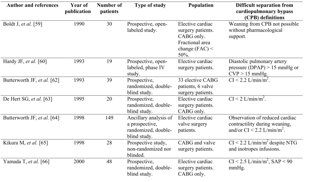
The literature confirms that difficult separation from CPB is a life-threatening condition because, if unsuccessful, it can lead to intraoperative mortality. [58] Several authors have studied and defined difficult separation from CPB. These definitions are summarized in Table 1. [10;17;19;51;59-73]
Butterworth et al. [64] defined difficult weaning from CPB as postoperative hemodynamic instability requiring the use of positive inotropic support such as infusions of dobutamine, epinephrine, or amrinone. Dopamine was considered a positive inotropic drug only if it was infused at rates of 5 µg/kg/min or greater. Patients received inotropic drugs based on the observation of reduced cardiac contractility during weaning from CPB, by measurement of a reduced cardiac index (< 2.2 liters/min/m2), or both. The right ventricle (RV) was directly inspected in the surgical field. The left ventricle (LV) was evaluated using TEE. Duration of drug use was not mentioned and TEE-related definition of RV or LV dysfunction was not identified. Surgenor et al. [74] defined heart failure after cardiac surgery as hypotension or low cardiac index requiring return under CPB, inotropic support or requirement for an IABP. Muller et al. [69] defined hemodynamic instability after cardiac surgery as ventricular dysfunction requiring the use of vasoactive agents based on direct visual inspection of the heart or through TEE examination or a cardiac index < 2 liters/min/m2. The term post-bypass inotropic support has been used as a synonym of difficult separation from CPB and defined as the use of dopamine, dobutamine or epinephrine for at least 12 hours in the intensive care unit. [17;58] The use of dopamine
from 0.5-3.0 µg/kg/min to increase urine output was not considered in the definition of inotropic support. [58] Finally, the term “low cardiac output syndrome” (LCOS) has been used in several studies [75-77] to describe the consequence of difficult separation from CPB. The term LCOS also covers the period in the intensive care unit. It is defined as a postoperative condition: 1) requiring an IABP to be weaned from CPB or in the intensive care unit because of hemodynamic compromise, or 2) requiring inotropic medication (dopamine, dobutamine, milrinone, or epinephrine) to maintain the systolic blood pressure at 90 mmHg and the cardiac output at 2.2 L/min/m² for 30 minutes in the intensive care unit after correction of all of the electrolyte and blood gas abnormalities and after adjusting the preload to its optimal value. The dosage of vasoactive drugs is not mentioned. The term LOF for low output failure has also been used to describe the need for one of the following: an IABP, return to CPB after initial separation or ≥ 2 inotropes at 48 hours
postoperatively. [78]
To summarize, in several of these studies, investigators have used variables such as 1) arterial pressure, 2) cardiac index, 3) filling pressures, 4) TEE findings, 5) amount and duration of vasoactive drugs, 6) subjective intraoperative assessment of reduced RV and LV contractility, 7) the need to return on CPB and 8) the use of mechanical devices to wean from CPB in their definition of difficult separation from CPB. There is also some overlapping in terms of the timing understood when using the phrase difficult separation from CPB. Some consider it to be an intraoperative event only, others a postoperative one, while other investigators include both periods in their definition (Table 1). In the setting of cardiac surgery and in this thesis, we define difficult separation from CPB as the process that may take place between the beginning of the weaning process of CPB and the moment the patient leaves the operating room.
Table 1 Various definitions of difficult separation from CPB proposed in the literature Author and references Year of
publication Number of patients Type of study Population Difficult separation from cardiopulmonary bypass (CPB) definitions Boldt J, et al. [59] 1990 30 Prospective,
open-labeled study. Elective cardiac surgery patients. CABG only. Fractional area change (FAC) < 50%.
Weaning from CPB not possible without pharmacological
support.
Hardy JF, et al. [60] 1993 19 Prospective, open-labeled, phase IV study.
Elective cardiac
surgery patients. Diastolic pulmonary artery pressure (DPAP) > 15 mmHg or CVP > 15 mmHg.
Butterworth JF, et al. [62] 1993 39 Prospective,
randomized, double-blind study. 33 elective CABG patients, 6 valve surgery patients. CI < 2.2 L/min/m2.
De Hert SG, et al. [63] 1995 20 Prospective,
randomized, double-blind study. Elective cardiac surgery patients. CABG only. CI < 2 L/min/m2.
Butterworth JF, et al. [64] 1998 149 Ancillary analysis of a prospective, randomized, double-blind study. Elective cardiac valve surgery patients.
Observation of reduced cardiac contractility during weaning, and/or CI < 2.2 L/min/m2. Kikura M, et al. [65] 1998 28 Prospective study,
non-randomized nor blinded.
CABG and valve
surgery patients. CI < 2.2 L/min/m
2 despite NTG and inotropes infusions.
Yamada T, et al. [66] 2000 48 Prospective,
randomized, double-blind study. Elective cardiac surgery patients. CABG only. CI < 2.5 L/min/m2, SAP < 90 mmHg.
Author and references Year of
publication Number of patients Type of study Population Difficult separation from cardiopulmonary bypass (CPB) definitions Suematsu Y, et al. [67] 2000 167 Retrospective
analysis. Elective cardiac surgery patients requiring CPB.
Intraoperative need for epinephrine and/or
norepinephrine exceeding 0.2 ug/kg/min.
Bernard F, et al. [17] 2001 66 Prospective
observational cohort study.
52 elective CABG alone, 14 combined procedures, valvular surgeries and re-operations.
SAP < 80 mmHg, DPAP > 15 mmHg during weaning from CPB, reinstitution of CPB or an IABP. Presence of significant vasopressor and/or inotropic support.
Van der Maaten JM, et
al. [68] 2001 34 Prospective, non-randomized clinical study.
Elective cardiac surgery patients. CABG only.
CI < 2.4 L/min/m2 and/or MAP < 60 mmHg.
Muller M, et al. [69] 2002 1471 Retrospective
analysis. Elective cardiac surgery patients, including CABG, valve and combined procedures.
Observation of reduced cardiac contractility during or after weaning (either by direct
observation of the right ventricle or with TEE) and/or CI < 2.0 L/min/m2.
Groban L, et al. [70] 2002 381 Post-hoc analysis of a randomized, masked clinical trial of insulin therapy.
Elective cardiac surgery patients. CABG only.
Inotropic, vasoactive and mechanical support (IABP, if needed) initiated if CI < 2.2 L/min/m2, DPAP > 20 mmHg and/or SAP < 90 mmHg.
Author and references Year of
publication Number of patients Type of study Population Difficult separation from cardiopulmonary bypass (CPB) definitions Wagner F, et al. [71] 2003 40 Prospective,
randomized, double-blind study.
Elective cardiac surgery patients. CABG only. FAC < 35% preoperatively.
Moderate to high dose inotropic and/or vasopressor therapy, or the need of a mechanical support (IABP).
Tsukui H, et al. [72] 2004 151 Retrospective
analysis. Elective cardiac surgery patients including ischemic heart disease, valvular and congenital pathologies, along with miscellaneous procedures. Epinephrine, norepinephrine, dopamine, dobutamine and milrinone were used if
hemodynamic instability during weaning from CPB. IABP was installed if instability persisted despite medical treatment.
McKinlay KH, et al. [73] 2004 1009 Retrospective
analysis. Elective cardiac surgery patients. CABG and complex procedures.
Inotropic support in the form of dopamine (> 5 ug/kg/min) or any dose of epinephrine, norepinephrine, dobutamine or milrinone, along with IABP vs. hypotension, low cardiac output and inability to separate from bypass.
Author and references Year of
publication Number of patients Type of study Population Difficult separation from cardiopulmonary bypass (CPB) definitions Surgenor SD et al. [78] 2006 8004 Prospective analysis CABG Low output failure: the need for
one of the following: an IABP, return to CPB after initial separation or ≥ 2 inotropes at 48 hours postoperatively
Robitaille A, et al. [10] 2006 1498 Retrospective
analysis. Elective cardiac surgery patients, all types combined (CABG, valve, complex and miscellaneous procedures). SAP < 80 mmHg, DPAP or wedge pressure > 15 mmHg during weaning from CPB, reinstitution of CPB or an IABP. Presence of significant
vasopressor and/or inotropic support.
CABG, coronary artery bypass graft; CI, cardiac index; CPB, cardiopulmonary bypass; DPAP, diastolic pulmonary artery pressure; FAC, fractional area change; IABP, intra-aortic balloon pump; MAP, mean arterial pressure; NTG, nitroglycerin; SAP, systolic arterial pressure; TEE, transesophageal echocardiography.
The first element of the definition is the systolic arterial pressure. Systolic pressure is routinely used and monitored in the operating room and the intensive care unit. It is used as an index of organ perfusion pressure and, therefore, tissue perfusion pressure. However the site of measurement of this parameter is very important. Systolic arterial pressure, when reduced in the hemodynamically unstable patient, has to be confirmed by central measurement, aortic or femoral. [79;80] This is a very important point and illustrated in Figure 2.
Figure 2 Radial to femoral artery pressure gradient during cardiac surgery
(A) Before cardiopulmonary bypass (CPB) a normal gradient between the radial (Rad) and femoral (Fem) artery was observed. (B) During the early part of CPB, no abnormality in gradient was observed. (C) The gradient appears during the later part of CPB. (D) After CPB, the systolic and mean femoral artery pressures were 118 mmHg and 81 mmHg, respectively. The systolic and mean radial artery pressures were 90 mmHg and 69 mmHg, respectively. (HR, heart rate; AP, arterial pressure; PAP, pulmonary artery pressure; CVP, central venous pressure) (With permission of Denault et al. [80]).
The appearance of a pressure gradient between the radial and femoral arteries can be commonly observed both in the cardiac operating room and in the intensive care unit in
patients who are thought to be hemodynamically unstable. Despite previous descriptions of this observation [79;81] in the literature, the mechanisms responsible for this gradient remain poorly understood [82] and its presence is not routinely recognized. The pressure gradient is normally < 20 mmHg between the aortic root and radial artery, being higher in the distal arteries. [83] In our clinical experience involving a large series of patients undergoing cardiac surgery, radial artery-aortic root systolic pressure gradients > 25 mmHg (the radial being lower than the aortic) occur in approximately 30-50% of cardiac procedures (Section 1.4.1). Maximum gradients are usually observed just after separation from CPB. In some patients, these gradients resolve towards the end of the procedure, but there is limited predictability regarding their dynamic variations. Transesophageal echocardiography and transthoracic echocardiography can also be used to detect an abnormal arterial gradient when clinically significant mitral regurgitation is present. [80] Early recognition of an abnormally wide aortic-radial arterial pressure gradient is therefore the first and most important step in excluding intraoperative hemodynamic instability as a cause of persistent hypotension in cardiac surgery.
The second element of the definition is cardiac filling pressure such as central venous pressure, diastolic pulmonary artery pressure and pulmonary capillary wedge pressure. As difficult separation from CPB represents cardiac dysfunction (either systolic, diastolic or both), filling pressures will be elevated in the presence of reduced systemic pressure. Elevated filling pressures are usually defined as either diastolic pulmonary artery pressure or pulmonary capillary wedge pressure >15 mmHg [60] or 20 mmHg. [70] This value is somehow arbitrary because it can depend on several factors, the most common being diastolic function or ventricular compliance. Ventricular compliance is unique to each surgical patient and is almost invariably altered after cardiac surgery. [39] Alteration in ventricular compliance after cardiac surgery has been described using echocardiography since the early 1990s. [84-92] If ventricular compliance is reduced after cardiac surgery, ventricular filling pressures will increase in order to maintain an appropriate preload and cardiac output. This observation explains why Reichert et al. [93] defined post-cardiac surgery hypovolemia as a pulmonary capillary wedge pressure value less than the
preoperative wedge pressure +10 mmHg. The “+ 10 mmHg” is a correcting factor based on the experience and observations of the authors, who noted that higher filling pressures were required after CPB in order to maintain an adequate preload and consequently an adequate cardiac output. Several of the studies pertaining to filling abnormalities or diastolic dysfunction after CPB examined a single echocardiographic parameter often limited to the left ventricle, [68;88;90;92;94-98] as opposed to biventricular systolic and diastolic evaluation. [40;43] This limitation could result in a misinterpretation of the actual change in cardiac function. This will be discussed in section 1.4.2.
The third element in the definition of difficult separation from CPB is the pharmacological intervention. The dosage and amount of vasoactive agents required for weaning from CPB needs to be quantified. The pharmacological approach on the use of vasoactive medication differs significantly from center to center, even in the same country. [99] At the MHI, significant vasopressive and/or inotropic support is defined by the use of norepinephrine > 0.06 µg/kg/min, epinephrine > 0.06 µg//kg/min, dobutamine > 2 µg/kg/min or the use of milrinone. [52] Returning on CPB can be secondary to hemodynamic or mechanical complications and is a severity criterion. The use of an IABP and VAD to wean from CPB implies a severe mechanical problem most likely related to the patient’s underlying condition. Finally, in order to standardize the vasoactive management during CPB (Appendix 2) and the weaning process, (Appendix 3) we developed algorithms to be applied in studies dealing with separation from CPB. [50;52;56]
In summary, the definition used to describe difficult separation from CPB varies significantly among investigators. Cleary defined hemodynamic variables, particularly the site of measurement of the arterial pressure, seem essential in detecting the true presence of difficult separation from CPB. Filling pressure indices have to be evaluated in relation with baseline measurements, as each patient can serve as his own control. A systematic echocardiographic approach would be useful to identify the mechanism at work in difficult separation from CPB. The use of vasoactive agents should follow a logical algorithm based on hemodynamic and echocardiographic information. Finally, a classification could be used as it appears that different grades of severity in separation from CPB can be present. The
worst form of difficult separation from CPB would be the one associated with the requirement for mechanical devices.
1.2 Predictors of difficult separation from CPB
Patients at risk of complications and death after cardiac surgery can be identified through the use of scores developed in several large-scale studies in which multivariate analysis identified variables associated with an increased risk of morbidity and mortality. Some of these scores include for instance the MHI score, [8] the Parsonnet score, [100] the EuroSCORE, [101] the Cardiac Anesthesia Risk Evaluation (CARE) score [102] and the Society of Thoracic Surgeons (STS) score. [103] These scores are useful because they can provide an estimation of mortality and morbidity. There is so far no score that enables the identification of patients at risk of difficult separation from CPB. It is likely that similar variables associated with an increased risk of morbidity and mortality will be associated with difficult separation from CPB. These variables can be classified as demographic, surgical, biochemical, hemodynamic and echocardiographic.
1.2.1 Demographic and surgical variables
Several demographic variables in relation with the type of surgery have been identified as important predictors of difficult weaning from CPB.
1.2.1.1 Coronary revascularization
In patients undergoing coronary revascularization, Surgenor et al. [74] identified reoperation, urgent surgery, peripheral vascular disease, diabetes and renal failure requiring dialysis as demographic and surgical variables associated with an increased mortality from heart failure. Other predictors of difficult separation from CPB in coronary bypass surgery are older age and female gender, [58] previous myocardial infarction and chronic pulmonary obstructive disease. [69] Rao et al. [75] retrospectively analyzed the risk of LCOS from a database of 4558 patients operated for coronary revascularization in Toronto between 1990 and 1993. The independent predictors of LCOS were determined by stepwise
logistic regression analysis. The prevalence of LCOS was 9.1%. The independent predictors were (odds ratio in parenthesis) left ventricular ejection fraction < 20% (5.7), repeat operation (4.4), emergency operation (3.7), female gender (2.5), diabetes (1.6), age > 70 year-old (1.5), left main coronary artery stenosis (1.4), recent myocardial infarction (1.4) and triple-vessel disease (1.3).
1.2.1.2 Valvular surgery
Valvular surgery is typically longer and more complex than coronary revascularization. It is not surprising that it is associated with an increased risk of postoperative inotropic requirement. In a study involving 1009 patients undergoing cardiac surgery, McKinlay et al. [73] identified coronary revascularization in association with mitral valve repair or replacement as an independent risk factor for postoperative inotropic support. Maganti et al. [77] retrospectively analyzed the risk of LCOS from a database of 2255 patients operated for isolated aortic valve replacement in Toronto between 1990 and 2003. The independent predictors were determined by stepwise logistic regression analysis. The prevalence of LCOS was 3.9%. The independent predictors were (odds ratio in parenthesis): renal failure (5.0), earlier year of operation (4.4), left ventricular ejection fraction < 40% (3.6), shock (3.2), female gender (2.8), and increasing age (1.02). Overall operative mortality was 2.9%. An additional factor associated with the requirement for inotropic drugs after valvular surgery is the anesthesiologist’s preference for the use of vasoactive medications. [64] In a study involving aortic valve replacement in combination with revascularization, Ahmed et al. [104] identified preoperative renal disease, elevated left ventricular end-diastolic pressure (≥ 20 mmHg), reduced left ventricular ejection fraction (≤ 40%) and low cardiac index (≤ 2.5 L/m/m? as predictors of postoperative inotropic requirements.
1.2.1.3 Duration and utilization of cardiopulmonary bypass
Both the duration of CPB and cross-clamping are surgical variables that predict hemodynamic complications in several studies. [10;17;58;69;70;72;73;105;106] We have also documented that hemodynamic complications in patients undergoing coronary
revascularization were observed in 53% of patients in whom CPB was used, as opposed to 14% of patients undergoing surgery with off-pump bypass. [10] The use of CPB was indeed an independent predictor of hemodynamic complications (p < 0.0001), and this finding was also observed by other authors. [58;72;107] As suggested by Butterworth et
al., [64] a longer CPB time can be associated with technical or mechanical difficulties or
associated procedures, including valvular surgery and coronary revascularization. As the CPB is longer, the patient and the myocardium are exposed to the effect of the inflammatory response with a potentially greater need for blood products. The latter is not only associated with LOF but also with increased mortality. [72;78]
1.2.2 Biochemical variables
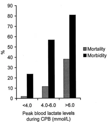
Among the biochemical variables, our group observed that an elevated veno-arterial PCO2 gradient before the cardiac surgical procedure was an independant variable associated with an increased risk of difficult separation from CPB. [105] Elevated veno-arterial PCO2 gradient is a marker of ischemia, [108] in the same manner as lactate. Not surprisingly, the intraoperative lactate level obtained during CPB has also been shown to correlate with difficult separation from CPB and mortality (Figure 3). [9]
Figure 3 Mortality and morbidity in relation with lactate level during CPB
Positive correlation in 1376 cardiac surgical patients between peak blood lactate levels during cardiopulmonary bypass (CPB) and the rate of postoperative morbidity and mortality (p < 0.001). (With permission of Demers et al. [9])
These two studies, conducted at the MHI, tend to support that measures of reduced oxygen transport or hypoperfusion before or during CPB could either be markers or determinants of hemodynamic instability and mortality after cardiac surgery. In that regard, Rao et al. [76] documented that, in 623 patients undergoing coronary revascularization, the only predictor of LCOS was the myocardial lactate release after 5 minutes of cross-clamping. Age and reduced left ventricular ejection fraction were the only two predictors of this metabolic abnormality after CPB. The rise in creatinine kinase (CK) was not a predictor of LCOS. Other authors have also confirmed that reduced myocardial pH [109] (Figure 4) or increased myocardial lactate measured during CPB [110] have been shown to be predictors of increased postoperative inotropic support and mortality. This abnormal lactate release could imply delayed recovery of normal aerobic myocardial metabolism. As
the myocardial metabolism is altered, myocardial function will be abnormal. Therefore, the risk of difficult separation from CPB is likely to correlate with indices of global or regional myocardial tissue hypoperfusion. In that regard, a recent paper by Turer et al. [111] explored the new field of metabolomics in cardiac surgery. The measurements of several metabolites produced from ischemia/reperfusion during retrograde cardioplegia were analyzed. An association between the duration of inotropic support and myocardial lactate was observed. This study suggests that patients with left ventricular dysfunction have limited myocardial metabolic reserve and flexibility after global ischemia/reperfusion stress.
Figure 4 Intramyocardial acidosis and inotropic requirement
Comparison of myocardial tissue pH37C between patients who needed inotropic support versus those who did not at 5 time points during surgery: Before aortic occlusion (AC), mean during AC, at 5 minutes of reperfusion, at 10 minutes of reperfusion, and at the end of reperfusion. (IABP, intra-aortic balloon pump).(Adapted from Kumbhani et al. [109])
1.2.3 Hemodynamic and echocardiographic variables
Among the hemodynamic data predicting post-CPB inotropic support and mortality after cardiac surgery, left ventricular systolic dysfunction is frequently found as the most important and frequently reported variable. [58;64;69;70;73;75;77;78;104;106;109] Left ventricular dysfunction is either defined by a history of congestive heart failure, by a cardiac variable such as reduced left ventricular ejection fraction (LVEF) or ventricular enlargement, or as its consequence on daily living, such as the New York Heart Association (NYHA) classification. All these definitions have been associated with postoperative inotropic requirement. [10;58;64;69;78] Left ventricular dysfunction will be associated with echocardiographic evidence of abnormal regional or global wall motion and can also be associated with an elevated left ventricular end-diastolic pressure (LVEDP). This parameter has also been reported as an independent predictor of inotropic requirement [58;104] and mortality. [11]
Right ventricular systolic and diastolic dysfunction may also be a predictor of mortality and morbidity. Maslow et al. [112] studied patients with reduced left ventricular systolic function (LVEF ≤ 25%) before coronary revascularization. Those without right ventricular dysfunction prior to surgery had less inotropic requirement after revascularization and a mortality rate of 9.7%. In contrast, patients with reduced LVEF associated with reduced right ventricular dysfunction experienced more frequent difficult separation from CPB and a mortality rate of 100% within 18 months. This study supports the hypothesis that preoperative right ventricular systolic dysfunction is a predictor of difficult weaning from CPB and mortality before cardiac surgery. However, right ventricular diastolic dysfunction may also be an important criterion to be evaluated. In a pilot study of 121 patients undergoing cardiac surgery, Carricart et al. [34] observed that preoperative abnormal hepatic venous flow, as a marker of right ventricular diastolic dysfunction, [113;114] was associated with difficult weaning from CPB. In a subset of patients undergoing valvular surgery only, abnormal hepatic venous flow before surgery was associated with a higher Parsonnet score, more atrial fibrillation, pacemaker
requirement, mitral valve replacement, reoperation, a lower systemic mean arterial (MAP) to mean pulmonary artery pressure (MPAP) ratio, a higher wall motion score index, a higher incidence of abnormal right ventricular systolic function and more frequent use of intravenous milrinone. However, abnormal hepatic venous flow before cardiac surgery was not found to be an independent predictor of difficult separation from CPB and worse outcome. In that study, pulmonary hypertension defined using the MAP/MPAP ratio was the best predictor of hemodynamic complications.
Pulmonary hypertension is another hemodynamic variable associated with an increased risk of difficult weaning from CPB, morbidity and mortality in cardiac surgery. [8;100;115-117] However, few studies have reported an association between pulmonary hypertension and difficult weaning from CPB. [10;34;46] This will be discussed in more detail in Chapter 6.
1.2.4 Patient-prosthesis mismatch
Aortic patient-prosthesis mismatch (PPM) is the result of a prosthesis too small for the patient’s body surface area (BSA). [118-125] The selection of the type and size of prosthetic valve is also very important, because it has been shown that, if the effective orifice area (EOA) of the valve is too small in relation to body size, then occurs a so-called PPM, which increases intraoperative and long-term mortality (Figure 5). [118-125]
Figure 5 Patient–prosthesis mismatch
A 71-year-old man with a body surface area of 1.89 m² was re-operated for symptoms of severe aortic valve stenosis (severe dyspnea, NYHA class IV and pulmonary hypertension of 60/15 mmHg). He had aortic valve replacement (AVR) 4 years ago with a Carbomedics #19 mechanical bileaflet prosthesis (effective orifice area (EOA) = 1.06 cm²). The preoperative mean gradient was 41 mmHg. The intraoperative aspect of the prosthetic valve was completely normal. (B) Example of an aortic root enlargement procedure in a 69-year-old patient with a reduced aortic diameter requiring AVR. (Courtesy of Dr. Michel Carrier with permission of Denault et al. [12])
From various studies, PPM can be found in 19-70% of patients undergoing aortic valve replacement (AVR). [119-122] In a study including 1266 patients who underwent AVR at the Quebec Heart and Lung Institute (QHLI), the prevalence of moderate PPM defined as an index EOA (iEOA) ≤ 0.85 cm2/m2 was 38%, and that of severe PPM (iEOA ≤ 0.65 cm2/m2) was 2%. After adjusting for other risk factors, moderate and severe PPM were associated with a 2.0-fold (95% confidence interval: 1.1-3.7) and 12.6-fold (95% confidence interval: 4.3-37.0) increase in mortality, respectively. It is possible that the increased LVEDP and left ventricular afterload with associated reduced coronary flow reserve [126] with PPM may predispose to difficult separation from CPB. In a study of 156 patients undergoing AVR and followed-up for a median period of 3.5 years, Brown et al. [127] observed that postoperative events and survival after AVR were more related to the severity of LV diastolic function than PPM. Finally, the link between aortic PPM and difficult separation from CPB has not been described.
PPM of the mitral valve has recently been described [128] and defined as an iEOA ≤ 1.2 cm²/m². In a study which included 929 consecutive patients undergoing mitral valve replacement, severe PPM was associated with a 3-fold increase in postoperative mortality after adjustment for other risk factors. As mitral PPM will be associated with postoperative pulmonary hypertension, right ventricular failure and consequently difficult separation from CPB could result from this condition. The relation between mitral PPM and difficult separation from CPB has not been described.
1.2.5 Other factors involved in the risk of difficult separation from CPB
Other factors could predispose to difficult separation from CPB in cardiac surgery. For instance, aberrant positioning of the cardioplegia cannula could be associated with inadequate myocardial protection (Figure 6).
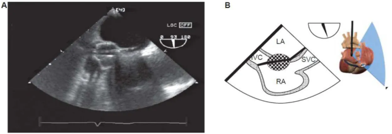
Figure 6 Retrograde cardioplegia cannula
(A, B) Bicaval view showing the retrograde cardioplegia cannula positioned toward the atrial septum through the patent foramen ovale. (IVC, inferior vena cava; LA, left atrium; RA, right atrium; SVC, superior vena cava). (Photo courtesy of Dr. Baqir Qizilbash with permission of Denault et al. [13]).
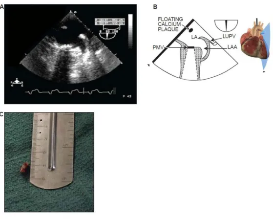
Coronary embolization from air or residual debris that can occur after CPB (Figure 7) could also be associated with difficult weaning from CPB.
Figure 7 Calcium emboli in valvular surgery
A 70-year-old man who underwent coronary revascularization and combined aortic and mitral valve replacement. (A,B) As weaning from cardiopulmonary bypass (CPB) proceeded, floating material was detected in the left atrium (LA) from this mid-esophageal two-chamber view. The attending surgeon went back immediately to full CPB. (C) This material was a 4 x 1 mm floating calcium plaque which was removed. The patient had no postoperative neurological complications (LAA, left atrial appendage; LUPV, left upper pulmonary vein; PMV, prosthetic mitral valve) (With permission of Denault et al. [13]).
Additionally, technical problems such as a residual paravalvular leak or dysfunctional prosthesis (Figure 8) could also contribute to difficult weaning from CPB.
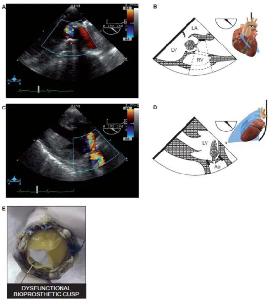
Figure 8 Dysfunctional AoV bioprosthesis after AVR
A 60-year-old man was reoperated after valve replacement (AVR) for periprosthetic aortic regurgitation (AR). (A–D) After the procedure, abnormal significant AR is still visible on the mid-esophageal long-axis and deep transgastric views. The new bioprosthesis was removed and replaced by another one. (E) Upon examination of the defective bioprosthesis, abnormal motion of one of the leaflets was noted (Ao, aorta; AoV, aortic valve; LA, left atrium; LV, left ventricle; RV, right ventricle). (Photo E courtesy of Dr. Tack Ki Leung, with permission of Denault et al. [13]).
All these conditions can be diagnosed and prevented with TEE. Finally, the reperfusion syndrome could also be associated with unexpected pulmonary hypertension upon weaning from CPB. This will be discussed in Chapter 6.
To summarize, there are several demographic, surgical, biochemical, hemodynamic and echocardiographic preoperative variables that can be associated with hemodynamic instability and difficult weaning from CPB after cardiac surgery. They are important to document if a new therapy is introduced, so that similar groups can be compared. Few of the demographic and surgical variables can be modified before planning cardiac surgery. The inclusion of left and right ventricular systolic and diastolic dysfunction, PPM and pulmonary hypertension as predictors of difficult separation from CPB is new and interesting because these variables could possibly be modified before and during cardiac surgery. Furthermore, the role of TEE is to monitor and to diagnose conditions that could result in difficult separation from CPB and could be modified through a medical or surgical approach. Table 2 and Table 3 summarize studies in which the primary endpoint was hemodynamic instability or difficult weaning from CPB after cardiac surgery.