Université de Montréal
Caractérisation de l’implication de β-caténine
dans les tumeurs surrénaliennes.
Présenté par Julien Durand
Département de Sciences Biomédicales Faculté de Médecine
Mémoire présenté à la Faculté des Études Supérieure en vue de l’obtention du grade de Maitre en Science (M.Sc) en Sciences Biomémicales.
Août 2010
Université de Montréal
Faculté des études supérieures et postdoctorales
Ce mémoire intitulé :
Caractérisation de l’implication de β-caténine dans les tumeurs surrénaliennes.
Présenté par : Julien Durand
A été évaluée par un jury composé des personnes suivantes : Président-rapporteur : DR Pangala.V. Bhat
Directeur de recherche : DR. Isabelle Bourdeau Membre du jury : DR. Réjean Lapointe
Table des matières
Abrégé/ Résumé ... vi
Abstract ... vii
List of figures ... viii
List of tables... ix
Abbreviation ... x
Remerciements ... xii
Chapter 1. Review of literature ... 1
Section 1. The Adrenal Gland ... 1
1.1. Adrenal gland physiology. ... 1
1.2. Adrenocortical Steroidogenesis . ... 2
1.2.1. Synthesis of Adrenal Steroids ... 2
1.2.2. Control of steroid production ... 4
1.3. Adrenal gland development ... 6
Section 2. Tumours of the adrenal cortex ... 8
2.1. Adrenocortical Adenomas ... 8
2.1.2. Cortisol-secreting adrenocortical adenomas ... 9
2.1.3. Aldosterone-secreting adenomas ... 9
2.2. Adrenal Hyperplasias. ... 10
2.2.1. ACTH-independent micronodular adrenal hyperplasias and PPNAD ... 10
2.2.2. ACTH-independent macronodular adrenal hyperplasia ... 10
2.2.3. Congenital hyperplasia (CAH) ... 12
2.3. Adrenocortical Carcinoma (ACC) ... 12
2.4. Genetic and pathway alterations of adrenocortical tumours. ... 12
2.4.1. Alterations of TP53 ... 13
2.4.2. Insulin Growth Factor 2 (IGF2) ... 14
2.4.3. PRKAR1A and other cAMP pathway alterations ... 16
2.4.4. Ras- pathway alterations. ... 17
2.4.5. MEN 1 ... 18
2.5. Adrenocortical tumourogenesis ... 19
2.5.1. Clonality of Adrenocortical tumours. ... 19
Section 3. Wnt/β-catenin signaling ... 22
3.1 Wnt signaling pathways. ... 22
3.1.1 Non-canonical or planar cell polarity (PCP) signaling ... 22
3.1.2 The Wnt-Ca2+ pathway ... 23
3.1.3. The Wnt/ β-catenin pathway ... 23
3.2. β-catenin ... 26
3.2.1. β-catenin in the nucleus... 27
3.2.2. Regulation of β-catenin targets genes ... 28
3.2.3. β-catenin transcription factors and alternative activation. ... 31
Section 4. Wnt implications in normal adrenal function and development ... 34
4.1. Wnt in the adult adrenal gland ... 34
4.2. Wnt signaling in adrenal steroidogenesis. ... 35
4.3. Wnt/β-catenin in adrenal development. ... 39
Section 5. Wnt / β-catenin in adrenal tumourogenesis. ... 41
5.1. Wnt / β-catenin signaling alterations in cancers ... 41
5.2. Implication of Wnt signaling alterations in adrenal tumors. ... 44
5.2.1. Implication of Β-catenin mutations. ... 46
5.2.2. Wnt/ β-catenin and cAMP pathway cross talk ... 48
5.2.3. Model of β-catenin driven adrenal tumourogenesis ... 49
5.3. β-catenin and human Adrenocortical cell lines. ... 50
5.3.1. H295R cells. ... 51
5.3.2. SW13 cells ... 51
5.3.3. HAC15 cells ... 52
Chapter II. Research Project ... 53
1. Basis for Research ... 53
2. Research Plan and objectives ... 55
3. Article 1 ... 57
Chapter III. Discussion ... 93
Chapter IV. Conclusions ... 100
Abrégé/ Résumé
Les lésions surrénaliennes surviennent dans la population générale à une fréquence d’environ 2-3%. Parmi les anomalies génétiques identifiées jusqu’à présent dans les tumeurs surrénaliennes, les mutations somatiques de β-caténine sont les plus prévalentes. Elles sont présentes dans environ 20% des adénomes et carcinomes cortico-surrénaliens. β-caténine est l’élément central de la voie canonique de WNT qui joue un rôle crucial dans le développement embryonnaire, l’homéostase et la tumourigenèse. Les mutations activatrices de β-caténine conduisent à l’accumulation nucléaire de β- caténine qui interagit avec les TCF/LEF-1 qui active la transcription des gènes cibles. Les gènes cibles de β-caténine, varient et dépendent du contexte cellulaire. Dans la glande surrénale, les gènes cibles de β-caténine sont inconnus. Nous avons effectué des études de microarray qui nous ont permis d’identifier 490 transcrits dérégulés dans les adénomes corticosurrénaliens porteurs de mutations ponctuelles de β-caténine. L’expression aberrante d’ISM1, RALBP1, PDE2A, CDH12, ENC1, PHYHIP et CITED2 dans les adénomes porteurs de mutations de β-caténine a été confirmée par PCR en temps réel. Le traitement des cellules humaines de carcinome cortico-surrénalien H295R (mutation de CTNNB1, Ser45Prol) avec les inhibiteurs de caténine/TCF (PKF115-584 et PNU74654) ont confirmé l'implication de β-caténine dans la régulation transcriptionelle d’ISM1, RALBP1, PDE2A, ENC1 et CITED2. En conclusion, nos travaux ont conduit à l’identification de nouveaux gènes cibles de β-catenin impliqués dans la tumourigenèse cortico-surrénalienne.
Mots clés : Glandes surrénales, Cortex surrénalien, Adénome cortico-surrénalien, Voie de signalisation de WNT, Microarray, Tumourigenèse, CTNNB1/β-caténine, PKF115-584, H295R
Abstract
Adrenal lesions occur in the general population at a prevalence of about 2-3%. Several mutations have been identified in adrenocortical tumours. β-catenin mutations were recently found to be the most frequent genetic alteration in both sporadic adrenocortical adenomas and carcinomas (20-30%). β-catenin is the central player in canonical Wnt signaling which plays a key role in organ/ gland development, maintenance of homeostasis and tumourigenesis. Activation of Wnt signaling by altered regulation of β-catenin levels evokes β-catenin accumulation in the nucleus, and interaction with the TCF/LEF-1 proteins that activates the transcription of target genes. These target genes are believed to be highly cell and context specific and are linked to developmental and cell cycling functions. β-catenin target genes in adrenocortical tumours are unknown. Using microarray technology, we found 490 transcripts that are deregulated in adrenocortical adenomas harbouring β-catenin activating mutations in comparison to non mutated adenomas and normal adrenal glands. These genes differ highly in function and many are poorly characterized genes. Differential expression of ISM1, RALBP1, PDE2A, CDH12, ENC1, PHYHIP and CITED2 in adenomas with activating β-catenin mutations was confirmed by real-time PCR. Treatment of human adrenocortical carcinoma cells, H295R (CTNNB1 Ser45Prol), with β-catenin/TCF inhibitors (PKF115-584 and PNU74654) further confirmed the implication of β-catenin on the transcriptional regulation of ISM1, RALBP1, PDE2A, ENC1 and CITED2. In conclusion, we have found new potential β-catenin target genes that may be involved in adrenocortical tumourigenesis.
Key words: Adrenal gland, adrenocortical adenoma, adrenocortical carcinoma, WNT signaling, microarray, tumourgenesis, CTNNB1/β-catenin, PKF115-584, H295R
List of Figures
Figure 1 : Adrenal gland and its zones. ... 2
Figure 2: Steroidogenesis pathway in the adrenal gland and gonads. (2) ... 4
Figure 3: Human cortisol production in the adrenal gland. ... 5
Figure 4: Developmental events of the adrenal gland from embryo to adult. ... 7
Figure 5 : Alterations of 11p15 locus and IGF-II overexpression in ACC. ... 15
Figure 6: Wnt pathways independant of β-Catenin. ... 23
Figure 7: (I) Wnt/β-catenin pathway ... 25
Figure 8: The 3 cellular states of β-catenin. ... 26
Figure 9: β‑catenin dependent gene transcription. . ... 27
Figure 10: WNT/β-catenin in adrenal function . ... 37
Figure 11: Common causes of Wnt signaling aberrant activation in cancer cells. ... 41
Figure 12 : Mutations of exon 3 of β-catenin in the literature. ... 42
Figure 13: Summary of β-catenin mutations and aberrant accumulation of beta-catenin in adrenocortical diseases.. ... 48
List of Tables
Table I: Classification of adrenal incidentalomas. ... 8
Table II: Genes /and associate familiale syndromes in adrenocortical tumours. ... 13
Table III: Genes that are upregulated directly by β-catenin/TCF in Humans ... 29
Table IV: Transcription factors other than TCF family members that may use β-catenin as a co-activator or co-repressor. ... 32
Table V: Nuclear β-catenin immunoreactivity in adrenal development ... 39
Table VI: Deregulation of Wnt signaling/target genes in adrenocortical ... 45
Table VII: Summary of β-catenin mutations found in adrenocortical tumors ... 47
Abbreviations
AA: Adrenocortical adenomaACC: Adrenocortical carcinoma
ACTH: Adrenal Corticotrophic Hormone
AIMAH: ACTH-independant macronodular adrenal hyperplasia Ang2: Angiotensin 2
ANP : Atrial natriuretic peptide APC : Adenomatous polyposis coli BWS: Beckwith-Wiedemann syndrome cAMP: Cyclic adenosine mono phosphate CGH: Comparative genomic hybridization CHIP: Chromatin immupopreciptation CK1: Cyclin kinase 1 CNC: Carney complex CRH: Corticotropin-releasing hormone CS: Cushing’s syndrome DHEA: Dehydroepiandrosterone DM: Deletion mutation
DNA: Deoxyribonucleic acid DSH or DVL: Dishevelled genes
EMSA: Electrophoresis mobility shift assay FAP: Familial adenomatous polyposis coli FCCM: Fat cell conditioned medium FISH: Fluorescence in situ hybridization FOXO:Forkead
GSK3β: Glycogen synthase kinase 3 beta KO: Knockout
LOH: Loss of heterozygosity LRP: Low density liprotein receptor miRNA: Micro ribonucleic acid NA: Normal adrenal gland PCP: Planar cell polarity
PI3K : Phosphatidylinositol 3-kinase PKA: Protein kinase A
PM: Point mutation
PPNAD: Primary pigmented adrenocortical disease RNA: Ribonucleic acid
RT: Reverse transcriptase SF-1: Steroidogenic factor 1 siRNA: Small interfering RNA
StAR: Steroidogenic acute regulatory protein TBE: TCF binding element
TCF: T-cell transcription factor UPD: Unipaternal disomy WNT: Wingless
WRE: Wnt responsive element Wt: Wild type
WT: Wilms tumour ZF: Zona fasciculate ZG: Zona glomerulosa ZR: Zona reticularis
Remerciements
Je désire premièrement remercier mes parents Danielle Laurin et Daniel Durand pour leurs supports émotionnels et financiers au cours de mes études. Sans eux je ne me serais jamais rendue jusqu’ici. Au reste de famille, je vous remercie tous pour votre aide et support.
Ensuite j’aimerai remercier mes anciens superviseurs Dr. Edward Ishiguro et Dr Hannah Nyugen qui ont su m’aider a développer mon esprit scientifique et qui m’ont enseigné tant de choses.
J’aimerai remercier mon superviseur de Maitrise Dr. Isabelle Bourdeau qui a su m’orienter dans la bonne direction dans ma recherche et qui a toujours encouragé mon initiative et mon vouloir dans faire plus. Je la remercie aussi pour sa patience. Je désire souligner mon appréciation pour son support et sa compassion durant certains moments difficiles de ma vie survenus au cours de ma maitrise.
Je remercie Dr. Antoine Lampron pour son aide au début de mes projets et pour le temps qu’il a pris pour discuter de mes résultats et des directions à prendre.
Je remercie spécialement Dr. Tania Mazzuco pour ses conseils, son aide direct et indirect dans mes projets et son encouragement. Je la remercie aussi pour son enthousiasme qui était tant contagieux.
Je remercie aussi ma blonde Melissa Lewis pour son support, pour le temps qu’elle a pris a corriger ce mémoire et surtout pour sa patience à cause des longues heures que j’ai passées au laboratoire et à la rédaction de ma thèse.
Finalement, je remercie tous ceux qui ont étés au labo 7-124 durant ma maîtrise, Audrey C, Mimi T, Daniel M, Sylvie O, Aurélia S, Fabien M, Eléonore B, Livia M, et Isabelle L qui on rendu ce temps à la maitrise très agréable.
Chapter 1. Review of literature
Section 1. The Adrenal Gland
1.1. Adrenal gland physiology
The adrenal gland is a small gland (4 grams in adults) situated at the upper extremity of both kidneys in mammals (Figure 1A, page 2). It is composed of two completely distinctive zones, the medulla and the cortex (Figure 1B, page 2) surrounded by a capsule. The medulla is the central part and represents 10-20% of the adrenal gland weight in humans. The medulla is composed primarily of chromaffin cells and secretes epinephrine and norepinephrin. The cortex is yellow in healthy individuals and is itself composed of three zones (zona glomerulosa, zona fasciculata, zona reticucularis) each responsible for the synthesis of different hormones. The cells in each layer have slightly different structures, reflecting their different functions (Figure 1D, page2). Note that the focus of this project was on aspects involving only the adrenal cortex.
The zona glomerulosa is responsible for aldosterone production and is incapable of producing the other hormones secreted by the cortex (see section 1.2). The zona fasciculata is the largest of three zones and makes up to 75% of the total cortex. The cells within this zone are much larger and contain high lipid content. The zona reticularis is a compact layer surrounding the medulla and produces androgens and cortisol along with the zona fasciculata.
A B
C D
Figure 1 : Adrenal gland and its zones. (A) Picture showing location of the adrenal gland above the kidney. www.nlm.nih.gov (B) Lateral cut of an adrenal gland showing location of the medulla and the cortex www.thyroidinstitute.org/images/adrenal_gland. (C,D) Hematoxylin and eosin staining of the adrenal cortex tissue (C) showing that it is separated into three zones; reticularis; fasciculata (zF); and, glomerulosa (D) http://www.ouhsc.edu/histology
1.2. Adrenocortical Steroidogenesis
Steroids are a type of organic compound that contain a core structure of 4 rings (cyclohexanes and one cyclopentane) that are joined to each other. Steroid structures are highly conserved in both the animal and plant kingdoms. Steroid specificity is derived from diverse modifications of the core structure catalyzed by different enzymes. In mammals the core structure is provided by cholesterol.
1.2.1. Synthesis of Adrenal Steroids
In the adrenal gland, cholesterol can be synthesized de novo but the majority of the cholesterol comes from plasma lipoproteins which are absorbed by the cells through membrane receptors (1). Once inside the cell, cholesterol is modified by cytochromes P450 enzymes. Steroid synthesis reactions are limited by the steroidogenic acute regulatory
protein (StAR) which controls cholesterol translocation to the mitochondrial outer membrane.
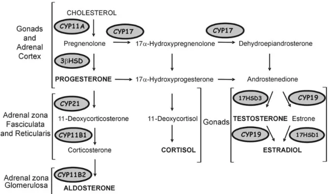
Adrenal steroidogenesis for the production of cortisol, aldosterone and sex hormones is depicted in Figure 2 (page 4). The CYP11A1 (cytochrome P450, family 11, subfamily A, polypeptide 1 or P450SC) enzyme which is expressed in all adrenal cells transforms the cholesterol to pregnenolone. Pregnenolone is then converted to progesterone or 17α-Hydroxypregnenolone by 3βHSD (3-β-hydroxysteroid dehydrogenase/Δ-5-4 isomerase) or CYP17 (17α-hydroxylase) respectively. Progesterone can also be converted to 17α-hydroxypregnenolone. In the adrenal zona fasciculata and reticularis cortisol and corticosterone are synthesized respectively from 17α-hydroxypregnenolone and progesterone by the enzymes CYP21 (21-hydroxylase) and CYP11B1 (steroid 11β-hydroxylase). The zona glomerulosa lacks the enzymes required for the production of sex hormones, and cortisol. Instead the zona glomerulosa is responsible for the conversion of corticosterone by CYP11B2 (aldosterone synthase) to aldosterone. CYP11B2 is only expressed in the adrenal zona glomerulosa which makes this zone specific for the production of aldosterone (2).
Figure 2: Steroidogenesis pathway in the adrenal gland and gonads. (2)
Although most sex hormones are produced by the gonads, the adrenal gland also produces the androgens DHEA, DHEA-S and androstenedione. DHEA is produced by both zona fasciculata and reticularis but only cells of the reticularis express SULT2A1 to produce DHEA-S; the active form of DHEA.
1.2.2. Control of steroid production
Being of pivotal importance to homeostasis, adrenal steroid secretion is under tight control by regulatory pathways. Control of the two main hormones, cortisol and aldosterone will therefore be briefly discussed.
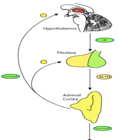
Cortisol, a glucocorticoid hormone is released in response to stress. Its primary functions are to increase blood sugar and glycogen storage in the liver (3), aid in fat, protein and carbohydrate metabolism (4), and to suppress the immune system (5). It is controlled by hypothalamic secretion of corticotropin-releasing hormone (CRH), which in turn
triggers pituitary secretion of Adrenal Corticotrophic Hormone (ACTH); ACTH is carried by the blood to the adrenal cortex where it triggers glucocorticoid secretion. The amount of cortisol produced and secreted in the blood undergoes diurnal variation, with peak production in the early morning and nadir during sleeping hours. Excess cortisol produced by the adrenal gland inhibits further production of CRH and ACTH leading to an inhibitory loop regulation (Figure 3).
Figure 3: Human cortisol production in the adrenal gland. Neural signals trigger CRF secretion by the hypothalamus. In turn, this signals to the pituitary to release ACTH which stimulates the adrenal gland to release cortisol. Cortisol acts on the hypothalamus and the pituitary to have a negative feedback effect on the release of CRH and ACTH respectively. Figure from http://models.cellml.org
Aldosterone is a mineralocorticoid hormone synthesized exclusively in the adrenal zona glomerulosa. Aldosterone regulates homeostasis by increasing the reabsorption of sodium and water and the secretion of potassium in the kidneys. This increases blood volume leading in part to increased blood pressure. Aldosterone secretion is controlled by the renin-angiotensin-aldosterone system (6). In the liver, renin activity (the rate limiting factor of the secretion of aldosterone) leads to the production of angiotensin I. Angiotensin is cleaved by the angiotensin-converting-enzyme to angiotensin II. Angiotensin II increases aldosterone secretion from the zona glomerulosa by increasing transcription of CYP11B2 (6). Angiotensin II itself also acts to inhibit renin production creating a negative feedback loop of regulation. ACTH also stimulates aldosterone production indirectly by stimulating the formation of deoxycorticosterone (7), a precursor of aldosterone (Figure 2, page 4).
1.3. Adrenal gland development
Adrenocortical cell development in human embryos is due to two independent events. 1) The adrenal cortex arises from adrenogonadal progenitors that first appear in the fourth week of gestation as a thickening of the coelomic epithelium between the urogenital ridge and the dorsal mesentery (8). Cells destined to generate the adrenal cortex migrate to the cranial pole of the mesonephros, which creates the foetal adrenal gland by the eighth week of gestation. This fetal adrenal gland contains an inner cluster of large cells, termed the foetal zone (Figure 4, page 7). 2) By week eight (Figure 4, page 7), a second group of cells form a compact outer zone of cells, termed the definitive zone (9). Cells in the definitive zone, unlike the foetal zone, lack expression of CYP17 (9).
At week nine (Figure 4, page 7), the foetal and definitive zone (foetal adrenal cortex) become encapsulated. The capsule is thought to provide growth factors that are
important in mediating adrenal growth and differentiation, such as insulin like growth factors 1 and 2 (IGF1 and 2) (9). Also at nine weeks, neural crest-derived cells migrate into the foetal adrenal cortex and differentiate, hence forming the catecholamine-producing chromaffin cells (10), which remain scattered until birth. This differentiation of the neural crest cells is triggered by the glucocorticoids secreted by the cortex (10).
At birth the adrenal gland is relatively larger than the adult gland due to the size of the fetal cortex (Figure 4, page 7). Preceding birth, further significant changes of the adrenal gland occurs. First the medulla is formed by compaction of the chromaffin cells (Figure 4, page 7). After three weeks there is full regression of the fetal zone followed by encapsulation of the medulla. Finally, the definitive zone of the human adrenal cortex differentiates into the three zones of the adrenal gland (the outer zona glomerulosa, the middle zona fasciculata, and the inner zona reticularis) (9) (Figure 4, page 7). It must also be noted that at this time events controlling adrenal cortex zonation are not fully understood.
Figure 4: Developmental events of the adrenal gland from embryo to adult. Modified from Hammer, G. D. et al.2005 (11)
Section 2. Tumours of the adrenal cortex
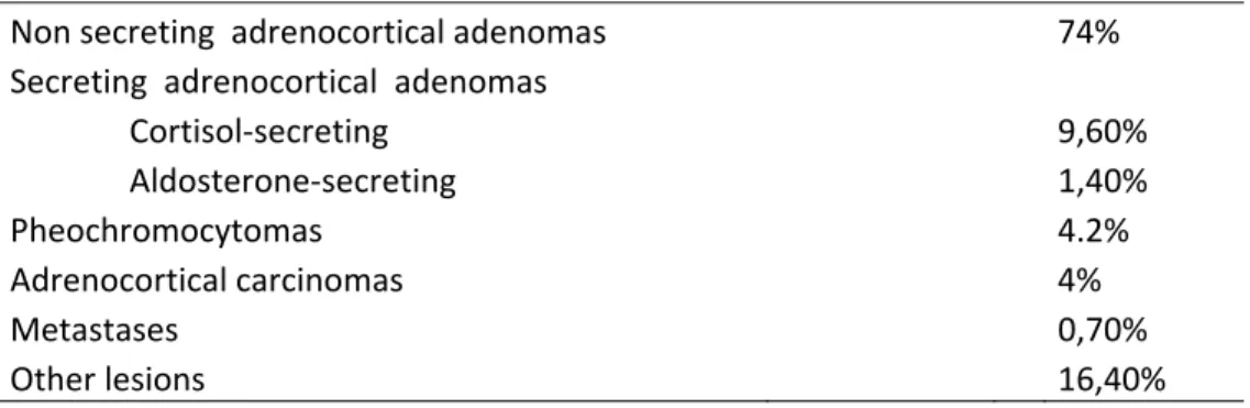
Adrenocortical adenomas are benign tumours of the adrenal cortex which are extremely common (present in 1-10% of persons at autopsy). They have been detected as incidental findings with increasing frequency in recent years, due to increased use of CT scans and magnetic resonance imaging in a variety of medical settings. As seen in table I, adenomas are the most common of adrenocortical tumours. About 30-50% of adrenal tumors are discovered from endocrine problems (such as in Cushing’s syndrome). Other lesions are found by chance with no clinical manifestations and are referred to as incidentalomas. Most of the incidentalomas are benign but about 4% of them are malignant (see table I).
Table I: Classification of adrenal incidentalomas. From a study of patients from le Study Group of the Italian Society of Endocrinology on Adrenal Incidentalomas (12)
Non secreting adrenocortical adenomas 74% Secreting adrenocortical adenomas
Cortisol-secreting 9,60% Aldosterone-secreting 1,40% Pheochromocytomas 4.2% Adrenocortical carcinomas 4% Metastases 0,70% Other lesions 16,40% 2.1. Adrenocortical adenomas
An adenoma is a benign tumour of glandular origin. Adenomas can grow from many organs including the colon, the adrenal glands, the pituitary gland, the thyroid, etc. Although benign, over time they may progress to become malignant in specific tissue such as in the colon, however this malignant transformation has never been described in the adrenal gland. Adrenocortical adenomas have the potential to cause serious health
complications by producing large amounts of hormones in an unregulated, non-feedback-dependent manner. Typically adenomas are well limited, encapsulated nodular lesions of the adrenal cortex ranging from a couple of millimeters to a few centimeters. Histological examination shows that cells in adenomas are similar to those of the normal cortex with high lipid content. Most adenomas are non-functional, meaning that they do not produce any glucocorticoids (cortisol), mineralocorticoids (aldosterone), and/or sex steroids (testoterone, estrogens). In about 15% of cases, adrenocortical adenomas are "functional", and secrete abnormally high levels of the glucocorticoid cortisol or the mineralcorticoid aldosterone which results respectively in endocrine disorders such as Cushing's syndrome and Conn's syndrome (hyperaldosteronism).
2.1.2. Cortisol-secreting adrenocortical adenomas
Overproduction of cortisol is the most common characteristic of functional adrenocortical adenomas. Elevated cortisol levels are the cause of Cushing’s syndrome which leads to high blood pressure, diabetes, osteoporosis, skin atrophy, infection and, many other symptoms. In most cases Cushing’s syndrome is due to elevated ACTH levels caused by improper functioning of the pituitary gland (pituitary tumour). Elevated ACTH levels can lead to overproduction of cortisol and an enlargement of the adrenal gland (13). In other cases, adrenocortical adenomas can produce hypercortisolism independently of ACTH (ACTH-independent Cushing’s syndrome) (14, 15).
2.1.3. Aldosterone-secreting adenomas
Adenomas, which secrete abnormally high levels of aldosterone cause primary aldosteronism. Hyperaldosteronism can be asymptomatic or associated with hypertension and hypokaliemia (16).
2.2. Adrenal Hyperplasias
Adrenal hyperplasias are much less common than adenomas. There are 2 main subtypes of primary bilateral ACTH-independant hyperplasias; 1) ACTH-independent macronodular adrenal hyperplasia (AIMAH) and 2) micronodular adrenal hyperplasia which includes primary pigmented nodular adrenocortical disease (PPNAD). Bilateral adrenal hyperplasias cause approximately 10 to 15% of adrenal Cushing’s syndrome (17, 18)
2.2.1. ACTH-independent micronodular adrenal hyperplasias and PPNAD
PPNAD is diagnosed mainly in young adults (19) and is characterized by normal sized adrenal glands containing several small cortical pigmented nodules (20). PPNAD may be isolated or associated with a multiple neoplasia syndrome, Carney complex (CNC), which can include spotty skin pigmentation, heart and skin myxomas and various endocrine tumours. PPNAD is the most frequent endocrine manifestation of Carney complex (19). Germline mutations of the PRKAR1A gene encoding for the types 1-α regulatory subunit of cAMP dependent protein kinase A are found in more than 50% of cases of CNC and are also frequent in isolated PPNAD (21-23). More recently, using a genome-wide scan approach, Horvath et al., identified germline mutations in the gene encoding phosphodiesterase 11A4 (PDE11A) in patients with CS secondary to micronodular adrenocortical hyperplasia (24).
2.2.2. ACTH-independent macronodular adrenal hyperplasia
AIMAH may rarely develop during the first year of life associated with McCune-Albright syndrome (25), but the majority of cases are detected during the fifth or sixth decade of life (26). The most frequent presentation of AIMAH is with clinical or
subclinical CS (18). Usually, computed tomography reveals clear enlargement of both adrenal glands. Histological examination is characterized by non-pigmented nodules composed of two types of cells, those with clear cytoplasm (lipid-rich) that form cordon nest-like structures, and those with a compact cytoplasm (lipid-poor) that form small nest or island-like structures (27).
AIMAH was most often reported as sporadic cases, but there have been increasing reports of familial AIMAH suggesting an autosomal dominant pattern of transmission (28). AIMAH may be rarely associated with syndromes where genetic defects have been identified such as MEN 1 (menin), familial adenomatous polyposis (APC) and hereditary leiomyomatosis and renal cell cancer disorder (fumarate hydratase) (27, 29). In addition, AIMAH is found in a subgroup of patients with Mc Cune-Albright syndrome, where activating mutations of the Gsα subunit occur in the adrenal gland during embryogenesis and lead to constitutive activation of the cAMP signaling and CS. Fragoso et al, identified gsp mutations in 3 of 5 patients with AIMAH and CS without any manifestations of Mc Cune-Albright syndrome (30). In a patient with AIMAH an ACTH receptor (MC2R) mutation was identified which lead to impaired desensitization and internalization of the receptor associated to apparent constitutive activity (31). Recently, two mutations in the same allele of MC2R was identified in a patient with clinical hypersensitivity to ACTH (32).
Although, there is some evidence of cylic AMP-dependent signaling aberrations, no mutations were found in PRKAR1A gene in AIMAH (33). However, somatic losses of the 17q22-24 region which contains PRKAR1A gene was found frequently in AIMAH samples (33).
2.2.3. Congenital hyperplasia (CAH)
Congenital hyperplasia (CAH) represents another type of adrenal hyperplasia and is one of the most frequent adrenal disorders observed in children at birth. CAH is characterized by deficiency in 21-hydroxylase expression or other adrenal specific steroidogenic enzymes in less frequent cases. Deficiency in cortisol production leads to excess of ACTH secretion which stimulates adrenal hyperplasia growth (34).
2.3. Adrenocortical carcinoma (ACC)
Adrenocortical carcinoma (ACC) is a rare, highly aggressive cancer of adrenal cortical cells, which may occur both in children and adults. ACC may be "functional", producing steroid hormones and consequent endocrine dysfunction similar to that seen in many adrenocortical adenomas, or non-functional. Due to their location deep in the retroperitoneum, most ACCs are not diagnosed until they have grown quite large. They frequently invade large vessels, such as the renal vein. ACC metastasizes via the lymphatics and through the blood, to the lungs and other organs. The overall prognosis of the disease is poor and present treatments are limited leading to poor survival. ACCs like adenomas are often sporadic of origin and can also be associated with familial syndromes like Li-Fraumeni (35) and Beckwith-Weidemann (36) syndrome. In the recent years, progress to determine key genes and pathways involved in the pathogenesis of ACCs has been slow, in part due to the rarity of these tumours.
2.4. Genetic and pathway alterations of adrenocortical tumours
Genetic alterations either hereditary or sporadic are the key to cancer development. Alterations that affect either tumour supressors or cause overexpression of oncogenes can
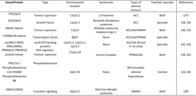
cause alterations of signaling pathway, resulting in uncontrolled cell growth and tumour formation. A number of hereditary syndromes with known genetic alterations are associated with adrenocortical tumours (Table II). Interestingly, a number of these genetic alterations may also be found in sporadic adrenocortical tumours like in the MEN, TP53 and PRKAR1A genes. The following section will discuss several pathways/genes thought to play a role in adrenal tumour development and progression, summarized in table II.
Table II: Genes /and associate familiale syndromes in adrenocortical tumours.
Gene/Protein Type Chromosomal location
Syndromes Types of adrenal tumours
Familial/ sporadic References
TP53/p53 Tumour supressor 17p13.1 Li-Fraumeni ACC Both (37)
IGF2/IGFII Growth factor 11p15.5 Beckwith-Weideman
syndrome ACC Sporadic (38, 39) MEN1/ Menin Tumour supressor 11q13 Multiple endocrine
neoplasia type 1 ACC/AA/AIMAH Both (40, 41) CTNNB1/B-catenin Transcription factor 3p21 None ACC/AA/PPNAD Sporadic
ras/RAS ( HRAS, KRAS,NRAS) small GTP binding proteins 11p15.5, 12p12.1, 1p13.2 None ACC/AA (N and
K-ras only) Sporadic (42, 43) PRKAR1A/ PRKAR1A
protein kinase
PKA regulator,
Tumour supressor 17q22-24 Carney Complex PPNAD/AA Both (44, 45)
PDE11A / Phosphodiesterase 11A PDE8B/ Phosphodiesterase 8B Phosphodiesterase 2q31-35 None Micronodular adrenal hyperplasias Familial (24, 46)
GNAS1/GNAS G protein signaling 20q13.2 McCUne-Albright
syndrome AIMAH Both (30)
2.4.1. Alterations of TP53
The TP53 tumour supressor gene codes for the protein p53. The p53 protein controls cell proliferation by controlling the G1/S cell cycle check point and can initiate program cell death in presence of DNA damage. TP53 mutations are highly common in human cancers (47). A hereditary germline mutations of TP53 located at 17p13.1 are the underlying genetic abnormality in 70% of Li-Fraumeni cancer syndrome cases (37). The Li-Fraumeni syndrome is a syndrome linked to an increased risk of breast cancer, soft tissue sarcoma, brain cancer, leukemia as well as ACC (48). ACC associated with
Li-Fraumeni syndrome occurs at an early age (children and young adults). In pediatric ACC, about 50-80% of sporadic cases are also associated with germline TP53 mutations (49, 50). In adults, 1/3 of sporadic ACCs have somatic TP53 mutations while in adenomas somatic mutations are uncommon suggesting that these alterations happen late in tumourogenesis (51-54). Furthermore ACC with somatic but not germline TP53 mutations are associated with poor prognosis and are considered to represent a more malignant phenotype.
2.4.2. Insulin Growth Factor 2 (IGF2)
The IGF pathway involves two ligands (IGF1 and IGF2), two receptors (IGFR1 and 2) as well as 6 IGF binding proteins (1 to 6)(55). IGF-2 is expressed during the embryonic and foetal stages of adrenal development and contributes to growth and differentiation, whereas expression is kept low in the adult adrenal (56-58). IGF-I and IGF-II, can both act as adrenocortical mitogens by promoting progression through the G1/S phase of the cell cycle via activation of phosphatidylinositol 3-kinase (PI3K) and the AKT protein kinase (59, 60).
Transcriptional analysis by microarray technology has shown IGF2 as the most over-expressed gene in human ACC compared with adrenocortical adenomas or normal tissues (61-63). IGF1 has not been observed to be overexpressed in ACC (38). In human ACC alterations at chromosome 11p15 by genetic or epigenetic, are present in about 90% of cases (64, 65) and this is believed to be the cause of overexpressed IGF2.
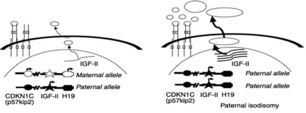
Figure 5 : Alterations of 11p15 locus and IGF-II overexpression in ACC. The imprinted 11p15 locus contains the CDKN1C (p57kip2), IGF-II, and H19 genes. In normal differentiated tissue (on the left), only the paternal allele of the IGF-II gene is expressed, whereas only the maternal alleles of CDKN1C and H19 are expressed. Paternal isodisomy is usually observed in adrenal cancers (on the right) with loss of the maternal allele at 11p15. This leads to the overexpression of IGF-II and decreased expression of CDKN1C and H19. Modified from Libe et al . 2007(65)
The IGF2 locus (11p15) (Figure 5) is parentally imprinted, and normally only the paternal allele is expressed (66). Two other imprinted genes in this region, H19 and p57KIP2, are expressed only from the maternal allele. The H19 mRNA is not translated and serves to inhibit IGF2expression. The p57KIP2 gene encodes a cyclin-dependent kinase inhibitor involved in the G1/ S phase of the cell cycle. Mutation and/or aberrant expression of imprinted genes in the IGF2 locus causes Beckwith-Wiedemann syndrome (BWS), a syndrome associated with adrenal hyperplasia and ACC (67).
Due to the significance and frequency of IGF-II upregulation in sporadic ACC, this growth factor and its receptors are considered candidates for molecularly targeted drug therapy. Overexpression of the IGF-1 receptor (IGF1R) has also been observed in ACC (39, 68). IGF1R antagonists cause growth inhibition in vitro and of human ACC xenografts in nude mice (60). Early clinical trials with humanized IGF1R antibodies have also shown to be promising in PHASE1 trials for ACC treatment (69)
2.4.3. PRKAR1A and other cAMP pathway alterations
The regulatory R1A subunit of protein kinase A (PRKAR1A) is the main regulator of protein kinase A (PKA) activity and a key component of the cAMP signaling pathway that has been the subject of many studies in endocrine tumourigenesis (70, 71). The PRKAR1A gene is located at the 17q22–24 locus and is, as previously mentioned implicated in the Carneycomplex (CNC; (21, 23) and PPNAD (44, 45)). Heterozygous inactivating germline mutations of PRKAR1A have been demonstrated in about 45 to 65% from two studies in CNC families (23),(72).Somatic PRKAR1A mutations have also been found in sporadic secreting adrenocortical adenomas,with similar characteristics to those of PPNAD (73). Mutations of PRKAR1A are not found in ACC although LOH at 17q is found in adenomas. In adenomas, LOH is restricted to the PRKAR1A locus (17q22–24) but in ACC LOH effect a larger area of 17q (65). This suggests that PRKAR1A alterations may play a role at the early onset of tumoural growth but that this effect is minimized in malignant growth.
More recently inactivation germline mutations in two phosphodiesterases (PDE) genes PDE11A and PDE8B have been found (24, 46) in adrenal micronodular adrenal hyperplasias. PDE11A is a dual specificity PDE which catalyzes hydrolysis of cAMP and cGMP (74). Of the four known isoforms of PDE11A (A1 to A4) only A4 is reported to be expressed in the adrenal gland (74). PDE11A is located at 2q31-35 and fluorescent in situ hybridization (FISH) studies of tumours have demonstrated 2q allelic loss in patients with PDE11A inactivating mutations. Strangely, inactivation mutations of PDE11A are also found in the general population although at a lower frequency. This suggests low penetrence of PDE11A mutations and that other PDE’s must be present in the adrenal gland to compensate for the lack of PDE11A activity.
The PDE8B gene codes for the PDE with the highest cAMP affinity (75). It is highly expressed in the adrenal gland and is harboured in the 5q13 locus, a region thought to be associated with micronodular adrenocortical disease (MAD)(46). In HEK293 cells, cAMP levels are significantly increased following transfection with mutant PDE8B (76).
Another type of cAMP pathway alteration involves activating somatic mutations in the gene coding for GNAS (Gsα). GNAS is a member of the G protein family, which form a heterotrimers with β, γ subunits that participate in signal transduction. GNAS has intrinsic GTpase activity and stimulates cAMP production (77). Mutants found of GNAS, prevent downregulation of cAMP signaling (27). GNAS mutations are associated with AIMAH and the McCune-Albright syndrome (MAS) which is primarily associated with fibrous dysplasias (78).
Up to date, alterations in multiple players of cAMP pathway leading to increased cAMP signaling appear to be the main cause of nodular adrenocortical disease. Clearly the adrenal gland seems highly susceptible to changes in the cAMP pathway with adrenal hyperplasias being the most predominant clinical manifestation in humans with germline mutations in PDE11A, PDE8B and PRKAR1A. In contrast, cAMP pathway alterations are not found in ACC and thus are not believed to lead to malignant growth, but may be associated with early tumor formation.
2.4.4. Ras pathway alterations
Ras proteins are membrane associated small GTPases proteins and are referred to molecular switches in signaling pathways that control proliferation, differentiation, death and mobility. The three ras proteins (H, N, and K) are the most commonly mutated oncogenes in human cancers (79).Controversial data is present in the literature; Lin et al, (42) found K-ras mutations in about 50% of tumoural tissues of Conn’s adenomas and
Yashiro et. al, who found N-ras mutations in 7 of 56 (12.5%) of all tumours tested (3 of 24 (12.5%) carcinomas and 4 of 32 (12.5%) adenomas (Yashiro et al 1994), while Moul et al. (80) and Ocker et al.(81) did not identify any Ras mutations. In a more recent study K-ras and N-ras mutations were each found in one out of 35 ACCs (43). In this study, 2 (5.7%) and 4 ACCs (11.4%) were also found to carry BRAF and EGFR TK domain mutations respectively (43). K-ras mutation found by Lin et al. were active in ACC (Lin, Hsu et al. 2000) and lead to increases in steroidogenic enzyme expression and an increase in cortisol production when transfected into normal adrenal cells (82). These data suggest that EGFR-Ras-Raf-MEK-ERK pathway alterations are present in only a subset of ACC.
2.4.5. MEN 1
Adrenal tumours are also associated with mutations in the MEN 1 gene which codes for the Menin protein. A heterozygous inactivating germlinemutation of MEN 1 can be found in about 90% of families affectedby multiple endocrine neoplasia type 1 (MEN 1). Menin Endocrine Neoplasia type 1 (MEN1) is a rare autosomal dominant hereditary cancer syndrome presented mostly by tumors of the parathyroids, endocrine pancreas and anterior pituitary. About 25–40% of MEN1 patients are affected by adrenocorticaltumours and/or hyperplasias (41, 83). In mostcases, these tumours are non-functional adenomas and are rarely seen in ACC. Somatic mutations in the MEN 1 gene are known but are veryrare in all adrenocortical tumours (40, 83). The 11q13 locus containing the MEN1 gene frequently losses heterozygosity at a greater frequency in ACC’s (90%) compared to adenomas ( 20%), however expression of the MEN1 gene does not differ between them (83), suggesting that the MEN1 gene does not play a role in the progression of sporadic adrenal tumours.
2.5. Adrenocortical tumourigenesis 2.5.1. Clonality of adrenocortical tumours
Determining tumour clonality is important to establish the origin as well as the mechanisms dictating tumour progression. Mono-clonality indicates that tumour progression is the end result of a or multiple genetic mutations, whereas polyclonality suggests that tumour cells are affected by local or systemic stimuli (59). Three X-chromosome inactivation studies have up to date been performed to determine the clonality of adrenocortical tumours. All three studies are in agreement andshow that ACC consists of monoclonal populations of cells, adenomas tumours are both monoclonal as well as polyclonal(64, 84, 85). Hyperplasias on the other hand are only polyclonal (84). Variation observed in adenomas tumours may be explained by different mechanisms or are due to observation of a multiple steps, within a common mechanism. These observations have been suggestive of a multistep (clonal) genetic model of adrenocortical tumour development.
2.5.2. Models of adrenocortical tumour development
Unlike other cancers, the factors and steps leading to adrenocortical tumour development is still under debate. In colon cancer progression of adenoma to carcinoma was proposed, however this sequence is not recognized in adrenocortical tumourogenesis.
Adrenocortical tumours comparative genomic hybridization (CGH) studies have shown a positive correlation between adrenocortical tumour size and the number of chromosomal alterations, supporting the hypothesis that chromosomal changes accumulate during tumour progression (65, 86). In benign lesions the average number of chromosomal
aberrations per tumour cell is low (87), with chromosomal gains occurring often of 9q (14%-22%) (87) (88) and 17q (26-35%) (88). Chromosomal losses also occur, although less often, with the most common being at chromosomal 1p (5%)(87) or 6q (9%) (88). In hyperplasias gain of complete or partial chromosome 17q is the most common chromosomal alteration. In hyperplasias similar chromosomal abnormalities to those in adenomas (e.i., gain of chromosome 17q), provides evidence that hyperplasias may evolve into adenomas (88). The hyperplasia-adenoma transition is also supported by clinical observation of adenomas presence within hyperplasias like PPNAD (89). In contrasts to benign adrenocortical neoplasms multiple chromosomal alterations in nearly all ACCs is observed (86-88, 90). In addition to deletions or amplifications, a high percentage of ACC cells exhibit loss of heterozygosity (LOH) at key loci which can result in an altered TP53 pathway of unipaternal disomy (UPD) causing IGF2 overexpresssion.
In adrenocortical tumourogenesis, only β-catenin mutations are found commonly in both benign and malignant neoplasm. In contrast, cAMP pathway genetic alterations are present in adrenocortical hyperplasias and some adenomas while the presence of IGF2 locus alteration is more specific to ACCs. In addition to these differing genetic alterations, clinically no clear progression of adenomas or hyperplasias to ACC has been observed. These facts support that different mechanisms lead to adrenocortical adenoma or ACC formation and that adenoma do not progress to malignant tumours.
On the other hand, some evidence refutes the latter. In mice model, overexpression of IGF2 does not lead to tumour formation (68, 91), suggesting that it plays a role later in tumourogenesis and serves as a pro-proliferative factor and not as an initiating element. Although this remains to be proven in humans, it suggests that IGF2 overexpression which is seen in the majority of ACCs does not causes tumour formation and that other genetic
alterations are involved in ACC formation. As mentioned earlier, somatic mutation of TP53 genes are found only rarely in non malignant adrenocortical neoplasm suggesting that these mutations may be gained during tumour progression and are not initiating events. Clonality status of adrenocortical tumours also suggests a multi-stage process of adrenocortical development. Finally, a case report described in 2003 supports the hypothesis of multistep adrenocortical tumourigenesis where an adrenocortical tumour had a central component of ACC surrounded by benign adrenocortical tissue (92). Unfortunately, clonal studies to determine if both parts originated from the same population of cells were not possible since the patient was a male. Although this is not proof of a multistep model it does suggest that a multistage process can occur, though rarely.
Section 3. Wnt/β-catenin signaling
3.1 Wnt signaling pathways
Wnt signaling is a highly complex and versatile process leading to a multitude of cellular effects. Wnt signaling is driven by a family of secreted glycoprotein called WNTS. Variability of Wnt effects are in part caused by the 19 different Wnt proteins present in humans. Wnt’s activate Wnt signaling by interaction with the frizzled family of membrane receptors (FZD1-10) which then activate dishevelled proteins (DVL1-3) to activate downstream events. There are three branches to the Wnt signaling pathway: 1) the canonical or Wnt/β-catenin pathway, 2) the non-canonical or the planar cell polarity pathway and the 3) Wnt/Ca2+ pathway. The canonical pathway involves stabilization of β-catenin leading to transcription activation of target genes while the other two pathways exert their effect independently of β-catenin. Since our studies involved Wnt/β-catenin, we will only briefly discuss the other two pathways.
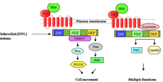
3.1.1 Non-canonical or planar cell polarity (PCP) signaling
In PCP signaling (Figure 6, page 23), Wnt signaling is transduced through Frizzled independent of LPR5/6 (LOW density lipoprotein receptor related protein) like in the canonical pathway. Utilizing the PDZ and DEP protein domains of DVL, this pathway mediates cytoskeletal changes through activation of the small GTPases Rho and Rac instead of leading to β-catenin stabilization. Hence, the PCP signaling pathway controls tissue polarity and cell migration/movement. Aberrant activation of Wnt/PCP signaling pathway in human cancer (i.e.WNT5a overexpression) leads to more malignant phenotypes (93). This pathway is also involved in organ development (94).
3.1.2. The Wnt-Ca2+ pathway
In the Wnt-Ca2+ pathway (Figure 6), Wnt signaling via Frizzled mediates activation of heterotrimeric G-proteins, which engage DVL, phospholipase C (PLC; not shown), calcium-calmodulin kinase 2 (CamK2) and protein kinase C (PKC), which modulates cell adhesion and motility (95).
Figure 6: Wnt pathways independant of β-Catenin. On the left, planar cell polarity signaling. On the right, WNT Ca2+ signaling.(95)
3.1.3. The Wnt/ β-catenin pathway
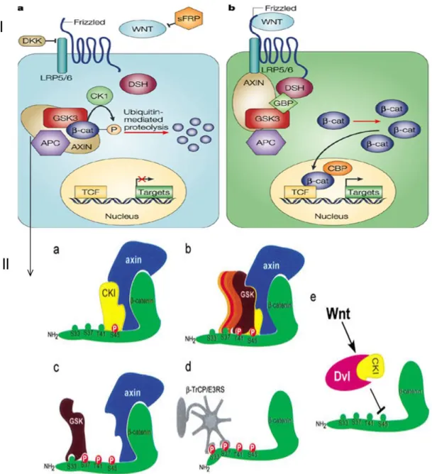
This pathway is commonly referred to as either being on (Figure 7-IB page 25), or off (Figure 7-IA, page 25). Normally the pathway is off, and cytosolic β‑catenin is constitutively degraded by the cytoplasmic degradation complex, which comprises axin, adenomatous polyposis coli (APC), glycogen synthase kinase 3β (GSK3β) and casein kinase1 (CK1). This degradation complex triggers SCF (SKP1, Cullin, F‑box)/β‑TrCP‑mediated polyubiquitylation leading to proteosomal degradation of β‑catenin.
The events are triggered through phosphorylation control of the of β-catenin NH2 terminus by multiple kinases (depicted on Figure 7II page 25). Initially β-catenin is bound by an Axin: APC complex. APC and Axin can be hyperphosphorylated by CK1α and GSK3β which increase the affinity to bind β-catenin. Once bound Axin recruits CK1α to the complex to phosphorylate β-catenin at serine 45 (S45). This acts a primer for further phosphorylation events by GSK3β at residues S37, S33, and T41. Phosphorylation of all four sites at the NH2 terminus are essential for proper degradation of β-catenin. APC, which acts as a scaffold like Axin, may also cleave the NH2 phosphorylated end and transfer β‑catenin to proteosomal complex (96). Ultimately the destruction complex maintains low levels of β-catenin when no activation signal is present.
Upon activation, WNTs interacts with Frizzled receptors and the co-receptor LRP5/6. DVL proteins are activated and cause dissociation of the destruction complex and thus β-catenin stabilization. CK1 also synergizes with DVL to mediate this event (97). CK2 another kinase also seems required to stabilize β-catenin by phosphorylation at residue T393 to inhibit the axin: β-catenin interaction (98). Upon stabilization β-catenin enters the nucleus and acts as a transcriptional activator via interaction of with T-cell transcription factor family members (Figure 7I, page 25).
Figure 7: (I) Wnt/β-catenin pathway. a) When pathway is off β-catenins is recruited to the destruction complex and targeted for degradation by ubiquitin mediated proteolysis. b) When pathway is activated via WNT interaction with frizzled recpetors, DSH/Dvl proteins interaction with the frizzled receptors and inhibit recrutement of β-catenin to the destruction complex which is termed β-catenin stabilization or active β-catenin. β-catenin is then exported to the nucleus and interacts with the TCF
transcription factor and activates transcription. (II) A model depicting the β-catenin phosphorylation–
degradation cascade by the destruction complex. (a) Axin recruits CKI to phosphorylate β-catenin at S45.
(b,c) S45 phosphorylation primes β-catenin for the succeeding GSK-3β phosphorylation cascade, finally hitting the S33/37 sites. (d) Phosphorylation at S33/37 creates a docking site for β-TrCP/E3RS, promoting the ubiquitination and degradation of β-catenin. (e) Wnt signaling, possibly through Dvl and CKI, regulates S45 phosphorylation. Figure from Amit et al. (99)
3.2. β-catenin
In humans β-catenin is a protein encoded by the CTNNB1 gene. β-catenin is a member of the armadillo family of proteins. These proteins have multiple copies of the so-called armadillo repeat domain which is specialized for protein-protein binding.
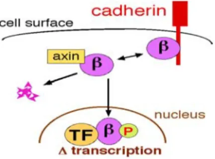
As mentioned previously, β-catenin is the centre player of the Wnt/β-catenin pathway. β-catenin itself exists in three states within the cell (Figure 8). 1) Complexed within the destruction complex. 2) Complexed with E-cadherin and other proteins that constitute adherens junctions (AJs). AJs are necessary for the creation and maintenance of epithelial cell layers by regulating cell growth and adhesion between cells. 3) In an active state (unphosphorylated at S45, T41, S37, S33) where it is either in the cytoplasm or the nucleus bound to transcription factors such as TCF/LEF (Figure 8) and SF1. Proper control of β-catenin levels and its state is essential for development and maintaining homeostasis. Improper control can lead to disease such as cancer (see Section 5).
Figure 8: The 3 cellular states of β-catenin. Within the cell β-catenin (β) exist within three states; bound by Cadherins (top right), complexed with the destruction complex (axin only is shown here, middle), or in the nucleus. TF= TCF; β= β-catenin; P= phosphorylated residues.
3.2.1. β-catenin in the nucleus
β-catenin has been involved in the transcriptional control of a multitude of genes mainly by co-activating gene transcription with the T-cell specific transcription factor (TCF) family of transcription factors (TCF-1, LEF-1(formally TCF-2), TCF-3, and TCF4 (TCF7L2). What is of great interest is that activation of genes by β-catenin/TCF is usually cell and context specific. TCF proteins contain a DNA binding domain but no trans-activation domain and can therefore not drive transcription by itself. TCF serves a dual role as both a repressor and activator. When unbound to β-catenin, TCF is still found bound to WRE (WNT responsive elements)/TBE’s (TCF binding elements) were it can act as a repressor upon interaction with the transcriptional repressor Groucho (Figure 9, left panel). When β-catenin enters the nucleus, it replaces Groucho and binds TCF. β-catenin can also interact with TCF that is yet unbound to DNA which then leads to DNA binding (Figure 9 right panel).
Figure 9: β‑catenin dependent gene transcription. This pathway centres on β‑catenin, which, together with the DNA‑binding T cell factor/lymphoid enhancer factor (TCF/LEF) family proteins, functions as a transcription factor to control Wnt target genes. A subset of these target genes are constitutively inhibited by pioneering nuclear TCF, which recruits transcriptional corepressors (left panel) to Wnt response elements (WREs). Once β-catenin is exported the nucleus it replaces TCF-bound corepressors (such as groucho; middle panel) or co-imports additional TCF to occupy WREs (right panel). Once bound to WREs through TCF, β‑catenin functions as a scaffold to recruit an auxiliary machinery of co‑activators that are involved in chromatin remodelling and control of RNA polymerase II to induce Wnt target gene expression.(100).
It is yet unclear if these two methods for the activation of β-catenin/TCF transcription are gene specific of or if both can occur for the expression of the same gene. Once bound to WREs through TCF, β-catenin functions as a scaffold to recruit an auxiliary machinery of co-activators that are involved in chromatin remodelling (i.e. BRG-1(101)) and control of RNA polymerase II to induce Wnt target gene expression. Regulation of β-catenin activity is quite complex inside the nucleus. In fact, there are over 25 non transcription factor proteins that are known to interact with β-catenin that act as either antagonist or agonist of its transcriptional activity (102). This supports the hypothesis that nuclear accumulation of β-catenin alone is not sufficient to state activity. Transcriptional activity hence, what genes are expressed is affected by the variation of co-factors present.
3.2.2. Regulation of β-catenin target genes
Multiple genes are known to be directly regulated by β-catenin/TCF (Table 3, page 29) and the list keeps growing. Most genes regulated by β-catenin/TCF were discovered in colon cancer cells. Two of the most studied genes in relation to β-catenin/TCF regulation have been CCND1 (CyclinD1) and AXIN2. CCND1 regulation by β-catenin/TCF was originally proven by reporter assays (103). Its regulation by β-catenin/TCF in colon has since been refuted as CyclinD1 reporter constructs do not appear to reflect endogenous gene regulation (104). It is still considered a target of β-catenin/TCF activity although caution should be warranted for its use as a marker of β-catenin/TCF activity.
One of the highly important genes regulated by β-catenin/TCF is AXIN2. Similarly to Axin1, the Axin2 protein reduces β-catenin stability leading to a feedback inhibition on Wnt-signaling (105). AXIN2 contains the most known TCF sites (6 in the first intron and 2 in the 5’flanking site) (105) and is suggested by leaders in the WNT field, to be the most
conserved gene to determine activation of Wnt/β-catenin signaling between different cell types and signals (100, 105). Its regulation by β-catenin/TCF differs in that it contains TCF binding sequences (n=6) within the first intron, while other genes, CCND1, MYC and ENC1 which are likely to be non universal targets, contain sites only within the 5’ flanking region of the transcriptional start site (103, 106, 107).
Table III: Genes that are upregulated directly by β-catenin/TCF in Humans
Genes Tissues/cell lines References
c-myc Colon cancer He, Sparks et al. 1998)(106)
Cyclin D Colon cancer Tetsu and McCormick 1999) (103)
Tcf-1 Colon cancer Tetsu and McCormick 1999 (103)
PPARdelta Colon cancer He, Chan et al. 1999(108)
c-jun Colon cancer Mann, Gelos et al. 1999 (108)
fra-1 Colon cancer Mann, Gelos et al. 1999) (108)
MMP-7 Colon cancer Brabletz, Jung et al. 1999 (109)
CD44 Colon cancer Wielenga, Smits et al. 1999 (110)
Gastrin Colon cancer Koh, Bulitta et al. 2000 (111)
ENC-1 Colon cancer Fujita, Furukawa et al. 2001 (107)
Claudin-1 Colon cancer Miwa, Furuse et al. 2001) (112)
Id2 colon cancer Rockman, Currie et al. 2001 (113)
Axin-2 Colon cancer Yan, Wiesmann et al. 2001; Lustig, Jerchow et al. 2002
(105, 114)
ITF-2 Colon cancer Kolligs, Nieman et al. 2002 (115)
Nr-CAM Colon cancer Conacci-Sorrell, Ben-Yedidia et al. 2002 (116)
BMP4 Colon cancer Kim, Crooks et al. 2002 (117)
Frizzled 7 EC cells Willert, Epping et al. 2002 (118)
LEF1 Colon cancer Filali, Cheng et al. 2002 (119)
Follistatin EC cells, ovary Willert, Epping et al. 2002 (118)
FGF18 Colon cancer Shimokawa, Furukawa et al. 2003 (120)
c-myc binding protein
Colon cancer Jung and Kim 2005 (121)
LGR5/GPR49 Intestine Barker, van Es et al. 2007 (122)
Gene list was obtained from http://www.stanford.edu/~rnusse/wntwindow.html (WNT homepage). Only genes with substantial evidence for implication of β-catenin in their transcription are described.
It is now well recognized that β-catenin target genes are cell specific. The use of microarray technology for transcriptome studies highly strengthens this point. Microarray
studies comparing gene expression profile of wild-type and tissues carrying β-catenin mutations in Wilms tumours (123, 124) and ovarian endometrioid adenocarcinomas (125) have shown a very different list of deregulated genes in comparison to table III (page 29). Two different studies of Wilms tumours (123, 124) even showed significant differences despite large cohorts studied (n=36, n=73 respectively).
Transcriptome studies have also highlighted differences in response to WNT molecules from different cells. In a study by Railo et al. (126), it was shown that upon WNT3a stimulation in mice NIH3T3 and rat PC12, only the expression of two genes were commonly activated and that only one (Disabled-2) was a direct target. The common target cyclinD1 (CCND1) was not observed to be activated by WNT3a in this study although cyclinD1 upregulation by WNT3A was demonstrated in human embryonic carcinoma cells (118). In another study with mice immature CD34+ thymocytes (127), only four genes were commonly activated between three methods to activate Wnt signaling; WNT3a stimulation, expression of a constitutive β-catenin mutant and inhibition of GSK3-β by lithium. This demonstrated that different methods of β-catenin activation lead to unique transcriptional effects. There are presently, three types of control of β-catenin gene expression, hypothesized.
1) Alternative isoforms of TCF: Alternative splicing of TCF7L2 (TCF4) often occurs and as shown in neonatal mice tissues these various transcripts lead to different gene promoter activation (128). In the mice developing pituitary gland, a TCF variant without DNA binding domain (TCF-4N) is expressed and reduces transcription of TCF/LEF dependent promoters by binding to β-catenin. Furthermore, TCF-4N redirects β-catenin, to activate non TCF/LEF dependent promoters by interaction with SF1 and the adipogenic transcription factor CCAAT/enhancer binding protein α (C/EBPα) (129). What controls the
expression of variants involved remains unknown, but it does appear that variants of TCF can be expressed at key times especially during developing to alter β-catenin target genes.
2) Nuclear antagonist: There exists several antagonist of β-catenin TCF transcriptional activity such as Chibby and ICAT proteins that appear to have a dedicated role in disrupting β-catenin interactions with TCF and promoting its nuclear export (130, 131). Other transcription factors such as members of the FOXO family or the vitamin D receptor can bind β-catenin which consequently inhibits β-catenin/TCF driven transcription (132, 133). For more details on β-catenin interaction with transcription factors, please see section 3.2.3.
3) Post translation modification of TCF. Post translation modification such as acetylation, phosphorylation, sumoylation, and ubiquitination/degradation of TCF family members can also occur and can either alter β-catenin/TCF transcription in an agonistic or antagonistic fashion (134, 135).
These factors are in part responsible for the variations of genes expression mediated by β-catenin but these factors cannot fully explain the observation made. Cell specific factors determining the variability seen in β-catenin target genes remain to be determined.
3.2.3. β-catenin transcription factors and alternative activation
Throughout literature the main interest in β-catenin driven gene transcription is with the transcription factor TCF/LEF. It is now clear that β-catenin can interact with a multitude of other transcription factors. Therefore, TCF proteins compete with several other transcription factors to bind to the limited pool of β-catenin in the nucleus (table IV).
Further regulatory pathways must be present in order to maintain balance, as interactions of β-catenin with other transcription factors leads to quite specific effects. Regulation of these pathways is not fully understood and beyond the scope of this review, but it is important to highlight that transcription factors other than TCF may interact with β-catenin (Table IV ) to understand the complexities of β-catenin’s roles.
Table IV: Transcription factors other than TCF family members that may use β-catenin as a co-activator or co-repressor.
Transcriptional Factors Effect on β-catenin/ TCF
Role of β-catenin References
AR(androgen
receptor) - Enhances AR transcriptional activity in prostate cancer in response to
androgens
Truica, Byers et al. 2000 (136); Yang, Li et al. 2002 (137)
ERα + Cross-talk between Wnt and estrogen
signaling pathways via functional interaction between β-catenin and Erα
Kouzmenko, Takeyama et al. 2004 (138)
Foxo3a, Foxo1,Foxo4
- Stress response Essers, de Vries-Smits et al. 2005
(139); Hoogeboom, Essers et al. 2008 (132)
HIF1a - Enhance HIF-1-mediated transcription,
promoting cell survival and adaptation to hypoxia
Kaidi, Williams et al. 2007 (140)
c-Jun + Tumour development. c-Jun and TCF4
cooperatively activated the c-jun promoter in reporter assays in a β-catenin-dependent manner.
Nateri, Spencer-Dene et al. 2005 (141)
Mitf - Self-renewal and maintenance of
melanocyte stem cells, Activate transcription of Mitf-specific target promoters.
Schepsky, Bruser et al. 2006 (142)
MyoD UNK Essentiel for MyoD transcription which
is a Transcription factor essential for muscle differentiation
Kim, Neiswender et al. 2008 (143)
p50 UNK Inflammatory response. Acts with p50
to activate CRP expression
Choi, Hur et al. 2007(144)
Pitx2 + Activation of specific cell cycle gene .
Activates CylinD1 and cyclinD2 with and without LEF respectively
Kioussi, Briata et al. 2002 (145)
Prop1 + and - Activates PIT1 (critical
lineage-determining transcription factor,) expression, Supresses Hesx1 (lineage-inhibiting transcription factor)
RAR - The activity of RA on RAR-responsive promoters was also potentiated by β-catenin. RA influences development, cell differentiation and cancer
Easwaran, Pishvaian et al. 1999 (147)
Runx2 - Control development and maturation.
Enhances LEF1-dependent repression of Runx2
Kahler and Westendorf 2003 (148)
Sox9 - Control of chondrocyte differentiation Akiyama, Lyons et al. 2004 (149)
VDR vitamine D
receptor - Enhances VDR activity. Involved in differentiation. 2001 (133) Palmer, Gonzalez-Sancho et al.
Steroidogenic
factor 1 (SF1) UNK Hormone producing cells only, Synergies SF1 driven expression of
StAR
(Hossain and Saunders 2003 (150); Kim, Reuter et al. 2008 (151)
Alternative transcription factors (non TCF) that are associated with β-catenin can be divided into two main groups. 1) Factors that lead to redirection of β-catenin resulting in decreased β-catenin/TCF transcription such as RAR, FOXO and AR. 2). Factors leading to increased β-catenin/TCF transcription such as c-jun, SF1, and PITX2. Both groups of transcription factors also lead to activation of other genes which are not direct TCF targets. Note that the interaction with SF1 has been observed with both decrease and increase in β-catenin/TCF transcription (150, 151).
Section 4. WNT implications in normal adrenal function and
development
4.1. WNT in the adult adrenal gland
Wnt/β-catenin signaling has been poorly studied in the normal adrenal gland function with little to no direct studies of the players involved in this pathway. In the normal adrenal gland β-catenin the central player of the pathway is restricted in location and activity to the outer cortex (capsule and ZG) (152-154). In 2003, Suwa et al., reported that DKK3 an agonist of WNT pathway and WNT4 expression was much higher in the ZF/ZG cells then in ZR cells (155). Further study of Wnt pathway demonstrated that FZD1,2, DVL 3 were also expressed in the adrenal cortex but other WNTs, FZDs, and DVL were not expressed at the mRNA levels. WNT4 is known to be a non transforming WNT (156) which means that it does lead to accumulation of β-catenin. In fact it has been recently shown that WNT4 redirects catenin to the cell membrane and thus inhibits β-catenin/TCF transcriptional activity (157). In human pituitary adenomas, WNT4 plays a role in cell survival/proliferation independently of β-catenin (158). This suggests that WNT4 expression may help to reduce levels of β-catenin transcriptional activity in the adrenal gland even upon activation by other WNT ligands. Furthermore, in vitro studies have demonstrated that WNT4 antagonizes the interaction between SF-1 and β-catenin which halts the synergistic activation of the StAR (159). In normal adrenal glands, it would thus appear that basal Wnt/β-catenin signaling is quite low but that the adrenal cortex cells can respond to WNT signals due to expression or the Wnt/β-catenin pathway proteins. In adults, adrenal cortex effects of WNT signaling and β-catenin driven transcription are largely unknown, however some studies have linked this pathway to steroidogenesis both in a canonical and non canonical fashion (151, 160-162).
4.2. WNT signaling in adrenal steroidogenesis
Wnt signaling also plays a role in adrenal steroidogenesis. Of the WNTs expressed in the adrenal gland, WNT4 has been linked to aldosterone synthesis. The initial evidence of involvement of WNT4 in adrenal glands was demonstrated by knock out studies of WNT4 that lead to a normally apparent adrenal gland (size and shape) but with an abnormal ZG associated with lower aldosterone levels (160). WNT4 overexpression in primary adrenocortical cells, stimulates production of aldosterone by increasing transcriptional levels of the enzymes CYP17, CYP21 (in a cAMP independent manner) and, CYP11B2 (aldosterone synthase) (163). Furthermore, WNT4 expression in the adrenal gland is stimulated by cAMP, ACTH, and slightly by AngII (161).
As discussed earlier in section 3.2.3, β-catenin can directly interact with several transcription factors including the steroidegenic factor-1(SF-1) (150). Studies of adipocytes effects on adrenal glands demonstrated that fat cells conditioned medium (FCCM) containing WNT10b and WNT3a, stimulated WNT transcriptional activity (β-cat/TCF activity) and StAR promoter in the adrenocortical cancer cell line H295R. In addition, cortisol and aldosterone secretion was stimulated (162). Overexpression of β-catenin levels (without FCCM) lead to similar effects on hormone production and was apparently synergistic with SF1. This study overexpressed β-catenin with an activating mutation (Serine45 deletion) in the H295R cells to determine β-catenin effects on StAR, SF-1 activity and aldosterone, cortisol secretion. However, it is now well known that the H295R cells carry a S45P (Serine 45 to proline) mutation in the exon 3 of β-catenin gene (153, 164) which lead to intrinsic constitutive activation of β-catenin (164) and its cytoplasmic and nuclear accumulation (personal observations). Moreover, this study showed no direct