Antibacterial properties of the pituitary
adenylate cyclase-activating polypeptide: A
new human antimicrobial peptide
Somia Debbabi, Marie-Christine GroleauID, Myriam Le´tourneau, Chitra Narayanan,
Laura-Lee Gosselin, Mustapha Iddir, Jacinthe Gagnon, Nicolas Doucet, Eric De´ziel*, David ChatenetID*
INRS–Institut Armand-Frappier, Universite´ du Que´bec, Ville de Laval, Canada
*david.chatenet@iaf.inrs.ca(DC);eric.deziel@iaf.inrs.ca(ED)
Abstract
The Pituitary Adenylate Cyclase-Activating Polypeptide (PACAP), a polycationic, amphi-philic and helical neuropeptide, is well known for its neuroprotective actions and cell pene-trating properties. In the present study, we evaluated the potent antibacterial property of PACAP38 and related analogs against various bacterial strains. Interestingly, PACAP38 and related analogs can inhibit the growth of various bacteria including Escherichia coli (JM109), Bacillus subtilis (PY79), and the pathogenic Burkholderia cenocepacia (J2315). Investigation of the mechanism of action suggested that a PACAP metabolite, identified as PACAP(9–38), might indeed be responsible for the observed PACAP38 antibacterial action. Surprisingly, PACAP(9–38), which does not induce haemolysis, exhibits an increased speci-ficity toward Burkholderia cenocepacia J2315 compared to other tested bacteria. Finally,
the predisposition of PACAP(9–38) to adopt aπ-helix conformation rather than anα-helical
conformation like PACAP38 could explain this gain in specificity. Overall, this study has revealed a new function for PACAP38 and related derivatives that can be added to its pleio-tropic biological activities. This innovative study could therefore pave the way toward the development of new therapeutic agents against multiresistant bacteria, and more specifi-cally the Burkholderia cenocepacia complex.
Introduction
Increased bacterial resistance to available antibiotics is a situation of global concern[1]. Based on their broad spectrum of activities and usually non-immunogenic action, antimicrobial peptides (AMPs) isolated from various species including plants, mammals, insects, and marine invertebrates[2], represent promising alternatives to traditional antibiotics. Natural antimicrobial peptides are relatively short polypeptides (fewer than 60 amino acid residues) that generally possess a positive net charge and amphipathic properties upon interaction with membranes[3]. Based on their structural (linear or helical) and biochemical properties (presence or absence of cysteine or over-representation of proline, arginine, tryptophan and
a1111111111 a1111111111 a1111111111 a1111111111 a1111111111 OPEN ACCESS
Citation: Debbabi S, Groleau M-C, Le´tourneau M,
Narayanan C, Gosselin L-L, Iddir M, et al. (2018) Antibacterial properties of the pituitary adenylate cyclase-activating polypeptide: A new human antimicrobial peptide. PLoS ONE 13(11): e0207366.https://doi.org/10.1371/journal. pone.0207366
Editor: Surajit Bhattacharjya, Nanyang
Technological University, SINGAPORE
Received: March 27, 2018 Accepted: October 30, 2018 Published: November 21, 2018
Copyright:© 2018 Debbabi et al. This is an open access article distributed under the terms of the Creative Commons Attribution License, which permits unrestricted use, distribution, and reproduction in any medium, provided the original author and source are credited.
Data Availability Statement: All relevant data are
within the paper.
Funding: This research was supported by a grant
from the Banting Research Foundation (to D.C.), a New University Researchers Start Up Program from the Fonds de Recherche du Que´bec – Nature et Technologies (to D.C., award number 188882), a Natural Sciences and Engineering Research Council of Canada (NSERC) Discovery Grant under award number RGPIN-2016-05557 (to N.D.), and
histidine), antimicrobial peptides can be divided into at least five different groups[4]. Sev-eral companies have intensified their efforts to commercially develop AMPs, many of which are now in clinical trials[5]. However, their clinical use for human treatment remains lim-ited to topical applications since AMPs are usually toxic or metabolically unstable when injected into the bloodstream[6]. Recently, the discovery of bacteria resistant tocolistin, an antibiotic of last resort for many multidrug resistant bacteria was reported[7], further prompting the discovery of new treatments to fight a growing number of infections evading the latest generations of antibiotics.
The pituitary adenylate cyclase-activating polypeptide is a 38 amino acid peptide (PACAP38) that belongs to the vasoactive intestinal polypeptide (VIP)-glucagon-growth hormone releasing factor-secretin superfamily[8]. This peptide, well known for its pleiotropic activities in human, mediates its biological activities through the activation of three G protein-coupled receptors, named PAC1, VPAC1, and VPAC2[8]. Notably, PACAP38 and its analogs, mostly synthetic ago-nists, are currently regarded as promising candidates for the treatment of neurodegenerative dis-orders and brain trauma[9]. The recent discovery of intracellular PACAP38 receptors in the brain and the testis as well as the physicochemical characteristics of PACAP38,i.e. an extended
amphi-pathicα-helix containing basic residues, prompted us to evaluate the propensity of PACAP38 to cross the plasma membrane in a receptor-independent manner[10]. We demonstrated the cell penetrating properties of PACAP38, which led to the development of a potent PACAP38-based cell penetrating peptide, [Arg17]PACAP(11–38), with no cytotoxic effect on various eukaryotic cells including CHO-K1[11]. Based on this unique property, and since most AMPs exhibit similar physicochemical and structural characteristics, we hypothesize that PACAP38 and its related derivatives can exert antimicrobial activities and therefore represent a new template for the devel-opment of a new class of innovative AMPs.
In this study, the antibacterial potential of PACAP38 as well as several selected derivatives was tested against various Gram-positive and Gram-negative bacteria. Our results demonstrate the potent antibacterial activity of PACAP38 against various bacterial strains, helping us iden-tify a lead template,i.e. PACAP(9–38), that appears to be specific against Burkholderia cenoce-pacia, an epidemic pathogen of cystic fibrosis patients.
Materials and methods
Materials
Fmoc-protected amino acids, Rink-amide AM resin and BOP (benzotriazol-1-yloxy)tris (dimethylamino)phosphonium hexafluorophosphate) reagents were purchased from Chem-Impex (Wood Dale, IL, USA). Solvents for solid phase peptide synthesis and purification were obtained from Tekniscience (Terrebonne, QC, CAN) whereas trifluoroacetic acid (TFA) was from PSIG (Montreal, QC, CAN). Na125I was purchased from Perkin Elmer (Montreal, QC, CAN). Other chemicals as well as cell culture media were obtained from Sigma-Aldrich (Mis-sissauga, ON, CAN) and Fisher Scientific (Nepean, ON, CAN). The culture media for bacteria were purchased from BD Difco (Mississauga, ON, CAN).
Peptide synthesis
The synthesis of PACAP38 and related analogs have been reported elsewhere[8]. Magainin-2 was synthesized by solid phase peptide chemistry using the Rink-amide AM resin as a solid support and standard Fmoc/tBu chemistry. Couplings of the protected amino acids were mediated by BOP (3 eq) and DIPEA (4.5 eq) in DMF for 1h. Coupling efficiency was moni-tored with the qualitative ninhydrin test and a 3-equivalent excess of the protected amino acids based on the original substitution of the resin (0.53 mmol.g-1) was used in most cases.
the Canadian Institutes of Health Research (to E. D.). C.N.’s stipend was supported by a grant from the National Institute of General Medical Sciences (NIGMS) of the US National Institutes of Health (NIH) under award number R01GM105978 (to N. D.). N.D. holds a Fonds de Recherche Que´bec – Sante´ (FRQS) Research Scholar Junior 2 Career Award.
Competing interests: The authors have declared
that no competing interests exist.
Abbreviations: CD, Circular dichroism; DPPC,
1,2-dipalmitoylphosphatidylcholine; PAC1, pituitary adenylate cyclase-activating polypeptide type 1 receptor; PACAP27, 27-amino acid isoform of PACAP; PACAP38, 38-amino acid isoform of PACAP; VIP, vasoactive intestinal peptide; VPAC1, VIP/PACAP type 1 receptor; VPAC2, VIP/PACAP type 2 receptor.
Fmoc removal was achieved with 20% piperidine in DMF for 20 min. Peptide cleavage was achieved at room temperature using a mixture of TFA/ethanedithiol/phenol/water (92/2.5/3/ 2.5) for 3h. The diethyl ether-precipitated crude peptides were purified on a preparative RP-HPLC using a Phenomenex C18Gemini column and the collected fractions were analyzed concomitantly by analytical RP-HPLC, performed on a Phenomenex C18Jupiter column, and MALDI-TOF mass spectrometry usingα-cyano-4-hydroxycinnamic acid as matrix (Voyager DE, Applied Biosystems). Fractions corresponding to the desired product and a purity higher than 95% were finally pooled and lyophilized.
Minimal inhibitory concentration (MIC) measurements
Assays were performed according to the CLSI microdilution method [12]. Briefly, the bacterial strains were inoculated and allowed to reach an OD600of 0.08–0.13 prior to dilution to 106CFU/ mL in Mueller-Hinton broth (MHB). A bacterial suspension (50μL) was then added to each well of a 96-well microplate containing serially diluted peptide solutions (50μL in MHB) and incu-bated at 37˚C for 20h. A well containing sterilized medium was used as a sterility control and another well containing no added peptide served as a growth control. The MIC endpoint, expressed inμg/mL, represents the lowest concentration of PACAP38 or related analogs at which no bacterial growth was observed after the incubation time, as determined by OD measurements.
Membrane and cytoplasmic proteins extraction
Burkholderia cenocepacia strains J2315 and K56-2 membrane and cytoplasmic protein
extrac-tion was performed as previously described with some modificaextrac-tions[13]. Bacteria were grown in 8 mL of Tryptic Soy Broth (TSB) until they reached an OD600of 2 and then PACAP38 (200μg/mL) was added to the cultures for 1h. The treated culture was then centrifuged at 7,000 xg for 15 min at 4˚C and the supernatant discarded. The resulting pellet was
resus-pended in 500μL of cold Tris-HCl (10 mM; pH 8) containing 20% sucrose (w/v), frozen at -20˚C and then thawed to facilitate bacterial cells disruption. Following the addition of DNase (50μg/mL), bacteria were lysed by FastPrep-24 (MPBio) using 0.1 mm glass beads (4.5 m/s, 60s). The suspension was then centrifuged at 10,000 xg for 30 min at 4˚C and the supernatant,
containing the cytoplasmic proteins, recovered and kept at 4˚C until use. The beads were then mechanically removed and the remaining pellet was ultracentifuged in a sucrose gradient (50% and 70%) at 100,000 xg overnight at 4˚C in order to separate the inner (eluting between
20% and 40% of the sucrose gradient) and outer (eluting between 50% and 70% of the sucrose gradient) bacterial membranes. The resulting membrane fractions were collected in a mini-mum volume of water and kept at 4˚C until use. A similar procedure was used to extract mem-brane and cytoplasmic proteins fromBacillus subtilis strains PY79 and 2597[14]. Briefly,
Bacillus subtilis strains were grown in 8 mL of Tryptic Soy Broth (TSB) to an OD600of 2 and then PACAP38 (200μg/ml) was added to the cultures for 1h before centrifugating at 8,000 x g for 10 min at 4˚C; finally, the pellet was washed twice with water. Bacterial cell lysis was then performed by FastPrep and the different protein extracts were isolated as described above, and kept at 4˚C until use.
Finally, the presence of PACAP or its metabolites in those different protein extracts was investigated by Matrix-Assisted Laser Desorption/Ionization Time-of-Flight (MALDI-TOF) mass spectrometry usingα-cyano-4-hydroxycinnamic acid as the matrix.
Haemolytic activity
All experimental procedures were performed in accordance with regulations and ethical guide-lines and approved by the institutional committee of the Institut National de la Recherche
Scientifique-Institut Armand-Frappier. Blood from healthy human volunteers was collected into sodium citrate-buffered vacutainers. Blood donations were obtained from informed and consenting individuals according to institutionally approved procedures. Red blood cells were pelleted (700 xg; 10 min), washed three times with 9 volumes of PBS by centrifugation (700 x g; 10 min), and then resuspended in PBS. Cell suspensions (1 mL of final suspension) were
incubated with different concentrations of peptide for 30 min or 3h, ranging from 10−9to 10−5 M (4.5 ng/mL to 45μg/mL for PACAP38 and 3.7 ng/mL to 37 μg/mL for PACAP (9–38)), at 37˚C with occasional mixing by inversion. PBS-incubated red blood cells were considered as negative control and maximum lysis of erythrocytes was obtained by incubating the cells with 0.1% (v/v) Triton X-100. At the end of the incubation period, cells were pelleted (700 xg, 10
min) and 100μL of each supernatant was used to evaluate the release of hemoglobin by mea-suring the absorbance at 545 nm. Results were obtained from 3 independent experiments per-formed in triplicate.
Cell culture
Chinese hamster ovary (CHO) cells stably and individually expressing human receptor PAC1, VPAC1 or VPAC2 were grown in Ham’s F12 medium supplemented with 10% of fetal bovine serum (FBS), 2 mM L-glutamine, 100 UI/mL of penicillin and streptomycin (P/S) and 400μg/ mL of G418. The cell line was maintained at 37˚C in a humidified atmosphere of 5% CO2and passages were performed by trypsinization when cells reached about 80% confluence.
Radioligand binding assay
Acetylated PACAP27 was radioiodinated using the chloramine-T oxidation technique and purified on aSep-Pak C18cartridge (Waters, Milford, MA, USA) [15]. Binding assays were per-formed using CHO cells stably transfected with the human PAC1, VPAC1 or VPAC2 receptor isoform. Cells were seeded at a density of 150,000 cells/well in 24-well plates. The next day, cells were washed with binding buffer (0.1% BSA, 25 mM Tris–HCl, 25 mM MgCl2, pH 7.4) for 10 min at room temperature. The solution was replaced with a fresh solution containing 0.05 nM of125I-Ac-PACAP27 and increasing concentrations of peptide (10−14to 10-5M). After 2h of incubation at room temperature, cells were washed twice with the binding buffer and then lysed with sodium hydroxide (0.1 M). Cell-bound radioactivity was quantified using a γ-counter. Results were expressed as percentage of specific binding of125I-Ac-PACAP27. Non-specific binding was determined in the presence of PACAP38 (10-5M).
Circular dichroism analysis
Circular dichroism (CD) spectra were recorded at room temperature from 200 to 250 nm, using a 1 mm optical path length with a 0.1 nm step, a 1 nm bandwidth, and an integration time of 4s on a Jasco J-815 Circular Dichroism (CD) Spectropolarimeter (Easton, MD, USA). Each spectrum represents the mean of three scans corrected for solvent contribution. A digital low-pass filter was used as a smoothing routine. Peptides were dissolved in 20 mM KH2PO4 (pH 7.0) at a final concentration 200μg/mL.
Molecular dynamic (MD) simulations of membrane-bound PACAP38 and
PACAP(9–38)
MD simulations of the DPPC membrane-bound conformations of PACAP38 and PACAP(9– 38) peptides were performed to characterize the secondary structure properties of the mem-brane-bound peptides. All simulations were performed under constant pressure, temperature
and number of molecule (NPT) conditions, using the GROMACS simulation package v4.5.4. [16] Peptides were simulated using the GROMOS96 53A6 forcefield[17], while the lipids were described using the modified Berger force field parameters[18] for use with the GROMOS96 53A6 force field. The peptides were aligned parallel to the surface of the lipid bilayer and sol-vated with SPC water. Chloride ions were added to neutralize the charge of the system. The simulation system consisted of 2 x 64 DPPC lipids, one peptide (PACAP38 or PACAP(9–38)), and ~ 7000 water molecules. The initial system box size was set to 6.4 nm x 6.4 nm x 9.5 nm. The systems were first energy minimized using the steepest descent method, followed by short equilibration runs before the long MD simulations. Two sets of simulations were performed for each of the two membrane-bound peptide systems (PACAP38 and PACAP(9–38)), corre-sponding to a total of 4 x 300 ns runs.
The temperature was set to 323 K using the Nose-Hoover thermostat with a coupling con-stant of 0.5 ps. The pressure was applied semi-isotropically and maintained using the Pari-nello-Rahman pressure coupling. All bonds and angles were constrained using the LINCS algorithm[19] and the particle mesh Ewald (PME)[20] method with a grid spacing of 0.16 nm was used for long-range interactions. Theα-helical coordinates of micelle-bound PACAP38 (PDB: 2D2P), determined by NMR, were used as the starting conformation of the peptide. The structure of PACAP(9–38) was obtained by truncating PACAP38 to remove the first eight resi-dues. The DPPC coordinates were taken fromhttp://moose.bio.ucalgary.ca. Analysis of sec-ondary structure propensity of the peptides along the simulation trajectory were performed using DSSP[21].
Results and discussion
Antibacterial activity of PACAP38 and PACAP-based derivatives
The physico-chemical characteristics of PACAP38,i.e. extended α-helix containing 11 basic
residues[8], as well as its cell-penetrating properties(10), a characteristic often associated with antimicrobial activity, prompted us to evaluate the propensity of PACAP to exert antibacterial activity. Hence, PACAP38 (1) and its related analogs were initially tested using the CLSI microdilution method for their capacity to inhibit the growth of various bacterial strains. Selected PACAP38 analogs were chosen based on their improved metabolic stability in human plasma and against DPP-IV,i.e. N-hexanoyl-PACAP38 (2), [Ala15]PACAP38 (3), [Ala20] PACAP38 (4), [Ala21]PACAP38 (5), [Ala14,20]PACAP38 (6)[22], or their inability to bind and activate PACAP38 cognate receptors,i.e. [Tic6]PACAP38 (7) and [Tic6]PACAP27 (8)[23]. As shown inTable 1, compounds 1–8 have a very weak or no antibacterial activity against Pseudomo-nas fluorescens (MF37), PseudomoPseudomo-nas aeruginosa (PA14), Burkholderia thailandensis (ATCC7003
88),Serratia marcescens (ATCC14756), Bacillus cereus (ATCC11778), and two Staphylococcus aureus strains (ATCC6538 and Newman), all showing a minimal inhibitory concentration (MIC)
over 200μg/mL. In the same assay, the reference AMP Magainin 2 (9), which assumes an amphi-philic helix when bound to acidic phospholipids just like PACAP38[24], was also unable to inhibit the growth of those strains (Table 1). Interestingly, we uncovered seven bacteria, including Pseudo-monas putida (KT2440), Escherichia coli (JM109), Burkholderia cenocepacia (J2315), Bacillus circulans (LSPQ3543), Actinobacillus pleuropneumoniae (4074), Bacillus subtilis (PY79), Bacillus velezensis, and Bacillus amyloliquefaciens (LMG22478), which are sensitive to PACAP38 and/or
its related analogs (Table 1). Notably,B. cenocepacia (J2315) and B. subtilis (PY79) are especially
sensitive to PACAP38 and its derivatives, displaying MIC values ranging from 2 to 40μg/mL.
In vivo, PACAP exists in two isoforms of 27 and 38 amino acids that share structural,
physi-cochemical and biological properties[8]. Both isoforms, containing between 5 and 11 basic res-idues, adopt an amphipathic helical conformation, starting around their ninth residue[9]. It is
well known that the C-terminal domain of PACAP38 is involved in the stabilization of the α-helix[8]. In the present work, removal of the C-terminal domain of compound 7 produces a PACAP analog,i.e. [Tic6]PACAP27 (8), which loses the ability to reduce bacterial growth (MIC > 160μg/mL). Since both derivatives, i.e. [Tic6]PACAP38 and [Tic6]PACAP27, possess an extended C-terminal helix, the number of positively charged residues decorating those ana-logs (5 residues for compound 8 and 11 for 7) might be an essential trait to exert their antibac-terial activity.
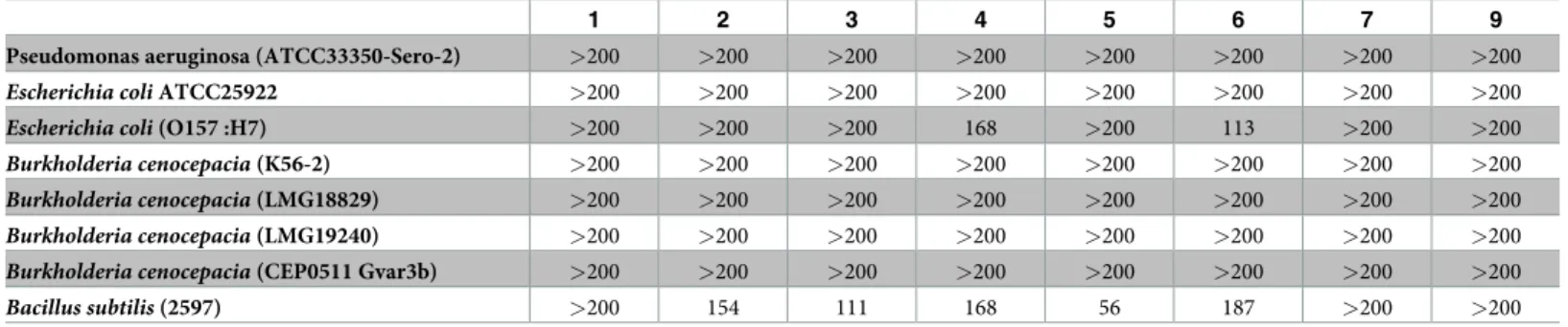
We next evaluated the specificity of PACAP activity against additional strains from the same species. While PACAP and its derivatives are highly active against strain J2315 ofB. ceno-cepacia, they were inactive against four other strains belonging to the same species (Table 2). Additional strains ofP. aeruginosa and E. coli were also evaluated with results mostly in
accor-dance with the previously observed effects (Tables1and2).
Table 2. MICaof PACAP and related analogs on homologous bacterial strains.
1 2 3 4 5 6 7 9
Pseudomonas aeruginosa (ATCC33350-Sero-2) >200 >200 >200 >200 >200 >200 >200 >200
Escherichia coli ATCC25922 >200 >200 >200 >200 >200 >200 >200 >200
Escherichia coli (O157 :H7) >200 >200 >200 168 >200 113 >200 >200
Burkholderia cenocepacia (K56-2) >200 >200 >200 >200 >200 >200 >200 >200
Burkholderia cenocepacia (LMG18829) >200 >200 >200 >200 >200 >200 >200 >200
Burkholderia cenocepacia (LMG19240) >200 >200 >200 >200 >200 >200 >200 >200
Burkholderia cenocepacia (CEP0511 Gvar3b) >200 >200 >200 >200 >200 >200 >200 >200
Bacillus subtilis (2597) >200 154 111 168 56 187 >200 >200 a
Minimum inhibitory concentration (MIC) represents the lowest concentration of an antimicrobial that will inhibit the visible growth of a microorganism after overnight incubation. Concentrations are expressed inμg/ml. Experiments were performed at least in triplicate.
https://doi.org/10.1371/journal.pone.0207366.t002
Table 1. Minimal inhibitory concentration (MIC)aof PACAP and related analogs on various bacterial strains.
1 2 3 4 5 6 7 8 9
Pseudomonas putida (KT2440) 113 77 93 56 186 113 33 >160 >200
Pseudomonas fluorescens (MF37) >200 >200 >200 112 >200 >200 >200 >160 >200
Pseudomonas aeruginosa (PA14) >200 >200 >200 112 >200 >200 >200 >160 >200
Escherichia coli (JM109) >200 116 93 28 112 37 >200 >160 165
Burkholderia cenocepacia (J2315) 10 10 9 5 20 8 2 >160 >200
Burkholderia thailendensis (ATCC700388) >200 >200 >200 >200 >200 >200 >200 >160 >200
Serratia marcescens (ATCC14756) >200 >200 >200 >200 >200 >200 >200 >160 >200
Bacillus circulans (LSPQ3543) 94 58 21 47 84 94 6 >160 >200
Bacillus cereus (ATCC11778) >200 >200 >200 >200 >200 >200 >200 >160 >200
Actinobacillus pleuropneumoniae 7.5 232 112 >200 >200 >200 >200 >160 >200
Bacillus subtilis (PY79) 28 12 5 3 42 3.5 7 >160 154
Bacillus velezensis 76 87 33 61 168 52 71 >160 >200
Bacillus amyloliquefaciens 42 26 10 7 20 9 21 >160 134
Staphylococcus aureus (ATCC6538#P) >200 >200 >200 >200 >200 >200 >200 >160 >200
Staphylococcus aureus (Newman) >200 >200 >200 >200 >200 >200 >200 >160 >200 a
Minimum inhibitory concentration (MIC) represents the lowest concentration of an antimicrobial that will inhibit the visible growth of a microorganism after overnight incubation. Concentrations are expressed inμg/ml. Experiments were done at least in triplicates.
Insight into the PACAP38 mode of action
The known cell penetrating properties of 1 as well as its ability to interact with biological mem-branes prompted us to evaluate its propensity to interact with the bacterial membrane and to penetrate inside bacteria. To do so, 1 was incubated with various bacterial strains for 1h, allow-ing the peptide to enter the cells. No bacterial death was observed after this incubation period (data not shown). The bacterial cells were then fractionated into compartments and the pres-ence of 1 or its metabolites in the outer/inner membranes or the cytoplasmic fraction was investigated by mass spectrometry (Table 3).
Table 3. Mass spectrometry analysis of bacterial compartments following incubation with PACAP38.
Cell fraction Observed mass (Da) Sequence Calculated mass (Da)
Bacillus subtilis (PY79)
Membrane 4534.1 HSDGIFTDSYSRYRKQMAVKKYLAAVLGKRYKQRVKNK 4534.3 Cytoplasm 4536.6 HSDGIFTDSYSRYRKQMAVKKYLAAVLGKRYKQRVKNK 4534.3 3667.9 SYSRYRKQMAVKKYLAAVLGKRYKQRVKNK 3661.3 Bacillus subtilis (2597) Membrane 4534.1 HSDGIFTDSYSRYRKQMAVKKYLAAVLGKRYKQRVKNK 4534.3 Cytoplasm - - -Burkholderia cenocepacia (J2315)
Outer membrane 4533.2 HSDGIFTDSYSRYRKQMAVKKYLAAVLGKRYKQRVKNK 4534.3
3666.6 SYSRYRKQMAVKKYLAAVLGKRYKQRVKNK 3661.3
Inner membrane - -
-Cytoplasm 4542.3� HSDGIFTDSYSRYRKQMAVKKYLAAVLGKRYKQRVKNK 4534.3
3663.3 SYSRYRKQMAVKKYLAAVLGKRYKQRVKNK 3661.3 Burkholderia cenocepacia (K56-2)
Outer membrane 4536.3 HSDGIFTDSYSRYRKQMAVKKYLAAVLGKRYKQRVKNK 4534.3
3669.2 SYSRYRKQMAVKKYLAAVLGKRYKQRVKNK 3661.3
Inner membrane - -
-Cytoplasm 4543.8� HSDGIFTDSYSRYRKQMAVKKYLAAVLGKRYKQRVKNK 4534.3
�Methionine oxidation
As described above, PACAP38 can reduce the growth ofB. subtilis strain PY79 but not of
strain 2597 (Tables1and2). Mass spectrometry analyses revealed that 1 (PACAP38) is found associated with the bacterial membrane in both strains but only present within the cytoplasmic fractions of PY79. Interestingly, a presumptive metabolite, identified as PACAP(9–38), was only observed associated with the sensitive PY79 strain. Furthermore, a similar pattern was also observed with theB. cenocepacia strains. While PACAP38 was observed on the outer
membrane and within the cytoplasm of bothB. cenocepacia strains, PACAP(9–38) was again
only present within the cytoplasmic fractions of the sensitive J2315 strain. Hence, presence of the PACAP(9–38) metabolite in the cytoplasm seems to correlate with sensitivity to the inhibitory activity of PACAP38. The inability of 1 to reduce the growth homologous strains of
Bacillus subtilis PY79 and Burkholderia cenocepacia J2315 might therefore point toward the
existence of different proteases in these bacterial strains. Hence, whileB. cenocepacia K56-2
and J2315 belong to the same clonal complex, they have several differences in their genomes and phenotypes[25]. Indeed, although there is only a draft genome of K-56-2 available (www. burkholderia.com), several differences in annotated proteases and peptidases are noted. Fur-ther experiments will be needed to confirm our assumption.
PACAP(9–38): A potentially selective antimicrobial peptide
To confirm the bactericidal activity of this PACAP38-derived metabolite, the analog was pre-pared by solid phase peptide synthesis. The pharmacological profile of this synthetic PACAP Table 4. Binding affinity of PACAP38 and PACAP(9–38).
HPLCa MSb
calc
MSc found
PAC1 VPAC1 VPAC2
IC50 (nM)d pIC50 IC50 (nM)d pIC50 IC50 (nM)d pIC50 PACAP38 98% 4534.3 4534.9 4.6 (2.7–7.7) 8.34± 0.11 3.6 (2.2–5.7) 8.45± 0.09 18 (4–78) 7.74± 0.28 PACAP(9–38) 98% > 10−6 5.87± 0.19 > 10−6 5.29± 0.37 > 10−6 5.77± 0.13
aPercentage of purity determined by HPLC using the eluent system: A = H
2O (0.1% TFA) and B = 60% CH3CN/40% A with a gradient slope of 1% B/min, at a flow rate
of 1 mL/min on a Vydac C18column. Detection at 229 nm.
bTheorical monoisotopic molecular weight as calculated with ChemDraw Ultra 7.0.1. cm/z value assessed by MALDI-TOF-MS.
dIC
50represents the concentration at 50% binding inhibition. Values in parentheses are 95% confidence limits.
https://doi.org/10.1371/journal.pone.0207366.t004
Table 5. MICavalues of PACAP(9–38) (10) against PACAP-sensitive bacteria.
10
Burkholderia cenocepacia (J2315) 19
Burkholderia cenocepacia (K56-2) >183
Pseudomonas putida (KT2440) >183
Escherichia coli (JM109) >183
Bacillus subtilis (PY79) >183
Bacillus subtilis (2597) >183
Bacillus amyloliquefaciens >183
Actinobacillus pleuropneumoniae >183 a
Minimum inhibitory concentration (MIC) represents the lowest concentration of an antimicrobial that will inhibit the visible growth of a microorganism after overnight incubation. Concentrations are expressed inμg/ml. Experiments were performed at least in triplicate.
(9–38) was evaluated using three CHO cell lines respectively co-expressing the human PAC1, VPAC1 or VPAC2 receptors. As depicted in earlier publications, the N-terminal domain of PACAP plays an essential role for binding affinity and biological activity[8,23,26,27]. Hence, compared to the native PACAP38, PACAP(9–38) is characterized by a dramatic reduction of its binding affinity towards all PACAP receptors (Table 4), much like PACAP(11–38)(11). PACAP(9–38) was then tested against several PACAP38-sensitive bacterial strains. Unexpect-edly, PACAP(9–38) was only able to reduce, with high efficiency (MIC: 19μg/mL), the growth of strain J2315 ofB. cenocepacia (Table 5), leaving unaffected several PACAP38-sensitive strains includingP. putida (KT2440), E. coli (JM109), and B. subtilis (PY79).
The percentage of haemolysis caused by various concentrations (4.5 ng/mL—45μg/mL) of PACAP38 and (3.7 ng/mL—37μg/mL) of PACAP(9–38) over 30 min or 3h at 37˚C is shown inFig 1At 37 or 45μg/mL, PACAP38 and PACAP(9–38) do not exert a very significant hae-molytic activity. While less prominent thanP. aeruginosa-associated infection among cystic
fibrosis patient,B. cenocepacia-related infection in such patients provokes a severe decline in
lung function that might end up into a life-threatening systemic infection known as cepacia syndrome [28]. Treatment of such infection is extremely difficult asB. cenocepacia is highly
resistant to multiple antibiotics or antibiotic combination [29]. With its apparent selectivity againstB. cenocepacia J2315 and a potentially good therapeutic index, PACAP(9–38)
repre-sents a suitable candidate for the development of antimicrobial agents against cystic fibrosis-associated infection involvingB. cenocepacia.
Relationships between the secondary structure and biological activity of
PACAP38 and PACAP(9–38)
The secondary structure of PACAP38 and PACAP(9–38) was investigated by circular dichro-ism to ensure the conservation of the C-terminalα-helix[9,23]. As shown inFig 2, PACAP38 and its truncated analog exhibit two negative minima at 208 and 222 nm in HFIP and 10% SDS (Fig 2B and 2C), characteristic of the presence of anα-helical structure. Conversely, in water, no structure was noticeable (Fig 2A).
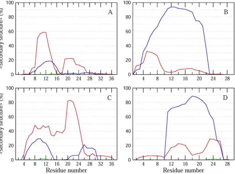
Molecular dynamics (MD) simulations provide atomistic insights into structural properties of proteins. Here, we performed MD simulations of PACAP38 and PACAP(9–38) bound to the surface of a 1,2-dipalmitoylphosphatidylcholine (DPPC) lipid bilayer to characterize the secondary structural propensities for the two membrane-bound peptides. DPPC was chosen to mimic the zwitterionic phosphatidylethanolamine-rich membranes of gram-negative bacteria [30]. Two sets of 300 ns simulations were performed for each of the two peptides, correspond-ing to a cumulative simulation time of 1.2μs. As previously demonstrated, this time scale pro-vides a good approximation of peptide reorganization and structural behavior in solution[31]. A comparison of the secondary structural propensities of the two peptides confirms the pre-dominant helical propensity in the two peptides (Fig 3). Interestingly, while PACAP38 showed a higher propensity for the formation ofα-helical conformations relative to π- and 310helices (Fig 3A and 3C), PACAP(9–38) consistently displayed a significantly higher propensity for π-helix conformations in the two independent simulations of the peptide (Fig 3B and 3D). This enhanced propensity of PACAP(9–38) to adoptπ-helix conformations may contribute towards its higher selectivity againstBurkholderia cenocepacia J2315. We note that while MD
Fig 1. Haemolytic activity of PACAP38 and PACAP(9–38) performed after 30 min (A) and and 3h (B) incubation with various concentrations of PACAP38 or PACAP(9–38). Statistical analysis was performed by using
ANOVA, followed by a Dunnett’s multiple comparison test, and differences were considered significant when�� �P < 0.001.
simulations provide important insights into the differences in the conformational properties of the two peptides, further experiments such as COSY and NOESY 2D NMR experiments would be necessary to confirm these observations.
Conclusions
We have demonstrated that the human neuropeptide PACAP38 possesses an antibacterial activity against a broad spectrum of bacteria. Notably, PACAP38 was able to inhibit the growth of the epidemic pathogenB. cenocepacia J2315 with a MIC of 10 μg/mL. Investigation about its
mechanism of action suggested that a PACAP metabolite, identified as PACAP(9–38), might indeed be responsible for the observed PACAP38 antibacterial action onB. cenocepacia J2315
andB. subtilis PY79. Surprisingly, PACAP(9–38) exhibited an increased specificity toward B. cenocepacia J2315 compared to other tested bacteria, a phenomenon that might be related to
Fig 2. Circular dichroism analysis of PACAP38 and PACAP(9–38). Each spectrum is the mean of 3 scans corrected
for solvent contribution.
https://doi.org/10.1371/journal.pone.0207366.g002
Fig 3. Secondary structural propensities of PACAP38 and PACAP(9–38). The average percentα-helix, 310andπ helix as a function of residue number along
the sequence are represented as red, green and blue lines, respectively, for two independent MD runs of PACAP38 (A, C) and PACAP9-38 (B, D). https://doi.org/10.1371/journal.pone.0207366.g003
its ability to cross the bacterial membrane. Indeed, differences in the bacterial wall composi-tion[32] might be responsible for the increased selectivity of PACAP(9–38), which was gener-ally generatedin situ within the bacterial cytoplasm following PACAP38 entry. Accordingly, it
was observed that the uptake efficacy of PACAP within eukaryotic cell cytoplasm was depen-dent on the expression of cell surface glycosaminoglycans[33]. Another possibility relies on the predisposition of PACAP(9–38) to adoptπ-helix conformations rather than α-helical confor-mations like PACAP38. While the mechanisms of action remain to be completely understood, we believe that PACAP38 and related derivatives can pave the way toward the development of new therapeutic agents against multidrug resistant bacteria, and more specifically the Burkhol-deria cepacia complex.
Author Contributions
Conceptualization: Nicolas Doucet, Eric De´ziel, David Chatenet.
Data curation: Somia Debbabi, Marie-Christine Groleau, Myriam Le´tourneau, Laura-Lee Gosselin, Jacinthe Gagnon.
Formal analysis: Somia Debbabi, Marie-Christine Groleau, Chitra Narayanan, Laura-Lee Gosselin, Mustapha Iddir, Jacinthe Gagnon.
Funding acquisition: Nicolas Doucet, Eric De´ziel, David Chatenet. Investigation: Somia Debbabi, Marie-Christine Groleau.
Methodology: Somia Debbabi, Marie-Christine Groleau, Myriam Le´tourneau, Chitra Narayanan.
Project administration: Nicolas Doucet, Eric De´ziel, David Chatenet. Resources: Eric De´ziel.
Software: Chitra Narayanan.
Supervision: Myriam Le´tourneau, Nicolas Doucet, Eric De´ziel, David Chatenet. Validation: Somia Debbabi.
Visualization: Somia Debbabi, Chitra Narayanan.
Writing – original draft: Somia Debbabi, Marie-Christine Groleau, Myriam Le´tourneau, Chitra Narayanan, Laura-Lee Gosselin, Jacinthe Gagnon, Nicolas Doucet, Eric De´ziel, David Chatenet.
Writing – review & editing: Somia Debbabi, Marie-Christine Groleau, Myriam Le´tourneau, Chitra Narayanan, Nicolas Doucet, Eric De´ziel, David Chatenet.
References
1. Watkins RR, Bonomo RA. Overview: Global and Local Impact of Antibiotic Resistance. Infect Dis Clin North Am. 2016; 30(2):313–22.https://doi.org/10.1016/j.idc.2016.02.001PMID:27208761
2. Wang Z, Wang G. APD: the Antimicrobial Peptide Database. Nucleic Acids Res. 2004; 32(Database issue):D590–2.https://doi.org/10.1093/nar/gkh025PMID:14681488
3. Splith K, Neundorf I. Antimicrobial peptides with cell-penetrating peptide properties and vice versa. Eur Biophys J. 2011; 40(4):387–97.https://doi.org/10.1007/s00249-011-0682-7PMID:21336522
4. Rotem S, Mor A. Antimicrobial peptide mimics for improved therapeutic properties. Biochim Biophys Acta. 2009; 1788(8):1582–92.https://doi.org/10.1016/j.bbamem.2008.10.020PMID:19028449
5. Fox JL. Antimicrobial peptides stage a comeback. Nat Biotechnol. 2013; 31(5):379–82.https://doi.org/ 10.1038/nbt.2572PMID:23657384
6. Midura-Nowaczek K, Markowska A. Antimicrobial peptides and their analogs: searching for new poten-tial therapeutics. Perspect Medicin Chem. 2014; 6:73–80.https://doi.org/10.4137/PMC.S13215PMID:
25374459
7. Liu YY, Wang Y, Walsh TR, Yi LX, Zhang R, Spencer J, et al. Emergence of plasmid-mediated colistin resistance mechanism MCR-1 in animals and human beings in China: a microbiological and molecular biological study. Lancet Infect Dis. 2016; 16(2):161–8.https://doi.org/10.1016/S1473-3099(15)00424-7
PMID:26603172
8. Vaudry D, Falluel-Morel A, Bourgault S, Basille M, Burel D, Wurtz O, et al. Pituitary adenylate cyclase-activating polypeptide and its receptors: 20 years after the discovery. Pharmacol Rev. 2009; 61(3):283– 357.https://doi.org/10.1124/pr.109.001370PMID:19805477
9. Bourgault S, Vaudry D, Dejda A, Doan ND, Vaudry H, Fournier A. Pituitary adenylate cyclase-activating polypeptide: focus on structure-activity relationships of a neuroprotective Peptide. Curr Med Chem. 2009; 16(33):4462–80. PMID:19835562
10. Doan ND, Chatenet D, Letourneau M, Vaudry H, Vaudry D, Fournier A. Receptor-independent cellular uptake of pituitary adenylate cyclase-activating polypeptide. Biochim Biophys Acta. 2012; 1823(4):940– 9.https://doi.org/10.1016/j.bbamcr.2012.02.001PMID:22343001
11. Doan ND, Letourneau M, Vaudry D, Doucet N, Folch B, Vaudry H, et al. Design and characterization of novel cell-penetrating peptides from pituitary adenylate cyclase-activating polypeptide. J Control Release. 2012; 163(2):256–65.https://doi.org/10.1016/j.jconrel.2012.08.021PMID:22922050
12. Clinical and Laboratory Standards Institute/NCCLS. 2006. Methods for dilution antimicrobial susceptibil-ity tests for bacteria that grow aerobically; approved standard, 7th ed. CLSI document M7-A7. Clinical and Laboratory Standards Institute, Wayne, PA.
13. Chung JW, Speert DP. Proteomic identification and characterization of bacterial factors associated with Burkholderia cenocepacia survival in a murine host. Microbiology. 2007; 153(Pt 1):206–14.https://doi. org/10.1099/mic.0.2006/000455-0PMID:17185549
14. Gerhardt P. Methods for general and molecular bacteriology. Washington, D.C.: American Society for Microbiology; 1994. xii, 791 p. p.
15. Poujol de Molliens M, Letourneau M, Devost D, Hebert TE, Fournier A, Chatenet D. New insights about the peculiar role of the 28–38 C-terminal segment and some selected residues in PACAP for signaling and neuroprotection. Biochem Pharmacol. 2018; 154:193–202.https://doi.org/10.1016/j.bcp.2018.04. 024PMID:29704474
16. Hess B, Kutzner C, van der Spoel D, Lindahl E. GROMACS 4: Algorithms for Highly Efficient, Load-Bal-anced, and Scalable Molecular Simulation. J Chem Theory Comput. 2008; 4(3):435–47.https://doi.org/ 10.1021/ct700301qPMID:26620784
17. Oostenbrink C, Villa A, Mark AE, van Gunsteren WF. A biomolecular force field based on the free enthalpy of hydration and solvation: the GROMOS force-field parameter sets 53A5 and 53A6. J Comput Chem. 2004; 25(13):1656–76.https://doi.org/10.1002/jcc.20090PMID:15264259
18. Berger O, Edholm O, Jahnig F. Molecular dynamics simulations of a fluid bilayer of dipalmitoylphospha-tidylcholine at full hydration, constant pressure, and constant temperature. Biophys J. 1997; 72 (5):2002–13.https://doi.org/10.1016/S0006-3495(97)78845-3PMID:9129804
19. Hess B, Bekker H, Berendsen H, Fraaije J. LINCS: A linear constraint solver for molecular simulations. J Comp Chem. 1997; 18:1463–72.
20. Darden T, York D, Pedersen L. Particle mesh Ewald: An N�log(N) method for Ewald sums in large sys-tems. J Chem Phys. 1993; 98:10089–92.
21. Kabsch W, Sander C. Dictionary of protein secondary structure: pattern recognition of hydrogen-bonded and geometrical features. Biopolymers. 1983; 22(12):2577–637.https://doi.org/10.1002/bip. 360221211PMID:6667333
22. Bourgault S, Vaudry D, Botia B, Couvineau A, Laburthe M, Vaudry H, et al. Novel stable PACAP ana-logs with potent activity towards the PAC1 receptor. Peptides. 2008; 29(6):919–32.https://doi.org/10. 1016/j.peptides.2008.01.022PMID:18353507
23. Bourgault S, Vaudry D, Segalas-Milazzo I, Guilhaudis L, Couvineau A, Laburthe M, et al. Molecular and conformational determinants of pituitary adenylate cyclase-activating polypeptide (PACAP) for activa-tion of the PAC1 receptor. J Med Chem. 2009; 52(10):3308–16.https://doi.org/10.1021/jm900291j
PMID:19413310
24. Gesell J, Zasloff M, Opella SJ. Two-dimensional 1H NMR experiments show that the 23-residue magai-nin antibiotic peptide is an alpha-helix in dodecylphosphocholine micelles, sodium dodecylsulfate micelles, and trifluoroethanol/water solution. J Biomol NMR. 1997; 9(2):127–35. PMID:9090128
25. Gislason AS, Turner K, Domaratzki M, Cardona ST. Comparative analysis of the Burkholderia cenoce-pacia K56-2 essential genome reveals cell envelope functions that are uniquely required for survival in species of the genus Burkholderia. Microb Genom. 2017; 3(11).
26. Bourgault S, Chatenet D, Wurtz O, Doan ND, Leprince J, Vaudry H, et al. Strategies to convert PACAP from a hypophysiotropic neurohormone into a neuroprotective drug. Curr Pharm Des. 2011; 17 (10):1002–24. PMID:21524253
27. Doan ND, Bourgault S, Dejda A, Letourneau M, Detheux M, Vaudry D, et al. Design and in vitro charac-terization of PAC1/VPAC1-selective agonists with potent neuroprotective effects. Biochem Pharmacol. 2011; 81(4):552–61.https://doi.org/10.1016/j.bcp.2010.11.015PMID:21114961
28. Drevinek P, Mahenthiralingam E. Burkholderia cenocepacia in cystic fibrosis: epidemiology and molec-ular mechanisms of virulence. Clin Microbiol Infect. 2010; 16(7):821–30. https://doi.org/10.1111/j.1469-0691.2010.03237.xPMID:20880411
29. Conway SP, Brownlee KG, Denton M, Peckham DG. Antibiotic treatment of multidrug-resistant organ-isms in cystic fibrosis. Am J Respir Med. 2003; 2(4):321–32. PMID:14719998
30. Clifton LA, Skoda MW, Daulton EL, Hughes AV, Le Brun AP, Lakey JH, et al. Asymmetric phospholipid: lipopolysaccharide bilayers; a Gram-negative bacterial outer membrane mimic. J R Soc Interface. 2013; 10(89):20130810.https://doi.org/10.1098/rsif.2013.0810PMID:24132206
31. Brice AR, Lazaridis T. Structure and dynamics of a fusion peptide helical hairpin on the membrane sur-face: comparison of molecular simulations and NMR. J Phys Chem B. 2014; 118(17):4461–70.https:// doi.org/10.1021/jp409412gPMID:24712538
32. Silhavy TJ, Kahne D, Walker S. The bacterial cell envelope. Cold Spring Harb Perspect Biol. 2010; 2(5): a000414.https://doi.org/10.1101/cshperspect.a000414PMID:20452953
33. Tchoumi Neree A, Nguyen PT, Chatenet D, Fournier A, Bourgault S. Secondary conformational conver-sion is involved in glycosaminoglycans-mediated cellular uptake of the cationic cell-penetrating peptide PACAP. FEBS Lett. 2014; 588(24):4590–6.https://doi.org/10.1016/j.febslet.2014.10.029PMID: