HAL Id: tel-02518809
https://pastel.archives-ouvertes.fr/tel-02518809
Submitted on 25 Mar 2020
HAL is a multi-disciplinary open access
archive for the deposit and dissemination of sci-entific research documents, whether they are pub-lished or not. The documents may come from teaching and research institutions in France or abroad, or from public or private research centers.
L’archive ouverte pluridisciplinaire HAL, est destinée au dépôt et à la diffusion de documents scientifiques de niveau recherche, publiés ou non, émanant des établissements d’enseignement et de recherche français ou étrangers, des laboratoires publics ou privés.
Role of the actomyosin cytoskeleton on the synaptic
effects of the type-1 cannabinoid receptor (CB1R)
Maureen Mcfadden
To cite this version:
Maureen Mcfadden. Role of the actomyosin cytoskeleton on the synaptic effects of the type-1 cannabi-noid receptor (CB1R). Neurons and Cognition [q-bio.NC]. Université Paris sciences et lettres, 2018. English. �NNT : 2018PSLET006�. �tel-02518809�
THÈSE DE DOCTORAT
de l’Université de recherche Paris Sciences et Lettres
PSL Research University
Préparée à l’Ecole Supérieure de Physique et de
Chimie Industrielles de la ville de Paris (ESPCI PARIS)
Role of the actomyosin cytoskeleton in the synaptic effects of the
type-1 cannabinoid receptor (CBtype-1R)
COMPOSITION DU JURY :
Dr. DIGREGORIO David
UPMC, President du jury
Dr. ANDRIEUX Annie
CHU de Grenoble, Rapporteure
Pr. KATONA István
Hungarian Academy of Sciences (MTA), Rapporteur
Dr. LEBRETON Stéphanie
Paris XI, Membre du jury
Dr. IZEDDIN Ignacio
ESPCI-PSL, Membre du jury
Dr. LENKEI Zsolt
Paris V, Membre du jury
Soutenue par Maureen
McFADDEN
le 20 avril 2018
h
Ecole doctorale
n°
158
Cerveau, Cognition, Comportement (ED3C)
Spécialité
Neurosciences
Dirigée par Zsolt LENKEI
Acknowledgments
First and foremost, I would like to thank my jury members Annie Andrieux, István Katona,
David DiGregorio, Ignacio Izeddin, and Stephanie Lebreton for accepting to take part in my
thesis defense and for their time and expertise in examining my work. I hope my thesis will be as enjoyable to read as it was to produce.
To my supervisor Zsolt Lenkei, I would like to thank you for accepting me within your team. Your open-mindedness and breadth of expertise has been an inspiration, and allowed me to develop both technically and personally. You have challenged me never to rest on my past knowledge and abilities but to develop new skills and expand my understanding, all the while remaining supportive and optimistic when things weren’t seeming too bright. For this and much more, I am very grateful to you.
To my team colleagues, I would like to thank you for your support and advice. Seeing you at the lab has always been a highlight of my day and has made my 4 years within the team a memorable experience I shall treasure. Diana Zala, you have been my mentor/ surrogate mother. Although you have been quite severe and harsh in your judgment at times, it has always been truthful and sometimes necessary to counteract my stubbornness, for which I apologize. Your integrity both in science and in life has truly been a great influence and an inspiration to me. To Jeremy Ferrier, my surrogate big brother, thank you for being my balcony/gym buddy. You were always happy to offer constructive criticism, even unrequested, which, when serious, has always been helpful. You have never hesitated to help me when you saw me struggling. Thank you for your patience and advice. To Sergio Leite, my evening buddy, you’re the only team member that had a time schedule similar to mine. You too have never hesitated to help in times of moral and/or technical need. Thank you for making the long nights and weekends at the lab so much brighter.
Thank you to all team members past and new: Ana Ricobaraza, for training me during my Masters internship and easing my entry into the team; Delphine Ladarre, for her advice in videomicroscopy; Sophie Pezet, for her experienced advice and support in all matters, Julie
Nguyen, for taking the cell culture off my shoulders, and her positive and sunny disposition; Michael Salerno, for being my go to reference for all English language and American culture
questions without complaint; Renata Santos, for helping me figure out my life plan. Thank you to the students for their positive attitude and great work. Particularly I would like to thank Lea Anselin for helping me out with my project and for our insightful conversations on culture and ethics; and Navid Barakzoy for taking on the continuation of my project and for his constant smile.
Thank you to all the members of my old lab unit ‘Plasticité du Cerveau’, where I spent 4 years, and its director Thomas Preat, for accepting me within the unit. My interactions there have always been pleasant and insightful, be it with the team leaders, researchers, postdocs
or students. I would also like to thank our new unit at the CPN and its director Thierry Galli for welcoming us in, and allowing me to defend my thesis there.
Last but certainly not least, I would like to thank my family and friends for their support and for generally making my life all the brighter even in hard times. In particular I would like to thank my mother, for always believing in me, even in the face of my undying pessimism, and my father, for his experience and advice in life, but especially in all things academic, statistics and programming. Your crash course in R programming has been a life saver.
1
Table of Contents
1 THE CHEMICAL SYNAPSE: A COMPUTATIONALLY AND PHYSIOLOGICALLY
PLASTIC UNIT ... 7
1.1 BRAIN WIRING: A LIFELONG PLASTIC PROCESS ... 7
1.2 SYNAPTIC STRUCTURE AND FUNCTION ... 8
1.2.1 PRESYNAPTIC STRUCTURE ... 9
1.2.2 PRESYNAPTIC FUNCTION: SYNAPTIC VESICLE RECYCLING ... 12
1.3 SYNAPTIC PLASTICITY ... 15
1.3.1 CA2+LEVEL REGULATION ... 16
1.3.2 KINASE RECRUITMENT AS A BIDIRECTIONAL SWITCH ... 16
1.3.3 PROTEIN SYNTHESIS ... 17
1.3.4 RELEASE MACHINERY MODULATION ... 17
1.3.5 SYNAPTIC VESICLE POOL MODULATION ... 18
1.3.6 STRUCTURAL MODULATION ... 18
2 THESIS AIM ... 20
3 THE ACTOMYOSIN CYTOSKELETON AND SYNAPTIC ACTIVITY ... 22
3.1 THE ACTOMYOSIN CYTOSKELETON ... 23
3.1.1 ACTIN FILAMENTS ... 23
3.1.2 NON-MUSCULAR MYOSIN II ... 24
3.1.3 ACTOMYOSIN UPSTREAM SIGNALING PATHWAYS ... 26
3.2 ACTOMYOSIN AT THE SYNAPSE ... 28
3.2.1 ACTIN IN THE AZ ... 29
3.2.2 ACTIN AND SYNAPTIC VESICLE POOLS ... 29
3.2.3 ACTOMYOSIN AND SYNAPTIC FUNCTION ... 31
4 CB1R AND THE ENDOCANNABINOID SYSTEM ... 35
4.1 RECEPTORS, LIGANDS, ET AL.:PROPERTIES OF THE ENDOCANNABINOID NEUROTRANSMITTER SYSTEM ... 36
4.1.1 RECEPTORS ... 36
4.1.2 LIGANDS ... 39
4.1.3 EXTRACELLULAR LIGAND RELEASE ... 40
4.2 CB1R DOWNSTREAM SIGNALING ... 41 4.2.2 GΒ/Γ SIGNALING ... 43 4.2.3 GI/O SIGNALING ... 43 4.2.4 GS SIGNALING ... 44 4.2.5 G12/13 SIGNALING ... 44 4.2.6 GQ/11 TYPE SIGNALING ... 44
4.3 CB1R-MEDIATED SYNAPTIC PLASTICITY ... 45
2
4.3.2 LONG-TERM DEPRESSION (LTD) ... 47
4.4 CB1R IN NEURAL DEVELOPMENT ... 49
4.4.1 CB1R EXPRESSION IN DEVELOPMENT ... 50
4.4.2 CB1R IN NEURONAL MORPHOGENESIS ... 50
ARTICLE 1 (PUBLISHED): CANNABINOID-INDUCED ACTOMYOSIN CONTRACTILITY SHAPES NEURONAL MORPHOLOGY AND GROWTH ... 53
5 RESULTS PART1: ACTOMYOSIN DYNAMICS MEDIATE CB1R-INDUCED INHIBITION OF VESICLE RELEASE IN CULTURE ... 77
5.1 IMAGING EXOCYTOSIS IN CULTURE WITH SYNAPTOPHLUORIN ... 78
5.2 CB1R-ACTIVATION INDUCES A DECREASE IN SYNAPTIC VESICLE EXOCYTOSIS ... 79
5.3 ACTOMYOSIN CONTRACTILITY THROUGH ROCK MEDIATES THE EFFECTS OF CB1R ON VESICLE EXOCYTOSIS ... 81
6 RESULTS PART2: STORM IMAGING REVEALS ACTOMYOSIN-INDUCED SYNAPTIC VESICLE REDISTRIBUTION UNDER CB1R ACTIVATION ... 83
6.1 STOCHASTIC OPTICAL RECONSTRUCTION MICROSCOPY (STORM) ... 84
6.2 CLUSTERING ANALYSIS FOR STORM IMAGES ... 86
6.3 PREDICTING THE ACTIVE ZONE LOCATION FROM THE POST SYNAPTIC DENSITY ... 89
6.3.1 PROPERTIES OF HOMER1 AND BASSOON APPOSITIONS ... 89
6.3.2 PREDICTING THE AZ ... 89
6.4 SYNAPTIC VESICLE IDENTIFICATION ... 91
6.4.1 IDENTIFYING SYNAPTIC VESICLES ... 91
7 RESULTS PART 3: THESIS ARTICLE ... 97
ARTICLE 2 (IN SUBMISSION): ACTOMYOSIN-MEDIATED NANOSTRUCTURAL REMODELING OF THE PRESYNAPTIC VESICLE POOL BY CANNABINOIDS INDUCES LONG-TERM DEPRESSION ... 98
8 DISCUSSION: ACTOMYOSIN CONTRACTILITY THROUGH ROCK MEDIATES CB1R INDUCED LTD ... 121
8.1 ACTOMYOSIN IN LTD VERSUS DSI/DSE ... 121
8.2 SIGNALING PATHWAY TO ACTOMYOSIN CONTRACTILITY IN CB1R-LTD ... 122
8.3 ACTOMYOSIN AND VESICLE RECYCLING UNDER CB1R ... 124
PERSPECTIVES ... 125
ANNEXES: R SCRIPTS ... 127
3
Abbreviations
2AG: 2-Arachidonyl glycerol ABP: Actin Binding Protein
ADP: Adenosine Diphosphate
AEA: Anandamide
ATP: Adenosine triphosphate
AZ: Active Zone
CB1R: Cannabinoid type-1 Receptor CB2R: Cannabinoid type-2 Receptor
DBSCAN: Density-Based Spatial Clustering of Applications with Noise
eCB: endocannabinoid
ELC : Essential Light Chain
EM: Electron Microscopy
GTP: Guanosine Triphosphate
LTD: Long-Term Depression
LTP: Long-term Potentiation
MLC: Myosin Light Chain
MLCK: Myosin Light Chain Kinase MLCP: Myosin Light Chain Phosphatase
MYH: Myosin Heavy Chain
NMII: Non-Muscle Myosin II
4
PALM: Photo-Activated Localization Microscopy
PSD: Post-Synaptic Density
RLC: Regulatory Light Chain
ROCK: Rho-associated, coiled-coil-containing protein kinase
STD: Short-Term Depression
STORM: Stochastic Optical Reconstruction Microscopy
5
Figures
Figure 1: Synaptic Structures
Figure 2: Synaptic vesicle Pools and Recycling
Figure 3: Synaptic plasticity and its potential mechanisms Figure 4: Actin cytoskeleton formations and binding proteins
Figure 5: Non-Muscle Myosin II (NMII) activation and filament formation Figure 6: Rho-GTPase downstream effectors in cytoskeletal modulation Figure 7: Actin at the synapse
Figure 8: Actin-like filament polymerisation at endocytic zone during synaptic activity Figure 9: Preferential signaling pathways of G-protein subunits
Figure 10: Mechanisms of CB1R-induced synaptic plasticity
Figure 11: Function of the synaptophysin-pHluorin probe in reporting exocytosis Figure 12: CB1R inhibits exocytosis at axonal boutons in hippocampal cultures Figure 13: Actomyosin contractility mediates CB1R-induced reduction in exocytosis. Figure 14: Novel discoveries accomplished through STORM imaging
Figure 15: Principles of DBSCAN and OPTICS.
Figure 16: Effect of parameter p on protein localisation clustering
Figure 17: Homer1 and Bassoon apposition properties in control and under CB1R activation Figure 18: Distribution of Vamp2 within the presynaptic compartment and at synaptic
vesicles
Figure 19: Effect of nested p value in the identification of clusters in randomized distributions
6
Figure 20: p value effect on simulated spheres of different densities Figure 21: Nested cluster diameters and axonal bouton properties
7
Introduction
1 The chemical synapse: a computationally and physiologically
plastic unit
1.1 Brain wiring: a lifelong plastic process
Animal behavior is inextricably linked to brain wiring. The brain regulates such basic but vital processes as breathing, heart rhythm, and stimuli perception to higher order processes such as learning and decision-making. The regulation of these processes ultimately depends on the activity of distinct neural networks. The great bulk of these networks is genetically programmed, forming distinct brain regions that are well conserved between members of a same species, and even between different phylum classes. However, in order to adapt to a constantly changing environment, the brain needs to conserve a certain deal of plasticity. Memory, for example, essentially depends on the capacity of rewiring neural networks to retain new information.
We now know that a great deal of plastic processes thought to stop after development remain in adulthood. Neurogenesis, for example, was found to occur throughout life both in the hippocampus and in the olfactory bulb. These newly formed neurons grow out and integrate into pre-formed neural networks by forming new connections to regulate network activity (Aimone et al. 2014). Beyond neurogenesis, the majority of neurons formed early in development can change their activity throughout life to adapt to changing requirements. This adaptation can be done either by increasing or decreasing the number of synapses a neuron will make onto other neurons within the network, or by changing the strength of the synapse itself, in a process known as synaptic plasticity.
This section will outline the structure and function of the synapse before introducing different forms of synaptic plasticity, focusing on presynaptically-induced plasticity and known mechanisms of action.
8
1.2 Synaptic structure and function
The synapse is a highly compartmentalized structure whose primary function is in intercellular signaling. Within the brain, it is typically formed between the axon of one neuron and the dendrite of a second neuron. While the main inputs to synapses are changes in the electrical potential of the membrane, the signal transduction that occurs at a neuronal synapse is principally chemical. The most widely accepted model of synaptic transmission goes as follows: An action potential arriving at a synapse will trigger the activation of voltage gated calcium channels (VGCCs), inducing Ca2+ entry into the presynaptic compartment. This presynaptic Ca2+ rise initiates synaptic vesicle fusion to the presynaptic membrane and the release of the neurotransmitters contained within into the synaptic cleft (Figure 1). These neurotransmitters go on to activate specific receptors at the post-synaptic membrane, where activation of ionotropic receptors leads to ion exchange with the extracellular medium. Depending on the neurotransmitter released and receptor activated, this process leads to depolarization or hyperpolarization of the postsynaptic membrane, corresponding to excitatory or inhibitory transmission, respectively.
These steps occur through highly regulated processes and specified structures contained within each compartment (Figure 1). At the synaptic cleft the postsynaptic and presynaptic sites are mirrored by the post synaptic density (PSD) and presynaptic active zone (AZ) respectively. These densely packed structures are easily identifiable by electron microscopy and are essential to evoked synaptic transmission (Figure 1B). The AZ docks and primes vesicles for fusion, and the postsynaptic density docks neurotransmitter receptors at the membrane. Furthermore, the presynaptic site contains a number of synaptic vesicles (SVs) separated into different functional pools, which can be tapped on depending on synaptic activity (Figure 1B).
This section will briefly review some of the steps and structures important in synaptic signaling with regards to the presynaptic compartment.
9 1.2.1 Presynaptic Structure
1.2.1.1.1 The Active Zone
Similarly to the post-synaptic density (PSD), the active zone (AZ) is the “site of action” of the presynaptic compartment, where SVs are docked and primed for fusion. It is precisely aligned to the postsynaptic density in order to ensure precise targeting of neurotransmitters to postsynaptic receptors and thus ensure fast and efficient signal transmission. Recent studies, using superresolution microscopy, have even observed what the authors call nanocolumns, a trans-synaptic alinement of AZ and PSD scaffolding proteins into virtual columns, the integrity of which affects synaptic efficacy (Tang et al. 2016).
The majority of the active zone is composed of scaffolding proteins, which help anchor and prime synaptic vesicles ready to be released (Figure 2), or the readily releasable pool (RRP). The precise function of all of these still remains to be fully understood, however a number of
10
them are known to interact directly with proteins of the synaptic vesicle membrane (Südhof 2012). For example, the AZ scaffolding protein Rim1α is known to interact directly with the SV protein Rab3A, docking the vesicles at the AZ (Haucke, Neher, and Sigrist 2011). Other known proteins such as bassoon and piccolo are also thought to play a role, if not in directly docking vesicles, at least in the maintenance of the AZ structure. It is further suspected that some of these proteins help prime vesicle for fusion with the plasma membrane, such as Munc-13 found to catalyze the SNARE complex essential for membrane fusion (Südhof 2012).
1.2.1.2 Synaptic Vesicle Pools
While a great number of synaptic vesicles are present at the presynaptic bouton, around 200 at hippocampal synapses as observed through electron microscopy (EM) (Harris and Sultan 1995; Schikorski and Stevens 1997), not all of them are competent for membrane fusion. A great majority of them are clustered within the bouton, bound by proteins such as synapsins which immobilize them by tethering them to each other and to the actin cytoskeleton (Siksou et al. 2007; Fornasiero et al. 2012). This observation among others has led to the categorization of different synaptic vesicle pools depending on their fusion-competence (Figure 2).
1.2.1.2.1 The readily releasable pool (RRP)
The readily releasable pool indicates the synaptic vesicles ready to be released upon evoked stimulation. It is typically qualified as the pool released upon low frequency stimulation, or when exposed to hypertonic sucrose solutions (Rosenmund and Stevens 1996). The RRP has typically been identified as the vesicles docked at the active zone, although it was found that certain docked vesicles do not undergo fusion (Darcy et al. 2006; Harata et al. 2001; Marra et al. 2012; Ratnayaka et al. 2012). Nonetheless, it has been shown that the number of docked vesicles correlates positively with the probability of release (Branco, Marra, and Staras 2010), as well as AZ and PSD sizes (Rosenmund and Stevens 1996). It contains on average between 5 and 15 synaptic vesicles at small hippocampal synapses (Harris and Sultan 1995; Schikorski et al. 1997).
11
1.2.1.2.2 The reserve pool, or recycling pool
The reserve pool indicates the synaptic vesicles that are recruited for release once the RRP is depleted. This recruitment typically occurs upon strong stimulation, or upon exposure to high concentrations of extracellular K+ (50-90mM). It is also typically thought to comprise mainly recently endocytosed vesicles. Indeed, studies find that RRP refilling during sustained stimulation will preferentially occur through newly recycled vesicles, which are more mobile (Gaffield, Rizzoli, and Betz 2006; Kamin et al. 2010) and preferentially relocate close to the active zone (Marra et al. 2012; Schikorski et al. 1997). This property has also given the reserve pool the name of ‘recycling pool’. It includes approximately 20-30% of the total pool, although this can vary greatly up to 70% depending on the synapse and stimulation paradigm (Annette Denker and Rizzoli 2010). Similarly to the RRP, its size has been found to correlate positively with probability of release (Murthy and Stevens 1998; Waters and Smith 2002).
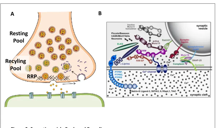
Figure 2. Synaptic vesicle Pools and Recycling
(A) Schematic drawing of the vesicle pools within a synapse. (B) Schematic representation of AZ and synaptic vesicle proteins known to interact during synaptic vesicle recycling (adapted from Sudhof et al. (2012))
A
B
RRP
Recyling
Pool
Resting
Pool
12
1.2.1.2.3 The resting pool
The resting pool indicates the synaptic vesicles which are not recruited upon evoked stimulation and it typically comprises a majority of the synaptic vesicles of the total pool. It is relatively unknown why such a large quantity of supposedly fusion incompetent vesicles resides within the bouton. One suggestion is that a portion of these vesicles may be precursor vesicles which are not neurotransmitter filled, but act as a buffer for proteins involved in vesicle recycling (Shupliakov 2009; A. Denker, Bethani, et al. 2011; A. Denker, Krohnert, et al. 2011). Furthermore, there is evidence of SV trading between the recycling pool and the resting pool (Kim and Ryan 2010; Ratnayaka et al. 2012), and certain forms of synaptic modulation have been found to recruit SVs from the resting pool as a form of potentiation (Tyler et al. 2006; J. Jung et al. 2014), suggesting that the resting pool may act as a resource in the modulation of synaptic activity.
Although the spatial distribution of vesicles within the presynaptic compartment appears to have a lot to do with their assignment to one of the pools, this does not appear to be a consistent rule as docked vesicles are not necessarily fusion competent and recycling vesicles can be found intermixed within the resting pool (Fowler and Staras 2015a). Some have therefore suggested there may be molecular markers distinguishing the different pools. SNARE proteins have been suggested to be good candidates, as it was found that VAMP7 is more present in resting vesicles, whereas VAMP2 may be preferentially located to the recycling pool (Hua et al. 2011), for example. Others have suggested that vesicle mobility within the presynaptic bouton may be a better indicator of pool assignment. Indeed, as mentioned previously, recycling vesicles are more mobile than resting pool vesicles (Gaffield, Rizzoli, and Betz 2006; Kamin et al. 2010). One reason for this may be the tethering of resting pool vesicles to the actomyosin cytoskeleton through synapsins, as inhibition of synapsin, either through genetic deletion or phosphorylation, increases mobility of vesicles within the presynaptic compartment (Orenbuch et al. 2012; Gaffield, Rizzoli, and Betz 2006). 1.2.2 Presynaptic function: synaptic vesicle recycling
While the basic principles underlying synaptic vesicle release upon stimulation have been known for some time, the knowledge concerning the different steps of synaptic vesicle recruitment, priming and recycling is relatively poor. One main reason for this paucity is the small size of the presynaptic compartment, especially at central small synapses, as well as the
13
minute size of its composing elements. Indeed, axonal boutons are on average around 1µm3 in volume, with synaptic vesicles being around 30-50nm in diameter, approximately 4x smaller than the resolution of conventional microscopy techniques. While this discrepancy can be overcome with electron microscopy (EM), EM does not lend itself well to studying the highly dynamic processes involved in synaptic vesicle cycling, which occur within a couple of seconds.
This section will briefly review what is known of the essential steps of the synaptic vesicle cycle within the presynaptic terminal.
1.2.2.1 Synaptic vesicle docking and priming
In order to rapidly respond to arriving stimuli, synaptic vesicles need to be positioned close to the presynaptic membrane and ready to be released. This is made possible by the docking of synaptic vesicles at the active zone (AZ). Although the specific mechanisms inducing SV docking are poorly understood, Rab3‐interacting molecules (RIMs) are thought to play an important role. Indeed, RIM proteins are known to bind to SV membrane proteins such as Rab3 (Haucke, Neher, and Sigrist 2011), by which they may tether SVs to the AZ. RIMs have also been found to interact with VGCCs (Deng et al. 2011; Han et al. 2011; Kaeser et al. 2011; K. S. Y. Liu et al. 2011). This interaction may allow them to dock SVs close to VGCCs, which would accelerate SV/membrane fusion initiation upon depolarization.
1.2.2.2 Exocytosis
The release of vesicle contents into the extracellular medium, known as exocytosis, occurs upon SV fusion to the presynaptic membrane. This reaction is known to occur through the formation of a SNARE complex between SNARE proteins of the SV membrane, such as VAMP2, and SNARE proteins of the plasma membrane, such as SNAP25. Formation of a complex between these proteins draws the two membranes together, forcing their fusion (Fasshauer et al. 1998).
While it is clear that Ca2+ entry upon depolarization initiates the fusion of synaptic vesicles, how this initiates the formation of the SNARE complex is relatively unclear. Synaptotagmin has been posited as a candidate as its calcium binding form is known to interact both with SNAREs and the plasma membrane, which may allow it to catalyze complex formation upon depolarization-induced Ca2+ entry (Chapman 2008).
14 1.2.2.3 Endocytosis
Endocytosis, or vesicle formation from the plasma membrane, is an important step of the synaptic vesicle cycle as it both compensates for the increase in membrane surface induced by exocytosis as well as replenishing the depleted synaptic vesicle pool. Importantly, endocytosis is often triggered by exocytosis, although the extent to which these mechanisms are linked is still under debate (Rizzoli 2014).
Upon SV fusion to the plasma membrane, the resulting SV membrane protein pool clusters and diffuses along the plasma membrane (Willig et al. 2006; Hoopmann et al. 2010; Opazo et al. 2010), clearing out of the synaptic cleft (Z. Li and Murthy 2001; Fernández-Alfonso, Kwan, and Ryan 2006; Wienisch and Klingauf 2006). There, these protein clusters are taken up through invagination of the plasma membrane to form novel synaptic vesicles.
Arguably the most widespread form of endocytosis is clathrin-dependent endocytosis. This process is relatively slow compared to exocytosis, causing a bottleneck for vesicle pool replenishment upon strong stimulations (Miller and Heuser 1984; Heuser et al. 1979; Heuser and Reese 1981). Initiation of the endocytotic process is elusive. Once plasma membrane invagination has started, clathrin-chain assemblies known as “triskelia” coat the forming vesicle. The vesicle is then detached from plasma membrane, through the action of ring-like dynamin assemblies, released of clathrin and refilled with neurotransmitter through specific transporters (Rizzoli 2014).
Other known forms of endocytosis have been described. One such form is bulk endocytosis, a process by which excessive exocytosis, for example under strong and repetitive stimulation, causes the presynaptic membrane to fold in onto itself. The resulting endosome is then budded off through clathrin-mediated endocytosis within the presynaptic terminal (Rizzoli 2014). Another form of vesicle recycling is performed through a process termed ‘kiss and run’. SVs undergoing this process do not completely fuse with the plasma membrane, but are opened just enough to release part of their content before reforming. This form of recycling has an advantage over conventional recycling as it is much faster than clathrin-mediated endocytosis (Q. Zhang, Li, and Tsien 2009; Park, Li, and Tsien 2012), meaning vesicle pools can be replenished more quickly. The relative part of ‘kiss and run’ events as compared to conventional SV recycling in synaptic signaling remains to be established however (Granseth et al. 2006; Granseth et al. 2009).
15
1.3 Synaptic plasticity
A great number of behavioral functions rely on the plasticity of neural systems. Arguably one of the simplest ways of regulating this is by modifying the activity of different neurons by modifying the activity of different synapses at different points in time, a phenomenon known as synaptic plasticity. While the difficulty of correlating synaptic plasticity with behavioral outputs in the past had struck a debate as to the in vivo relevance of known forms of plasticity (Malenka and Bear 2004), advances in the past decade have overcome these doubts, with a number of studies showing the direct involvement of synaptic plasticity in a number of behavioral processes, including monocular deprivation, reward seeking, and fear conditioning (reviews in (Malenka and Bear 2004) and (Monday and Castillo 2017)).
Synaptic plasticity is the process by which the output of a synapse to depolarizing stimuli is changed. An increase in the output is termed a potentiation, while a decrease is named a depression. The induction of these changes typically occurs through activation of G-protein coupled receptors (GPCRs). Indeed, forms of plasticity have been described for GPCR activation of most of the major neurotransmitter systems, including dopaminergic, serotoninergic, glutamatergic and GABAergic. Indeed, GPCR-induced plasticity englobes most forms of plasticity described, with one major exception being NMDA-induced forms (Atwood, Lovinger, and Mathur 2014).
The induction of synaptic plasticity has been described both at presynaptic and postsynaptic compartments. Postsynapticly-induced plasticity results in decreases or increases in postsynaptic ionic currents in response to neurotransmitter release. Presynapticly-induced plasticity results in an increase or decrease in the probability of neurotransmitter release. Synaptic plasticity can be transient, occurring on a timescale of milliseconds to a couple of minutes, in which case it is termed short-term plasticity. Changes can further persist from 30min to several weeks (Malenka and Bear 2004), in which case it is known as long-term plasticity (Figure 3A). Importantly, a number of GPCRs known to induce plasticity are known to enact both short- and long-term forms (Atwood, Lovinger, and Mathur 2014). What mediates the induction of a short term rather than a long-term form of plasticity in these cases is unknown, although it is suggested that previous synaptic activity and duration of GPCR activation may be mediating factors (Atwood, Lovinger, and Mathur 2014).
16
Indeed, the mechanisms underlying short-term forms of plasticity are often tied to relatively simple molecular cascades, such as the direct modulation of ion channels, regulating membrane excitability. Long-term forms of plasticity however recruit complex signaling cascades and the mechanisms regulating their long-term maintenance remain often unknown, particularly concerning the presynaptic forms (Figure 3B). This section will focus on long-term forms of presynaptic plasticity, looking at what is known of the common molecular or structural mechanisms they induce that ultimately lead to changes in synaptic strength.
1.3.1 Ca2+ level regulation
As opposed to short-term forms of plasticity, it would appear that both long-term potentiation (LTP) and long-term depression (LTD) depend on increases in presynaptic Ca2+ levels (Lüscher and Malenka 2012). However, studies have suggested that the magnitude of the increase determines the orientation of the plasticity, towards depression or potentiation. This was found to be the case in at hippocampal mossyfiber/CA3 synapses, where strong or weak activity would induce LTP or LTD respectively presumably by modulating the level of presynaptic Ca2+ (Tzounopoulos et al. 1998). This necessity for Ca2+ holds true when considering that both LTP and LTD have been found to recruit kinases and phosphatases for induction and/or maintenance, which either directly depend on Ca2+ for activation or recruit calcium dependent processes.
1.3.2 Kinase recruitment as a bidirectional switch
One of the most observed mediators of long-term plasticity is the cAMP/PKA signaling pathway, although the specific mechanisms underlying its effect on neurotransmitter release remains elusive. Indeed, increases and decreases of cAMP/PKA signaling have been reported in LTP and LTD respectively (Ying Yang and Calakos 2013). Many forms of LTP and LTD are dependent on GPCRs most often coupled to Gs and Gi/o type proteins, respectively, both of
which have opposite effects on cAMP/PKA signaling. Thus, Gs coupled GPCRs such as the
dopamine-1 receptor (D1R) are found to induce LTP in a PKA-dependent manner (C. Li and Rainnie 2014), whereas Gi/o coupled receptors such as mGluR2/3, D2R or CB1R are found to
induce LTD through PKA (Ying Yang and Calakos 2013). This model favors a common target mechanism in the bidirectional regulation of synaptic plasticity at the synapse, although the nature of this mechanism remains to be determined.
Other kinases found to affect presynaptic forms of plasticity are mitogen-activated protein kinases, found downstream of LTD induction (Morrison and Davis 2003). Their downstream
17
mechanisms in plasticity are poorly understood, however these kinases have been known to be involved in the expression of immediate early genes, which may help to induce protein synthesis for long-term maintenance.
1.3.3 Protein synthesis
Protein synthesis has long been established as essential in a number of postsynaptic forms of synaptic plasticity (Santini, Huynh, and Klann 2014), and has therefore been postulated as a potential mediator of presynaptic forms. Indeed, some forms of presynaptic plasticity have been found to rely on protein synthesis (Yin et al. 2006; Y. Y. Huang, Li, and Kandel 1994; Calixto et al. 2003; Younts et al. 2016). Similarly to Ca2+-dependence however this does not appear to be bidirectional as both LTP and LTD forms have been found to depend on protein synthesis.
While the proteins synthesized are most likely different for both LTP and LTD their nature in both cases is as of yet unknown, but may include the production of kinases or phosphatases necessary for LTP/LTD maintenance, as well as certain structural proteins such as AZ or vesicle proteins that could be used to strengthen the synapse.
1.3.4 Release machinery modulation
One of the most promising candidates suggested to be the substrate of long-term plasticity is the AZ protein RIM1α. Among its several known functions, RIM1α is known to bind to the SV Rab3 proteins, docking them to the AZ, as well as to VGCCs, bringing them closer to the AZ and synaptic vesicles (Deng et al. 2011; Han et al. 2011; Kaeser et al. 2011; K. S. Y. Liu et al. 2011). Rim1α is also known to interact with a number of other AZ scaffolding proteins, such as Munc-13, which may help it catalyze vesicle priming (Südhof 2012). As such, RIM1α appears to be a good target to regulate synaptic plasticity and indeed, with its interacting proteins Munc13 and Rab3A, it was found to be necessary for a number of forms of presynaptic LTP and LTD (Ying Yang and Calakos 2013).
Although PKA is known to be able to phosphorylate RIM1α, which would elegantly link the cAMP/PKA and Rim1α dependence of many of these forms of plasticity, attempts to show an interaction between the two pathways has been relatively mixed (Lonart et al. 2003; Simsek-Duran, Linden, and Lonart 2004; Kaeser et al. 2008; Y. Yang and Calakos 2010; Castillo et al. 2002).
18 1.3.5 Synaptic vesicle pool modulation
Another candidate target for bidirectional long-term plasticity is in the recruitment/depletion of the synaptic vesicles from different SV pools. Although findings in this area are relatively scarce, an increase/reduction in RRP size has been found to mediate certain forms of LTP and LTD respectively (Fowler and Staras 2015b). Furthermore, bidirectional recruitment of SVs to and from the resting pool was found to mediate plasticity, with CDK5- dependent recruitment of SVs from the resting pool thought to mediate NMDA-dependent LTP (Fowler and Staras 2015b). Conversely, eCB-LTD was found to require calcineurin (Heifets, Chevaleyre, and Castillo 2008), which has been found to mediate the transfer of vesicles from the recycling pool to the resting pool (Marra et al. 2012).
1.3.6 Structural modulation
Much like postsynaptic spines, a number of studies have shown that presynaptic sites can show extensive structural modulation. Indeed it has been shown that, while a majority of axonal boutons will remain stable over the course of weeks to months, particularly in adulthood (De Paola et al. 2006; Qiao et al. 2016), bouton turnover can occur on a timescale of minutes to hours at certain neurons (Kuhlman and Huang 2008; Marik et al. 2010; Keck et al. 2011; Fu et al. 2012; Schuemann et al. 2013), often in an activity dependent manner (Fu et al. 2012; Kuriu, Yanagawa, and Konishi 2012; Schuemann et al. 2013).
Furthermore, axonal boutons have been shown to change size in an activity-dependent manner. Stimulation of individual boutons through glutamate uncaging was found to increase bouton volume along with spine size, although the change occurred relatively slowly, increasing by 45% over 3 hours, difference to control only becoming significant after 130min (Meyer, Bonhoeffer, and Scheuss 2014). Given the relatively slow time course described, it could be suggested that structural changes may start to occur upon plasticity induction, but may occur on a nanoscopic scale. This hypothesis needs further research.
19
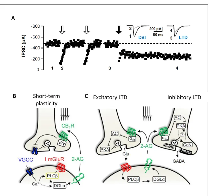
Figure 3. Synaptic plasticity and its potential mechanisms
(A) Operational definitions for distinct forms of synaptic depression (adapted from Atwood et al. (2014)) (B) Potential mechanisms of presynaptic plasticity. Both LTP and LTD have been found to occur through mediated changes in Ca2+, G-protein recruitment, protein synthesis, changes in release machinery and structural changes, potentially occurring through actin cytoskeleton modulation. (adapted from Monday and Castillo (2012) and Yang and Calakos (2013))
B
G-protein dependent Ca2+ level dependentA
Structural ChangeLocal protein synthesis
Release Machinery
20
2 Thesis Aim
While there is little information on presynaptically induced synaptic plasticity, that is not to say that it does not hold importance within behavior. Indeed, the cannabinoid type-1 receptor (CB1R) is one known presynaptically located modulator of synaptic plasticity, and is one of the most abundant transmembrane receptors in the brain. It has long been known to affect both short-term forms of synaptic plasticity as well as long-term forms, and its activity has been tied to a number of behavioral correlates, including effects on memory, mood, motor-activity and perception . Nonetheless, although the mechanisms driving short-term effects of CB1R induced plasticity are well established, those mediating its long-term effects remain poorly understood, with a number of candidate mechanisms having been suggested but none properly confirmed.
In parallel, CB1R has been found to affect neural development. Early studies, including one issued by my hosting team, have shown CB1R to be highly expressed in the axons of developing projection neurons (Romero et al. 1997; Berghuis et al. 2007; Vitalis et al. 2008). Looking further into individual neuron development in cultures, studies showed that modulation of CB1R activity had a significant effect on neuronal morphology, including axon and dendrite length, as well as dendrite number (Berghuis et al. 2005; Berghuis et al. 2007; Vitalis et al. 2008). Furthermore, a number of studies would specifically find CB1R-activation to have a repulsive effect on axonal growth cone pathfinding (Berghuis et al. 2007; Argaw et al. 2011). Combined, these findings would point towards a downstream effect of CB1R activation on the actomyosin cytoskeleton (Berghuis et al. 2007), known effector of axonal outgrowth (Dent, Gupton, and Gertler 2011), although the specific molecular pathway employed had yet to be determined.
When I arrived within the hosting team at the start of my thesis, the team held important results showing a downstream pathway linking CB1R activation to contraction of the actomyosin cytoskeleton and growth cone retraction, results to which I contributed to before publishing (Roland et al., 2014). Importantly, these results provided a novel effector pathway downstream of neuronal CB1R through RhoA/ ROCK activation and phosphorylation of the cytoskeletal motor non-muscle myosin II.
21
Given the elusive nature of CB1R-induced plasticity and the molecular pathway described in our article, one obvious question given these results was whether CB1R-activation might recruit actomyosin contractility to induce long-term plasticity. The aim of my thesis has therefore been in providing answers to this question.
To start providing answers, two initial questions were formulated:
(1) Is there evidence that the actomyosin cytoskeleton may play a role in synaptic plasticity?
(2) Is there evidence that CB1R may recruit the actomyosin cytoskeleton at synapses? The following sections will explore evidence that provides answers to these questions before arriving at the main results obtained during my thesis.
22
3 The Actomyosin Cytoskeleton and Synaptic Activity
Cells hold a plethora of different proteins allowing them to perform essential functions for organism survival. These functions could not be carried out however without a specific structural organization allowing them both to quickly and efficiently cycle essential proteins to their necessary locations (a process more efficient than random diffusion) as well as a core structure allowing them to hold or adapt depending on the necessities of the organism. Cells are therefore composed of a cytoskeleton which gives them structure. This cytoskeleton is composed of different types of filaments which hold both similar and different roles. These are mainly microfilaments, or filamentous actin (F-actin) composed of actin, microtubules composed of α- and β- tubulin, and intermediate filaments, which may be composed of various different proteins including formin.
Focusing on the two best described components, actin filaments and microtubules vary in their filament structure and dynamics. While actin filaments are relatively thin, around 7nm in diameter, microtubules are larger, around 25nm. Given their simple structure, actin filaments are therefore more adapted to mediate processes requiring fast assembly and disassembly, while the more stable nature of microtubules is more adapted to providing structural stability, a property which is reflected in their preferential location within cell processes. Indeed, while microtubules are more prominent in the cell body and at the dendritic shaft of polarized dendrites, actin filaments are often preferentially located in dynamic processes, such as migration rings in endothelial cells or growth cones in neurons. This is further reflected at the synapse, with one study revealing actin to be twice more prevalent than tubulin in synaptosomes (Wilhelm et al., 2014). By this logic, synapses are structurally dynamic compartments, requiring actin dynamics in their functions (Kevenaar and Hoogenraad 2015). This chapter will introduce the fundamentals of actin and myosin in the composition of the actomyosin cytoskeleton before outlining the importance of actomyosin dynamics in synaptic structure and function, with a particular focus on what is known of presynaptic actomyosin and how it may contribute to presynaptically-induced synaptic plasticity.
23
3.1 The actomyosin cytoskeleton
3.1.1 Actin filaments
An actin filament (F-actin) is composed of 2 chains intertwined in a helical structure. These chains are composed of monomeric G-actin, the asymmetric structure of which endows a polarized nature to actin filaments, of which a barbed, or (+), end and a (-) end can be distinguished. F-actin formation starts with the nucleation of G-actin monomers into dimers and trimers. G-actin coupling to ATP induces the polymerization of G-actin monomers, upon which the actin-coupled ATP is hydrolyzed to ADP. ADP release leads to depolymerization of actin, which must be recharged with ATP in order to polymerize once again.
While polymerization of the actin filaments is possible at both (+) and (-) ends, polymerization occurs 10 times faster at the barbed end (T. D. Pollard and Mooseker 1981; Thomas D. Pollard 1986), with depolymerization of the filaments occurring preferentially at the (-) end. G-actin will therefore preferentially travel back to the barbed end of the filament
before polymerizing once again. This polar property of F-actin
polymerization/depolymerization has given the name of “treadmilling” to F-actin dynamics (Wegner 1976).
F-actin formation occurs intrinsically in a buffered solution of G-actin and ATP, an assay often used to study F-actin targeted signaling and dynamics in vitro. Nonetheless, a number of proteins are capable of binding to actin in order to regulate this treadmilling process, either by catalyzing or stabilizing the polymerization/depolymerization reaction. Indeed, while the F-actin filament is relatively stable, G-F-actin dimers and trimers are relatively unstable. A number of actin binding proteins (ABP) are available therefore to bind these assemblies and connect them to the F-actin filament before denucleation. Some ABPs may also block the reaction depending on the cell’s needs. For example, cofilin promotes G-actin nucleation, thus catalyzing F-actin polymerization, while thymosine-β4 capping of G-actin monomers abates polymerization (Figure 4) (Thomas D. Pollard 2016).
Furthermore, certain proteins allow the formation of specific F-actin structures that would be impossible with F-actin filaments alone. For example, Arp2/3 allows new F-actin filaments to branch out from pre-existing filaments (Figure 4), creating F-actin networks necessary for the fast trafficking of a number of proteins within the cell, or for the development of filopodia necessary for cell motility (Thomas D. Pollard and Cooper 2009). Myosin II also confers
24
specific architecture to actin filaments within cells. A good example of this is the sarcomere, the basic unit of striated muscle fibers, whose actomyosin organization gives the muscle its fibrous appearance. This property of myosin II as well as its contractile functions will be expanded upon below.
3.1.2 Non-muscular myosin II
Myosins are a eukaryotic superfamily of actin-binding molecular motors, of which 18 classes have been established. Of these, 11 classes have been identified in humans (Richards and Cavalier-Smith 2005). Phylogenetically related to kinesins, the microtubule-binding motor proteins, many of the myosin classes function as cargo trafficking motors, with their C-terminal binding cargo vesicles, while their N-C-terminals transiently bind to F-actin in an ATP dependent manner, conferring them a ‘walking’ mechanism along F-actin tracts. The myosin II class differs from these, as, while its N-terminal maintains actin binding properties, its C-terminal mostly binds other myosin II motors, forming thick myosin filaments capable of contracting the actin cytoskeleton. Nonetheless, myosin II is the most common class of myosin found in eukaryotic cells, responsible for generating most cellular contractile forces (Vicente-Manzanares et al. 2009).
25
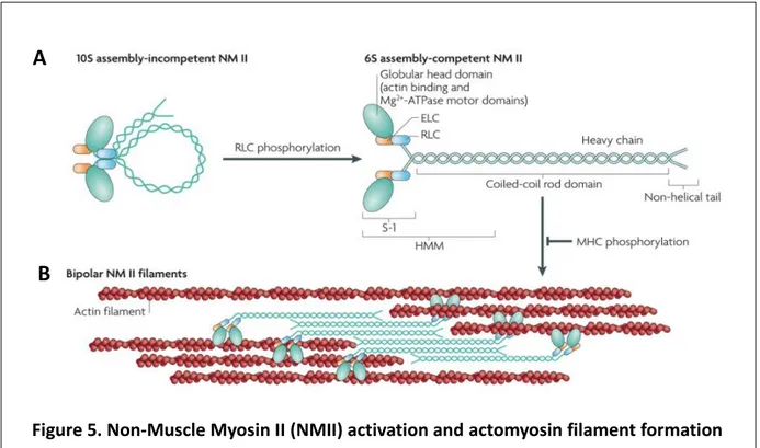
Figure 5. Non-Muscle Myosin II (NMII) activation and actomyosin filament formation (A) Phosphorylation of the NMII regulatory light chain (RLC) is required to unfold NMII into an assembly competent formation. (B) Binding of NMII units by their heavy chains forms bipolar NMII filaments which can bind to actin filaments and form actomyosin bundles (adapted from Vicente-Manzanares et al. (2009))
A
B
The myosin II class can further be separated into muscle and non-muscle (NMII) myosin II subtypes. While the muscular subtype is mostly restricted to striated muscle, where it is responsible for muscle tone and contraction, NMII is found in all eukaryotic cells, contributing to fundamental cellular functions, such as maintaining cellular tension, adhesion and migration during development (Vicente-Manzanares et al. 2009).
Like muscle myosin, NMII is formed by the dimerization of two chain units, each composed of a globular head and three peptide chains: a heavy chain, a regulatory light chain (RLC) and an essential light chain (ELC) (see Fig). Dimerization into NMII occurs through the helical intertwining of the heavy chains. At the resting state this heavy chain helix folds onto itself in an incompetent formation. Phosphorylation of the RLC is then necessary to unfold the dimer and render the myosin functional (Figure 5A). Once unfolded, the heavy chain domains of NMII units can self-associate to form isoform-specific, anti-parallel myosin filaments (Figure 5B) (Vicente-Manzanares et al. 2009), which can contain up to 20 NMII copies. These filaments crosslink actin filaments, forming the actomyosin cytoskeleton, and translocate them towards each other upon motor activity (Kneussel and Wagner 2013). These properties
26
can be regulated by phosphorylation of the NMII heavy chains, leading to dissociation of the filaments or preventing their formation.
Actin binding occurs at the globular head domain, where an Mg2+-ATPase unit is also located. Actin binding occurs with high affinity while in an ADP bound state. Upon ADP release and ATP binding, actin unbinds from the head. ATP hydrolysis then leads to conformational change of the myosin head and light chains, which, upon Pi release and actin binding, leads to a lever-like power stroke of the myosin head towards the barbed end of the actin filament. This motor activity can further be regulated by phosphorylation of the RLC, which increases myosin head ATPase activity by controlling myosin head conformation (Kneussel and Wagner 2013; Vicente-Manzanares et al. 2009).
In mammals, three NMII isoforms have been identified, NMIIA, NMIIB and NMIIC, distinguished uniquely by their heavy chains, and encoded by the MYH9, MYH10 and MYH14 genes, respectively. These isoforms differ in cellular distribution, Mg2+-ATPase activity, and actin affinity. All three isoforms have been found to be expressed in the brain, although their preferential expression patterns appear to differ between cell type, with NMIIA and NMIIB being the most expressed in neurons (Y. Zhang et al. 2014).
3.1.3 Actomyosin upstream signaling pathways
A number of signaling pathways have been described that lead to regulation of actomyosin formation and activity.
3.1.3.1 Small GTPases
Small GTPases are the best known regulators of the actomyosin cytoskeleton. They are a superfamily composed of around 200 proteins which can be further separated into 6 subcategories: Rho, Ras, Rab, Arf, Sar and Ran (Colicelli 2004). Their activation is dependent on their specific binding to GTP. Among these subcategories, Rho-GTPases are known to regulate a number of cytoskeletal processes (Spiering and Hodgson 2011). Specifically, Rho-GTPase subfamilies are known to conduct opposite effects on the actomyosin cytoskeleton (Figure 6). RhoA proteins are generally associated with negative growth (Luo 2002), particularly through the action of ROCK and phosphorylation of the myosin light chain. Rac and Cdc42 generally promote growth (Albertinazzi et al. 1998; Albertinazzi et al. 2003; Hall and Lalli 2010), by catalyzing actin polymerization and inhibiting NMII, for example.
27 3.1.3.2 Myosin Kinases
Regulation of NMII phosphorylation is an important target in the regulation of actomyosin cytoskeleton crosslinking and contraction. This can be achieved through the phosphorylation/ hydrolysation of the myosin light chain (MLC).
Over a dozen kinases have been reported to phosphorylate NMII RLC isoforms. These include myosin light chain kinase (MLCK), Rho-associated, coiled coil-containing kinase (ROCK), citron kinase, leucine zipper interacting kinase (ZIPK; also known as DAPK3) and myotonic dystrophy kinase-related CDC42-binding kinase (MRCK; also known as CDC42bP). Both ROCK and ZIPK are activated by RhoA while MLCK is activated by Ca2+-calmodulin. ROCK can also increase MLC activity by inhibiting its two principal phosphatases, protein phosphatase 1 (PP1) and myosin light chain phosphatase (MLCP) (Vicente-Manzanares et al. 2009).
3.1.3.3 G-proteins coupled receptors (GPCRs)
Although a direct link between GPCRs and recruitment of the actomyosin cytoskeleton has not consistently been shown, a number of GPCRs have been found to induce structural effects known to depend on actomyosin contraction. One such assay is in the study of changes in cell
28
morphology such as cell rounding and process outgrowth, which are critically dependent on actomyosin dynamics.
Globally, Gi/o coupled receptors have been found to produce negative effects on cell
outgrowth. In neurons, axonal growth cone retraction can be inhibited by activation of GABAB (Xiang et al. 2002) and somatostatine type-1 receptor (SST1) (Cai et al., 2008). Neurite branching has also been found to be inhibited by activation of dopamine 2 receptors (D2R) and serotonin 5-HT1B receptors (Parish et al. 2001; Parish et al. 2002; Gaspar, Cases, and Maroteaux 2003), as well as activation of the chemokine receptor type 4 (CXCR4) (Lysko, Putt, and Golden 2011).
3.1.3.4 cAMP/PKA
In neurons, cAMP/ PKA has been found to induce the attractive cues for axonal outgrowth induced by BDNF and netrin-1 (De La Torre et al. 1997; Ming et al. 1997; Song, Ming, and Poo 1997). While the link to actomyosin is not described in these studies, studies in non-neuronal cells find that cAMP//PKA activity can induce morphological changes through inhibition of RhoA (Aburima et al. 2013; Oishi et al. 2012). In particular, one study shows platelet shape change through phosphorylation of RhoGDIα by PKA, and subsequent sequestration of RhoA into RhoA-RhoGDIα complexes (Oishi et al. 2012).
3.2 Actomyosin at the synapse
The actin cytoskeleton is a predominant component of the synaptic cytoskeleton at both pre- and postsynaptic compartments (Figure7A) (Cingolani and Goda 2008). At the presynaptic compartment, it has been found to compose 2% of protein content (Wilhelm et al. 2014). Despite a number of efforts from studies using electron microscopy, the specific structure of the actomyosin cytoskeleton at the presynaptic compartment remains under debate. Part of the reason for this might be that as the actin cytoskeleton is highly dynamic in nature, its specific structure may vary widely depending on experimental conditions, such as stages of synaptic vesicle recycling or synapse maturity. Furthermore, sample preparation may further affect the cytoskeleton, with different fixation protocols potentially producing different effects. Nonetheless, several studies seem to corroborate the presence of actin both at the active zone and within the synaptic vesicle pool.
29 3.2.1 Actin in the AZ
Direct evidence for actin filaments at the AZ is relatively scarce. GFP-βactin was found to colocalize well with Bassoon, as observed with conventional microscope (Miguel Morales, Colicos, and Goda 2000) and immunogold staining of actin shows actin staining of the AZ in electron micrographs (Figure7C) (Bloom et al. 2003).
Nonetheless, a number of reports have been produced of different filaments connecting synaptic vesicles to the AZ (Landis et al. 1988; Hirokawa et al. 1989). Some of these filaments were found to be short, interconnecting docked vesicles or directly docking vesicles at the AZ (Landis et al. 1988; A. A. Cole, Chen, and Reese 2016). Furthermore, short filaments extending from the active zone are found to tether vesicles (Cole, Chen, and Reese 2016; Hirokawa et al. 1989) most likely to facilitate vesicle replenishment during synaptic activity. However, the morphological identification of these filaments is difficult, with some studies suggesting these filaments might be composed of fodrin, rather than actin (Hirokawa et al. 1989).
3.2.2 Actin and Synaptic vesicle pools
A number of EM studies show long filaments extending either from the active zone or plasma membrane into the central synaptic vesicle pool (Hirokawa et al. 1989; Landis et al. 1988; Perkins et al. 2010; A. A. Cole, Chen, and Reese 2016). Although their nature is not specifically analyzed, they can be morphologically identified due to their helical structure (Figure7B) (Hirokawa et al. 1989).
Within the vesicle pool, actin is known to interact with a number of vesicle binding proteins including β-catenin and synapsin, among others (Bamji et al. 2003; Takamori et al. 2006; Fernández-Busnadiego et al. 2010). Although the specific function of this tethering remains under debate, a number of reports suggest their importance in keeping synaptic vesicles within the presynaptic bouton as well as immobilizing diffusing vesicles to restrain vesicle recycling (T A Ryan et al. 1996; Siksou et al. 2007; Fornasiero et al. 2012). Furthermore, functional studies have provided evidence for a role of actin itself in vesicle recycling. These functions will be reviewed in later sections.
31
3.2.2.1 Presynaptic NMII
Direct evidence of the presence of non-muscle myosin II in presynaptic terminals is scarce. It has been shown that the NMIIB isoform is present in presynaptic sites at superior cervical ganglion neurons (SCGNs) (Takagishi et al. 2005), and both NMIIA and NMIIB isoforms have been found at mouse neuromuscular junctions (Vega-Riveroll et al. 2005). Blocking NMII activity has been found to affect vesicle recycling in certain models. These functions will be explored further below.
3.2.3 Actomyosin and synaptic function
Due to the difficulty of access of the presynaptic compartment, particularly at small central synapses, a consensus concerning the specific roles of the actomyosin cytoskeleton in neurotransmitter release remains to be reached. Part of the reason for this might be the diversity of models used to study presynaptic function, which include studies at hippocampal cultures, calyx of Held, neuromuscular junctions, and the lamprey reticulospinal synapse. 3.2.3.1 Actin cytoskeleton and endocytosis
A number of reports indicate activity-induced polymerization of the actin cytoskeleton (Bernstein, DeWit, and Bamburg 1998; Shupliakov et al. 2002; Trifaró et al. 2002; Bloom et al. 2003; Sankaranarayanan, Atluri, and Ryan 2003). The most coherent role suggested for this has been in endocytosis. In lamprey synapses, stabilization of actin filaments with phalloidin blocks vesicle recycling as observed through FM1-43 dye uptake (Bleckert, Photowala, and Alford 2012). Furthermore, at both the Calyx of Held and snake neuromuscular junctions, disruption of the actin cytoskeleton was found to inhibit RRP recovery under high frequency stimulation, without affecting low frequency evoked transmission (Kuromi and Kidokoro 1998; J. C. Cole, Villa, and Wilkinson 2000; Sakaba and Neher 2003; Lee et al. 2013; Miki et al. 2016). As RRP refilling is strongly dependent on the recycling pool, which principally arises from evoked endocytosis, these studies strongly suggest a role for actin filaments in vesicle endocytosis during recycling. Indeed, in drosophila expressing the shibire mutation, a temperature sensitive dynamin homolog, it was found that disruption of actin polymerization with cytochalasin D did not have a direct effect on the RRP but rather on the size of the recycling pool (Kuromi and Kidokoro 1998). While studies at central synapses are ambivalent, there is evidence that actin polymerization might
32
mediate certain types of endocytosis at hippocampal synapses, specifically compensatory endocytosis (Watanabe et al. 2013).
Furthermore, certain EM studies find the formation of elongated filaments in endocytic zones after stimulation (Figure 8) (Shupliakov et al. 2002; Bloom et al. 2003), and stabilization of the actin cytoskeleton was found to cause changes in the structure of clathrin-coated pits (Shupliakov et al. 2002). In addition, it was found in hippocampal synapses that, while recycling vesicles preferentially relocate close to the active zone, stabilization of the actin cytoskeleton prevented this relocation, and slowed sustained exocytosis during prolonged stimulation (Marra et al. 2012). These results strongly suggest a role for actin in redistributing vesicles to the recycling vesicle pool after endocytosis.
3.2.3.2 Actomyosin and synaptic vesicle tethering
Going seemingly against the studies reported above, certain studies have found facilitation of transmission upon actin filament disruption. In cultured chick sympathetic neurons it was found that actin depolymerization occurs in presynaptic terminal after prolonged stimulation, and preventing this depolymerization with phalloidin significantly reduced sustained release (Bernstein, DeWit, and Bamburg 1998). In hippocampal cultures, inducing depolymerization with latrunculin A was found to increase the frequency of small neurotransmitter induced currents (Miguel Morales, Colicos, and Goda 2000). Furthermore, at cultured frog neuromuscular junctions, it was found that depression of transmission induced by prolonged stimulation was prevented by latrunculin A, which was accompanied by microfilament disruption (Wang, Zheng, and Poo 1996). Taken together these studies suggest a second role for actin at the presynaptic compartment in what has been called a ‘barrier’ model, preventing excessive depletion of the vesicle pool upon sustained stimulation.
Several other trails of evidence support this role. A number of studies show that a majority of synaptic vesicles are immobile at the synapse, with recycling vesicles showing the most mobility (Gaffield, Rizzoli, and Betz 2006; Kamin et al. 2010). Depolymerization of the actin cytoskeleton has been found to increase the mobility of vesicles (Shtrahman et al. 2005; R. Jordan, Lemke, and Klingauf 2005), suggesting the integrity of the cytoskeleton at rest might restricts vesicle movement.
33
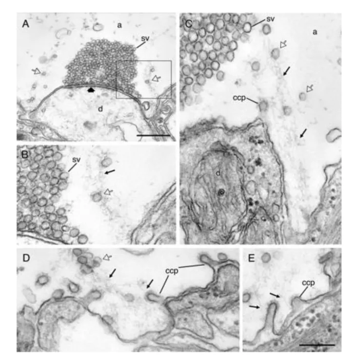
Figure 8. Actin-like filament polymerization at endocytic zone during synaptic activity (A–C) Electron micrographs from two different synapses in axons that were microinjected with phalloidin and stimulated before fixation. Synaptic vesicles (open arrows) are tethered along filaments (thin arrows) extending from the endocytic zone toward the margin of the vesicle cluster. (D and E) Clathrin-coated pits (ccp) at the plasma membrane of the endocytic zone are attached by the neck to actin-like filaments (thin arrows). (Scale bars: A, 0.5 μm; B–E, 0.2 μm.) (adapted from Shupliakov et al. (2002))
34
As mentioned previously, the actomyosin cytoskeleton is known to bind to a number of vesicle binding proteins, which tether vesicles to each other as well as to the cytoskeleton (Landis et al. 1988; Hirokawa et al. 1989). One such binding protein that has been extensively studied is synapsin, which forms short filaments of 30-40nm long (Hirokawa et al. 1989), which tether vesicles together and to the actin cytoskeleton (Peters and Kaiserman‐Abramof 1970; Landis et al. 1988; Hirokawa et al. 1989). Deletion of synapsin was found to reduce vesicle pool clustering (T A Ryan et al. 1996; Siksou et al. 2007; Fornasiero et al. 2012), with vesicles spreading out to extrasynaptic areas. Furthermore, synapsin deletion or phosphorylation has been found to increase vesicle mobility (Orenbuch et al. 2012; Gaffield, Rizzoli, and Betz 2006). Although changes in synapsin tethering do not necessarily indicate a similar role for actin, it could be inferred that disruption of the cytoskeleton integrity might unbind synapsin bound vesicles and therefore produce similar results.
3.2.3.3 Presynaptic NMII function
Much like the actin cytoskeleton, evidence for a role of non-muscle myosin II at the synapse is scarce and contradictory. At the Calyx of Held, inhibition of MLCK leads to increase in RRP size (Srinivasan, Kim, and von Gersdorff 2008). Conversely, at motorneuron excitatory synapses, activation of MLCK through LPA/LPA1 leads to decrease in transmission and reduces the number of vesicles in the RRP, observed through EM (García-Morales et al. 2015).
Alternatively, inhibition of NMII ATPase activity with the selective inhibitor blebbistatin was found to decrease transmission during prolonged activity in hippocampal cultures (Peng et al. 2012), as well as inhibit facilitation of transmission at cerebellar parallel fiber to MFI synapses (Miki et al. 2016). Furthermore, various MLCK inhibitors have been shown to inhibit transmission, particularly during sustained transmission both at hippocampal cultures (Timothy A. Ryan 1999; Yue and Xu 2014; L. Li et al. 2016) and SCGN cholinergic synapses (Mochida et al. 1994).
These studies also show contradiction with some showing preferential effects on exocytosis (Timothy A. Ryan 1999), while others show effects on endocytosis (Yue and Xu 2014). Furthermore, some studies show that the inhibitors used might not be specific to MLCK, with one study showing an effect of ML-7 produced through VGCCs rather than MLCK (Tokuoka and Goda 2006).
35
4 CB1R and the endocannabinoid system
While the psychoactive and therapeutic effects of cannabinoids have been known and exploited for thousands of years, the physiological mechanisms underlying these effects have only truly started to unravel in the past 20 years. With the main psychotropic compound of the cannabis plant, (−)-Δ9-tetrahydrocannabinol (Δ9-THC), being discovered in the mid-1960s ((Gaoni and Mechoulam 1964; Mechoulam and Gaoni 1967), it would be another 20 years before the necessary technological advancements could bring us the cloning and molecular identification of THC’s main psychotropic agent in the brain, the cannabinoid-type 1 receptor (CB1R) (W. A. Devane et al. 1988; Bidaut-Russell, Devane, and Howlett 1990; Lisa A. Matsuda et al. 1990). Later research would identify this receptor not only as one of the most abundant transmembrane proteins in the brain (Y. Zhang et al. 2014), but as a major regulator of neuronal function through the endocannabinoid neurotransmitter system.
The endocannabinoid system is highly conserved throughout vertebrate species, especially among mammals. This is especially true when looking at the properties of CB1R expression and distribution. Not only is there 97-99% homology in the cnr1 gene amino acid identity between mammals, but CB1R concentrations between brain areas is also well conserved between rodents (immunostaining) and humans (PET imaging). Furthermore, intracellular distributions of endocannabinoid metabolic enzymes has also been well conserved between rodents and humans (Ludányi et al. 2011). What these findings point to is not only a conserved evolutionary function for endocannabinoid signaling in vertebrates, but furthermore, they suggest a primary role for the ECS in fundamental brain function.
This chapter will introduce the components of the endocannabinoid system, its receptors, ligands and metabolic pathways, before focusing on the known functions of CB1R in development and synaptic transmission, and the known and putative signaling pathways mediating these functions.