Pr
oceedings
of
the
Nutrition
Society
The Nutrition Society Scottish Section meeting was held at Pollock Halls, Edinburgh on 18–19 March 2013
Conference on
‘PUFA mediators: implications for human health’
Symposium 1: PUFA: health effects and health claims
Ageing and apoE change DHA homeostasis: relevance to age-related
cognitive decline
Marie Hennebelle
1,2, Mélanie Plourde
1,2,3, Raphaël Chouinard-Watkins
1,2,
Christian-Alexandre Castellano
1,2, Pascale Barberger-Gateau
4and Stephen C. Cunnane
1,2,3*
1
Research Center on Aging, Université de Sherbrooke, Sherbrooke, QC, Canada,
2
Physiology and Biophysics, Université de Sherbrooke, Sherbrooke, QC, Canada,
3
Departments of Medicine, Université de Sherbrooke, Sherbrooke, QC, Canada
4
INSERM, ISPED, Centre INSERM U897-Epidemiologie-Biostatistique, F-33000 Bordeaux, France
Epidemiological studies fairly convincingly suggest that higher intakes of fattyfish and n-3 fatty acids are associated with reduced risk of Alzheimer’s disease (AD). DHA in plasma is normally positively associated with DHA intake. However, despite being associated with lowerfish and DHA intake, unexpectedly, plasma (or brain) DHA is frequently not lower in AD. This review will highlight some metabolic and physiological factors such as ageing and apoE polymorphism that influence DHA homeostasis. Compared with young adults, blood DHA is often slightly but significantly higher in older adults without any age-related cognitive decline. Higher plasma DHA in older adults could be a sign that theirfish or DHA intake is higher. However, our supplementation and carbon-13 tracer studies also show that DHA metabolism, e.g. transit through the plasma, apparent retroconversion and β-oxidation, is altered in healthy older compared with healthy young adults. ApoE4 increases the risk of AD, possibly in part because it too changes DHA homeostasis. Therefore, inde-pendent of differences in fish intake, changing DHA homeostasis may tend to obscure the relationship between DHA intake and plasma DHA which, in turn, may contribute to making older adults more susceptible to cognitive decline despite older adults having similar or sometimes higher plasma DHA than in younger adults. In conclusion, recent development of new tools such as isotopically labelled DHA to study DHA metabolism in human subjects highlights some promising avenues to evaluate how and why DHA metabolism changes during ageing and AD.
Alzheimer’s disease: Docosahexaenoic acid: Ageing: ApoE: Cognition
The cognitive and psychological health of older adults is
now a major preoccupation for healthcare services and
researchers alike. Alzheimer
’s disease (AD) is the main
form of cognitive decline in older persons in Western
countries
(1). Age is the main risk factor associated with
AD
(1), but other factors also have an effect such as a
pre-disposing genetic polymorphism, i.e.
ε4 allele of apoE4
(2),
vascular risk factors including hypertension, obesity and
type 2 diabetes
(1), and lifestyle including physical activity
and dietary habits
(3,4). Among the nutrients closely
asso-ciated with brain function, the n-3 fatty acids, especially
DHA, have attracted special attention. Fatty
fish and
seafood are the most important dietary sources of both
DHA and EPA. DHA is by far the predominant n-3
fatty acid in the brain and is present mostly in various
membrane phospholipids (PL) of neurons, especially in
synapses
(5). In contrast to other common dietary
long-chain fatty acids, DHA is highly conserved and poorly
β-oxidised
(6–8). In human subjects, DHA synthesis is
rela-tively inef
ficient, especially in comparison to rodents
(9).
Low intake of n-3 fatty acids has long been associated
with higher risk of CVD
(10), and also of suboptimal brain
*Corresponding author: Professor S.C. Cunnane, fax (+1) 819-829-7141, email [email protected]
Abbreviations: AD, Alzheimer’s disease; CE, cholesteryl esters;13C-DHA, carbon-13 labelled DHA; PL, phospholipids.
Pr
oceedings
of
the
Nutrition
Society
development
(11). Much effort has been focused over the
past decade on whether a higher DHA intake could
decrease the risk of cognitive decline in older adults, or
reduce the progression from mild cognitive impairment
towards AD. In general, these studies polarise in two
directions: randomised clinical trials that are largely
negative and epidemiological studies that are more
posi-tive
(12)about the DHA
’s role in maintaining cognition
during ageing. Thus, in general, DHA supplementation
trials in AD (with or without EPA) have not so far
pro-duced any truly positive results
(12–15). Methodological
issues such as dose of n-3 fatty acid, duration of
treat-ment or selection criteria may well have affected the
out-comes of these trials. DHA supplementation may have a
greater positive effect on memory and learning in healthy
adults
(16), elderly with subjective cognitive complaints
(17)or with mild cognitive impairment
(18)than in those
with AD
(12,14,19). However, prospective epidemiological
studies have been more positive; they broadly show
that habitually low intake of
fish and/or DHA is
associ-ated with higher risk of developing AD
(12,20,21). These
results are supported by the neuroprotective role of
DHA reported for non-human models of
neurodegenera-tive disease
(22–26).
Studies
with
biological
samples
(human
blood
and brain) may be able to provide useful leads to
ex-plain the divergent results between randomised clinical
trials and epidemiological studies. We have previously
reviewed at some length the methodological limits on
observational or intervention studies on DHA
sup-plementation in older adults or in AD
(12–15,20,21,27); so
we will review them here only brie
fly. We will also
pre-sent an emerging framework showing that DHA
homo-eostasis changes in older adults and differs in carriers
from non-carriers of apoE4, probably before the onset
of cognitive decline.
DHA in plasma and post-mortem human brain
Brain DHA
In primate, pig and rodent models, when n-3 intake is
severely de
ficient for extended periods, brain DHA also
decreases across all cell types and regions, in association
with
lower
scores
on
cognitive
and
behavioural
tests
(25,28–31).
AD
is
now
widely
associated
with
lower
fish and DHA intake, so it would be logical that
post-mortem brain samples of patients with a de
finitive
diagnosis of AD also contained lower DHA. Indeed, in
the hippocampus, which is central to memory processing
and learning, AD patients reportedly do have lower
DHA
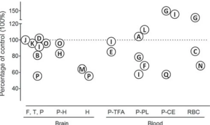
(32,33). However, in the temporal and frontal
cor-tices which are also affected in AD, DHA is almost
always the same as in the controls (
Fig. 1
). Studies
re-porting lower DHA in the AD brain show that other
fatty
acids
are
also
lower,
particularly
n-6
PUFA
(35,41,44,46). Thus, the effect of AD is not speci
fic
to DHA which is contrary to what would be expected
if only n-3 fatty acid intake were de
ficient.
There are many potential methodological reasons for
the observed lack of agreement between the apparently
low DHA intakes in AD yet frequently normal DHA
levels in the brain
(12). Crucial among these are the
‘healthy’ controls against which the AD cases are
com-pared as well as the very marked extent of regional
brain atrophy associated with ageing regardless of the
presence of neurological disease
(44,50). Furthermore, the
basis for classifying a patient as having AD, i.e. whether
on clinical cognitive criteria or on neuropathological
score, may not give consistent results since senile plaques
are increasingly recognised as being present in a signi
fi-cant proportion of cognitively normal elderly
per-sons
(51–53). Hence, there is a risk that post-mortem
samples from brain banks for which cognitive status is
not known at the time of death could be misclassi
fied if
based solely on neuropathological scores. It also appears
that membrane PL in the cortex can tenaciously retain
DHA and that a more discrete and speci
fic subcellular
pool or membrane pool of DHA may have to be
measured
(43). Brain membrane DHA cycles rapidly
be-tween PL and free DHA via DHA-CoA
(54,55), and the
deteriorating ef
ficacy of this process could theoretically
contribute to the neurodegenerative processes. Thus, the
key issue in relation to post-mortem tissue analysis is
that the time to lipid extraction is rarely less than 4
–5h
yet DHA turnover is on the order of minutes, if not
seconds. Since the turnover of DHA towards resolvins
and neuroprotectins are orders of magnitude lower than
the amount of DHA in the brain NEFA pool, truly
‘physiological’ amounts of these products are extremely
dif
ficult to measure, especially in human subjects
(23,56).
As also noted elsewhere, these and other issues severely
constrain the validity and hence the utility of DHA
measurements on human post-mortem brain samples
(57).
Fig. 1. Summary of the published literature on brain and blood
DHA in Alzheimer’s disease. The symbols represent the results of
individual studies using each study’s control group as the
reference (100 %; dotted line). The papers from which these DHA
data are obtained are as follows: A, Arsenaultet al.(34); B, Astarita
et al.(35)
; C, Boston et al.(36); D, Brooksbanket al.(37); E, Cherubini
et al.(38)
; F, Conquer et al.(39); G, Corrigan et al.(40); H, Corrigan
et al.(41)
; I, Cunnaneet al.(42); J, Fraser et al.(43); K, Guanet al.(44);
L, Laurinet al.(45); M, Prasadet al.(46); N, Selleyet al.(47); O, Skinner
et al.(48)
; P, Söderberg et al.(32); Q, Tully et al.(49). F,T,P, frontal,
temporal and/or parietal cortex; P-H, para-hippocampus;
H, hippocampus; P-TFA, plasma total fatty acids; P-PL, plasma phospholipids; P-CE, plasma cholesteryl esters; RBC, red blood cells.
Pr
oceedings
of
the
Nutrition
Society
Blood DHA
Lower DHA would normally be expected in the blood
of those with habitually low DHA intake (whether
diag-nosed with AD or not). In some AD studies, lower DHA
is indeed reported for plasma total lipids
(38), PL
(39,40,42),
cholesteryl esters
(49)and NEFA
(58). However, many
other AD studies show no difference in plasma DHA,
whether in PL or total fatty acids
(34,38,42,45,59). Some
even report higher DHA in plasma PL
(45)or cholesteryl
esters (CE)
(40,42). Similar inconsistencies are present
across DHA levels reported for the erythrocytes in AD
(
Fig. 1
)
(36,40,47). Prospective studies also show this
inconsistency: some found a strong association between
lower blood DHA level and slower cognitive decline
(60)or lower risk of dementia
(61), whereas other did
not
(45,59,62). It may be that the cognitive domain
studied
(63,64)and apoE4 genotype
(65,66)contribute to
this scatter in the data.
DHA homeostasis during ageing and apoE
We propose that even when collected under
hypotheti-cally ideal conditions (zero delay; perfectly matched,
cognitively healthy controls, etc.), data obtained from
single blood samples are too limited to fully understand
possible changes in DHA metabolism due to genotype,
ageing or neurodegenerative disease. However,
isotopi-cally labelled DHA is emerging as a useful tool to assess
how the metabolism of DHA changes with age. Indeed,
in a relatively simple study design, it was clear that the
clearance of a 50 mg oral dose of uniformly carbon-13
labelled
DHA
(
13C-DHA)
from
the
blood
over
1 month was much slower in healthy 76-year-old
com-pared with 27-year-old adults
(67). These results were
simi-lar to our earlier report that the increase in plasma DHA
during a short-term treatment with
fish oil was higher
in healthy older persons
(68).
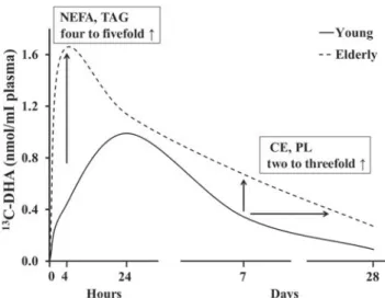
13C-DHA enrichment in
plasma NEFA and TAG of older adults was most
affected (four- to
fivefold higher than in the young
adults) but its enrichment in PL and CE
13C-DHA was
also affected. The doubling of
13C-DHA enrichment in
plasma PL and CE emerged only after about 7 d
post-dose, suggesting slower DHA clearance through plasma
lipid classes, i.e. an altered plasma
‘DHA wave’ in
older adults (
Fig. 2
)
(67).
Clearly, therefore, healthy ageing seems to change
DHA metabolism and, hence, homeostasis in human
subjects. Notwithstanding the limited extent to which
the kinetic behaviour of a tracer can be compared with
a single plasma fatty acid measurement, the difference
in
13C-DHA homeostasis in the elderly seems to re
flect
the results observed in two studies in which lower
DHA was reported in plasma PL yet higher DHA was
reported in plasma CE of AD patients
(40,42). The
mini-mally invasive nature of this type of experiment makes
it dif
ficult to invoke a particular mechanism but one
could speculate that the changing sensitivity of
endo-thelial lipoprotein lipase could be involved in this
age-associated difference in DHA homeostasis
(69).
ApoE4 carriers are at signi
ficantly higher risk
of AD
(70,71). It is now emerging that the apoE4 status
also affects DHA metabolism in human subjects
(27,72,73).
This interaction may help explain why the protective
association of higher dietary intake of
fish
(74,75)or higher
erythrocyte total n-3 fatty acids
(65)is generally limited to
non-carriers of apoE4. Measuring expired
13C-CO
2after
dosing with
13C-DHA permits the estimation of the
whole body half-life of DHA in healthy older adults,
which is of the order of 32 d in carriers of apoE4 and
140 d in non-carriers of apoE4
(72). Ageing seems not to
affect the whole body half-life of DHA although the
small sample size makes these results still somewhat
preliminary
(67).
Using positron emission tomography and the tracer,
11
C-DHA, human brain turnover of DHA has been
esti-mated to be about 4 mg/d, giving rise to a half-life of
brain DHA of about 2
·5 years
(54). Hence, the half-life of
brain DHA is much longer than its whole body half-life.
Perhaps further assessments of the brain or whole body
half-life of DHA could provide some insight into the
cur-rent ineffectiveness of DHA supplements in AD despite
the fact that these supplements typically supply several
fold the brain
’s apparent daily turnover of DHA
(12).
ApoE4 seems also to supress the plasma DHA
response to a
fish oil supplement
(73)and the metabolism
of an oral dose of
13C-DHA
(72), an effect somewhat
op-posite to that observed with healthy ageing. For up to
28 d after a single oral dose of
13C-DHA, carriers of
apoE4 have a slightly lower concentration of plasma
13
C-DHA compared with non-carriers
(72). When the
tra-cer is given both before and again after a 5-month period
Fig. 2. Delayed plasma clearance of carbon 13-labelled DHA
(13C-DHA) during healthy ageing, adapted from Plourde et al.(67).
Plasma 13C-DHA concentration was followed over 28 d after the
oral administration of a single 50 mg dose of 13C-DHA in young
(27 years; n 6) and elderly (76 years; n 6) participants. In older
adults, plasma tracer concentration in NEFA and TAG was four to
fivefold higher 4h after giving the oral dose and about twofold
higher 1–4 weeks later in phospholipids (PL) and cholesteryl
Pr
oceedings
of
the
Nutrition
Society
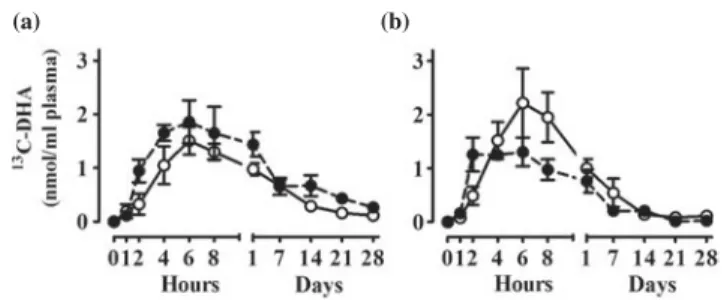
of DHA+EPA supplementation, the apoE4 carriers had
a greater accumulation of the tracer in plasma after
sup-plementation compared with the non-carriers, again
suggesting slower clearance of DHA to and/or use by
tissues (
Fig. 3
). Hence, two established risk factors for
AD (ageing and apoE4) both signi
ficantly change DHA
homeostasis but possibly in different ways.
Linking dietary and plasma DHA
Habitual DHA intake is commonly estimated to be
<250 mg/d
(76–78)but this is a dif
ficult and laborious
measurement and subject to high day-to-day variability
depending on the frequency of
fish or shellfish
con-sumption. Hence, it would be useful if DHA intake
and plasma DHA were highly correlated because plasma
DHA measurement is now technically simple and
reliable, so it could potentially be a surrogate for dietary
DHA measurement. There is indeed a good positive
correlation between dietary and plasma intake for
DHA consumption, especially at DHA intakes towards
1000 mg/d, during which plasma DHA rises to a
maxi-mum of about 4 % in plasma total lipids. However,
DHA always seems to be present in plasma, even when
DHA intake is negligible; thus, vegans consuming no
known dietary sources of DHA still have about 0
·5 %
DHA in plasma total lipids
(79). The problem is that
there are relatively few reports on which to build the
relationship of dietary to plasma DHA. At DHA intakes
between 0 and 50 mg/d, DHA is between 0
·5 and 1·2 % of
plasma total lipids, but the spread in these data is
large
(57). A value of 0
·5 % DHA in plasma total lipids
therefore seems to be at or close to the lower limit
poss-ible of plasma DHA in healthy adults.
The AD cases we have studied had 1
·0 % DHA in
plasma total lipids, which empirically corroborates very
low DHA intake, yet they had
‘normal’ DHA in the
PL of brain cortical grey matter
(42). Plasma DHA does
not rise in human subjects given EPA or
α-linolenic
acid supplements, even in vegans
(80–82). The conundrum
therefore is: how are plasma (or brain) DHA levels
main-tained when DHA intakes are very low to negligible? We
speculate that with changes in DHA metabolism and
homeostasis during age-related cognitive decline, the
diet
–plasma relation of DHA may shift, explaining why
with lower DHA intake, a population with age-related
cognitive decline appears to have the same plasma
DHA concentration as healthy elderly even though the
availability of DHA to the tissues may be reduced
(83).
The lack of an established reference lipid class in blood
(PL, CE, TAG or NEFA, erythrocytes, etc.) for DHA
measurements relative to intake still hampers the extent
to which plasma DHA data from various reports can
be compared in relation to ageing, genotype and risk of
cognitive decline. This area clearly needs further research
but suf
fice it to say that it is becoming increasingly
important to take into account changing DHA
homeo-stasis in the study of ageing population, especially in a
context of age-related cognitive decline or AD.
Conclusion
We have sought to brie
fly highlight some of the
meth-odological challenges and potential future directions for
the study of DHA in ageing and AD. The emerging
evi-dence for changing DHA half-life in older adults and in
carriers of apoE4 should encourage more basic research
on DHA metabolism in human subjects. Molecular,
cel-lular and animal models have contributed enormously to
understanding the complexity of DHA biology, but none
of them seem to represent the changes in DHA
homeo-stasis reported in elderly human subjects. The human
brain is able to strongly retain DHA in membrane PL
despite very low DHA intake and advanced AD
(42), so
the classical dietary n-3 de
ficiency model used to probe
the function of DHA in the animal brain appears
inap-propriate for research into AD. In human subjects,
DHA in the post-mortem brain is unlikely to correctly
re
flect what is happening during ageing and AD, due
to its fast turnover in neuronal membrane PL.
Ageing-and apoE4-associated changes in DHA metabolism
strongly suggest altered DHA homeostasis involving a
decrease in plasma DHA clearance during age-related
cognitive decline and AD. Therefore, a shift in the
relationship between plasma and dietary DHA may be
occurring during age-related cognitive decline and AD,
one that needs to be considered when looking at plasma
DHA as a measure of dietary DHA intake. In the future,
the availability of innovative tools for studies of DHA
half-life and metabolism in human subjects will be
needed to understand in a better manner the changes in
DHA metabolism occurring during human ageing and
AD and the potential protective role of DHA on
cogni-tive decline. As shown in prospeccogni-tive studies, a proteccogni-tive
role of DHA in cognitive health of older persons may
depend on consuming a healthy diet throughout adult
life.
Acknowledgements
Mélanie Fortier, Conrad Filteau, Christine
Rioux-Perreault, Jennifer Tremblay-Mercier and Sébastien
Tremblay provided excellent technical support.
(a) (b)
Fig. 3. Plasma carbon 13-labelled DHA (13
C-DHA) concentration
over 28 d after a single oral dose of 40 mg 13C-DHA. Results
expressed as means (SEM) show the plasma 13C-DHA status (a)
pre- (n 6) and (b) post- (n 4) supplementation of 5 months with 1·8
g/d EPA +1·4g/d DHA in apoE ε4 carriers (apoE4+; ○) and
Pr
oceedings
of
the
Nutrition
Society
Financial Support
CIHR, NSERC, FQRNT (CFQCU), INSERM, CFI and
CRC provided
financial support for S. C. C.’s research.
Con
flicts of Interest
None.
Authorship
M. H. and S. C. C. conceived and wrote the
first draft
with all the authors contributing to the revisions and
final version of the manuscript.
References
1. Blennow K, de Leon MJ & Zetterberg H (2006) Alzheimer’s disease. Lancet 368, 387–403.
2. Corder EH, Saunders AM, Strittmatter WJ et al. (1993) Gene dose of apolipoprotein E type 4 allele and the risk of Alzheimer’s disease in late onset families. Science 261, 921–923.
3. Gomez-Pinilla F (2008) Brain foods: the effects of nutrients on brain function. Nat Rev Neurosci 9, 568–578.
4. Alles B, Samieri C, Feart C et al. (2012) Dietary patterns: a novel approach to examine the link between nutrition and cognitive function in older individuals. Nutr Res Rev 25, 207–222.
5. Crawford MA, Bloom M, Broadhurst CL et al. (1999) Evidence for the unique function of docosahexaenoic acid during the evolution of the modern hominid brain. Lipids 34, Suppl, S39–S47.
6. Cunnane SC, Ryan MA, Nadeau CR et al. (2003) Why is carbon from some polyunsaturates extensively recycled into lipid synthesis? Lipids 38, 477–484.
7. Gavino GR & Gavino VC (1991) Rat liver outer mito-chondrial carnitine palmitoyltransferase activity towards long-chain polyunsaturated fatty acids and their CoA esters. Lipids 26, 266–270.
8. Leyton J, Drury PJ & Crawford MA (1987) Differential oxidation of saturated and unsaturated fatty acids in vivo in the rat. Br J Nutr 57, 383–393.
9. Plourde M & Cunnane SC (2007) Extremely limited syn-thesis of long chain polyunsaturates in adults: implications for their dietary essentiality and use as supplements. Appl Physiol Nutr Metab 32, 619–634.
10. Skeaff CM & Miller J (2009) Dietary fat and coronary heart disease: summary of evidence from prospective cohort and randomised controlled trials. Ann Nutr Metab 55, 173–201.
11. Innis SM (2007) Dietary (n-3) fatty acids and brain devel-opment. J Nutr 137, 855–859.
12. Cunnane SC, Plourde M, Pifferi F et al. (2009) Fish, doc-osahexaenoic acid and Alzheimer’s disease. Prog Lipid Res 48, 239–256.
13. Yaffe K (2010) Treatment of Alzheimer disease and prog-nosis of dementia: time to translate research to results. JAMA 304, 1952–1953.
14. Mazereeuw G, Lanctot KL, Chau SA et al. (2012) Effects of omega-3 fatty acids on cognitive performance: a meta-analysis. Neurobiol Aging 33, 1482, e1417–e1429.
15. Sydenham E, Dangour AD & Lim WS (2012) Omega 3 fatty acid for the prevention of cognitive decline and dementia. Cochrane Database Syst Rev 6, CD005379. 16. Stonehouse W, Conlon CA, Podd J et al. (2013) DHA
supplementation improved both memory and reaction time in healthy young adults: a randomized controlled trial. Am J Clin Nutr 97, 1134–1143.
17. Yurko-Mauro K, McCarthy D, Rom D et al. (2010) Beneficial effects of docosahexaenoic acid on cognition in age-related cognitive decline. Alzheimers Dement 6, 456–464.
18. Sinn N, Milte CM, Street SJ et al. (2012) Effects of n-3 fatty acids, EPA v. DHA, on depressive symptoms, quality of life, memory and executive function in older adults with mild cognitive impairment: a 6-month randomised con-trolled trial. Br J Nutr 107, 1682–1693.
19. Quinn JF, Raman R, Thomas RG et al. (2010) Doco-sahexaenoic acid supplementation and cognitive decline in Alzheimer disease: a randomized trial. JAMA 304, 1903–1911.
20. Fotuhi M, Mohassel P & Yaffe K (2009) Fish consump-tion, long-chain omega-3 fatty acids and risk of cognitive decline or Alzheimer disease: a complex association. Nat Clin Pract Neurol 5, 140–152.
21. Barberger-Gateau P, Feart C, Letenneur et al. (2013) Dietary patterns and dementia. In Chronic Medical Disease and Cognitive Aging: Toward a Healthy Body and Brain pp. 197–224 [Yaffe K, editor]. United States: Oxford University Press.
22. Kim HY, Akbar M & Kim YS (2010) Phosphatidylserine-dependent neuroprotective signaling promoted by docosahexaenoic acid. Prostaglandins Leukot Essent Fatty Acids 82, 165–172.
23. Bazan NG, Molina MF & Gordon WC (2011) Docosahexaenoic acid signalolipidomics in nutrition: sig-nificance in aging, neuroinflammation, macular degener-ation, Alzheimer’s, and other neurodegenerative diseases. Annu Rev Nutr 31, 321–351.
24. Sidhu VK, Huang BX & Kim HY (2011) Effects of doco-sahexaenoic acid on mouse brain synaptic plasma mem-brane proteome analyzed by mass spectrometry and (16)O/(18)O labeling. J Proteome Res 10, 5472–5480. 25. Calon F & Cole G (2007) Neuroprotective action
of omega-3 polyunsaturated fatty acids against neuro-degenerative diseases: evidence from animal studies. Prostaglandins Leukot Essent Fatty Acids 77, 287–293. 26. Boudrault C, Bazinet RP & Ma DW (2009) Experimental
models and mechanisms underlying the protective effects of n-3 polyunsaturated fatty acids in Alzheimer’s disease. J Nutr Biochem 20, 1–10.
27. Barberger-Gateau P, Samieri C, Feart C et al. (2011) Dietary omega 3 polyunsaturated fatty acids and Alzheimer’s disease: interaction with apolipoprotein E gen-otype. Curr Alzheimer Res 8, 479–491.
28. Brenna JT & Diau GY (2007) The influence of dietary docosahexaenoic acid and arachidonic acid on central nervous system polyunsaturated fatty acid composition. Prostaglandins Leukot Essent Fatty Acids 77, 247–250. 29. Oster T & Pillot T (2010) Docosahexaenoic acid and
synap-tic protection in Alzheimer’s disease mice. Biochimica et Biophysica Acta– Mol Cell Biol Lipids 1801, 791–798. 30. Connor WE, Neuringer M & Reisbick S (1991) Essentiality
of omega 3 fatty acids: evidence from the primate model and implications for human nutrition. World Rev Nutr Diet 66, 118–132.
31. Novak EM, Dyer RA & Innis SM (2008) High dietary omega-6 fatty acids contribute to reduced docosahexaenoic
Pr
oceedings
of
the
Nutrition
Society
acid in the developing brain and inhibit secondary neurite growth. Brain Res 1237, 136–145.
32. Soderberg M, Edlund C, Kristensson K et al. (1991) Fatty acid composition of brain phospholipids in aging and in Alzheimer’s disease. Lipids 26, 421–425.
33. Lukiw WJ, Cui JG, Marcheselli VL et al. (2005) A role for docosahexaenoic acid-derived neuroprotectin D1 in neural cell survival and Alzheimer disease. J Clin Invest 115, 2774–2783.
34. Arsenault LN, Matthan N, Scott TM et al. (2009) Validity of estimated dietary eicosapentaenoic acid and docosahexaenoic acid intakes determined by interviewer-administered food frequency questionnaire among older adults with mild-to-moderate cognitive impairment or dementia. Am J Epidemiol 170, 95–103.
35. Astarita G, Jung KM, Berchtold NC et al. (2010) Deficient liver biosynthesis of docosahexaenoic acid correlates with cognitive impairment in Alzheimer’s disease. PLoS ONE 5, e12538.
36. Boston PF, Bennett A, Horrobin DF et al. (2004) Ethyl-EPA in Alzheimer’s disease–a pilot study. Prostaglandins Leukot Essent Fatty Acids 71, 341–346.
37. Brooksbank BW & Martinez M (1989) Lipid abnormalities in the brain in adult Down’s syndrome and Alzheimer’s dis-ease. Mol Chem Neuropathol 11, 157–185.
38. Cherubini A, Andres-Lacueva C, Martin A et al. (2007) Low plasma N-3 fatty acids and dementia in older persons: The InCHIANTI study. J Gerontol A, Biol Sci Med Sci 62, 1120–1126.
39. Conquer JA, Tierney MC, Zecevic J et al. (2000) Fatty acid analysis of blood plasma of patients with Alzheimer’s dis-ease, other types of dementia, and cognitive impairment. Lipids 35, 1305–1312.
40. Corrigan FM, Van Rhijn AG, Ijomah G et al. (1991) Tin and fatty acids in dementia. Prostaglandins Leukot Essent Fatty Acids 43, 229–238.
41. Corrigan FM, Horrobin DF, Skinner ER et al. (1998) Abnormal content of n-6 and n-3 long-chain unsaturated fatty acids in the phosphoglycerides and cholesterol esters of parahippocampal cortex from Alzheimer’s disease patients and its relationship to acetyl CoA content. Int J Biochem Cell Biol 30, 197–207.
42. Cunnane S, Schneider J, Tangney C et al. (2012) Plasma and brain fatty acid profiles in mild cognitive impairment and Alzheimer’s disease. J Alzheimer’s Dis 29, 691–697. 43. Fraser T, Tayler H & Love S (2010) Fatty acid composition
of frontal, temporal and parietal neocortex in the normal human brain and in Alzheimer’s disease. Neurochem Res 35, 503–513.
44. Guan Z, Wang Y, Cairns NJ et al. (1999) Decrease and structural modifications of phosphatidylethanolamine plas-malogen in the brain with Alzheimer disease. J Neuropathol Exp Neurol 58, 740–747.
45. Laurin D, Verreault R, Lindsay J et al. (2003) Omega-3 fatty acids and risk of cognitive impairment and dementia. J Alzheimer’s Dis 5, 315–322.
46. Prasad MR, Lovell MA, Yatin M et al. (1998) Regional membrane phospholipid alterations in Alzheimer’s disease. Neurochem Res 23, 81–88.
47. Selley ML (2007) A metabolic link between S-adenosylhomocysteine and polyunsaturated fatty acid metabolism in Alzheimer’s disease. Neurobiol Aging 28, 1834–1839.
48. Skinner ER, Watt C, Besson JA et al. (1993) Differences in the fatty acid composition of the grey and white matter of different regions of the brains of patients with Alzheimer’s disease and control subjects. Brain 116(Pt 3), 717–725.
49. Tully AM, Roche HM, Doyle R et al. (2003) Low serum cholesteryl ester-docosahexaenoic acid levels in Alzheimer’s disease: a case-control study. Br J Nutr 89, 483–489.
50. Svennerholm L, Boström K & Jungbjer B (1997) Changes in weight and compositions of major membrane com-ponents of human brain during the span of adult human life of Swedes. Acta Neuropathol 94, 345–352.
51. Aizenstein HJ, Nebes RD, Saxton JA et al. (2008) Frequent amyloid deposition without significant cognitive impairment among the elderly. Arch Neurol 65, 1509–1517. 52. Braak H & Braak E (1997) Frequency of stages of Alzheimer-related lesions in different age categories. Neurobiol Aging 18, 351–357.
53. Duyckaerts C & Hauw JJ (1997) Prevalence, incidence and duration of Braak’s stages in the general population: can we know? Neurobiol Aging 18, 362–369; discussion 389–392.
54. Umhau JC, Zhou W, Carson RE et al. (2009) Imaging incorporation of circulating docosahexaenoic acid into the human brain using positron emission tomography. J Lipid Res 50, 1259–1268.
55. Chen CT, Green JT, Orr SK et al. (2008) Regulation of brain polyunsaturated fatty acid uptake and turnover. Prostaglandins Leukot Essent Fatty Acids 79, 85–91. 56. Serhan CN & Petasis NA (2011) Resolvins and protectins
in inflammation resolution. Chem Rev 111, 5922–5943. 57. Cunnane SC, Chouinard-Watkins R, Castellano CA et al.
(2013) Docosahexaenoic acid homeostasis, brain aging and Alzheimer’s disease: can we reconcile the evidence? Prostaglandins Leukot Essent Fatty Acids 88, 61–70. 58. Wang DC, Sun CH, Liu LY et al. (2012) Serum fatty acid
profiles using GC-MS and multivariate statistical analysis: potential biomarkers of Alzheimer’s disease. Neurobiol Aging 33, 1057–1066.
59. Ronnemaa E, Zethelius B, Vessby B et al. (2012) Serum fatty-acid composition and the risk of Alzheimer’s disease: a longitudinal population-based study. Eur J Clin Nutr 66, 885–890.
60. Heude B, Ducimetiere P & Berr C (2003) Cognitive decline and fatty acid composition of erythrocyte membranes–the EVA Study. Am J Clin Nutr 77, 803–808.
61. Schaefer EJ, Bongard V, Beiser AS et al. (2006) Plasma phosphatidylcholine docosahexaenoic acid content and risk of dementia and Alzheimer disease: the Framingham Heart Study. Arch Neurol 63, 1545–1550.
62. Kroger E, Verreault R, Carmichael PH et al. (2009) Omega-3 fatty acids and risk of dementia: the Canadian Study of Health and Aging. Am J Clin Nutr 90, 184–192.
63. Dullemeijer C, Durga J, Brouwer IA et al. (2007) n 3 fatty acid proportions in plasma and cognitive performance in older adults. Am J Clin Nutr 86, 1479–1485.
64. Beydoun MA, Kaufman JS, Satia JA et al. (2007) Plasma n-3 fatty acids and the risk of cognitive decline in older adults: the Atherosclerosis Risk in Communities Study. Am J Clin Nutr 85, 1103–1111.
65. Whalley LJ, Deary IJ, Starr JM et al. (2008) n-3 Fatty acid erythrocyte membrane content, APOE varepsilon4, and cognitive variation: an observational follow-up study in late adulthood. Am J Clin Nutr 87, 449–454.
66. Samieri C, Feart C, Proust-Lima C et al. (2011) omega-3 fatty acids and cognitive decline: modulation by ApoEepsilon4 allele and depression. Neurobiol Aging 32, 2317, e2313–e2322.
67. Plourde M, Chouinard-Watkins R, Vandal M et al. (2011) Plasma incorporation, apparent retroconversion and
Pr
oceedings
of
the
Nutrition
Society
beta-oxidation of 13C-docosahexaenoic acid in the elderly. Nutr Metab (Lond) 8, 5.
68. Vandal M, Freemantle E, Tremblay-Mercier J et al. (2008) Plasma omega-3 fatty acid response to afish oil supplement in the healthy elderly. Lipids 43, 1085–1089.
69. Millar JS, Lichtenstein AH, Cuchel M et al. (1995) Impact of age on the metabolism of VLDL, IDL, and LDL apoli-poprotein B-100 in men. J Lipid Res 36, 1155–1167. 70. Jack CR Jr, Knopman DS, Jagust WJ et al. (2010)
Hypo-thetical model of dynamic biomarkers of the Alzheimer’s pathological cascade. Lancet Neurol 9, 119–128.
71. Jack CR Jr, Petersen RC, Xu YC et al. (1998) Hippocampal atrophy and apolipoprotein E genotype are independently associated with Alzheimer’s disease. Ann Neurol 43, 303–310.
72. Chouinard-Watkins R, Rioux-Perreault C, Fortier M et al. (2013) Disturbance in uniformly 13C-labelled docosahexaenoic acid metabolism in elderly humans carry-ing apolipoprotein E epsilon 4 allele. Br J Nutr 30, 1–9. 73. Plourde M, Vohl MC, Vandal M et al. (2009) Plasma n-3
fatty acid response to an n-3 fatty acid supplement is modu-lated by apoE epsilon4 but not by the common PPAR-alpha L162 V polymorphism in men. Br J Nutr 102, 1121–1124.
74. Huang TL, Zandi PP, Tucker KL et al. (2005) Benefits of fatty fish on dementia risk are stronger for those without APOE epsilon4. Neurology 65, 1409–1414.
75. Barberger-Gateau P, Raffaitin C, Letenneur L et al. (2007) Dietary patterns and risk of dementia: the Three-City cohort study. Neurology 69, 1921–1930.
76. Harris WS, Mozaffarian D, Lefevre M et al. (2009) Towards establishing dietary reference intakes for
eicosapentaenoic and docosahexaenoic acids. J Nutr 139, 804S–819S.
77. Kris-Etherton PM, Grieger JA & Etherton TD (2009) Diet-ary reference intakes for DHA and EPA. Prostaglandins Leukot Essent Fatty Acids 81, 99–104.
78. Meyer BJ (2011) Are we consuming enough long chain omega-3 polyunsaturated fatty acids for optimal health? Prostaglandins Leukot Essent Fatty Acids 85, 275–280. 79. Sanders TA, Hinds A & Pereira CC (1989) Influence of n-3
fatty acids on blood lipids in normal subjects. J Intern Med Suppl 731, 99–104.
80. James MJ, Ursin VM & Cleland LG (2003) Metabolism of stearidonic acid in human subjects: comparison with the metabolism of other n-3 fatty acids. Am J Clin Nutr 77, 1140–1145.
81. Fokkema MR, Brouwer DA, Hasperhoven MB et al. (2000) Short-term supplementation of low-dose gamma-linolenic acid (GLA), alpha-gamma-linolenic acid (ALA), or GLA plus ALA does not augment LCP omega 3 status of Dutch vegans to an appreciable extent. Prostaglandins Leukot Essent Fatty Acids 63, 287–292.
82. Horrobin D, Fokkema MR & Muskiet FA (2003) The effects on plasma, red cell and platelet fatty acids of taking 12 g/day of ethyl-eicosapentaenoate for 16 months: dihomogammalinolenic, arachidonic and docosahexaenoic acids and relevance to Inuit metabolism. Prostaglandins Leukot Essent Fatty Acids 68, 301–304.
83. Lopez LB, Kritz-Silverstein D & Barrett Connor E (2011) High dietary and plasma levels of the omega-3 fatty acid docosahexaenoic acid are associated with decreased demen-tia risk: the Rancho Bernardo study. J Nutr Health Aging 15, 25–31.