EVIDENCE OF CHITOSANASE INVOLVEMENT IN THE PROTECTION OF BACTERIA AGAINST THE ANTIMICROBIAL ACTIVITY OF THE CHITOSAN
by
Mariana Gabriela Ghinet
Thesis presented to the Department of Biology in Fulfillment of the Requirements for the Degree of Philosophise Doctor (Ph. D.)
FACULTE DES SCIENCES UNIVERSITE DE SHERBROOKE
1*1
Library and Archives Canada Published Heritage Branch 395 Wellington Street Ottawa ON K1A 0N4 Canada Bibliotheque et Archives Canada Direction du Patrimoine de I'edition 395, rue Wellington Ottawa ON K1A 0N4 CanadaYour file Vote reference ISBN: 978-0-494-64216-0 Our file Notre reference ISBN: 978-0-494-64216-0
NOTICE:
The author has granted a
non-exclusive license allowing Library and Archives Canada to reproduce, publish, archive, preserve, conserve, communicate to the public by
telecommunication or on the Internet, loan, distribute and sell theses
worldwide, for commercial or non-commercial purposes, in microform, paper, electronic and/or any other formats.
AVIS:
L'auteur a accorde une licence non exclusive permettant a la Bibliotheque et Archives Canada de reproduire, publier, archiver, sauvegarder, conserver, transmettre au public par telecommunication ou par I'lnternet, prefer, distribuer et vendre des theses partout dans le monde, a des fins commerciales ou autres, sur support microforme, papier, electronique et/ou autres formats.
The author retains copyright ownership and moral rights in this thesis. Neither the thesis nor substantial extracts from it may be printed or otherwise reproduced without the author's permission.
L'auteur conserve la propriete du droit d'auteur et des droits moraux qui protege cette these. Ni la these ni des extraits substantiels de celle-ci ne doivent etre imprimes ou autrement
reproduits sans son autorisation.
In compliance with the Canadian Privacy Act some supporting forms may have been removed from this thesis.
Conformement a la loi canadienne sur la protection de la vie privee, quelques formulaires secondaires ont ete enleves de cette these.
While these forms may be included in the document page count, their removal does not represent any loss of content from the thesis.
Bien que ces formulaires aient inclus dans la pagination, il n'y aura aucun contenu manquant.
1*1
Canada
LA CHITOSANASE EN TANT QUE FACTEUR DE PROTECTION DES BACTERIES CONTRE L'ACTIVITE ANTIMICROBIENNE DU CHITOSANE
par
Mariana Gabriela Ghinet
These presentee au Departement de biologie en vue de l'obtention du grade de docteure es sciences (Ph. D.)
FACULTE DES SCIENCES UNIVERSITE DE SHERBROOKE
Le 21 mai 2010
lejury a accepte la these de Madame Mariana Ghinet dans sa version finale.
Membres du jury
Professeur Ryszard Brzezinski Directeur de recherche Departement de biologie
Professeur Denis Lebel Membre
Departement de biologie
Monsieur Denis Groleau Membre externe
Institut de recherche en biotechnologie
Professeur Claude Dery President rapporteur Departement de biologie
To my wonderful parents, Gheorghe and Sabina, who have raised me to be the person I am today. Despite the huge distance that separates us, you have been with me every step of the way, through good and bad times. To my brother Sebastian, my inspiration and model in life
and to my sister- in-law Florenta, who encouraged me throughout the years. To my fiance, Johny, with whom I have spent the most wonderful years of my life, filled with love and
happiness.
SUMMARY
Chitosan, a biopolymer composed of P-(l,4)-linked D-glucosamine and JV-acetyl-D-glucosamine residues has multiple industrial applications. Recently, chitosan has gained great interest due to its antimicrobial activity. Chitosan has antimicrobial activity against a wide range of target organisms such as bacteria, fungi and viruses. This antimicrobial activity is based on its cationic character, and is mediated by the chitosan's positively charged amino groups interactions with negatively charged residues in the bacterial cell wall. Enzymes with chitosanase activity catalyzing the hydrolysis of glycoside linkages in chitosan are found in many organisms, including bacteria, fungi, and plants. In the last three decades, chitosanases have been intensively studied as tools for biotechnological transformation of chitosan. However, less is known about their physiological functions in chitosanase-producing microorganisms. Previous reports have characterized chitosanases as metabolic enzymes allowing bacteria to use chitosan as carbon and nitrogen sources.
The aim of this research project was to examine chitosanases significance as possible resistance factors against the antimicrobial effect of chitosan. Our work, as well as previous studies realized in our laboratory, showed that expression of a heterologous chitosanase gene in the Gram-negative bacterium Escherichia coli (naturally devoid of chitosanase activity) increases the level of resistance against chitosan. Interestingly, the resistance level to chitosan was influenced by the relative activity of the heterologous chitosanase. The expression of inactive heterologous chitosanase did not confer any resistance to chitosan supporting our hypothesis that chitosanases may have a role in the protection against the antimicrobial effect of chitosan.
In order to obtain more direct evidence sustaining our hypothesis, we inactivated the chitosanase gene from Streptomyces lividans TK24. Hence, we developed a new system for gene disruption and replacement in Streptomyces with cytosine deaminase as negative selection marker. The disruption of the chitosanase gene in S. lividans TK24 resulted in an
increased susceptibility of the mutant strain towards the toxic effect of chitosan. Our in vivo experiments showed that, in the presence of chitosan, growth of this mutant strain as well as its ability for xylose uptake were impaired compared to the wildtype strain. This represents the first genetical proof for the protective role of a chitosanase against the bactericidal effect of chitosan.
In our quest to discover chitosanases with new characteristics, we determined the biochemical properties of the chitosanase CsnA from Streptomyces coelicolor A3(2). Our studies revealed that CsnA was, in many aspects, very similar to the chitosanase CsnN174 from Streptomyces sp. N174. An interesting feature of the CsnA is its secretion. The signal peptide of the CsnA has a Tat-dependent motif. The CsnA is the first studied chitosanase to be secreted via the Tat pathway. These studies also contributed to a better understanding of the chitosanase secretion.
Evidence concerning the role of chitosanases in the protection of bacteria against the bactericidal effect of chitosan was also brought by the study of cell localization of the exo-P-D-glucosaminidase (CsxA) from Amycolatopsis orientalis. CsxA has a carbohydrate-binding module (CBM35) with an unusual affinity. This module appended to CsxA recognizes as substrate glucuronic acid, a component of the Gram-positive bacterie cell wall. Thereby, we analyzed by epifluorescence and confocal microscopy the cellular localization of the CsxA-CBM35 in Amycolatopsis orientalis cells grown in the presence of chitosan. The microscopy analysis showed that CsxA is anchored to the bacterial cell wall via CBM35's interaction with glucuronic acid. Due to its strategic localization, the exo-P-D-glucosaminidase from Amycolatopsis orientalis could also contribute to protection against the antimicrobial effect of chitosan.
The results obtained during this study improved our knowledge concerning the physiological role of chitosanases in bacteria. Independent of their cellular localization, intracellular, cell wall-anchored or secreted, the chitosanases play an important role in bacterial resistance to chitosan. Furthermore, our results contribute to a better understanding of the chitosanase
SOMMAIRE
Le chitosane, un biopolymere compose de residus de D-glucosamine et JV-acetyl-D-glucosamine lies par liaisons P-(l,4), possede des applications industrielles multiples. Recemment le chitosane a recu beaucoup d'interet grace a son activite antimicrobienne. Le chitosane montre une activite antimicrobienne envers des nombreux microorganismes comme les bacteries, les champignons et les virus. Cette activite antimicrobienne est basee sur son caractere cationique. Grace a ses groupements amines charges positivement, le chitosane interagit avec les residus charges negativement qui se trouvent au niveau de la paroi cellulaire bacterienne. Les enzymes possedant une activite chitosanase, catalysant l'hydrolyse des liens glycosidiques dans le chitosane, ont ete trouvees chez beaucoup d'organismes, incluant des bacteries, des champignons et des plantes. Au cours des trois dernieres decennies, les chitosanases ont ete intensivement etudiees comme outils pour la transformation biotechnologique du chitosane. Cependant, leurs fonctions physiologiques chez les micro-organismes qui les produisent sont peu connues. Des etudes precedentes ont caracterise les chitosanases en tant qu'enzymes metaboliques permettant aux bacteries d'utiliser le chitosane comme sources de carbone et d'azote.
Le but de ce projet de recherche etait d'examiner la signification des chitosanases comme facteurs possibles de resistance contre l'effet antimicrobien du chitosane. Nos travaux, ainsi que d'autres travaux effectues dans ce laboratoire, ont montre que l'expression d'un gene heterologue de chitosanase dans Escherichia coli, une bacterie Gram-negative naturellement depourvue d'activite chitosanolytique, augmente le niveau de resistance au chitosane. De plus, le niveau de resistance au chitosane est influence par l'activite relative de la chitosanase heterologue. L'expression d'une chitosanase heterologue inactive n'a pas confere de resistance au chitosane, fait qui confirme notre hypothese selon laquelle les chitosanases peuvent jouer un role dans la protection contre l'effet toxique du chitosane. Afin d'obtenir des preuves directes soutenant ce role protecteur des chitosanases contre le chitosane, nous avons inactive le gene codant pour la chitosanase CsnA de Streptomyces lividans TK24. Pour ce faire, nous
avons developpe un nouveau systeme permettant l'interruption genique et la deletion en cadre de lecture pour les streptomycetes, systeme utilisant le gene de la cytosine desaminase comme marqueur negatif. L'interruption du gene de la chitosanase de S. lividans TK24 a eu comme resultat une plus grande sensibilite de la part du mutant envers l'effet toxique du chitosane. Nos experiences realisees in vivo ont prouve, qu'en presence du chitosane, la croissance du mutant ainsi que sa capacite a utiliser le xylose comme source de carbone ont ete alterees en comparaison avec la souche sauvage. Ces resultats representent les premieres preuves genetiques du role protecteur d'une chitosanase contre l'effet bactericide du chitosane.
Au cours de la recherche de chitosanases poseedant de nouvelles caracteristiques, nous avons determine les proprietes biochimiques de la chitosanase CsnA de Streptomyces coelicolor A3 (2). Nos etudes ont montre que CsnA est, sur beaucoup d'aspects, tres semblable a la chitosanase CsnN174 de Streptomyces sp. N174. Une caracteristique interessante de CsnA est son mode de secretion. Le peptide signal de CsnA a un motif Tat-dependant. CsnA est la premiere chitosanase etudiee a etre secretee par la voie de secretion Tat-dependante. Ces etudes ont contribue a une meilleure comprehension du processus de secretion des chitosanases.
De nouvelles preuves concernant le role des chitosanases dans la protection des bacteries contre l'effet bactericide du chitosane ont ete apportees egalement par les etudes de la localisation cellulaire de l'exo-P-D-glucosaminidase (CsxA) d'Amycolatopsis orientalis. CsxA a un module de liaison aux hydrates de carbone (CBM35) caracterise par une affinite peu commune. Ce module attache a la CsxA reconnait comme substrat l'acide glucuronique, un composant de la paroi cellulaire des bacteries Gram-positives. De ce fait, nous avons analyse par microscopie a epifluorescence et microscopie confocale la localisation cellulaire de CsxA-CBM35 chez les cellules d'Amycolatopsis orientalis cultivees en presence de chitosane. L'analyse par microscopie a prouve que CsxA est ancre a la paroi cellulaire bacterienne par l'intermediaire de CBM35 qui interagit avec l'acide glucuronique. Grace a cette localisation strategique, l'exo-P-D-glucosaminidase d'Amycolatopsis orientalis pourrait egalement
Les resultats obtenus pendant cette etude ont ameliore nos connaissances sur le role physiologique des chitosanases chez les bacteries. Independamment de leur localisation cellulaire; intracellulaire, ancre a la paroi cellulaire ou secrete, les chitosanases jouent un role important dans la resistance bacterienne au chitosane. De ce fait, nos resultats ont permis de mieux comprendre la polyvalence des fonctions des chitosanases chez les bacteries.
ACKNOWLEDGEMENTS
First, I would like to express my sincere appreciation to my research director, Dr. Ryszard Brzezinski for his priceless advice and guidance, and for his endless moral support. I would also like to thank him for the opportunities he has provided me through the course of my graduate studies especially the privilege to participate in international conferences. Thank you for your disponibility and for helping me to fight my demons (oral presentations).
I am extremely grateful to my advisor committee members Dr. Denis LeBel and Dr. Claude Dery for their enduring assistance and useful suggestions during my PhD studies. I would like to thank Dr. Denis Groleau for evaluating and criticizing this thesis.
To our collaborators: Dr. Rolf Morosoli, Dr. Alisdair Boraston, Dr. Sebastien Roy, thank you very much for your contribution to the development and the realization of this project. I would like to acknowledge Mr Gilles Grondin for his assistance with the microscopy work.
I would also like to take this opportunity to thank my friends: Nathalie, Melanie, Mina, Nancy, Marie Piere, Marie-Eve, Edith ... who have delighted my study and research in the laboratory.
Finally, I would like to thank my parents, for their unconditional love, guidance, and support they have always given me, for helping me to succeed and for instilling in me the confidence that I am capable of doing anything I put my mind to. A special thank goes to my brother Sebastian and his wife Florenta, for all the good moments that we've spend together, for their love and encouragements and especially for their patience. Last but not least, to Johny, my soul mate and confidant, thank you for always being there for me. Thank you for your endless love, support, and patience as I went through this journey. I could not have made it through without you by my side.
TABLE OF CONTENTS
SUMMARY I
SOMMAIRE Ill
ACKNOWLEDGEMENTS VI
LIST OF ABREVIATIONS X
LIST OF TABLES XII
INTRODUCTION 1
CHITIN AND CHITOSAN 1
CHITOSANASE 2 OTHER ENZYMES HYDROLYZING THE CHITOSAN 3
CHITINASE 4
EXO-B-D-GLUCOSAMINIDASES 4
CELLULASE AND LYSOZYME 5
OCCURRENCE OF CHITOSANASES IN NATURE 8
CHITOSANASES: MODE OF ACTION 1 4
CLEAVAGE SPECIFICITY 16
CHITOSANASE SECRETION 17
BIOLOGICAL ROLES OF CHITOSANASES 24 MECHANISM OF THE ANTIMICROBIAL EFFECT OF CHITOSAN 28
GENERAL HYPOTHESIS OF THIS P H D PROJECT: CHITOSANASES AS RESISTANCE FACTORS
AGAINST THE ANTIMICROBIAL EFFECT OF CHITOSAN 3 5
RESULTS 37
CHAPTER 1 37
1.0 ROLE OF HETEROLOGOUS CHITOSANASE EXPRESSION IN THE PROTECTION AGAINST
THE ANTIMICROBIAL EFFECT OF CHITOSAN ON ESCHERICHIA COLI 3 7
1.1 MOLECULAR WEIGHT MODULATES THE ANTIMICROBIAL EFFECT OF CHITOSAN ON
ESCHERICHIA COM 39
2.0 DEVELOPMENT OF A NEW SYSTEM FOR GENE DISRUPTION AND REPLACEMENT IN STREPTOMYCES AND OTHER ACTINOBACTERIA WITH CYTOSINE DEAMINASE AS A
NEGATIVE SELECTION MARKER 5 4 2.1 CYTOSINE DEAMINASE AS A NEGATIVE SELECTION MARKER FOR GENE DISRUPTION
AND REPLACEMENT IN STREPTOMYCES AND OTHER ACTINOBACTERIA 5 7
CHAPTER III 85
3.0 BIOCHEMICAL AND PHYSIOLOGICAL STUDIES OF THE CHITOSANASE SCO0677 FROM
STREPTOMYCES COELICOLOR A3(2) 85
3.1 CHITOSANASE FROM STREPTOMYCES COELICOLOR A3(2): BIOCHEMICAL PROPERTIES AND ROLE IN PROTECTION AGAINST
ANTIBACTERIAL EFFECT OF CHITOSAN 88
CHAPTER IV 117
4.0 C E L L WALL LOCALISATION OF THE EXO-CHITOSANASE C S X A FROM AMYCOLATOPSIS
ORIENTALIS 117
EVIDENCE OF CHITOSANASE LOCALIZATION IN THE CELL WALL OF THE ACTINOMYCETE
AMYCOLATOPSIS ORIENTALIS. 119
4.1.1 INTRODUCTION 120 Cell wall localisation of the exo-chitosanase CsxA from Amycolatopsis orientalis 120
4.2 MATERIAL AND METHODS 123 4.2.1 Bacterial strains, plasmids and media. 123
4.2.2 Exo-chitosanase CsxA production and purification 123 4.2.3 Polyclonal anti-CsxA antibodies production and purification. 123
4.2.4 Anti-CsxA and anti-CBM35 antibodies validation by Western blotting 124
4.2.5 Immunofluorescence microscopy 125
4.3 RESULTS 127 4.3.1 Cell wall localisation of the exo-chitosanase CsxA from Amycolatopsis orientalis. 127
4.3.2 Production and purification ofanti-CsxA antibodies. 127 4.3.3 In vivo studies of the cell wall localization of the exo-chitosanase CsxA 129
4.3.4 In vivo studies of the cellular localization of an anchored protein (CsxA) and a secreted protein
4.4 DISCUSSION 134
ACKNOWLEDGEMENTS 136
BIBLIOGRAPHY 137 CONCLUSION 140
ANNEXE 1 147
ALIGNMENT OF THE AMINO ACID SEQUENCES OF THE CHITOSANASES MEMBERS OF GH46
FAMILY 147
LIST OF ABREVIATIONS Ap: bp: CBM: CFU: Cm: DA: DDA: DiBAC4: DMSO: DP: EC: 5-FC: 5-FU: GH: GlcN: GlcNAc: Hm: IPTG: kb: K-cat'-kDa: Kan: Km: LB: MAM: MIC: Ampicillin base pair carbohydrate-binding module colony-forming unit chloramphenicol degree of acetylation degree of deacetylation
bis-(l,3-dibarbituric acid)-trimethine oxanol dimethyl sulfoxide degree of polymerization Enzyme Commission 5' -Fluorocytosine 5'-Fluorouracil glycoside hydrolase D-glucosamine Af-acetyl-D-glucosamine hygromycin isopropyl-beta-D-thiogalactopyranoside kilo base catalytic constant kilo Dalton kanamycin Michaelis constant Luria-Bertani medium minimal agar medium
Mw: molecular weight neo: neomycin
NPN: l-TV-phenylnaphthylamine pKa: acid dissociation constant
PBS: phosphate buffered saline PCR: polymerase chain reaction RPM: revolutions per minute
SDS-PAGE: sodium dodecyl sulfate polyacrylamide gel electrophoresis S. aureus: Staphylococcus aureus
Tat-pathway: Twin-arginine translocation pathway TY: Tryptone/ yeast extract medium U: unit
Vmax: maximal velocity
v/v: volume per volume w/v: weight per volume
YME: yeast/ malt extract medium
LIST OF TABLES
INTRODUCTION
Table 1: Substrate specificity of chitosanases from different sources 15 Table 2: Minimal inhibitory concentration of chitosan against various bacterial strains.
29
CHAPTER I
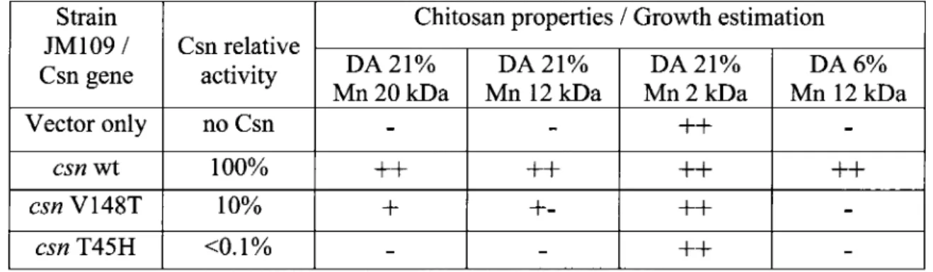
Table 1: Effect of heterologous Csn activity expression in E. coli on growth on chitosan media 46
CHAPTER II
Table 1: Bacterial strains and plasmids used in this work 63 Table 2: Minimal inhibitory concentrations (MICs) of 5-fluorocytosine and 5-fluorouracil
determined for wild type strains 70 Table 3: Minimal inhibitory concentrations (MICs) determined for Streptomyces lividans
TK24 derivatives constructed in this study 72 Table 4: Occurence of pMP-vectors polylinker restriction sites in completely sequenced
actinobacterial genomes 75
CHAPTER III
Table 1: Purification of CsnA from a culture of recombinant Streptomyces lividans 10-164
LIST OF FIGURES INTRODUCTION
Figure 1: Chitosan synthesis from chitin 1 Figure 2: Molecular structure of cellulose and chitosan 6
Figure 3: Comparisons of the tripartite structure of Sec and Tat signal peptide sequence 20 Figure 4: Representation of Tat signal peptides with the conserved residues forming the
twin-arginine motif. 21 Figure 5: Phylogenetic analysis of primary sequences GH46 family members 23
CHAPTER I
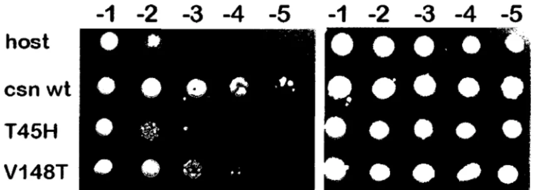
Figure 1: Spot test of growth of E. co/j strain JM109 47 Figure 2: Minimal inhibitory concentration of chitosan for E. coli DH5a™ strains expressing
chitosanases with different relative activities 48 Figure 3: Cell counts of E. coli DH5a™ strains expressing or not a heterologous chitosanase
49 Figure 4: Influence of heterologous chitosanases expression by E. coli and 5". lividans strains
on the survival of chitosanase-negative E. coli in the presence of chitosan 50
CHAPTER II
Figure 1: Insertional inactivation of S. lividans SC02657 homolog gene (2657h) via double
crossing over 73 Figure 2: Elements of pMP201 vector and derivatives 76
Figure 3: Restriction maps of the vectors constructed in this work 78
CHAPTER III
Figure 1: Phylogenetic analysis of primary sequences GH46 family members 98
Figure 2: SDS-PAGE (12%) analysis of CsnA 100 Figure 3: Comparison of recombinant chitosanase production directed by Sec or Tat
Figure 4: Effect of the pH and temperature on CsnA activity 102 Figure 5: Thermal unfolding of CsnA and CsnN174 chitosanases at pH 4.1 or pH 5.5 in the
absence or presence of chitosan 104 Figure 6: Alignment of primary sequences of the chitosanases from S. lividans TK24 and S.
coelicolor A3(2) 105 Figure 7: Determination of minimal inhibitory concentration of chitosan on agar plates 106
Figure 8: Effect of chitosan on xylose uptake by S. lividans strains 107
CHAPTER IV
Figure 1: Validation of chicken anti-CsxA and rabbit anti-CBM35 antibodies 128 Figure 2: Epifluorescence microscopy analysis of the cell wall localization of CsxA and
CBM35 130 Figure 3: Confocal microscopy analysis of the cell wall localization of CsxA and CBM35.. 131
Figure 4: Confocal microscopy analysis of the cellular localization of a secreted protein and an
anchored protein 132 Figure 5: Epifluorescence microscopy analysis of the CBM35 ligand specificity 133
INTRODUCTION
Chitin and chitosan
Chitin is the second most abundant renewable natural resource after cellulose (Deshpande, 1986). This insoluble polymer composed of P-(l,4)-linked 7V-acetyl-D-glucosamine residues is widely distributed in nature particularly in outer skeleton of insects, marine invertebrates, fungi and algae (Muzzarelli, 1977; Ruiz-Herrera and Xoconostle-Cazares, 1995; Cauchie, 2002). The partially JV-deacetylated derivative of chitin is called chitosan.
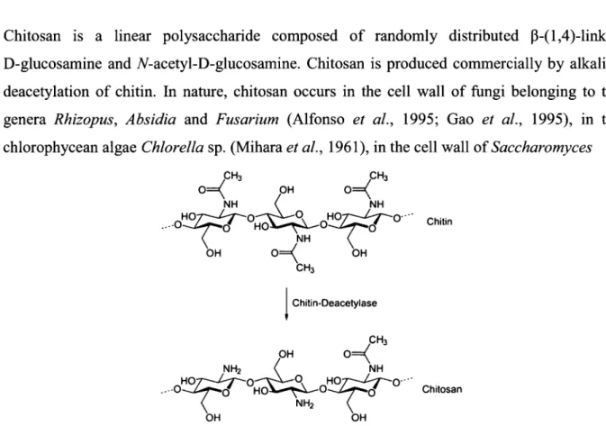
Chitosan is a linear polysaccharide composed of randomly distributed p-(l,4)-linked D-glucosamine and 7V-acetyl-D-glucosamine. Chitosan is produced commercially by alkaline deacetylation of chitin. In nature, chitosan occurs in the cell wall of fungi belonging to the genera Rhizopus, Absidia and Fusarium (Alfonso et oil., 1995; Gao et ah, 1995), in the chlorophycean algae Chlorella sp. (Mihara et ah, 1961), in the cell wall of Saccharomyces
..-o
Chitin-Deacetylase
.- -0
Figure 1: Chitosan synthesis from chitin.
Chitin
• 0 - "
cerevisiae spores (Briza et ah, 1988) and transitorily in insects' cuticles (Aruchami et al., 1986). The chitin deacelylase is the key enzyme for chitosan occurrence in nature (Figure 1) (Davis and Bartnicki-Garcia, 1984). Compared to cellulose and chitin, chitosan is much less abundant in Nature.
Chitosanase
Generally, chitosanases have been recognized as enzymes that hydrolyze chitosan but not chitin. In 2004, the Enzyme Commission amended the definition of chitosanase as being the enzyme performing endo-hydrolysis of P-(l,4) linkages between D-glucosamine residues in a partially JV-acetylated chitosan.
Chitosanases (EC 3.2.1.132) were independently discovered by two groups: Monaghan et ah, (1972) in a study of the possible use of lytic enzymes to combat pathogenic fungi, and Ramirez-Leon and Ruiz-Herrera (1972) during a fundamental study of the cell wall architecture.
Chitosanases are members of the glycoside hydrolases group which is divided into 115 families. Based upon their amino acids sequences, chitosanases have been classified into six glycoside hydrolase families: GH5, GH7, GH8, GH46, GH75 and GH80. The families GH5, GH7 and GH8 contain a variety of glycoside hydrolases such as chitosanase, cellulase, licheninase, and endo-1,4-0 xylanase while GH46, GH75 and GH80 are currently exclusively composed of chitosanases.
Other enzymes hydrolyzing chitosan
Interestingly, many enzymes with different original specificities have also been reported for their ability to hydrolyze chitosan and its derivatives. In the beginning, most of those double specificities of the enzymes were attributed to contaminations of protein extracts by chitosanases. With time, due to the increased number of reports describing enzymes with specificities for more than one substrate, the term bifunctional enzymes was adopted. These enzymes may be divided into three classes according to their specificity for chitosan hydrolysis.
The first class comprises enzymes with high specificity for chitosan hydrolysis such as: endo-chitinases (EC 3.2.1.14 - GH18 and GH19) (Mitsutomi et ah, 1995), exo-p-D-glucosaminidases (also called exo-chitosanases) (EC 3.2.1.165 - GH2 and GH35) (Nanjo et al., 1990) and 7V-acetyl-glucosaminidases (EC 3.2.1.52 - GH3, GH20, GH84) (Muzzarelli, 1993). The members of this class are very important for chitosan assimilation by soil microorganisms. Thus, together with the endo-chitosanases (EC 3.2.1.132), they contribute to the total hydrolysis of the partially JV-acetylated chitosan into monomers of D-glucosamine and JV-acetyl D-glucosamine. The endo-chitinases and endo-chitosanases liberate chitooligomers by an endo-type of action (hydrolysis occurs inside of chitosan chain). Then, chitooligomers are further hydrolyzed by exo-P-D-glucosaminidases and ./V-acetyl-glucosaminidases. Those enzymes release a single glucosamine or JV-acetyl-glucosamine residue from the non-reducing end of the oligomers at a time and the resulting monomers as well as short oligomers may be transported and further metabolized by the bacteria (exo-type of action).
Chitinase
By definition, chitosan is a partly TV-deacetylated derivative of chitin, so it is not surprising to find bacterial endo-chitinases able to recognize chitosan as substrate and to specifically hydrolyze it. The chitinases degrading chitosan belong to the GH18 and GH19 families, and their hydrolytic action towards chitosan is influenced by the JV-acetylation degree of this polymer. The endo-chitinases hydrolyze chitosan with high to moderate degree of iV-acetylation. For instance, previous studies on chitinases Al and D (GH18) from Bacillus circulans WL-12 (Mitsutomi et ah, 1995) showed that these chitinases efficiently hydrolyzed the iV-acetyl-p-D glucosaminidic bonds in 50% JV-acetylated chitosan. Similarly, the chitinases I (GH18) and II (GH19) from Burkholderia gladioli CHB101 (Shimosaka et al, 2001) were efficient at hydrolyzing 30% JV-acetylated chitosan. Those bacteria also secrete endo-chitosanases which hydrolyze, with maximal efficiency, chitosan with 0-30% TV-acetylation degree (Mitsutomi et al, 1998; Shimosaka et ah, 2000). The contribution of these two types of glycoside hydrolases confers to the bacterial host the capacity to use chitosan with different TV-acetylation degrees as carbon source.
Exo-p-D-glucosaminidases
In most cases, chitosanases are endo-type enzymes. The exo-P-D-glucosaminidases are also known as exo-chitosanases and have been isolated from few microorganisms such as the fungi Aspergillus oryzae IAM2660 (Zhang et al., 2000), Aspergillus fumigatus KH-94 (Kim et al., 1998), Aspergillus flavus IAM2044 (Ji et al., 2003), Hypocrea jecorina (formely Trichoderma reesei PC-3-7; Nogawa et al., 1998), Penicillum funiculosum KY616 (Matsumura et al., 1999) and the bacterium Amycolatopsis orientalis (Nanjo et al., 1990). All these enzymes have been purified and characterized from a biochemical point of view. Based on their amino acid
Trichoderma reesei PC-3-7 (Ike et ah, 2006) belong to the GH2 family, while the exo-P-D-glucosaminidase from Thermococcus kodakaraensis KOD1 belongs to GH35 (Tanaka et ah, 2003). The culture supernatants of Aspergillus and Amycolatopsis strains contained endo-chitosanases along with exo-P-D-glucosaminidases, implying the cooperation of these enzymes in chitosan metabolism. The exo-P-D-glucosaminidase from Amycolatopsis orientalis is the most studied in terms of structure and function (Cote et ah, 2006, Fukamizo et ah, 2006; van Bueren et ah, 2009). Interestingly, the exo-P-D-glucosaminidase from Thermococcus kodakaraensis KOD1 was reported to be involved in a novel chitinolytic pathway (Tanaka et ah, 2004). This new chitino lytic pathway is based on the concerted action of the exo-p-D-glucosaminidase and of a diacetylchitobiose deacetylase. The GlcNAc dimers produced from chitin by the chitinase from Thermococcus kodakaraensis KOD1 are further N-deacetylated by a diacetylchitobiose deacetylase at the non-reducing end. GlcN-GlcNAc dimers are then hydrolyzed by the exo-p-D-glucosaminidase to GlcN and GlcNAc residues which are easily metabolized by bacteria (Tanaka et ah, 2004). Moreover, the exo-chitosanases from Hypocrea jecorina PC-3-7 and Aspergillus oryzae IAM2660 are inducible only by GlcNAc while the one secreted by A. orientalis is inducible by chitosan and by GlcN, which suggests that the fungal and bacterial exo-P-D-glucosaminidases may possess different physiological functions.
Cellulase and lysozyme
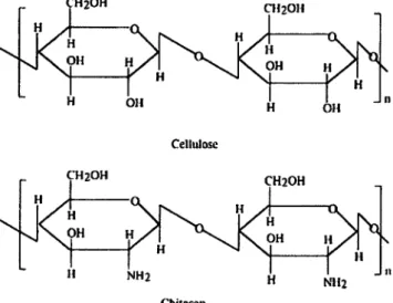
The second class of enzymes capable of hydrolyzing chitosan includes enzymes with moderate selectivity for chitosan, such as cellulase and lysozyme. Cellulases (EC 3.2.1.4) are enzymes hydrolyzing the p-(l,4)-D-glucosidic linkages in cellulose. Most of the cellulases have a significant hydrolytic activity towards chitosan and some of them may hydrolyze chitosan with an activity almost equivalent to that of chitosanases. This special behavior may be explained by the structural similarity of chitosan and cellulose. Cellulose is a polymer of P-(l,4)-linked D-glucose residues. Thus, chitosan may be considered as an aminated derivative
of cellulose obtained by the replacement of the C-2 hydroxyl group with amino groups (Figure2). Therefore, this capacity of cellulase to hydrolyze chitosan seems to be attributed to a nonspecific recognition of the group at the C-2 position in glucose or glucosamine, when the enzyme-substrate complex is formed. It is known that the specificity of the enzymatic reaction catalyzed by glycoside hydrolases is mostly determined by the characteristics of the substrate binding subsites. Consequently, in order to understand this phenomenon we have to analyze the key amino acids for substrate-binding in chitosanases and cellulases. The chitosanases from the GH46 family recognize partially JV-acetylated chitosan with high selectivity. In their case, the key amino acids involved in substrate binding are invariably carboxylic acids such as: spartic acid and glutamic acid (Katsumi et ah, 2005) which can electrostatically interact with the positive charges of the chitosan chain. In the cellulase case, substrate binding sites are occupied by positively charged amino acids such as histidine, arginine and hydrophobic amino acids such as tryptophan, which may explain the less selective recognition of chitosan by these enzymes (Mulakala and Reilly, 2005).
Cellulose
CH2OH CH2OH H y a I
" N« I Mil J" Chitosan
Figure 2: Molecular structure of cellulose and chitosan.
The existence of bifunctional enzymes with cellulase-chitosanase activity is well documented. In many cases, the term bifunctionality may be confusing, when it cames to the classification of the respective enzymes. Depending on the experimental conditions such as the choice of
substrate, temperature, buffer and pH, one type of activity may be favoured to the detriment of the other. For example, the bifunctional cellulase-chitosanase produced by the Gram-negative microorganism Myxobacter sp. AL-1 (Pedraza-Reyes and Gutierrez-Corona, 1997) showed a better chitosanase activity at 70°C than at 42°C, when chitosan hexamer was used as substrate, but a greater cellulase activity at 42°C than at 70°C, when carboxymethyl- cellulose was used as substrate. Under their respective optimal conditions, the chitosanase and cellulase activities were very similar: 61U/mg and 48 U/mg, respectively.
Bifunctional cellulases from various sources show different substrate specificities. The enzymes from B. cereus D-ll (Gao et ah, 2008), Myxobacter sp. AL-1 (Pedraza-Reyes and Gutierrez-Corona, 1997) and Bacillus circulans WL-12 (Mitsutomi et ah, 1998) show comparable cellulase and chitosanase activities. Moreover, Cel8A from Lysobacter sp. IB-9374 (Ogura et ah, 2006) displays a chitosanase activity that is 40% of its cellulase activity. Interestingly, most of the bacterial bifunctional cellulase-chitosanases belong to the GH8 family and are secreted when carboxymethyl-cellulose or glucan is used as carbon source. Moreover, analysis of their primary structure showed high homology with glucanases from GH8 but not with the highly selective chitosanases of the GH46 family.
Another enzyme known to catalyze chitosan hydrolysis with moderate selectivity is lysozyme (EC 3.2.1.17). Ordinarily, lysozyme catalyzes the hydrolysis of 1,4-beta-linkages between N-acetyl-D-glucosamine and TV-acetylmuramic acid residues in peptidoglycans found in the bacterial cell wall. There is a structural resemblance between chitosan and peptidoglycans. The ability of lysozyme to hydrolyze partially iV-acetylated chitosan is well documented. Thus, it was shown that hen egg white lysozyme and human lysozyme (Sashiwa et al., 1991; Nordtveit et al., 1994) can both hydrolyze soluble chitosan. The 7V-acetylation degree of chitosan is a key parameter influencing the capacity of lysozyme to hydrolyze chitosan. Hence, it was shown that lysozyme is more active against chitosans with a high 7V-acetylation degree. Interestingly, hen egg white lysozyme and GH46 chitosanase share the same structural
organization of the substrate binding and catalytic cleft (Monzingo et al., 1996). Together, this explains the capacity of lysozyme to bind and hydrolyze chitosan. The moderate selectivity is due to the presence of basic and hydrophobic amino acids such as arginine and tryptophan in the substrate binding cleft of lysozyme instead of carboxylic acids characteristic of chitosanase (Maenaka et ah, 1994). Thus, the presence of arginine will cause a slight repulsion of the positively charged chitosan, while tryptophan participates to non-polar interactions with the pyranose rings of the substrate.
Finally, the third class is constituted of enzymes hydrolyzing partially iV-acetylated chitosan with low selectivity. In 2005 Kittur et al. isolated a pectinase (EC 3.2.1.15) from Aspergillus niger which showed endo- and exo- chitosanase activities towards chitosan. Other enzymes such as a lipase from Mucor circinelloides (Struszczyk et al., 2008) and papain (Terbojevich et ah, 1996) have been also reported for their ability to hydrolyze chitosan. It is matter of discussion whether or not the chitosanase activities of these enzymes was due to the proteins themselves or to some minor impurities in the preparations.
Occurrence of chitosanases in Nature
Chitosanases are widely distributed in Nature. Since their discovery in 1972, chitosanases have been isolated from numerous prokaryotes, fungi, viruses and plants.
Among prokaryotes, most chitosanase activities have been identified in actinomycetes and bacilli. Actinomycetes are soil-living, Gram-positive bacteria known for their important role in the decomposition of organic matter such as cellulose and chitin. So, it is not surprising that as much as fifteen actinomycete strains such as Streptomyces sp. strain 6 (Price and Storck,
Streptomyces sp. strain N174 (Boucher et ah, 1992), Kitasatospora sp. N106 (formerly known as Nocardioides sp. N106; Masson et ah, 1995), Amycolatopsis sp. CsO-2 (formerly known as Nocardioides sp.; Okajima et ah, 1995; Saito et ah, 2009), Streptomyces griseus HUT 6037 (Tanabe et ah, 2003) and Microbacterium sp. OU01 (Sun et ah, 2006) have been isolated as chitosanase producers and their enzymes characterized from a biochemical point of view. Other actinomycetes strains such as S. coelicolor A3(2) (Bentley et ah, 2002), S. lividans TK24 (GenBank accession number GQ438786.1), S. avermitilis MA-4680 (Ikeda et ah, 2003), S. sclerotialus (GenBank accession number AB 196768.1), Streptomyces sp. AM-7161 (Ichinose et ah, 2003), and Renibacterium salmoninarum ATTC 33209 (GenBank accession number ABY24857) are also known to possess chitosanase activities. The amino acid sequences for all the strains mentioned above are known and, with the exception of the chitosanase from Streptomyces griseus HUT 6037 (GH5), they all belong to the GH46 family.
Bacilli are also Gram-positive bacteria. Bacillus strains producing enzymes with chitosanase activity are abundantly present in soil. To date, more than thirty five Bacillus strains have been identified as high chitosanase producers. Bacillus megaterium PI (Pelletier and Sygusch,
1990), Bacillus circulans MH-K1 (Ando et ah, 1992), Bacillus circulans WL-12 (Mitsutomi et ah, 1998), Bacillus subtilis strain 168 (Rivas et ah, 2000), Bacillus coagulans CK108 (Yoon et ah, 2002), Paenibacillus fukuinensis D2 (Kimoto et ah, 2002), Bacillus sp. DAU101 (Lee et ah, 2006), Bacillus sp. strain S65 (Su et ah, 2006), Bacillus thuringiensis (Lee et ah, 2007), and Bacillus cereus D-ll (Gao et ah, 2008) are just a few of those that have been characterized from a biochemical point of view. In fact, the real number of soil Bacillus strains with chitosanase activities is still to be determined. Most of the Bacillus chitosanases are regrouped into the GH8 family with a few exceptions whose primary structure has a high homology with most of the actinomycetes chitosanases, thus they belong to GH46 family.
Thus, it is not surprising that the majority of the chitosanases that have been studied extensively in terms of their catalytic features, protein structures and enzymatic mechanisms,
have been isolated from actinomycetes and bacilli (Boucher et al., 1995; Marcotte et al., 1996; Fukamizo et al, 2005; Saito et al, 1999).
Gram-negative bacteria such as Myxobacter sp. AL-1 (Pedraza-Reyes and Gutierrez-Corona, 1997), Burkholderia gladioli CHB101 (Shimosaka et al., 2000), Pseudomonas sp. A-01 (Ando et al., 2008), Mitsuaria chitosanitabida (Amakata et al., 2005) and Serratia marcescens TKU011 (Wang et al, 2008) were reported to efficiently produce chitosanases. Moreover, a gene coding for a chitosanase activity was identified in the genome sequence of the cyanobacterium Nostoc punctiforme PCC 73102 (GenBank accession number ACC80641.1). The chitosanase from Myxobacter sp. AL-1 belongs to the GH5 family, those from Burkholderia gladioli CHB101, Pseudomonas sp. A-01 and Nostoc punctiforme PCC 73102 are members of the GH46 family, while the one from Mitsuaria chitosanitabida belongs to GH80.
An interesting group of microorganisms producing chitosanases is represented by fungi. Fungi are known to possess chitosan, the chitosanase's substrate, in their cell wall. The first fungal chitosanase was isolated by the team of Fenton and Eveleigh in 1981. They produced the chitosanase from Penicillium islandicum by cultivating this fungus in the presence of Rhizopus rhizopodiformis hyphae as substrate. Since then, as much as twenty three chitosanase producing fungal strains have been isolated and enzymes with chitosanase activity were further characterized. Mucor rouxii (Alfonso et al., 1992), Fusarium solani (Shimosaka et al., 1993), Aspergillus oryzae (Zhang et al., 2001), Aspergillus aculeatus F50, Aspergillus fumigatus (Cheng et al., 2006), Trichoderma reesei (Ike et al., 2007), and Gongronella sp. JG
(Wang et al., 2008) are just some of those chitosanolytic fungal strains. Fungal chitosanases are regrouped in the GH75 and GH7 families, and among them, the one originating from Fusarium solani is the best characterized (Shimosaka et ah, 1996). The fungal chitosanases have no sequence similarities with the bacterial ones. Interestingly, in some cases, the addition
chitosanases may play a different role than the utilization of the exogenous chitosan as nutrient.
The virus strains' group for which chitosanase activity was detected is less representative, compared to the bacterial one. To date, two CVK2 virus strains infecting Chlorella viridis (Yamada et ah, 1997) and PBCV-1 infecting Paramecium bursaria (Sun et ah, 1999; Han et ah, 2002), were isolated for their ability to degrade the rigid, chitosan containing, cell wall of the algae from Chlorella species. Remarkably, analysis of the viral chitosanases amino acid sequences corresponding to the catalytic segment revealed that these enzymes have a high homology with chitosanases produced by Streptomyces sp. N174, Kitasatospora sp. N106 and B. subtilis (Fukamizo and Brzezinski 1997), therefore, they belong to the GH46 family. The chitosanase from the CVK2 virus strain was purified and a role of this chitosanase in the lytic cycle of the virus was proposed.
Interestingly, enzymes with chitosanase activity have also been isolated from leaf, seed, roots, and fruit extracts of several plant species such as Cucumis sativus (cucumber), Citrus sinensis (sweet orange), Nicotiana tabacum (tobacco) and Lycopersicon esculentum (tomato) (El Ouakfaoui and Asselin., 1992a; El Ouakfaoui and Asselin, 1992b; Osswald et ah, 1994; Brunner et ah, 1998; Pozo et ah, 1998). So far, the corresponding enzymes have not been characterized at the primary sequence level.
Therefore, the taxonomical distribution of the chitosanase producers is remarkable. Regarding their primary structure, the chitosanases from different taxonomic groups are well distributed in the six glycoside hydrolase families. The GH7 and GH75 families regroup chitosanases from fungi. The GH8 family is reserved essentially to chitosanases produced by members of the Bacillus group. The GH5 and GH46 families are the most diversified from a taxonomical
point of view, as they contain chitosanases originating from actinobacteria, bacilli, Gram-negative bacteria and viruses. Finally, the GH80 family includes only bacterial chitosanases.
Moreover, multiple forms of chitosanases were detected in representatives of all the groups described above. For example, in 1990 Pelletier and Sygusch showed that B. megaterium PI secretes three chitosanases. Later on, other microorganisms in which multiple forms of chitosanases were detected, have been added to the list: Mucor rouxii (Alfonso et al., 1992), Bacillus licheniformis UTK (Uchida et al., 1992), Chlorella virus CVK2 (Yamada et al.,
1997), Streptomyces coelicolor A3(2) (Bentley et al, 2002), Aspergillus sp. CJ22-326 (Chen et al., 2005), and Microbacterium sp. (Sun et al., 2006). In plants, multiple chitosanase isoforms were detected as well; up to six chitosanases were seen in the various organs of the plant Cucumis sativus (El Ouakfaoui and Asselin, 1992a). In Citrus sinensis, four proteins with chitosanase activity were also identified by Osswald et al. in 1994. In most cases, these chitosanase activities are encoded by different genes (Alfonso et al., 1992; Bentley et al., 2002) with the exception of Chlorella virus CVK2 in which the vChta-1 gene was shown to produce, by a mechanism of alternate gene expression, two chitosanases with different roles in viral infection (Yamada et ah, 1997).
More interesting, some chitosanases have hydrolytic activity on substrates other then chitosan. After examples of chitosanases that are capable of hydrolyzing chitin can be found in the literature. In Nature, chitin is found in the shell of marine invertebrates, insect's cuticles, and in the cell wall of fungi and algae (Muzzarelli, 1977) where it plays an important role in the maintenance of shape or cellular integrity. Following chitin synthesis by chitin synthetase, this polymer is iV-deacetylated by chitin deacetylase (Davis and Bartnicki-Garcia, 1984) to a certain degree which confers some flexibility to the chitin; flexibility required for example when marine invertebrates or insects are growing, or when the fungal hyphal branches are developing. Therefore, chitosanases from Enterobacter sp. G-l (Yamasaki et ah, 1993) and
Mucor rouxii (Alfonso et ah, 1992) are able to hydrolyse chitin proccesing a certain degree of iV-deacetylation.
Chitosanases with cellulase activity have been isolated from various sources such as: B. cereus SI (Kurakake et ah, 2000), B. sp. KCTC 0377BP (Choi et ah, 2004), Streptomyces griseus HUT 6037 (Tanabe et ah, 2003) and Trichoderma reesei (Ike et ah, 2007). Interestingly, the chitosanases B and C from B. megaterium PI (Pelletier and Sygusch, 1990) possess comparable enzymatic activities toward chitosan, chitin and cellulose. Even if the structural resemblance between those substrates (polymers of P-(l,4)-linked derivates of glucose) may explain this multiple substrate specificity, the real hydrolysis mechanism needs to be further studied.
Interestingly, for most of the bifunctional cellulase-chitosanases, a single substrate-binding domain is thought to be responsible for interaction with the two substrates, but the mechanism behind this is still unclear. However, Liu and Xia proposed that the bifunctional cellulase-chitosanase isolated from a commercial cellulase preparation, has two different substrate binding domains (Liu and Xi, 2006). However, their binding mechanism and the domain location in the protein are unknown.
Another example of multiple substrate specificity is that of the 40 kDa chitosanase from Bacillus circulans WL-12 (Mitsutomi et ah, 1998). Substrate specificity analysis revealed that this chitosanase hydrolyzed chitosan (30% JV-acetylated) and lichenan (P-l,3-l,4-glucan) with similar efficiency. Interestingly, eight years before, this enzyme has been reported as a
p-1,3-1,4-glucanase by Bueno et ah (1990). Therefore, in order to determine which was the major substrate for this enzyme, Mitsutomi and coworkers analysed the production of this protein by B. circulans WL-12 in media supplemented with chitosan and lichenan. The study showed that production of this enzyme was induced by chitosan (30% iV-acetylated) but not by lichenan.
Thus, this enzyme is first a chitosanase and, it according to its primary structure, has been included into the GH8 family. As most of the chitosanases belonging to the GH8 family, this chitosanase/p-l,3-l,4-glucanase has also the capacity to hydrolyze CM-cellulose ((3-(l,4)-glucan), which may indicate that in lichenan the enzyme hydrolyzes only p-(l,4)-linkages.
The fact that chitosanases show different specificities for substrates made Schindler et al. hypothesize that: "Evolutionary changes in substrate structure may have influenced the development of the active site of lysozyme so that it could function most efficiently with the particular natural substrate encountered by each species" (Schindler et al., 1977).
Chitosanases: mode of action
Based on the observations presented in the previous sections, two questions may be raised: are there any differences among the chitosanases produced by organisms belonging to the groups described above? And why does microorganism need to secrete more than one chitosanase when the availability of chitosan in soil as nutrient is not even comparable with other carbon sources such as cellulose and chitin? The answer comes from the characteristics of chitosan itself.
An important property of chitosan is its degree of 7V-acetylation. Chitosan wholly jV-deacetylated or with different JV-deacetylation degrees (DDA) can be obtained from chitin by various, well controlled chemical methods (Muzzarelli, 1985; Varum and Smidsrod, 2004). Fully JV-deacetylated chitosan is not found in nature (Bartnicki-Garcia, 1968). Natural chitosan has a DDA varying from 55 to 95%. The chitosan present in the cell wall of fungi from different genera is characterized by various degrees of 7V-deacetylation: Absidia coerulea and
albicans 84% DDA, Fusarium oxysporum 83% DDA, Penicillium citrinum 79% DDA, Rhizopus oryzae 90-78%) DDA, Cunninghamella blakesleeana 65%, and Mucor rouxii 95-55% DDA (Briza et ah, 1988; Miyoshi et ai, 1992; Pochanavanich and Suntornsuk, 2002). These natural chitosans are probably synthesized by the sequential action of chitin synthetase and chitin deacetylase, as shown for Mucor rouxii and Colletotrichum lindemuthianum (Davis and Bartnicki-Garcia, 1984). Hence, there is diversity in the substrate available for the microorganisms living in soil.
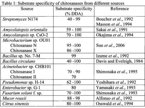
Consequently, the microorganisms adapted to this substrate diversity by producing chitosanases with different substrate specificity. The effect of DDA on chitosanase activity has been well documented (Table 1).
Table 1: Substrate specificity of chitosanases from different sources Source Streptomyces N174 Amycolatopsis orientalis Amycolatopsis sp. CsO-2 Microbacterium sp. OU01 Chitosanase N Chitosanase X Bacillus sp. No. 7-M Bacillus circulans Acinetobacter sp. CHB101 Chitosanase I Chitosanase II Pseudomonas sp. H-14 Enterobacter sp. G-l Fusarium solani f. sp. Mucor rouxii Citrus sinensis Substrate specificity (% DDA) 4 0 - 9 9 59 - 100 70 - 100 95 -100 86-100 99 40 -100 7 0 - 9 0 70 62 -100 80 70-100 88-98 80-100 Reference Boucher et al., 1992 Masson et al, 1994 Sakaiefa/., 1991 Okajima ef a/., 1994 Sun et al, 2006 Izume et al., 1992
Davis and Eveleigh, 1984 Shimosaka et al., 1995 Yoshihara et al., 1992 Yamasaki et al., 1993 Shimosaka et al., 1993 Alfonso etal., 1992 Osswald etal, 1994
The chitosanases from Bacillus sp. No. 7-M (Izume et al., 1992), Pseudomonas sp. H-14 (Yoshihara et ah, 1992), Amycolatopsis sp. CsO-2 (Okajima et al., 1994) and Bacillus sp.
PI-7S (Seino et al, 1991) showed a preference for chitosan that is approximately 100% ./V-deacetylated. Chitosanases from Penicillium islandicum (Fenton and Eveleigh, 1981) and Streptomyces sp. N174 (Boucher et al, 1992) could depolymerise chitosan substrates having a wide range of JV-deacetylation degrees: 40-70% and 79-99%, respectively.
Interestingly, chitosanases from Fusarium solani (Shimosaka et al., 1993) and Amycolatopsis orientalis (Sakai et al., 1991) can act optimally only on chitosan having 70% JV-deacetylation whereas that from Enterobacter sp. G-l (Yamasaki et al., 1993) has its best activity on chitosan having 80% 7V-deacetylation. This special behaviour is an indication that the JV-acetyl glucosamine residues are important in the recognition and reaction mechanism of enzymes on various substrates.
Cleavage specificity
Partially TV-acetylated chitosan contains four types of linkage between its subunits: GlcN-GlcN, GlcNAc-GlcN-GlcN, GlcN-GlcNAc and GlcNAc-GlcNAc. The proportion of these linkages varies in function of the degree of N-acetylation and their distribution is random in the polymer structure. According to their cleavage specificity, chitosanases are divided into three subclasses. Enzymes of subclass I cleave GlcN-GlcN and GlcNAc-GlcN linkages and includes chitosanases from Bacillus pumilus (Fukamizo et ah, 1994), Penicillium islandicum (Fenton and Eveleigh, 1981) and Streptomyces sp. N174 (Fukamizo et ah, 1995). Subclass II contains chitosanases from Bacillus sp. No 7-M(Izume et ah, 1992) Pseudomonas sp. H-14 (Yoshihara et al, 1992), Amycolatopsis sp. CsO-2 (Okajima et al., 1994) and Bacillus sp. PI-7S (Seino et al., 1991) that recognize only GlcN-GlcN linkages. Subclass III includes enzymes from Bacillus circulans MH-KI (Saito et al., 1999), Nocardia orientalis (Sakai et al., 1991), and Bacillus circulans WL-12 (Mitsutomi et al, 1998) which recognize GlcN-GlcN as well as
chitosan made possible the clear differentiation between chitosanases and chitinases. To date, all studied chitosanases have a high specificity for the cleavage of GlcN-GlcN linkages. In contrast, the chitinases cannot cleave the linkage between two glucosamine residues as they require at least one GlcNAc residue in the cleaved linkage (Fukamizo et al., 1994).
This classification was changed this year by the addition of a fourth subclass following the discovery of an enzyme from the fungus Alternaria alternata that specifically cleaves only the GlcNAc-GlcN linkage (Kohlhoff et al., 2009). Due to its new cleavage specificity, this enzyme was called a chitinosanase. This is half a chitinase and half a chitosanase enzyme which cannot be considered a chitinase because it does not cleave GlcNAc-GlcNAc linkages nor a chitosanase because it does not cleave GlcN-GlcN linkages.
Chitosanase secretion
The targeting and transport of proteins to and across biological membranes is a very important characteristic of cellular life. Interestingly, it is thought that 25-30% of the genes coding for proteins from the model Gram-negative organism E. coli, or the Gram-positive organism Streptomyces coelicolor, encode proteins that will be located either partially or completely outside the cytoplasm. In prokaryotes, two major pathways exist for the export of proteins across the cytoplasmic membrane the Sec pathway and the Tat pathway.
The Sec pathway is the general Secretion route for proteins in most bacteria. This pathway is generally responsible for the transport of newly synthesized proteins out of the cytosol in an unfolded state; thereby they fold into their native structure once released from the membrane. The Sec secretion machinery (also called Sec-translocase) is present in the cytoplasmic membrane of all bacteria, archaea, the thylakoid membrane of plant chloroplasts and the
endoplasmic reticulum of eukaryotes. In bacteria, the Sec pathway is responsible for the secretion of most extracellular proteins with diverse functions in metabolism, substrate uptake and excretion, cell envelope structure, sensing and cell communication (Lee and Schneewind, 2001, Natale era/., 2008).
The Twin-arginine translocation pathway, also called Tat pathway, is involved in the translocation of proteins in their folded state. Tat-translocation systems have been first identified in thylakoid membranes of plant chloroplasts. Functional Tat secretion systems have also been found in the cytoplasmic membranes of many bacteria and archaea (Muller et ah, 2005). In bacteria, proteins secreted by this system are found to be involved in energy metabolism, quorum sensing and motility, cell division and biogenesis of the cell envelope, symbiosis and pathogenesis (Berks et a\., 2005, Stevenson et ah, 2007). In plant chloroplasts, the Tat pathway is involved in the assembly of the oxygen-evolving complex and the cytochrome b(f complex within the thylakoid membrane (Mould and Robinson, 1991; MMik et ah, 2001).
Since the discovery of the Tat secretion pathway in 1996 by Berks, numerous studies were dedicated to the analysis of the distribution and utilization of this system by organisms of all Kingdoms. Lately, specialized programs such as TATFIND 1.2 (Dilks et ah, 2003) and TATscan (Li et ah, 2005) were developed for the computational prediction of Tat dependent proteins. In bacteria, most of the secreted proteins are translocated via the general Sec pathway, whereas only a subset of bacterial proteins is exported via the Tat-pathway. For example, genomic data combined with computational predictions showed that for microorganisms such as Rickettsia prowazekii and Staphylococcus aureus only one and two Tat substrates have been identified, respectively (Dilks et ah, 2003). Moreover, Fusobacterium nucleatum and Lactococcus lactis lack a Tat system. Interestingly, in other bacteria such as Caulobacter crescentus, Sinorhizobium meliloti and Streptomyces coelicolor
have been identified, respectively (Dilks et al., 2003; Li et al., 2005). Moreover, it was shown that in chloroplasts, the number of Tat substrates is equivalent to that of Sec substrates (Muller et al., 2005). Thus, even if the Sec pathway is the general secretion pathway in the majority of organisms, it seems that the degree to which the Tat pathway is used is quite variable, even among related organisms.
Secreted proteins are synthesized in the cytoplasm in a precursor form with an N-terminal extension named signal peptide. This signal sequence is required for correct targeting of the protein to the secretion systems on the cytoplasmic site of the membrane and is cleaved after secretion by a specialized protease. The signal peptide sequence has a tripartite structure: a positively charged amino-terminal (n-region), a hydrophobic core (h-region) and a polar carboxyl terminal (c-region) region.
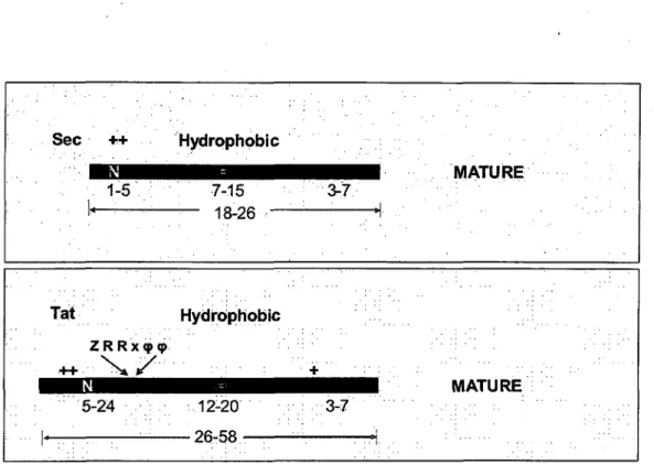
There are several significant differences between Sec- and Tat-specific signal peptides (Figure 3). Tat signal sequences are on average longer than Sec signals, due to larger n-regions (5-24 residues compared to 1-5 residues in Sec signal peptides). The h-region of Tat signals comprises 12-20 uncharged residues thus being slightly longer and less hydrophobic than h-regions of Sec-substrates (7-15 residues). Furthermore, Tat signal sequences are characterized by a conserved pattern of amino acids which includes two important arginine residues, the twin-arginine motif, located at the junction between the n- and h-regions. In bacteria, this motif has been described as Z-R-R-x-cp-cp, where Z stands for any polar residue and cp for hydrophobic residues.
Sec ++ Hydrophobic 1-5 : 7-15 • i.« • • - 18-26 Tat Hydrophobic ZRRx<pq> 5-24 ; 12-20 I OR CO 3-7 . !"• • • + ' " .; 3-7 ':""' . ..,. . MATURE MATURE
Figure 3: Comparison of the tripartite structure of Sec and Tat signal peptide sequence.
This Tat motif is extremely well conserved and is crucial for the role of Tat signal peptides (Figure 4). Genetic studies showed that replacement of the Arg-Arg pair with a Lys-Lys motif completely abolished Tat-specific export, while substitution of a single Arg by Lys affected only the rate of translocation via the Tat system (DeLisa et al, 2002). Tat-specific c-regions frequently contain a positively charged amino acid residue (+ Figure 3), which was proposed to function as a "Sec-avoidance signal" (Bogsch et al, 1997) and a proline residue acting as a 'helix breaker' to facilitate peptidase recognition of the cleavage site. The signal peptidase recognition site is conserved in Sec- and Tat-dependent peptides and is represented by an A-X-A motif found at end of the c-region.
Generic signal peptide structure - j n ^ h c J
N [ + + 1 ; . •"
Twin-arginine signal peptides
S u f i MSlJSRRQFJ^ftSGIALCAGAVPLKASA otaaaaaaaaaocaaaaaa
TorA jMNHM)LFQAfeRRRFLflbLGGLTVAGMLGPSLLTPRRATA
ocaaaaaaaaaaaaaa acta caxaaaa DmeA MK3np:KPAVI&&^yl5^
a a a a a a a a a a a a a a a a a a a a a a a a a
HyaA j MNlJEETFTQAMRRQGVlniRSFLK^CSIAATSLGLGAGMAPKI AWA
aaacuaaaaaaaa cuxaaaaaaaaaacca a a a a a
Figure 4: Representation of Tat signal peptides with the conserved residues forming the twin-arginine motif (boxed). (From Palmer et al., 2005, modified), n-region is represented in green, h-region in orange and c-region in purple. The "Sec-avoidance motif, represented by the highly conserved positively charged amino acids found within the c-regions, is shown in bold. Amino acid sequences of twin-arginine signal peptides from E. coli DmsA (dimethyl sulfoxidereductase); Sufi (copper oxidase); TorA (trimethylamine N-oxide (TMAO) reductase) and HyaA (hydrogenase).
This motif is recognized by the type I signal peptidase, a membrane-bound enzyme that cleaves the signal sequence from the mature secreted protein domain during or shortly after translocation. Interestingly, when the A-X-A site of a Sec-dependent signal sequence was mutated, no protein secretion was observed (Page et al., 1996), suggesting the importance of this conserved motif for the maturation of proteins translocated via the Sec pathway. In contrast, mutations of the A-X-A signal peptidase recognition site of the Tat-dependent signal peptide had no effect on the precursor processing rate (Li et al., 2006, Sambasivarao et al., 2000). These observations suggest that there is probably a major difference in the processing of the Sec- and Tat-dependent precursors and that processing is probably not carried out by the same signal peptidase I.
It has been shown that the Sec and Tat pathways are also involved in translocation and insertion of membrane proteins into the cytoplasmic membrane. In the case of the Sec-pathway, membrane proteins do not have cleavable N-terminal signal peptides. The integration into the cytoplasmic membrane of those proteins may occur via the Sec-dependent or a Sec-independent pathway, both mechanisms involving the YidC protein (Samuelson et al, 2000). Interestingly, in both bacteria and chloroplasts, Tat-targeted integral membrane proteins are synthesized with an N-terminal signal peptide and contain single internal or C-terminal transmembrane helices. Furthermore, integration of the transmembrane helices into the lipid bilayer is strictly Tat-dependent (Hatzixanthis et al, 2003).
Most of the bacterial and fungal chitosanases are secreted extracellularly (Fenton and Eveleigh, 1981; Pelletier and Sygusch, 1990; Boucher et al, 1992; Yamasaki et al, 1993; Okajima et al, 1994; Wang et al, 2008). Little is known of the specific pathway responsible for the chitosanase secretion. Computational analysis of the signal peptide sequences showed that most chitosanases (for which the amino acid sequence had been published) present a Sec-dependent signal peptide and very few had a Tat-Sec-dependent signal peptide (R. Brzezinski unpublished data). Among the Tat-dependent chitosanases, five are from Streptomycetes strains: SCO0677 from Streptomyces coelicolor A3(2), CsnA from Streptomyces lividans TK24, SAV2015 from Streptomyces avermitilis MA-4680, SAMR0713 from Streptomyces ambofaciens ATCC 23877 (all belonging to GH46) and ChoII from Streptomyces griseus HUT 6037 (GH5). Three other Tat-dependent chitosanases are from p-proteobacteria strains: Herbaspirillum sp. 9, Mitsuaria chitosanitabida 3001 and Mitsuaria sp. 67 (GH80). To date, only the chitosanase SCO0677 from Streptomyces coelicolor was confirmed to be a real Tat-dependent protein by genetical and biochemical studies (Li et al, 2005). Interestingly, SCO0677 is very similar at the molecular level to the chitosanase from Streptomyces sp. N174 a Sec-dependent chitosanase, as shown by the unrooted phylogenetic tree (Figure 5) generated following alignment of amino acid sequences of all the chitosanases belonging to the GH46 family (Annexe I).
Figure 5: Phylogenetic analysis of the primary sequence of GH46 family members. The amino acid sequences of the respective proteins have been extracted from the following database entry. STRN174 - Streptomyces N174 (P33665); NOCN106 - Kitasatospora sp. N106 (P48846); AMY CS02 - Amycolatopsis sp. CsO-2 (Q9LBG4); BAC_amMJl - Bacillus amyloliquefaciens MJ-1 (Q0PVM7); BAC_CIRC - Bacillus circulans MH-K1 (P33673); BACJCFB - Bacillus sp. KFB-C04 (Q9RC18); BAC_SUBT - Bacillus subtilis subsp. subtilis str. 168 (007921); BUR GLAD Burkholderia gladioli CHB101 (Q9XDS6); SAV2015 -Streptomyces avermitilis 4680 (BAC69726.1); SAV6191 - -Streptomyces avermitilis MA4680 (BAC73902.1); SCO0677 Streptomyces coelicolor A3(2) (Q9RJ88); PSE_A01 -Pseudomonas sp. A-01 (Q8KZM5); STRAM - Streptomyces sp. AM-7161 (Q7WT07); CHR_VIOL Chromobacterium violaceum ATCC 12472 (AAQ61593.1); PAENIBAC -Paenibacillus sp. BH-2005 (Q2PWA1); PAEEHIM - -Paenibacillus ehimensis EAG1 (024825); MICROB - Micro bacterium sp. OU01 (A7KBW5); CHV_CVK2 - Chlorella virus CVK2 (012288); CHVPBU - Paramecium bursaria Chlorella virus 1 (Q84608); REN_SALM Renibacterium salmoninarum ATCC 33209 (A9WUI6); STRPRIS -Streptomyces pristinaespiralis ATCC 25486 (NW002063180.1); STR_CLAV - -Streptomyces clavuligerus ATCC 27064 (NW002063046.1); STRAMBO - Streptomyces ambofaciens ATCC 23877 (A0AD68); STRLIV - Streptomyces lividans TK24; STRSCL - Streptomyces sclerotialus ISP 5269 (B1Q2K4); PLAV0939 - Parvibaculum lavamentivorans DS-1 (A7HRM9); DARO 2852 Dechloromonas aromatica RCB (Q47C49); DARO2340 -Dechloromonas aromatica RCB (NC007298.1); CELLJAP - Cellvibrio japonicus Uedal07 (B3PI04); CBEI0949 Clostridium beijerinckii NCIMB 8052 (A6LS03); NPUNR2009 Nostoc punctiforme PCC 73102 (B2J4Y1); BACamFZ - Bacillus amyloliquefaciens FZB42 (A7Z8H9); BAC_DAU - Bacillus sp. DAU101 (A0EQW7).
As shown in Figure 5, the thirty four chitosanases from the GH46 family define four different clusters. The first cluster (in red) is composed mostly of chitosanases produced by bacteria belonging to the Actinomyces genus. Inside this cluster, there are two interesting subclusters: the first is formed by the chitosanases from Streptomyces sp. N174 (Boucher et al., 1992) which is the most studied chitosanase for its structure and function and which is very similar to the CsnN106 from Kitasatospora sp. N106 (Masson et ah, 1995), and the chitosanase from Amycolatopsis sp. CsO-2 (Saito et al., 2009). The second subcluster, or the Tat subcluster is composed of SCO0677 from S. coelicolor, CsnA from S. lividans TK 24, SAV2015 from Streptomyces avermitilis MA-4680, SAMR0713 from S. ambofaciens ATCC 23877 and the chitosanase from Streptomyces sclerotialus ISP 5269 which is the only member of this subcluster that does not have a typical Tat-dependent signal peptide. The second cluster (in green) and the forth cluster (in blue) reunite chitosanases from Bacillus species and few Gram-negative bacteria. In between, there is the cluster (in violet) formed by the chitosanases produced by the virus strains Chlorella virus CVK2 and Paramecium bursaria Chlorella virus
1.
There are also intracellular chitosanases. They have been found in plants (El Ouakfaoui and Asselin, 1992a,b; Osswald et al., 1994), in fungi such as Mucor rouxii (Alfonso et al., 1992) and Mucor cirnelloides (Struszczyk et al., 2008); and in Chlorella virus CVK2 (Yamada et al., 1997). This different cellular localization of chitosanases suggests that they may have different biological roles.
Biological roles of chitosanases
The main function of the microbial extracellular hydrolases is without any doubt the release of nutrients from various substrates, in order to sustain microbial life. In this context,
chitosanases may be used for a metabolic purpose by microorganisms growing in an environment with chitosan or chitin containing substrates.
In Nature, chitosan is found mostly in the cell wall of zygomycetes and other species of fungi (Alfonso et al., 1995; Gao et al., 1995). Studies dedicated to the analysis of natural chitosan showed that the degree of iV-acetylation of this polymer varies from one strain to another (Briza et al, 1988; Miyoshi et ah, 1992; Pochanavanich and Suntornsuk, 2002). In response to the presence in soil of chitosan with different degrees of 7V-acetylation, soil microorganisms had to adapt. Thereby, they express chitosanases that preferentially hydrolyze different kinds of linkages in chitosan (GlcN-GlcN/ GlcNAc-GlcN/ GlcN-GlcNAc). This allows microorganisms to better recycle fungal cell walls and to use the chitosan as carbon and nitrogen sources (Fenton and Eveleigh, 1981; Boucher et al., 1992; Okajima et al., 1994).
In the meantime, many fungal species with extracellular and/or intracellular chitosanase activities have been characterized. For most species, chitosanase production was induced by chitosan, implying a metabolic role for these hydrolases. Interestingly, it has been shown that growth of chitosanase-producing fungi such as Fusarium solani and Aspergillus fumigatus (Shimosaka et al., 1993, Cheng et al., 2006) was strongly inhibited by addition of chitosan into the culture medium. This may indicate that the main function of these secreted chitosanases does not consist in the degradation and utilization of exogenous chitosan, but they are rather involved in cell division and autolysis. This physiological role had also been proposed for the intracellular chitosanases from Mucor rouxii (Alfonso et al., 1992). Moreover, intracellular enzymes with endo-chitosanase, endo- and exo-chitinase and chitinase deacetylase activities were isolated from this fungus. This battery of enzymes contributes to cell wall transformation during the fungal life cycle, from the initiation of hyphal branches, cell separation during growth and to cell wall degradation of the aged mycelium (Alfonso et al, 1992).