O
pen
A
rchive
T
OULOUSE
A
rchive
O
uverte (
OATAO
)
OATAO is an open access repository that collects the work of Toulouse researchers and
makes it freely available over the web where possible.
This is an author-deposited version published in :
http://oatao.univ-toulouse.fr/
Eprints ID : 18529
To link to this article : DOI:10.1016/j.otsr.2016.02.004
URL :
https://doi.org/10.1016/j.otsr.2016.02.004
To cite this version : Najdi, Hassan and Thévenin-Lemoine, Camille
and Sales de Gauzy, Jérôme and Accadbled, Franck Arthroscopic
treatment of intercondylar eminence fractures with intraepiphyseal
screws in children and adolescents. (2016) Orthopaedics &
Traumatology: Surgery & Research, vol. 102 (n°4). pp. 447-451. ISSN
1877-0568
Any correspondence concerning this service should be sent to the repository
administrator:
staff-oatao@listes-diff.inp-toulouse.fr
Arthroscopic treatment of intercondylar eminence fractures with
intraepiphyseal screws in children and adolescents
H. Najdi
∗, C. Thévenin-lemoine , J. Sales de gauzy , F. Accadbled
Département d’orthopédie et de traumatologie pédiatrique, hôpital des enfants, 330, avenue de Grande Bretagne, TSA 70034, 31059 Toulouse cedex 9, France
Keywords: Tibia Intercondylar eminence Fracture Arthroscopy Arthrofibrosis Growth stunting Children
a b s t r a c t
Introduction: Tibial intercondylar eminence fracture rarely occurs in childhood. Its treatment requires anatomic reduction to provide knee stability and a rigid fixation to minimize postoperative immobiliza-tion time.
Hypothesis: Arthroscopy combined with fluoroscopy with intra-epiphyseal ASNIS screw fixation can meet the requirements of this treatment.
Material and methods: The series comprised 24 patients (mean age: 11 years) with Meyers and McKeever type II tibial intercondylar eminence fractures (n = 15) or type III (n = 9), operated on between 2011 and 2013. Fixation with 4-mm ASNIS screws was placed arthroscopically. The demographic data, associated lesions, radiological union, stability, functional result, and the Lysholm score were evaluated.
Results: With a mean follow-up of 2 years, the mean Lysholm score was 99.3 for type II and 98.6 for type III fractures. At the 6th postoperative week, range of motion in the operated knees was identical to the healthy knees. At the 12th postoperative week, there was no sign of anterior laxity. Twelve cases included meniscal entrapment, but no significant difference was observed in the functional results. Discussion, conclusion: ASNIS screw fixation under arthroscopy can be successfully applied in the treat-ment of types II and III tibial intercondylar eminence fractures in children. This technique provides excellent stability, allows early weigh-tbearing, and preserves function of the knee and its growth. Level of evidence: IV, retrospective study.
1. Introduction
Fracture of the tibial intercondylar eminence in children is a relatively rare lesion with an incidence of 3 per 100,000 childhood fractures[1]. It corresponds to the anterior cruciate ligament (ACL) tear in adults.
In children, most often the tibial intercondylar eminence is not completely ossified and the collagen fibers of the ACL are continu-ous with the perichondrium of growth cartilage. This is why fewer ligament lesions and more avulsion fractures are found in children, with a high incidence between 8 and 17 years of age[1].
Meyers and McKeever published the classification of these fractures in 1959 [2]. Treatment remains orthopaedic for type I non-displaced fractures[3,4], it is surgical for type II fractures with
extension to the joint surface or in cases that show knee insta-bility[4–6]. Type III/A fractures (displaced), type III/B (turned and displaced), and type IV (comminuted) always call for arthrotomy.
The objective of this study was to analyze and assess the results of arthroscopic intra-epiphyseal screw fixation for types II and III tibial intercondylar eminence fractures in children. We hypothe-sized that this technique without arthrotomy preserves sufficient stability and allows early recovery, significantly reducing the onset of stiffness and arthrofibrosis.
2. Material and methods
This retrospective single-center study was conducted in our institution between 2012 and 2013, on children treated arthroscop-ically for Meyers and McKeever types II and III tibial intercondylar eminence fractures. We excluded type I fractures (non-displaced) and types II and III fractures that were treated with arthrotomy. We collected data on the different parameters on the patients’ demo-graphics, the fracture mechanism, treatment, associated lesions, and the duration of immobilization.
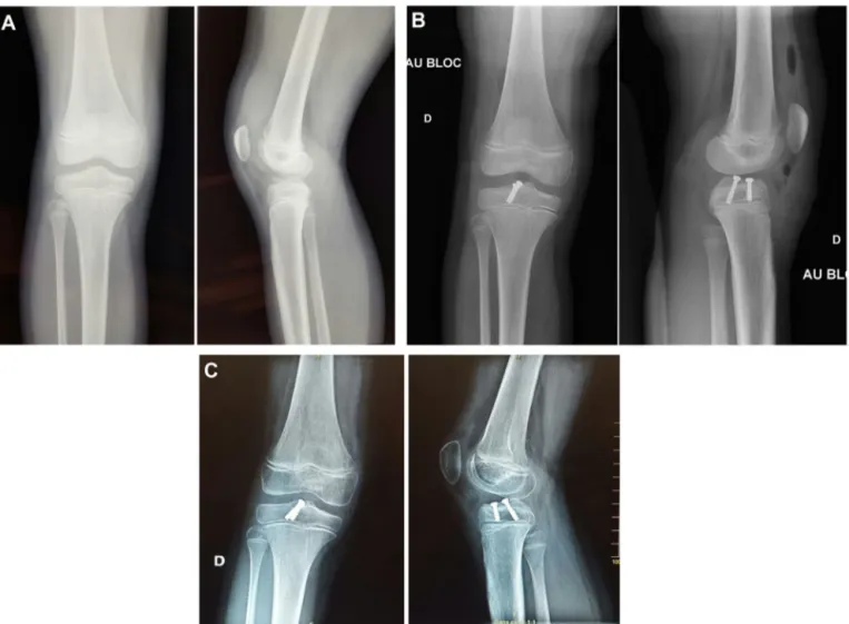
Fig. 1. A. AP and lateral radiographs of a type II tibial intracondylar eminence fracture in an 8-year-old female after a skiing accident. B. Postoperative radiographs: arthroscopic reduction and osteosynthesis using 2 ASNIS 4-mm-diameter screws in the intraepiphyseal. C. Radiographs 6 weeks after the operation.
Table 1
Associated lesions by fracture type.
Type 2 Type 3 Entrapment of anterior horn of medial meniscus 4 3 Entrapment of intermeniscal ligament 1 3
Another lesion 1 0
No lesions 9 3
Twenty-four fractures in 24 patients (17 males, 7 females) were treated with this method and followed up for 2 years (range: 1.5–3 years). The mean age at injury was 11 years (range: 6–15 years), with 13 left sides and 11 right sides. Type II (15 cases) and type III (9 cases) fractures were defined on AP and lateral radio-graphs (Fig. 1A). A CT was done in 4 patients and MRI in one. The injury mechanisms were all related to sports, skiing (7 cases), rugby (7 cases), cycling (6 cases), football (2 cases), and high jump (2 cases).
Arthroscopy was carried out under general anesthetic, in the dorsal decubitus position, legs dangling, with a tourniquet at the root of the lower limb and a vice at the knee. The image intensifier was installed laterally before draping. Using standard anterolateral and anteromedial approaches, we performed hemarthrosis evacua-tion and abundant lavage, and the exploraevacua-tion of associated lesions found in 12 patients (Table 1). The fracture was identified and the surrounding area cleaned, and the recess reamed with an arthro-scopic reamer so as to obtain at least anatomic reduction or even McLennon counter-sinking[7]. Through the parapatellar medial
accessory entry, the fractured fragment was reduced using a metal-lic cannula and a guidewire was inserted through the motorized cannula. Osteosynthesis was provided using one or two 4-mm-diameter ASNIS (Stryker) screws through the medial parapatellar approach in the intraepiphyseal position with arthroscopic and lat-eral radioscopic guidance (no drilling, self-boring and self-tapping ASNIS screws). In most cases, we used 12- to 20-mm-long screws with long threading in cancellous bone of the proximal epiphysis of the tibia, which is relatively flexible. The fixation was deemed satisfactory when the distal end of the screw did not reach the growth plate, whereas its proximal end was located below the joint cartilage. One case of a lesion in the posterior segment of the lat-eral meniscus with 2 cm in the red-white zone was repaired using two vertical FastFix sutures. One case of an osteochondral lesion related to the injury was noted, but it did not require reduction or fixation. At the end of surgery, drainage was put in place and Ropi-vacaine applied within the joint. Immobilization was maintained for 4 weeks with a long-leg cast for patients less than 10 years of age, in the extension position, or a Zimmer knee brace in older children. Then physical therapy was prescribed two or three times a week. Partial weightbearing was authorized at this time, which became total weight-bearing at the 6th postoperative week. Return to sports was authorized only at the 4th month. The screw was removed only if it crossed the growth plate, no earlier than the 3rd postoperative month. The patients were clinically and radio-logically assessed at 6 weeks, 3 months, 6 months, and 1 year after the operation. Knee stability was confirmed by a physical exam-ination including the Lachmann, anterior drawer, and Pivot shift
tests. The functional result was assessed using the Lysholm score [8], pain evaluation, satisfaction with treatment, return to prior level of activity, onset of complications, and the need for surgical revision.
3. Results
The postoperative x-rays (Fig. 1B) showed anatomical correc-tion of all fractures. The 6-week x-ray showed complete healing (Fig. 1C). At 3 postoperative months, all patients walked with-out limping, with dry, stable knees, and with symmetrical range of movement. The radiograph showed complete healing with the material in place. The mean Lysholm score was 99.3 in type II frac-tures and 98.6 in type III fracfrac-tures. Similar results were found at 6 months and 1 year.
Return to daily activities and sports was noted after 6 months in all patients except one, who presented anterior laxity and under-went revision surgery to remove the screw and then for ligament reconstruction 1 year later. At the mean follow-up of 2 years (range: 1–3 years), the range of motion of the operated knee was identical to the healthy side in all patients, except one patient whose flexion was limited to 125◦. The Lysholm score and the satisfaction score varied from very good to excellent in all patients (Table 2). There was no notable residual pain or complications such as secondary fracture dislocation, malunion or union delay, growth stunting, or epiphyseal lesion.
4. Discussion
Treatment of non-displaced fractures (type I) is clearly a simple orthopaedic treatment. Problems can arise in treatment of types II and III fractures, given that anatomical reduction and reconstruc-tion of the joint surface are required to preserve knee funcreconstruc-tion.
Tibial intracondylar eminence fracture surgical management techniques include both open approaches and arthroscopic tech-niques. Fixation of fragments can be performed using Kirchner wires[9], steel wire cerclage[10], intraosseous sutures[11], screws with washers[4], intrafocal fixation[4], and retrograde compres-sion screws anchored in bone[12]. It is important to consider the size and fragmentation of the revulsed eminence: if the size of the avulsion is sufficiently large, screw fixation is recommended because it is the most stable fixation method[13]; if the frag-ment size is small or if the avulsed eminence is fragfrag-mented, the fixation can be done using intraosseous sutures or thin steel wire looped through the cruciate ligament and then threaded through holes drilled in the proximal end of the tibia[11,14]. In a study on 17 patients with type II or III tibial intracondylar eminence frac-tures, arthroscopically treated with either screws or suture, Hunter et al.[15]found no significant difference between these two meth-ods in terms of results. Comparing their result to ours, we realize that these two series are not comparable, with a much wider age range in their series (mean: 26.6 years; range: 7.5–60.1 years). In a retrospective study, Senekovic et al.[16]demonstrated good ther-apeutic results for tibial intracondylar eminence fractures with arthroscopic treatment, using cannulated screws with washers, which allowed immediate weight-bearing. However, countersunk screws could cause problems if revision surgery is required as well as in ACL reconstruction. However, it is not impossible to insert an interference screw next to the original screw.
Entrapment of the anterior horn of the medial meniscus or the intermeniscal ligament under the fragment of the tibial intra-condylar eminence was reported by Falstie-Jensen et al.[17]and others[18,19]. The entrapped structures prevent reduction of 65% of type III fractures and 26% of type II fractures. Kocher et al. [20]reported 54% entrapment of the anterior horn of the medial
meniscus, the intermeniscal ligament, or of the anterior horn of the lateral meniscus. However, interposition of soft tissues had no significant effect on the long-term results because all the avul-sions were anatomically reduced after removal (with retraction or resection) of the interposed soft tissues. Twelve of our 24 patients (50%) showed soft tissue interposition between the avulsed frag-ment and the tibia. We solved this interposition in the 12 cases and obtained anatomical reduction, with no difference in the results when we compared these patients with those with no soft tissue interposition. In the literature, we found 14% associated ligament lesions[21], whereas the prevalence of associated meniscal lesions is rarer (6–8%) and therefore does not seem to be associated with anterior meniscal or intermeniscal ligament entrapment[20]. Despite anatomic reduction, anterior instability, stiffness (notably loss of knee extension), and pain with knee extension are common complications [22]. Stiffness (with or without extension deficit) is the most frequent complication. This may be related to post-operative cast or brace wear over a long period of time. In the present study, none of the patients had developed stiffness at the last follow-up. As a consequence, the early exercises that we favored in our study because of the rigid internal fixation are rec-ommended to preserve knee function and reduce the incidence of stiffness.
In children, the appearance of more than a 50% increase in the initial length of the ACL fiber before tibial intracondylar eminence fracture is clear proof of laxity[22]. Kocher et al.[23]reported the persistence of anterior laxity in 6 patients, with a positive Lachman score in 5 cases, a click on the pivot shift test in 2 cases, and an increase in KT-1000 laxity in four patients, despite excellent func-tional results in these 6 patients. These results are consistent with those reported by Baxter and Wiley[24,25]for 17 patients, with excellent functional results without instability, despite a positive Lachman exam in 51% of the patients and a 3.5-mm increase in the mean differential laxity. This objective laxity could be related to the interstitial lesion and lengthening of the ACL at the time of tibial intercondylar spine fracture[23]. We observed that nearly all of our patients returned to the same level of sports, without an impres-sion of the knee giving way, with no instability episodes and with no delayed meniscus lesions. A single patient underwent a new inter-vention for instability with ACL reconstruction. The residual laxity reported in the literature results from several factors, including insufficient reduction, because anatomic reduction with no inter-nal fixation can result in secondary displacements and pathological laxity[7].
One of the potential limitations of the study is the relatively low number of cases studied. However, it is well known that tibial intra-condylar eminence fractures are rare, but the series that we present herein is comparable with other studies on the same type of fracture [13,15,26]. We propose a therapeutic alternative that can stabilize types II and III tibial intracondylar eminence fractures. On the other hand, the arthroscopic screw fixation technique is not adapted to treatment of type IV comminute fractures. Another of the study’s limitations is that only standard x-rays and in a few cases CT com-prised the essential preoperative imaging protocol. However, other authors have reported results of tibial spine fracture in children (or adults) using classic standard x-rays and arthroscopy only as diagnostic tools[7,27].
ASNIS screw fixation for tibial intracondylar eminence fracture with arthroscopy undertaken in cases with good indications, per-formed with precision and caution, is an adapted surgical method for the treatment of types II and III tibial intracondylar eminence fractures in children. The method provides sufficient stability and early weight-bearing, with no need to immobilize the knee for a long period of time. Use of 4-mm-diameter ASNIS screws in the epiphysis spares the knee from mechanical joint problems and biological disturbance at the growth plate.
Table 2
Results of the 24 patients who completed the follow-up questionnaire after internal arthroscopic fixation.
Patient Age (years) Gender Side Investigations Type Associate lesions Follow-up (years)
Movement Complication Lysholm score Satisfaction Return to previous level of activity (months)
1 12
Male
Left Rx + CT 3 Entrapment AHMM 2 Normal None 100 10 6
2 10 Female Right Rx 3 None 2 Normal None 100 10 6
3 9 Male Right Rx 3 None 1 Normal None 100 10 6
4 7 Female Right Rx 2 None 1 Normal None 100 10 6
5 14 Male Left Rx 2 Entrapment AHMM 1 Normal None 100 10 6
6 9 Male Left Rx 3 None 2 Normal None 100 10 6
7 15 Female Left Rx 2 None 2 Normal None 100 10 6
8 7 Female Left Rx 3 Entrapment AHMM 2 Flexion: 125 Flexion: 125◦ 95 8.5 6
9 13 Male Right Rx + CT 2 None 3 Flexion: 130 ACL laxity
(ligament reconstruc-tion); patellofemoral pain 95 8.5 24
10 14 Male Right Rx 2 Entrapment AHMM 2 Normal None 100 10 6
11 12 Male Left Rx 3 Entrapment AHMM 2 Normal None 95 10 6
12 9 Male Left Rx 2 None 3 Normal None 100 10 6
13 13 Male Left Rx + CT 2 Entrapment IML 2 Normal None 100 10 6
14 12 Male Left Rx + MRI 2 A vertical lesion PSLM 2 Normal None 100 10 6
15 14 Male Left Rx 3 Entrapment IML 2 Normal None 98 9 6
16 13 Male Right Rx 2 None 2 Normal None 100 10 5
17 14 Male Right Rx 3 Entrapment IML 2 Normal None 100 10 6
18 8 Male Left Rx 2 None 2 Normal None 100 10 3
19 14 Male Left Rx + CT 2 None 2 Normal None 100 10 6
20 11 Female Right Rx 2 None 2 Normal None 95 10 6
21 9 Male Left Rx 2 Entrapment AHMM 2 Normal None 100 10 5
22 10 Female Right Rx 2 Entrapment AHMM 2 Normal None 100 10 4
23 8 Female Right Rx 2 None 2 Normal None 100 10 6
24 13 Male Right Rx 3 Entrapment IML 2 Normal None 100 10 6
Disclosure of interest
The authors declare that they have no competing interest.
References
[1]Kocher MS, Mandiga R, Klingele K, Bley L, Micheli LJ. Anterior cruciate liga-ment injury versus tibial spine fracture in the skeletally immature knee: a comparison of skeletal maturation and notch width index. J Pediatr Orthop 2004;24:185–8.
[2]Meyers MH, Mc KF. Fracture of the intercondylar eminence of the tibia. J Bone Joint Surg Am 1959;41A:209–20.
[3]Gronkvist H, Hirsch G, Johansson L. Fracture of the anterior tibial spine in children. J Pediatr Orthop 1984;4:465–8.
[4]Reynders P, Reynders K, Broos P. Pediatric and adolescent tibial eminence frac-tures: arthroscopic cannulated screw fixation. J Trauma 2002;53:49–54.
[5]Kendall NS, Hsu SY, Chan KM. Fracture of the tibial spine in adults and children. A review of 31 cases. J Bone Joint Surg Br 1992;74:848–52.
[6]Mylle J, Reynders P, Broos P. Transepiphysial fixation of anterior cruciate avul-sion in a child. Report of a complication and review of the literature. Arch Orthop Trauma Surg 1993;112:101–3.
[7]McLennon JG. Lessons learned after second-look arthroscopy in type III frac-tures of the tibial spine. J Pediatr Orthop 1995;15:59–62.
[8]Lysholm J, Gillquist J. Evaluation of knee ligament surgery results with special emphasis on use of a scoring scale. Am J Sports Med 1982;10:150–4.
[9]Zaricznyj B. Avulsion fracture of the tibial eminence: treatment by open reduc-tion and pinning. J Bone Joint Surg Am 1977;59:1111–4.
[10]Gaspar L, Farkas C, Csernatony Z. Acute arthroscopy. Acta Chir Hung 1997;36:100–3.
[11]Oohashi Y. A simple technique for arthroscopic suture fixation of displaced fracture of the intercondylar eminence of the tibia using folded surgical steels. Arthroscopy 2001;17:1007–11.
[12]Mosier SM, Stanitski CL. Acute tibial tubercle avulsion fractures. J Pediatr Orthop 2004;24:181–4.
[13]Delcogliano A, Chiossi S, Caporaso A, Menghi A, Rinonapoli G. Tibial intercondy-lar eminence fractures in adults: arthroscopic treatment. Knee Surg Sports Traumatol Arthrosc 2003;11:255–9.
[14]Kogan MG, Marks P, Amendola A. Technique for arthroscopic suture fixation of displaced tibial intercondylar eminence fractures. Arthroscopy 1997;13:301–6.
[15]Hunter RE, Willis JA. Arthroscopic fixation of avulsion fractures of the tibial eminence: technique and outcome. Arthroscopy 2004;20:113–21.
[16]Senekovic V, Veselko M. Anterograde arthroscopic fixation of avulsion fractures of the tibial eminence with a cannulated screw: five-year results. Arthroscopy 2003;19:54–61.
[17]Falstie-Jensen S, Sondergard Petersen PE:. Incarceration of the meniscus in fractures of the intercondylar eminence of the tibia in children. Injury 1984;15:236–8.
[18]Burstein DB, Viola A, Fulkerson JP. Entrapment of the medial meniscus in a fracture of the tibial eminence. Arthroscopy 1988;4:47–50.
[19]Chandler JT, Miller TK. Tibial eminence fracture with meniscal entrapment. Arthroscopy 1995;11:499–502.
[20]Kocher MS, Micheli LJ, Gerbino P, Hresko MT. Tibial eminence fractures in children: prevalence of meniscal entrapment. Am J Sports Med 2003;31:404–7.
[21]Iborra JP, Mazeau P, Louahem D, Diméglio A. Fractures of the intercondylar eminence of the tibia in children. A propos of 25 cases with a 1–20 year follow-up. Rev Chir Orthop 1999;85:563–73 [Article in French].
[22]Park HJ, Urabe K, Naruse K, Aikawa J, Fujita M, Itoman M. Arthroscopic eval-uation after surgical repair of intercondylar eminence fractures. Arch Orthop Trauma Surg 2007;127:753–7.
[23]Kocher MS, Foreman ES, Micheli LJ. Laxity and functional outcome after arthro-scopic reduction and internal fixation of displaced tibial spine fractures in children. Arthroscopy 2003;19:1085–90.
[24]Wiley JJ, Baxter MP. Tibial spine fractures in children. Clin Orthop Relat Res 1990;255:54–60.
[25]Baxter MP, Wiley JJ. Fractures of the tibial spine in children. An evaluation of knee stability. J Bone Joint Surg Br 1988;70:228–30.
[26]Mulhall KJ, Dowdall J, Grannell M, McCabe JP. Tibial spine fractures: an analysis of outcome in surgically treated type III injuries. Injury 1999;30:289–92.
[27]SchmitgenGF, Utukuri MM. Arthroscopic treatment of tibial spine fractures in children: a review of three cases. Knee 2000;7:115–9.