EVALUATION OF INHIBITORY MEASURES FOR FOOD SPOILER YEAST CANDIDA KRUSEI DURING
FERMENTATION PROCESS BY CHEMICAL, BIOCHEMICAL AND NANOPARTICLE APPROACHES
Indrani Bhattacharya,
1Jyothi Bezawada,
1Jay Shankar Singh Yadav,
1Song Yan,
1R. D. Tyagi,
*1and R.Y. Surampalli
2Address(es): R.D.Tyagi,
1 Institut national de la recherche scientifique, 490, Rue de la Couronne, Québec, Canada G1K 9A9. 2 Department of Civil Engineering, University of Nebraska-Lincoln, PO Box 886105 Lincoln, USA. *Corresponding author: [email protected]
ABSTRACT
Keywords: H2O2, Spoiler yeast, Inhibition, Killer protein, Ag-KT4561 bio-molecule nanoparticles, Green chemistry
INTRODUCTION
Cheese whey (a byproduct of cheese processing industries) has been efficiently exploited for the production of single cell protein (SCP) over the years as cheese whey has an immense nutritional value (Ayoola et al., 2008; Carvalho et al., 2013). It contains 4.5-5% (w/v) of lactose, 0.6-0.8% (w/v) of soluble proteins, 0.4-0.5% (w/v) of lipids and 8-10% (w/v) of mineral salts of the dried extract. Efficient utilization of cheese whey for SCP conversion reduces the biochemical oxygen demand (BOD) by 75% and thus decreases the disposal problem (Eyster, 1950; Prazeres et al., 2012). Mostly lactose-consuming organisms, such as
Kluyveroymyces spp. and Lactobacillus spp. grow in cheese whey (Koleva et al.,
2008; Orru et al., 2010). Production of SCP from cheese whey serves dual purpose by reducing environmental pollution and generating a valuable product (i.e. proteinaceous biomass which is used as animal feed and food ingredients) (Koleva et al., 2008). Likewise, Kluyveroymyces marxianus has been grown in cheese whey as a mono-culture for SCP production (Yadav et al., 2012). However, on a large scale industrial process, contamination is a major problem for SCP production despite the treatment of large volume of cheese whey. To eliminate this problem, certain extreme fermentation parameters have been applied, such as low pH (3.0-4.0) and high temperature (40-45 °C). At these conditions, most of the pathogenic microorganisms cannot survive (Maneesri and Maneesri, 2009; Ahariz et al., 2010) and therefore a safe food or feed product is ensured during fermentation process. Additionally, the extreme fermentation conditions help to make the process economical due to reduced operating cost of maintaining sterility.
However, certain food spoilers (contaminant) e.g. Candida krusei still survive under extreme conditions (pH 3.0 and 45 °C) (Guo and Bhattacharjee, 2006). The opportunistic Candida species exist as commensal in healthy individuals (Heard and Fleet, 1988). During the production of SCP, C. krusei emerges as a contaminant while it grows along with K. marxianus and this is a concern for food safety. C. krusei is known as a food contaminant and an opportunistic pathogen (Siso, 1996; Hornbæk et al., 2006; Maneesri and Maneesri, 2007; Kim and Lee, 2012). However, C. krusei is reported to be present in many dairy and fermented food products, but yet does not come under generally recognized as safe (GRAS) microorganism (Walker and Dijck, 2006; Walker et al., 2008).
Therefore, the eradication of C. krusei is essential from food products to meet safety regulations.
Certain chemical and biochemical approaches were reported to employ for selective inhibition of C. krusei. The chemical (NaCl, H2O2) and the biological inhibitors (medicinal plants, such as Lupinus angustifolis, Syzigium aromaticum (clove) oil; nisin and Williopsis saturnus and synergistic effect of W. saturnus and H2O2) to inhibit C. krusei have been reported (Ayoola et al., 2008; Dingman, 2008; Adeniyi et al., 2010; Da Silva et al., 2011). It was reported that H2O2 inhibited C. krusei (Morgulis et al., 1926). Apparently, C. krusei has also been tested against a wide range of essential oils where ethanol 70% v/v served as control (Nel et al., 2006; Souza et al., 2008; Waema et al., 2009). Another significant approach to inhibit C. krusei was using NaCl; however it depends on the sensitivity of the organism and the concentration of NaCl used. The sensitive strain of C. krusei undergoes cell death at 2 M concentration of NaCl (Aguiar and Lucas, 2000).
Yeasts such as Aspergillus furnigatus, W. saturnus (major yeast from yogurt) have the capacity to produce killer proteins (Fang et al., 2002; Brock, 2008). The mycotoxins/killer proteins produced by W. saturnus have a broad spectrum of inhibitory activity at wide range of pH and temperature (Buzzini et al., 2004). These could be used as the versatile anti-spoilage agents for food and feed production (Kao et al., 1999; Liu et al., 2006). Another killer protein is nisin, which is used for food preservation and is produced by Lactobacillus spp. or lactic acid bacteria (LAB) (Guwy et al., 1999). Nisin is used to stabilize food products and is often added to the cheese for inhibiting toxin production by
Clostridium botulinum. It was also reported to inhibit C. krusei efficiently
(Lowes et al., 2000; Russell and Jarvis, 2001).
In certain industrial fermentation processes, stress of pH shock was encountered to inhibit certain food spoiler yeasts (Siso, 1996; Pinheiro et al., 2002). At pH 2.0, C. krusei did not grow well (Lowes et al., 2000). In a mixed culture, where the presence of other yeast strains was also reported, effective utilization of any inhibitor (i.e. chemical or biochemical inhibitor against C. krusei) depends entirely upon whether the other type of yeast was also inhibited by the specific inhibitor being used. Another effective way of inhibition of pathogens is by the usage of metal nanoparticles (NPs) or biomolecule based nanoparticles (Dingman, 2008). It has already been observed that silver NPs can kill pathogens Screening of chemical, biochemical and biomolecule-nanoparticle methods for the inhibition of Candida krusei were evaluated without hampering the growth of dairy yeast Kluyveromyces marxianus. The effective inhibition was observed with the help of H2O2, Williopsis
saturnus, at specific combination of pH and temperature (pH 5.0 and 40 °C) and Ag-KT4561 nanoparticles among the various methods
used. However, the most efficient inhibition was observed with Ag-KT4561 nanoparticles. In general H2O2 works best at pH range 4.0 to 10.0 and at temperature 30 °C or above. H2O2 concentration of 4000 ppm at 45 °C and pH 5.5 exhibited significant inhibition of C.
krusei, while K. marxianus remains unaffected. But, when used with lyophilized supernatant of W. saturnus, 2400 ppm H2O2 was effective. Further, nanoparticle with silver was synthesized to reduce the quantity of killer protein and enhance the efficiency of protein. Complete inhibition of C. krusei was observed at 350 µM of synthesized silver nano-particle (AgNPs) of the killer protein from W.
saturnus, with little effect on K. marxianus concentration. A stability test confirms the effect of protein silver nanoparticles on C. krusei
for more than 20 weeks without any change in pH and temperature. Thus, the nanoparticles could be potentially used for inhibition of C.
krusei without affecting the growth of K. marxianus and the process could be run non-aseptically.
ARTICLE INFO Received 17. 2. 2015 Revised 7. 1. 2016 Accepted 13. 1. 2016 Published 1. 6. 2016 Regular article doi: 10.15414/jmbfs.2016.5.6.509-517
at very low concentrations and biomolecule based nanoparticles do not exert any toxic effects on human cells. Apart from that, silver NPs do not cause any microbial resistance and also there is no specific site of action for inhibition of the microbial cells (Panacek et al., 2009). Hence, the aim of the present study was to evaluate different inhibition methods to inhibit C. krusei alone as well as in a mixed culture system without affecting the growth of K. marxianus. MATERIALS AND METHODS
Chemicals
Analytical grade chemicals were used in the experiments. NaCl (Quelab Lab Inc., Montréal, Canada), H2O2 (Laboratoire Mat, Québec), yeast Extract (Fisher Scientific, USA), malt extract (Oxoid Ltd., Basingstoke, England), meat peptone (Organotechnie SA., La Courneuve, France), glucose, ethyl alcohol 95% (Fisher Scientific, USA), agar (Quebact Lab Inc., Montréal, Canada), cheese whey (Agropur, Canada), and AgNO3 (Fisher Scientific, Ottawa).
Microorganisms
K. marxianus strain used in the study was isolated and characterized from the
SCP production plant using cheese whey as substrate. C. krusei strain was also isolated and identified as a contaminant during SCP production employing cheese whey. W. saturnus strain DBVPG 4561 was obtained from the Industrial Yeasts Collection DBVPG of Perugia (Italy). Strains were sub-cultured on YEPD (yeast extract 10 g/L, peptone 10 g/L, dextrose 20 g/L) agar slants and stored at 4 oC for further use.
Inhibition studies for C. krusei Chemical Methods
Inhibition by NaCl
Pre-culture broth of K. marxianus of 2.0x10⁸ CFU/mL and C. krusei of 4.0x10⁷ CFU/mL were prepared in 100 mL YEPD media in 500 mL Erlenmeyer flasks. The medium pH was adjusted to 3.5 and sterilized at 121 oC for 15 min. After sterilization, 1.5 M and 2 M NaCl were added in different sets of flasks. C. krusei is a non-lactose assimilating organism, while K. marxianus is a lactose assimilating organism. Therefore, sterilized YEPD media were inoculated with 30 μL (from stock culture) of K. marxianus and 50 μL of C. krusei. Inoculated flasks were incubated at 40 °C for 24 h. Samples were taken at regular intervals for the analysis of total cell count. Total cell concentration was measured using standard agar plate technique (Logothetis et al., 2007; Goretti et al., 2009;Kosseva et al., 2009).
Inhibition by H2O2
A pre-culture was prepared for C. krusei and K. marxianus as above. After that, culture media of cheese whey powder 4.5% (w/v) and urea 0.22% (w/v) were prepared and pasteurized at 80 °C for 20 min. The pasteurized cheese whey culture media at different pH (3.5, 4.5, 5.5, and 6.0) was added to different 500 mL flasks and inoculated with 1% (v/v) inoculum of C. krusei and K. marxianus. Different concentrations of H2O2 (100, 200, 300 and 400 ppm) were added to these flasks. After inoculation, flasks were incubated at 28 °C and 40 °C in an orbital incubator shaker for 24 h. Samples were drawn at regular intervals to analyze the total cell count.
Simultaneously, two different set of experiments were conducted, where in the primary set of experiments the H2O2 concentration were varied (0, 300, 400, 500, 600 and 800 ppm) and applied directly on the fermenter broth containing K.
marxianus and C. krusei, which was collected from commercial continuous SCP
production plant. 100 mL of fermented broth of K. marxinaus (3.0x10⁶ CFU/mL) severely contaminated with C. krusei (1.8x10⁶ CFU/mL) was taken in 500 mL 2 sterilized flasks. Flasks were incubated at pH 3.5, 150 rpm and 40 °C in an incubator shaker.
In the secondary set of experiments, variation in H2O2 concentration (2400, 3200 and 4000 ppm) were considered and applied directly to the fermenter broth and flasks were incubated at pH 5.0, 150 rpm and 45 °C in an incubator shaker. Biochemical Methods
Inhibition by S. aromaticum oil
A set of experiments were conducted in which 0.4% (v/v) of clove oil was added in fermenter broth which contains C. krusei and K. marxianus. The initial cell count of C. krusei and K. marxianus was 5.0x10⁶ CFU/mL and 6.0x10⁶ CFU/mL, respectively. The flasks were placed in an orbital incubator shaker at 28 °C at 150 rpm for 6 h. Sampling was performed at an interval of 2 h. Samples were analyzed for total cell count using standard agar plate technique.
Inhibition with nisin
The culture of C. krusei and K. marxianus were grown separately in MRS broth at 35 oC for 24 h. Bioassay MRS media with 0.75% of Bacto agar and 1% Tween-20 were prepared. Media were sterilized at 121 oC for 15 min. A solution of nisin (1,000 IU/mL) was prepared by adding 0.025 g of commercial nisin (Sigma-Aldrich, Milwaukee, USA) into 25 ml of sterile solution of 0.02 N HCl. Sterilized media were cooled down to 40 oC and inoculated with 1% (v/v) of the 24 h culture of C. krusei and K. marxianus in two sets (duplicate). Then the bioassay agar (25 mL) was aseptically poured into sterile petri dishes (100x15 mm) and allowed to solidify for 1 h. On each plate, four or five holes were bored, using a 7 mm outer diameter stainless steel borer with a slight suction. An aliquot (50 µL and 100 µL) of standard nisin solution was placed into a well and the bioassay agar plate was incubated right away at 35 oC for 24 h. The control for each plate was prepared using sterile distilled water in wells. Zone of inhibition was observed in control and test samples.
Inhibition study with W. saturnus Preparation of W. saturnus culture broth
YEPD (100 mL) was prepared in 500 mL flasks and sterilized at 121 oC for 15 min. The sterilized flask was inoculated with loopful of W. saturnus and incubated in an orbital incubator shaker at 150 rpm and 28 oC for 48 h. Samples were taken at regular time intervals for total cell count.
Well assay method
Pre-culture of C. krusei was prepared in YEPD as described above using 1% (v/v) inoculum. After 24 h, C. krusei sample was diluted 102, 103 and 104 times in saline solution and different diluted samples were spread plated in YEPD agar plates. After spread plating, wells were made in agar plates using borer and 60
µL of W. saturnus (48 h) culture was added in each well. The plates were
incubated in an orbital incubator at 28 oC for 24 h. The plates were visually observed after 24 h.
To differentiate the morphology of C. krusei from W. saturnus, Methylene Blue Citric-Phosphate agar (MBA) plates were prepared and spread plated using C.
krusei and W. saturnus. Plates were incubated for 24 h at 28 oC and were visually examined to check the morphology.
Inhibition by W. saturnus
W. saturnus was grown in YEPD and cheese whey medium for 24 h. YEPD and
cheese whey powder 4.5% (w/v) with 0.22% (w/v) urea were prepared in two flasks of 2 L capacity each containing 500 mL medium. After sterilization, each flask was inoculated with 2% (v/v) W. saturnus and incubated in an orbital incubator shaker at 150 rpm and 28 °C. The culture was harvested at 48 h. The culture broth was centrifuged at 10 000 x g and the supernatant was lyophilized to obtain the powder which contained extracellular proteins. The extracellular proteins specifically contain a particular protein KT4561 ( ̴ 62 kDa protein), which has anti-mycotic activity (Buzzini et al., 2004). Simultaneously, another set of flasks containing W. saturnus were grown, where no centrifugation was performed. Henceforth, the cultures were directly taken for lyophilization. Pre-cultures were prepared by growing C. krusei, K. marxianus and W. saturnus in YEPD medium for 24 h. One hundred milliliters of fresh cheese whey powder 4.5% (w/v) with urea 0.22% (w/v) solution was added to each five hundred milliliters Erlenmeyer flask (two flasks) and pasteurized at 80 °C for 20 min. After pasteurization, media were aseptically adjusted to different pH (3.5 and 4.5) followed by inoculation with 1% (v/v) mixed culture (C. krusei and K.
marxianus).
Inhibition by lyophilized supernatant from W. saturnus
Various concentrations of lyophilized supernatant of W. saturnus were considered (well plate assay method) and the zone was created by the inhibitory effect of the killer protein. The inhibition zones were measured after 24 h of incubation at 30 ºC. A linear equation (y = 0.30x-0.36) was sketched out between the diameter of the clear zone (measured in millimeters, x axis) and the logarithm of the quantity of the killer protein (measured in nanograms, y axis). This method was used to determine the killer protein concentration required for the inhibition of C. krusei which is similar to the technique mentioned in (Chen et al., 2000). Lyophilized supernatant prepared in YEPD media was served as the control and lyophilized cheese whey was the experimental product.
Inhibition of C. krusei by synergistic effect of H2O2 and W. saturnus
To study inhibition of C. krusei, different H2O2 concentrations were used along with W. saturnus (entire organism lyophilized supernatant powder, as described
above). 300 ppm of H2O2 was used along with 1% (v/v) of W. saturnus (inoculum from pre-culture) for the inhibition of C. krusei in a mixed culture of
C. krusei and K. marxianus at pH 6.0 and 28 °C. Similar sets of experiments were
conducted with a variation in pH (3.5-4.5) at 28 °C.
Two different set of experiments were conducted, where in the primary set of experiments was conducted where lyophilized W. saturnus was used by varying the H2O2 concentration directly on the fermenter broth containing K. marxianus and C. krusei, collected from commercial continuous SCP production plant. 100 mL of fermented broth of K. marxinaus (3.1x10⁶ CFU/mL) grossly contaminated with C. krusei (1.5x10⁶ CFU/mL) was taken in 500 mL 2 sterilized flasks. The lyophilized powder of W. saturnus (200 mg/mL) along with different concentrations of H2O2 (2400 and 4000 ppm) was then added to each flask. Flasks were incubated at pH 5.0, 150 rpm and 40 °C in an incubator shaker. Whereas in secondary set of experiments, about 400 mg/mL of lyophilized supernatant of W. saturnus was used along with 2400 ppm of H2O2 in a mixed culture by adjusting the pH of fermenter broth to 5.0. As the killer protein produced by lyophilized W. saturnus is highly effective at pH range of 4.5-10.0 and temperature from 25 to 45 oC (Goretti et al., 2009). Flasks were kept at 150 rpm and 40 °C in an orbital incubator shaker.
Inhibition of C. krusei by Ag-KT4561 NPs
Synthesis of nanoparticles (Ag-KT4561) was carried out in the previous study (Bhattacharya et al., 2015). However a bulk preparation of the same has been conducted in this study. During the scale-up process, 20 mL of 0.1 M AgNO3 solution is continuously stirred along with 18 mL of W. saturnus supernatant at 25 °C for 48 h. Ag+ ions were completely reduced at 48 h of stirring. After which the bulk nanoparticle solution was taken for lyophilization and the lyophilized product was tried against C. krusei in 4.5% (w/v) cheese whey and 0.22% (w/v) of urea. From the lyophilized product different concentration of Ag-KT4561 ranging from 10 µM - 1 mM were tried at pH 5.5 and 30 °C in shake flasks. In these experiments, mixed culture of C. krusei (2% (v/v)) and K. marxianus (2% (v/v)) were tested for 12 h and total cell concentration (CFU/mL) was measured at 3 h time intervals.
Analytical methods Cell count
Total cell count as CFU (colony forming units) was estimated by standard agar plate technique in YEPD agar plates (Nathan et al., 1978). The appropriately diluted samples were plated on agar plates and incubated at 30 C overnight to form fully developed colonies. The colonies of K. marxianus, C. krusei and W.
saturnus were identified based on its morphology by visible examination.
Protein estimation
The soluble protein concentration was determined by Lowry’s method (Lowry et al., 1951) using bovine serum albumin as standard.
UV-Vis Spectroscopy
The bulk sample of AgNO3 and W. saturnus supernatant were prepared at 48 h and samples were collected at every 6 h to analyze for nanoparticle formation at 300-700 nm in Spectrophotometer (Carry 100 Bio®, Varian USA).
Statistical method
For each set, samples were analyzed in triplicates and mean values are taken in account. Further standard deviation of the colonies in log units (Log10 CFU/mL) were calculated for each experimental point in Microsoft excel 2013 external package similar to the method of De Oliveira et al., 2014.
RESULTS Inhibition by NaCl
Different concentrations (1.5- 2 M) of NaCl were tested to inhibit the growth of
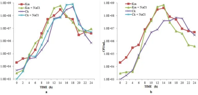
C. krusei in mono-culture and the results were presented in Figure 1. It was clear
that NaCl concentration of 2 M showed significant inhibition of C. krusei compared to 1.5 M without having any effect on K. maxianus. This is due to the fact that C. krusei exhibited salt-stress (Aguiar and Lucas, 2000) and got killed at pH 3.5 and 40 °C. No inhibition of C. krusei was observed when NaCl concentration was less than 2 M at pH 3.5 and 40 °C.
Figure 1 Impact of a)1.5 M NaCl and b) 2.0 M on C. krusei (Ck) and K. marxinaus (Km) in YEPD medium at pH 3.5 and 40 °C (Shake flask experiments)
Inhibition by H2O2
Viability of individual cultures of C. krusei, and K. marxianus at different concentrations of H2O2 (100 - 400 ppm) in cheese whey at pH 6.0 and 28 °C was studied (Table 1). It showed that C. krusei was not inhibited at lower concentration of H2O2. However, inhibition occurred at 300 ppm H2O2. K.
Table 1 Impact of various concentrations of H2O2 on C. krusei and K. marxianus in cheese whey powder at pH 6.0 and temperature 28 °C (Shake flask)
Individual Organisms (Log 10 CFU/mL) ± Standard Deviation H2O2 (ppm) 0 100 200 300 400 Time (h) Ck Km Ck Ck Ck Ck Km 0 5.73±.07 4.83±.01 5.81±.01 5.81±.01 5.92±.01 5.15±.03 5.81±.02 12 6.52±.02 9.33±.01 6.08±.03 5.23±.01 5.40±.04 NG 9.26±.02 24 8.51±.02 8.31±.01 6.12±.05 5.18±.05 NG NG 8.28±.02 Legend: NG- No Growth Observed, Ck – C. krusei, Km – K. marxianus
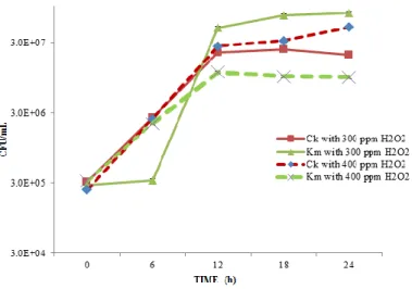
The inhibition of C. krusei in mixed cultures (C. krusei and K. marxianus) in cheese whey was studied and the results were presented in Figure 2. The concentration of H2O2 used was 300 ppm and 400 ppm (from previous results in Table 1). C. krusei was not inhibited at 300 - 400 ppm of H2O2. On the contrary,
C. krusei dominated over K. marxianus in a mixed culture at 24 h.
Figure 2 Impact of H2O2 on C. krusei (Ck), K. marxianus (Km) in cheese whey at pH 6.0 and 28 °C with 300 ppm and 400 ppm H2O2
Variations in pH along with 300 ppm H2O2 concentration
pH variations were carried out and it was lowered to 3.5 from 6.0 and studies of mixed culture (C. krusei, K. marxianus and W. saturnus 1% and 2% (v/v)) were also carried out maintaining similar parameters in cheese whey. C. krusei was not inhibited at these parameters, whereas growth of K. marxianus and W. saturnus remains unchanged.
A mixed culture study at pH 4.5 and 28 °C in cheese whey powder with 300 ppm H2O2 exhibited a partial inhibition of C. krusei (Figure 3a). However when bio-preservative W. saturnus was added at the similar condition, the growth of C.
krusei decreased by one log-unit at 12 h (Figure 3b), but the growth accelerated
after 12 h. As W. saturnus produces killer protein (KT4561) but the concentration of the killer protein remains low which is insufficient to inhibit C. krusei in a large-scale fermentation. Hence, this parameter could be considered for the inhibition of C. krusei.
Figure 3 Impact of H2O2 and W. saturnus (Ws) on the mixed culture (C. krusei (Ck) and K. marxianus (Km)) in cheese whey at pH (3.5, 4.5) and 28 °C (Shake flask). a) With 300 ppm H2O2 only b) With 300 ppm H2O2 and 1 % (v/v) W. saturnus
C. krusei was efficiently inhibited in mixed cultures (C. krusei and K. marxianus)
and (C. krusei, K. marxianus and W. saturnus) at pH 4.0, 28 °C and 400 ppm of H2O2. Lower CFU/mL of 1.7x10³ was observed for K. marxianus at pH 4.0 and 28 °C. On the contrary, when C. krusei was grown along with K. marxianus and
W. saturnus, K. marxianus was observed at high CFU/mL of 2.4 x 10³ (as
compared to C. krusei when grown along with K. marxianus). K. marxianus showed remarkable growth at pH 5.5 and 40 °C rather than at other pH values, hence pH 4.5-5.5 was ideal for K. marxianus.
Higher ranges of H2O2 concentrations
After deducing the optimum amount of H2O2 used for the complete inhibition of
C. krusei, similar concentration was applied for industrial scale fermenter broth
to eliminate C. krusei without affecting the K. marxianus. So, 300 ppm of H2O2 was the optimum concentration for inhibiting C. krusei in the mixed culture in shake flask experiments. When concentration of H2O2 was increased from 300 to 800 ppm in the lab scale fermenter broth, no significant inhibition of C. krusei
was observed at pH 3.5 and 40 °C (Table 2). K. marxianus degraded H2O2 at pH 3.5, making H2O2 ineffective for C. krusei inhibition (Pinheiro et al., 2002). In fermented broth, higher concentration (2400 ppm, 3200 ppm, and 4000 ppm) of H2O2 was considered at pH 5.0 and 45 °C. Study was conducted for 6 h, as H2O2 got degraded into H2O and O2 after 6 h (Table 3). A very high concentration 4000 ppm of H2O2 finally could kill C. krusei completely in the fermented broth. Higher concentration of H2O2 was required due to simultaneous degradation of H2O2 by catalase action of K. marxianus (Pinheiro et al., 2002).
Table 2 Impact of varying concentration of H2O2 on the mixed culture in the fermenter broth at pH 3.5 and 40 °C (Shake flask) H2O2 (ppm)
0 300 400 500 600 800
Individual Organisms (Log 10 CFU/mL) ± Standard Deviation Time (h) Ck Km Ck Km Ck Km Ck Km Ck Km Ck Km 0 7.88±.01 9.04±.02 8.26±.02 8.97±.02 8.42±.02 8.71±.01 7.17±.03 7.67±.01 6.02±.02 6.14±.05 6.17±.11 5.87±.02 3 7.91±.01 8.89±.01 8.34±.02 8.72±.01 8.12±.02 8.76±.01 6.71±.01 6.15±.05 6.47±.10 6.87±.01 5.34±.03 6.49±.02 6 8.18±.04 7.05±.01 8.18±.03 8.69±.01 7.96±.01 7.96±.01 6.85±.01 6.11±.03 6.04±.04 6.95±.04 5.36±.04 6.87±.02 9 7.78±.01 7.79±.01 8.28±.02 8.80±.01 8.18±.03 8.18±.03 6.77±.01 7.28±.03 6.85±.04 7.32±.07 6.04±.02 6.70±.08 12 7.18±.04 8.12±.06 8.80±.02 8.18±.02 8.32±.02 8.32±.02 6.72±.01 6.90±.01 8.25±.13 8.00±.02 6.47±.03 7.47±.02 24 8.40±.02 8.08±.09 8.45±.02 8.45±.03 8.42±.02 8.42±.02 8.04±.13 8.04±.02 8.45±.06 7.41±.07 8.41±.05 7.98±.02 Legend: Ck- C. krusei, Km – K. marxianus
Table 3 Impact of higher concentrations of H2O2 on the mixed culture in the fermenter broth at pH 5.0 and 45 °C (Shake flask) Time (h)
H2O2 (ppm)
2400 3200 4000
Individual Organisms (Log 10 CFU/mL) ± Standard Deviation
Ck Km Ck Km Ck Km
0 6.18±.04 6.50±.02 5.31±.02 6.31±.02 NG 5.18±.05 2 6.31±.01 6.58±.01 6.18±.03 6.42±.01 NG 5.31±.01 4 6.47±.02 6.57±.01 6.31±.01 6.48±.01 NG 5.31±.03 6 6.52±.01 6.81±.01 6.37±.02 6.54±.01 NG 5.39±.01
Legend: NG- No Growth, Ck – C. krusei, Km – K. marxianus Inhibition by S. aromaticum oil
A study of the mixed culture (C. krusei and K. marxianus) at pH 3.5 and 28 °C along with various concentrations of clove oil was performed. It was observed that using clove oil concentration 0.5% (v/v) at pH 3.5 and 28 °C is ideal for C.
krusei inhibition without affecting much the growth of K. marxianus (1.6x10⁷
CFU/mL) in a mixed culture. However when concentration of clove oil was brought down to 0.4% (v/v) and was used in the fermented broth. C. krusei was inhibited at 0 h and K. marxianus (1.7x 10⁷ CFU/mL) growth was unhampered at 6 h (Table 4). Clove oil 0.4% (v/v) at similar set of pH and temperature used above was ideal for C. krusei inhibition in a mixed culture. Candida are associated with infections as they form biofilms, S. aromaticum extracts worked against biofilm formation and thus, inhibit the growth of C. krusei (Kim and Lee, 2012).
Table 4 The inhibition performed by using 0.4% (v/v) of clove oil at pH 3.5, 28°C in fermenter broth (100 mL)
Time (h)
Individual Organisms (Log 10 CFU/mL) ± Standard Deviation Ck Km 0 7.21±.03 6.78±.01 2 NG 6.91±.01 4 NG 7.08±.04 6 NG 7.26±.03
Legend: NG- No Growth Observed, Ck – C. krusei, Km – K. marxinaus
Inhibition by nisin
After 24 h of incubation the plates were observed and no yeast species were inhibited by nisin.
Inhibition by W. saturnus
A primary test was conducted to investigate the interaction between W. saturnus and C. krusei, along with K. marxianus. From the plate technique, it was concluded that W. saturnus could inhibit C. krusei but not K. marxianus. It is necessary to check whether C. krusei is an inducer for the production of the killer protein in W. saturnus or the latter naturally produces extracellular protein KT4561.
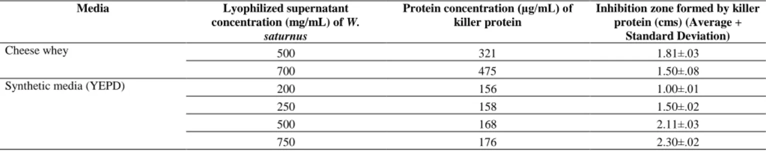
Usage of W. saturnus lyophilized powder
A minimum of 156 µg/mL of lyophilized protein in YEPD media is equivalent to 321.9 µg/mL of lyophilized protein in cheese whey needed for the inhibition of
C. krusei (Table 5). W. saturnus did not show any effect below pH 4.5 and it
grows well at 25-45 °C. Also W. saturnus grow well at pH 3.5 but failed to produce killer protein at the same pH.
Table 5 The inhibition zone created by the minimum concentration of the killer protein along with varying concentration from the lyophilized supernatant from W. saturnus
Media Lyophilized supernatant concentration (mg/mL) of W.
saturnus
Protein concentration (µg/mL) of killer protein
Inhibition zone formed by killer protein (cms) (Average +
Standard Deviation)
Cheese whey 500 321 1.81±.03
700 475 1.50±.08
Synthetic media (YEPD) 200 156 1.00±.01
250 158 1.50±.02
500 168 2.11±.03
750 176 2.30±.02
Inhibition by synergistic effect of H2O2 and lyophilized W. saturnus/ supernatant from W. saturnus
4000 ppm of H2O2 could inhibit the growth of C. krusei in the fermented broth (mono-culture) obtained from continuous aerated fermentation (Table 3); and 200 mg/mL (killer protein concentration is 156 mg/L) was the concentration of lyophilized powder needed for the inhibition of C. krusei (obtained from Table 5). A synergistic effect of H2O2 and lyophilized powder of W. saturnus was studied. The set of experiments conducted at pH 5.0 and 40 °C, where 4000 ppm of H2O2 and 200 mg/mL of lyophilized W. saturnus powder was added. W.
saturnus was highly effective in killing C. krusei, but in these set of experiments;
such an inhibition did not occur because W. saturnus possesses peroxidase activity, which along with K. marxianus degraded H2O2 at a much faster rate than
K. marxianus alone (Buzzini et al., 2004).
2400 ppm of H2O2 was considered along with 200 mg/mL of lyophilized supernatant of W. saturnus grown in cheese whey at pH 5.0 and 40 °C. At 24 h, cell concentration of C. krusei was reduced (Figure 4). By increasing the concentration of lyophilized supernatant of W. saturnus to 300 mg/mL, complete inhibition did not take place in a mixed culture. Simultaneously when 2400 ppm H2O2 and 400 mg/mL of lyophilized supernatant of W. saturnus was applied, H2O2 was degraded between 0-6 h because of the catalase-peroxidase enzymatic activity from K. marxianus and W. saturnus, but lyophilized supernatant of W.
saturnus showed activity till 24 h. C. krusei (CFU/mL) lowered and showed
drastic reduction in cell concentration at 24 h, whereas K. marxianus (1.8x10⁷ CFU/mL) remained unaffected.
Figure 4 Impact of 2400 ppm H2O2 with 200 - 400 mg/mL (156 – 200 µg/mL killer protein) of lyophilized supernatant W. saturnus (Ws) powder on the mixed culture (C. krusei (Ck) and K. marxianus (Km)) in the fermenter broth at pH 5.0 and 40 °C (Shake flask)
Inhibition of C. krusei by Ag-KT4561 NPs
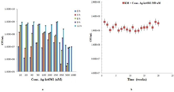
Higher concentration of the Ag-KT4561 was observed at 48 h than at 12 h (Figure 5a). Therefore NPs formed at 48 h were considered for this study. A concluding study of the mixed culture (C. krusei and K. marxianus) along with silver-KT4561 nanoparticles (Ag-KT4561NP) at pH 5.5 and 30 °C showed that 350 µM of Ag-KT461 could efficiently inhibit C. krusei. At concentration of 350 µM (Ag-KT4561), the conjugate consists of 1 ppm of reduced Ag.
Figure 5 a) UV-VIS spectroscopy showed peak at 410 nm at 12 h and maximum at 48 h during bulk preparation of Ag-KT4561; b) A 12 h study of C. krusei when various concentrations of Ag-KT4561 was mixed with cheese whey
While growth curves of K. marxianus slightly decreases from 8.9x10⁸ to 2.6x10⁸ (Figure 5b). The decrease was less than a log-unit and this might be due to the presence of silver in the Ag-NPs. The other concentrations of the Ag-KT4561 used are as less as 10 µM and as maximum as 1 mM. In any food and feed grade products, a very high concentration of biomolecule based nanoparticle may be toxic for consumption but at a lower concentration of 350 µM (with 1 ppm of reduced silver ions), Ag-KT4561 is an efficient bio-preservative. Another effective approach to use biomolecule based nanoparticle is, no pH adjustment and no temperature adjustment is required. Ag0 has anti-microbial effects against
combination of killer protein from W. saturnus which specifically targets C.
krusei. A synergistic effect of both (reduced Ag ion and killer protein) can kill C. krusei and K. marxianus remains partially affected (Figure 5b and 6a). A stability
test of Ag-KT4561(350 uM) was performed on cheese whey till 12 h for 20 weeks and every time C. krusei was killed after being inoculated at 0 h and K.
marxianus showed growth at a maximum of 2.3x10⁸ CFU/mL. Though K.
marxianus growth was affected it did not perish away with the concentration of
Ag in Ag-KT4561 (Figure 6b). A tabular representation (Table 6) shows the economics of bio-inhibitor (Ag-KT4561) production in a bulk amount of 20,
Figure 6 a) A 12 h study of K. marxianus when various concentrations of Ag-KT4561 was mixed with cheese whey; b) A stability test done for 20 weeks representing growth of K. marxianus (KM) at a minimum of 2.1 x 10⁸ and no traces of C. krusei
Table 6 Bio preservative (Ag-KT4561 conjugate) production of 20,000 L S. No Items Required (amount) Cost of Production
(CAD $) Reagents and culture medium for stock
1 Silver nitrate (271.6 g) for 40 L d.H20 242 2 Culture medium of W. saturnus 360 L 306
Preparation cost
3 Mechanical stirring of AgNO3 reagent (36W Input power) for 48 h ( ̴ 1.728 kwh) 12.19 4 Centrifugation (700W) for 15 mins ( ̴ 0.175
kwh)
1.23 5 Freeze drying (1200W) for 24 h ( ̴ 28 kwh) 197
Total 760
6 10% cost of man-power 76
DISCUSSION
The results indicated that 2 M NaCl could effectively inhibit C. krusei in a monoculture of K. marxianus at temperature 40 °C, pH 3.5 without affecting the growth of K. marxianus. However, C. krusei showed lower NaCl tolerance than any other yeast species e.g. Saccharomyces had different sensitivity towards osmotic stress, but C. krusei was inhibited efficiently at 2 M without affecting the growth of K. marxianus (Lynum and Nauth, 2000; Uchida et al., 2005). The reported concentration is used to discriminate K. marxianus as it is sensitive up to a concentration of 3 M NaCl. Stress-induced by salt induction results into two different phenomena, primarily, ion toxicity and secondly, osmotic stress. Apparently, other physiological changes can also take place such as: a) efflux of intracellular H2O, i.e. total cell volume deduction; b) transient increase in glycolytic intermediates and finally triggering the hyper osmotic glycerol signaling pathway. Specific species, such as Saccharomyces and Klyuveromyces can develop systems to counteract to osmotic stress by NaCl. Special features of
Saccharomyces and Klyuveromyces species are that they produce intracellular
trehalose under stress conditions to maintain the membrane integrity and stabilizing the proteins (Kuhn et al., 2004; Wang and Wu, 2008; Davey, 2011). However, in large-scale fermenters, it was not possible as it would lead to high utilization of NaCl for the inhibition of C. krusei. At industrial scale, such inhibitions performed by utilization of NaCl is difficult not because of the market price which is 16-20 USD per Kg; but the volume of NaCl required was more than 500 Kg for 40 000 L industrial reactor (Goretti et al., 2009; Kosseva et al., 2009). Therefore, NaCl was not a suitable approach for C. krusei inhibition. Apparently, H2O2 was found to be effective at 300 ppm when C. krusei inhibition was performed at shake flask level. In a shake flask study, it has been shown that 200 ppm could efficiently inhibit C. krusei (Nel et al., 2006). Other yeasts, such as W. saturnus and K. marxianus have shown no inhibition in the presence of H2O2 as both the yeasts showed catalase activity. Maximum oxidative stress was observed in case of Saccharomyces spp. which was nearly 2-folds more than K.
marxianus (Kang et al., 2011). Every organism possesses specific
antioxidant-defense systems. When W. saturnus was added along with K. marxianus and C.
krusei, no inhibition of C. krusei was bound to happen as W. saturnus even
possessed peroxidase activity (Buzzini et al., 2004). Apart from catalase, when
K. marxianus was introduced to H2O2 in the exponential phase, other enzymes, such as superoxide dismutase and glutathione reductase content were even increased to 2-fold (Nilsson, 2011). These were specific antioxidant defensive agents present in Klyuveromyces spp. (Meurman et al., 2007).
When pH was brought down from 6.0 to 3.5, it did not affect the inhibition of C.
krusei either. However, catalase was widely active in a vast range of pH (3.5-10).
Apparently, decomposition of peroxidase lowered the pH in the medium (Pinto et al., 2009; Warnke et al., 2009; Guevara-Flores et al., 2010). While varying the pH, temperature was increased to 40 °C and C. krusei was efficiently inhibited. When it came to the effect of catalase activity on thermal capacity, 55 °C was the critical temperature and beyond which catalase enzyme was completely destroyed (Erdei et al., 2011). The factors that shifted the physiological process of the glutathione reductases in K. marxianus showed higher pH of 6.8 along with temperature (37-40 °C) where it possesses more antioxidant activity (Ghaly et al., 2005; Pinto et al., 2009).
In an aerated continuous fermenter, 4000 ppm of H2O2 was required without affecting the growth of K. marxianus and W. saturnus as both the microorganisms possessed catalase and peroxidase activity which efficiently degraded H2O2 during 6 h of inoculation. With such an extreme concentration of 4000 ppm of H2O2, K. marxianus still had the capacity to resist it. However when fermenter conditions were considered, it was well-stated that 100 ppm of H2O2 could cause corrosion of fermenter frame. The other catalysts for corrosion were O2 and higher temperature (Sathishkumar et al., 2010). Again, •OH, H2O2, O2, H2, •O2 could interact with the surroundings; and therefore led to corrosive behavior of many materials including stainless steel (McMahon et al., 2007; Siddique and Wahid, 2012). Though, H2O2 might be an inexpensive ingredient for C. krusei inhibition, fermenter inner body could have corrosive effects. Therefore, lower concentration of H2O2 might be an ideal approach. The use of chemicals depends entirely on the form of free radicals being produced and the damage they may or may not have on the fermenter.
Simultaneously, it was observed that 0.45% (v/v) clove oil could inhibit C. krusei without affecting other yeasts. The factor responsible for inhibiting the growth of
C. krusei was eugenol. Eugenol is the component present in clove oil which can
kill C. krusei at optimum concentrations (Noori, 2012). S. aromaticum (clove oil) 0.4% (v/v) can efficiently bring down the concentration of C. krusei in a mono-culture of K. marxianus. However, on a large-scale fermentation, it would not be an approachable or economical aspect for inhibition. When the prices were compared, it is observed that 200 Kg of wholesale clove oil will cost USD 12 834.00, and definitely in large scale fermentations, the volume of clove oil required was 160 L, which will not only make the final product oily, but also very expensive. Therefore, it was not an ideal approach for C. krusei using clove oil. Similarly when nisin was used for the inhibition as it is one of the biochemical approaches, nisin could not inhibit C. krusei at all. Henceforth, the focus was shifted to W. saturnus killer protein.
W. saturnus was found to be an effective species which could kill C. krusei in a
mono-culture of K. marxianus without having any implications on K. marxianus. Killer protein produced by KT4561 at a concentration of 200 mg/mL (where killer protein concentration is 156 ug/mL) when grown in glucose rich medium;
and 400 mg/mL when grown in a lactose-efficient medium (cheese whey) can inhibit C. krusei. The purpose of production of lyophilized supernatant from W.
saturnus is to justify that W. saturnus produces naturally occurring extracellular
killer protein KT4561, which strongly inhibited C. krusei. The killer protein produced by W. saturnus caused cell membrane damage and an independent energy link in between the cell wall receptor and KT4561 at the region of (1→6)-β-D-glucan complex (Fang et al., 2002).
More efficient inhibition of C. krusei was possible if W. saturnus would have been grown in a glucose-rich medium where efficient production of the killer protein could have inhibited C. krusei. This study revealed a real understanding of the different microbial species dealt with and different behavioral patterns with respective to the varied inhibitors used for the inhibition of C. krusei. The factor to be considered when biochemical approaches such as H2O2 were used is whether it again had any effect on the organic matter present in cheese whey. As over the years, approaches have been made to protect the food and humans from consuming it against any oxidative damage. Free radicals such as hydroxyl, peroxyl, and superoxide have been bound to release when biochemical methods are used for inhibiting the food pathogens (Erdemoglu et al., 2007).
Killer protein-based nanoparticle showed an effective inhibition for C. krusei. It was observed that 350 µM of Ag-KT4561 (with 1 ppm of Ag) could bring in effective inhibition of C. krusei within 3 h. But beyond 350 µM concentration could affect the growth K. marxianus. K. marxianus growth was affected due to the presence of Ago but killer protein has no effect on it. Other significant consideration was that no pH or temperature was adjusted, because Ag ion was effective against almost all pathogens. Other benefit of using biomolecule based nanoparticle was that metal nanoparticles were toxic for human or animal consumption but biomolecule based nanoparticles had shown no toxicity so far (Nel et al., 2006; Da Silva et al., 2011).
CONCLUSION
Biomolecule based nanoparticle approach (Ag-KT4561) for inhibition of C.
krusei served to be better method than other chemical and biochemical methods
used in this study. Other suitable alternative approach might have been ultra-filtration, however, on an industry scale it was an expensive approach. Therefore, Ag-KT4561 was the effective and economic inhibitory approach towards C.
krusei (non-Candida albicans spp.) and it even supported green chemistry.
Although the composition of cheese whey was known, further verification and prolonged usage of killer protein-based silver nanoparticle to sustain the antimicrobial effect need to be investigated further.
Acknowledgements: The authors are sincerely thankful to the Natural Sciences and Engineering Research Council of Canada (Grant A4984, RDCPJ 379601, Canada Research Chair) for financial support. The views and opinions expressed in this article are strictly those of the authors.
REFERENCES
ADENIYI, C.B.A., ODUMOSU, B.T., AIYELAAGBE, O.O., KOLUDE, B. 2010. In-vitro antimicrobial activities of methanol extracts of Zanthoxylum
xanthoxyloides and Pseudocedrela kotschyi. African Journal of Biomedical Research, 13(1), 61-68.
AGUIAR, C., LUCAS, C. 2000. Yeasts killer/sensitivity phenotypes and halotolerance. Food Technology and Biotechnology, 38(1), 39-46.
AHARIZ, M., COURTOIS, P. 2010. Candida albicans susceptibility to lactoperoxidase – generated hypoiodite. Clinical, Cosmetic and Investigational
Dentistry, 2, 69-78. http://dx.doi.org/10.2147/cciden.s10891
AYOOLA, G.A., COKER, H.A.B., ADESEGUN, S.A., ADEPOJU-BELLO, A.A., OBAWEYA, K., EZENNIA, E.C., ATANGBAYILA, T.O. 2008. Phytochemical screening and antioxidant activities of some selected medicinal plants used for malaria therapy in southwestern Nigeria. Tropical Journal of
Pharmaceutical Research, 7(3), 1019-1024.
http://dx.doi.org/10.4314/tjpr.v7i3.14686
BHATTACHARYA, I., BEZAWADA, J., YAN, S., TYAGI, R.D. 2015. Optimization and production of silver-protein conjugate as growth inhibitor.
Journal of Bionanoscience, 9(4), 261-269.
http://dx.doi.org/10.1166/jbns.2015.1309
BROCK, M. 2008. Physiology and Metabolic Requirements of Pathogenic Fungi.
The Mycota, 6(1), 63-82. http://dx.doi.org/10.1007/978-3-540-79307-6_4 BUZZINI, P., CORAZZI, L., TURCHETTI, B., BURATTA, M., MARTINI, A. 2004. Characterization of the invitro antimitotic activity of a novel killer protein from Williopsis saturnus DBVPG 4561 against emerging pathogenic yeasts.
FEMS Microbiology Letters, 238(2), 359-365. http://dx.doi.org/10.1111/j.1574-6968.2004.tb09777.x
CARVALHO, F., PRAZERES, A.R., RIVAS, J. 2013. Cheese whey wastewater: Characterization and treatment. Science of the Total Environment. 445-446: 385-396. http://dx.doi.org/10.1016/j.scitotenv.2012.12.038
CHEN, W.B., HAN, Y.F., JONG, S.C., CHANG, S.C. 2000. Isolation, purification, and characterization of a killer protein from Schwanniomyces
occidentalis. Applied and Environmental Microbiology, 66(12), 5348-5352.
http://dx.doi.org/10.1128/aem.66.12.5348-5352.2000
DA SILVA, B.F., PÉREZ, S., GARDINALLI, P., SINGHAL, R.K., MOZETO, A.A., BARCEIO, A. 2011. Analytical chemistry of metallic nanoparticles in natural environments. TrAC Trends in Analytical Chemistry. 30(3), 528-540. http://dx.doi.org/10.1016/j.trac.2011.01.008
DAVEY, H.M. 2011. Life, death, and in-between: meanings and methods in microbiology. Applied and Environmental Microbiology, 77(16), 5571-5576. http://dx.doi.org/10.1128/aem.00744-11
DE OLIVEIRA, B.P., LINS, C. C. d. S. A., DINIZ, F. A., MELO, L.L., DE CASTRO, C.M.M.B. 2014. In Vitro antimicrobial photoinactivation with methylene blue in different microorganisms. Brazilian Journal of Oral Sciences, 13(1), 53-57. http://dx.doi.org/10.1590/1677-3225v13n1a11
DINGMAN, J. 2008. Nanotechnology: It’s Impact on food safety. Journal of
Environment Health, 70(6), 47-50.
ERDEI, É., MOLNÁR, M., GYÉMÁNT, G., ANTAL, K., EMRI, T., PÓCSI, I., NAGY, J. 2011. Trehalose overproduction affects the stress tolerance of
Kluyveromyces marxianus ambiguously. Bioresource Technology, 102(14),
7232-7235. http://dx.doi.org/10.1016/j.biortech.2011.04.080
ERDEMOGLU, N., OZKAN, S., TOSUN, F. 2007. Alkaloid profile and antimicrobial activity of Lupinus angustifolius L. alkaloid extract.
Phytochemistry Reviews, 6(1),197-201. h ttp://dx.doi.org/10.1007/s11101-006-9055-8
EYSTER, H.C. 1950. Effect of temperature on catalase activity. Ohio Journal of
Science, 50(6), 273-277.
FANG, Y.Z., YANG, S., WU, G. 2002. Free radicals, antioxidants, and nutrition. Nutrition, 18(10), 872-879. http://dx.doi.org/10.1016/ S0899-9007(02)00916-4
GHALY, A.E., KAMAL, M., CORREIA, L.R. 2005. Kinetic modeling of continuous submerged fermentation of cheese whey for single cell protein production. Bioresource Technology, 96, 1143-1152. http://dx.doi.org/10.1016/j.biortech.2004.09.027
GORETTI, M., TURCHETTI, B., BURATTA, M., BRANDA, E., CORAZZI, L., VAUGHAN-MARTINI, A., BUZZINI, P. 2009. In vitro antimycotic activity of a
Williopsis saturnus killer protein against food spoilage yeasts. International
journal of food microbiology, 131(2-3), 178-182.
http://dx.doi.org/10.1016/j.ijfoodmicro.2009.02.013
GUEVARA-FLORES, A., ARENAL, I.P.D., MENDOZA-HERNÁNDEZ, G., PARDO, J.P., FLORES-HERRERA, O., RENDÓN, J.L. 2010. Mitochondrial thioredoxin-glutathione reductase from larval Taenia crassiceps (Cysticerci).
Journal of Parasitology Research, 2010, 719-856.
http://dx.doi.org/10.1155/2010/719856
GUO, S., BHATTACHARJEE, J.K. 2006. Novel lysine biosynthetic gene sequences (LYS1 and LYS5) used as PCR targets for the detection of the pathogenic Candida yeast. Applied Microbiology and Biotechnology. 72(2), 416-420. http://dx.doi.org/10.1007/s00253-006-0470-y
GUWY, A.J., MARTIN, S.R., HAWKES, F.R., HAWKES, D.L. 1999. Catalase activity measurements in suspended aerobic biomass and soil samples. Enzyme
and Microbial Technology, 25, 669-676. http://dx.doi.org/10.1016/s0141-0229(99)00115-5
HEARD, G.M., FLEET, G.H. 1988. The effects of temperature and pH on the growth of yeast species during the fermentation of grape juice. Journal of
Applied Bacteriology, 65, 23-28. http://dx.doi.org/10.1111/j.1365-2672.1988.tb04312.x
HORNBÆK, T., BROCKHOFF, P.B., SIEGUMFELDT, H., BUDDE, B.B. 2006. Two subpopulations of Listeria monocytogenes occur at subinhibitory concentrations of leucocin 4010 and nisin. Applied and Environmental
Microbiology, 72, 1631-1638.
http://dx.doi.org/10.1128/aem.72.2.1631-1638.2006
KANG, K., WONG, K.S., FONG, W.P., TSANG, P.W.K. 2011. Metergoline-induced cell death in Candida krusei. Fungal Biology, 115, 302-309. http://dx.doi.org/10.1016/j.funbio.2011.01.001
KAO, A.S., BRANDT, M.E., PRUITT, W.R., CONN, L.A., PERKINS, B.A., STEPHENS, D.S., BAUGHMANN, W.S., REINGOLD, A.L., ROTHROCK, G.A., PFALLER, M.A., PINNER, R.W., HAJJEH, R.A. 1999. The epidemiology of candidemia in two United States cities: results of a population-based active surveillance. Clinical Infectious Diseases, 29(5), 1164-1170. http://dx.doi.org/10.1086/313450
KIM, Y., LEE, H.S. 2012. Anticandidal effect of Syzygium aromaticum on biofilm formation, cell surface hydrophobicity, and cell cycle. Journal of
Medicinal Plants Research, 6(10), 1926-1939.
KOLEVA, D.I., PETROVA, V.Y., KUJUMDZIEVA, A.V. 2008. Comparison of enzymatic antioxidant defense systems in different metabolic types of yeasts.
Canadian Journal of Microbiology. 54(11), 957-963. http://dx.doi.org/10.1139/w08-093
KOSSEVA, M.R., PANESAR, P.S., KAUR, G., KENNEDY, J.F. 2009. Use of immobilized biocatalysts in the processing of cheese whey. International Journal
of Biological Macromolecules, 45, 437-447.
KUHN, D.M., MUKHERJEE, P.K., CLARK, T.A., PUJOL, C., CHANDRA, J., HAJJEH, R.A., WARNOCK, D.W., SOLL, D.R., GHANNOUM, M.A. 2004.
Candida paralopsilosis characterization in an outbreak setting. Emerging
Infectious Diseases, 10(6),
1074-1081. http://dx.doi.org/10.3201/eid1006.030873
LIU, H.J., LI, Q, LIU, D.H., ZHONG, J.J. 2006. Impact of hyperosmotic condition on cell physiology and metabolic flux distribution of Candida krusei.
Biochemical Engineering Journal, 28(1), 92-98.
http://dx.doi.org/10.1016/j.bej.2005.08.038
LOGOTHETIS, S., GRAEME, W., ELIAS, T.N. 2007. Effect of salt hyperosmotic stress on yeast cell viability. Zbornik Matice Srpske za Prirodne
Nauke, 113, 271-284. http://dx.doi.org/10.2298/zmspn0713271l
LOWES, K.F., SHEARMAN, C.A., PAYNE, J., MACKENZIE, D., ARCHER, D.B., MERRY, R.J., GASSON, M. J. 2000. Prevention of yeast spoilage in feed and food by the yeast mycocin HMK. Applied and
Environmental Microbiology, 66(3), 1066-1076.
http://dx.doi.org/10.1128/aem.66.3.1066-1076.2000
LOWRY, O.H., ROSEBROUGH, N.J., FARR, A.L., RANDALL, R.J. 1951. Protein measurement with the Folin Phenol reagent. Journal of Biological
Chemistry, 193(1): 265-275.
LYNUM, M., NAUTH, K.R. 2000. Stabilization of cream cheese compositions using nisin-producing cultures. US Patent 6,110,509. Draft endorsed on 31 August.
MANEESRI, J., MASNIYOM, P. 2007. Induction and inhibition of film yeast from fermented bamboo shoot by seasoning plants. Songklanakarin. Journal of
Science and Technology, 29(4), 1135-1143.
MANEESRI, J., MANEESRI, J. 2009. Effect of chemical factors and clove oil to decrease the growth of film yeast on fermented bamboo shoots. Asian Journal of
Food and Agro-Industry. 2(4), 159-167.
MCMAHON, M.A.S., XU, J., MOORE, J.E., BLAIR, I.S., MCDOWELL, D.A. 2007. Environmental stress and antibiotic resistance in food-related pathogens.
Applied and Environmental Microbiology, 73(1), 211-217.
http://dx.doi.org/10.1128/aem.00578-06
MEURMAN, J.H., SIIKALA, E., RICHARDSON, M., RAUTEMAA, R. 2007. Non-Candida albicans Candida yeasts of the oral cavity. Applied Microbiology (Eds.), 719-731.
MORGULIS, S., BEBER, M., RABKIN, I. 1926. Studies on the effect of temperature on the catalase reaction. Journal of biological Chemistry, 68(3), 521-533.
NATHAN, P., LAW, E. J., MURPHY, D. F., MACMILLAN, B. G. 1978. A laboratory for the selection of topical antimicrobial agents. Burns, 4(3), 177-178. http://dx.doi.org/10.1016/s0305-4179(78)80006-0
NEL, A., XIA, T., MADLER, L., LI, N. 2006. Toxic potential of materials at the nanolevel. Science, 311, 622-627. http://dx.doi.org/10.1126/science.1114397 NILSSON, O. 2011. Radiation induced corrosion of steel. Thesis, Department of Chemistry, Nuclear chemistry: Royal Institute of Technology, 2011, 1-16p. NOORI, S. 2012. An overview of oxidative stress and antioxidant defensive system. Open Access Scientific Reports, 1(8), 1-9. http://dx.doi.org/10.4172/scientificreports.413
ORRÙ, G., NERO, S.D., TUVERI, E., CIUSA, M.L., PILIA, F., ERRIU, M., ORRÙ, G., LICIARDI, G., PIRAS, V., DENOTTI, G. 2010. Evaluation of antimicrobial-antibiofilm activity of a hydrogen peroxide decontaminating system used in dental unit water lines. Open Dentistry Journal, 4, 140-146. http://dx.doi.org/10.2174/1874210601004010140
PANACEK, A., KOLAR, M., VECEROVA, R., PRUCEK, R., SOUKUPOVA, J., KRYSTOF, V., HAMAL, P., ZBORIL, R., KVITEK, L. 2009. Antifungal activity of silver nanoparticles against Candida spp. Biomaterials, 30, 6333-6340. http://dx.doi.org/10.1016/j.biomaterials.2009.07.065
PINHEIRO, R., ISABEL, B., MOTA, M. 2002. Oxidative stress response of
Kluyveromyces marxianus to hydrogen peroxide, paraquat and pressure. Applied
Microbiology and Biotechnology, 58(6), 842-847.
http://dx.doi.org/10.1007/s00253-001-0927-y
PINTO, E., VALE-SILVA, L., CAVALEIRO, C., SALGUEIRO, L. 2009. Antifungal activity of the clove essential oil from Syzygium aromaticum on
Candida, Aspergillus and dermatophyte species. Journal of Medical Microbiology. 58, 1454-1462. http://dx.doi.org/10.1099/jmm.0.010538-0 PRAZERES, A.R., CARVALHO, F., RIVAS, J. 2012. Cheese whey management: a review. Journal of Environmental Management, 110, 48-68. http://dx.doi.org/10.3923/jest.2012.155.167
RUSSELL, J.B., JARVIS, G.N. 2001. Practical mechanisms for interrupting the oral-fecal lifecycle of Escherichia coli. Journal of Molecular Microbiology and
Biotechnology, 3(2), 265-272.
SATHISHKUMAR, T., SHANMUGAM, S., RAMESHKUMAR, S., RAJAVELAN, G., HARIDOSS, V. 2010. Characterization of salivary glutathione reductase in normal individuals and its implications on smokers. Researcher, 2(4), 74-81.
SIDDIQUE, N.I., WAHID, Z.A. 2012. Application of chemical and biological coupled treatment technology in POME and petroleum waste water as biodegradation alternative. Journal of Environmental Science and Technology, 5(3), 155-167. http://dx.doi.org/10.3923/jest.2012.155.167
SISO, M.I.G. 1996. The biotechnological utilization of cheese whey: a review.
Bioresource Technology, 57, 1-11. http://dx.doi.org/10.1016/0960-8524(96)00036-3
SOUZA, E.L.D., STAMFORD, T.L.M., LIMA, .E.D.O, FILHO, J.M.B, MARQUES, M.O.M. 2008. Interference of heating on the antimicrobial activity and chemical composition of Origanum vulgare L. (Lamiaceae) essential oil.
Cienc Tecnol Aliment, 28(2), 418-422. http://dx.doi.org/10.1590/s0101-20612008000200023
UCHIDA, S., SHIGENAKA, N., TACHIBANA, M., WADA,Y., SAKAI, M., AKAMINE, K., OHSUMI, K. 1998. Effects of Hydrogen Peroxide on Intergranular Stress Corrosion Cracking of Stainless Steel in High Temperature Water, (I). Journal of Nuclear Science and Technology, 35(4), 301-308.
http://dx.doi.org/10.1080/18811248.1998.9733860
WAEMA, S., MANEESRI, J., MASNIYOM, P. 2009. Isolation and identification of killer yeast from fermented vegetables. Asian Journal of Food
and Agro-Industry, 2(4), 126-134.
WALKER, G., DIJCK, P.V. 2006. Physiological and Molecular Responses of Yeasts to the Environment. Yeasts in Food and Beverages, 111-152. http://dx.doi.org/10.1007/978-3-540-28398-0_5
WALKER, L.A., MUNRO, C.A., BRUIJN, I.D., LENARDON, M.D., MCKINNON, A., GOW, N.A.R. 2008. Stimulation of chitin synthesis rescues
Candida albicans from Echinocandins. PLoS Pathogens, 4(4), 1-12.
http://dx.doi.org/10.1371/journal.ppat.1000040
WANG, J., WU, J. 2008. Antifungal activity of 25-azalanosterol against Candida species. European Journal of Clinical Microbiology & Infectious Diseases. 27(11), 1131-1136. http://dx.doi.org/10.1007/s10096-008-0554-y
WARNKE, P.H., BECKER, S.T., PODSCHUN, R., SIVANANTHAN, S., SPRINGER, I.N., RUSSO, P.A.J., WILTFANG, J., FICKENSCHER, H., SHERRY, E. 2009. The battle against multi-resistant strains: Renaissance of antimicrobial essential oils as a promising force to fight hospital-acquired infections. Journal of Cranio-Maxillofacial Surgery, 37(7), 392-397.
http://dx.doi.org/10.1016/j.jcms.2009.03.017
YADAV, J.S.S., BEZAWADA, J., YAN, S., TYAGI, R.D., SURAMPALLI, R.Y. 2012. Candida krusei: biotechnological potentials and concerns about its safety. Canadian Journal of Microbiology, 58(8), 937-952. http://dx.doi.org/10.1139/w2012-077