Ragged N-termini and other
variants
of class A
f-lactamases
analysed by chromatofocusing
Andre MATAGNE,* Bernard JORIS,t JozefVAN
BEEUMENt
and Jean-Marie FRERE*§*Laboratoired'Enzymologie andtCentrefor Protein Engineering, Universitede Liege,Institut deChimie, B6, B-4000 SartTilman (Liege 1), Belgium, and
tLaboratorium
voorMicrobiologieenmicrobiele Genetica, Rijksuniversiteit-Gent, Ledeganckstraat 35, B-9000Gent, BelgiumFour
f6-lactamases
excreted by Gram-positive bacteria exhibited microheterogeneity when analysed bychromatofocusing orion-exchange chromatography. Ragged N-terminiwerein partresponsible for thecharge variants,butdeamidation ofan asparagine residue wasalso involved, atleast for the Bacilluslicheniformis enzyme.Theactivity ofacontaminating proteinase could also be demonstrated in the caseof theActinomadura R39
,8-lactamase.
With that enzyme, proteolysis resulted inpartial inactivation, but the inactivatedfragmentswereeasily separated from the active forms. With these, as with theother enzymes, the kinetic parameters ofthe major variants were identical with those of the mixture within the limits of experimental error, so that the catalytic properties of these enzymes can be determined with the 'heterogeneous' preparations.INTRODUCTION
Excretion of extracellular
fl-lactamases exhibiting
various extents ofmicroheterogeneity by Gram-positive bacteria seems tobearathercommonphenomenon (Ambler&Meadway, 1969; Thatcher, 1975a; Dehottay et al., 1987; Lenzini et al., 1988). The most widely studied case is that of the enzyme produced by Bacillus licheniformis, where a 'ragged' N-terminus was first demonstrated (Ambler & Meadway, 1969; Thatcher, 1975b; Simons et al., 1978; Izui et al., 1980). The same phenomenon appearedtobeatleastpartially responsiblefor the microhetero-geneityofthe,-lactamases ofStreptomycesalbus andS. cacaoi cloned and produced in S. lividans (Dehottay et al., 1987; Lenzini et al., 1988). The present study was undertaken after discoveringthat distinct activefractions ofvariousGram-positivefl-lactamases
could be separated by chromatofocusing on a MonoP column (Dehottay et al., 1987; Matagne et al., 1990). Although we were interested in elucidating the origin of the variations at the molecular level, we thought that it was even more important to compare the kinetic parameters of the individual variants with those ofthe mixtures to determine if meaningful results could be obtained without resolving pure preparations into individual components.To allow easy reference to the sequences of other class A /-lactamases, we usetheABLnumbering scheme. Inthat system, aresiduecan be referredtobytwonumbers: oneis itsposition in the natural protein or precursor sequence and the second (calledABL) is basedon Ambler's(1979) alignmentof the first four enzymes whose sequencewasdetermined.Forinstance,the active serine residue in the Actinomadura R39
/3-lactamase
is no. 86 inthat enzyme and ABL 70.MATERIALS AND METHODS
II-Lactam
compoundsBenzylpenicillin was from Rhone-Poulenc (Paris, France), ampicillin from Bristol Benelux
(Brussels, Belgium),
,3-iodo-penicillanicacidfrom Pfizer Central Research
(Sandwich, Kent,
U.K.), andcephaloridine
andcephalothin
were from EliLilly
and Co.
(Indianapolis, IN,
U.S.A.).
These antibioticswerekindly
given bythe respective companies. Penicillin V was a gift from Professor H. Vanderhaeghe and Professor P. Claes (Katholieke Universiteit Leuven, Leuven, Belgium). Nitrocefin was purchased from Oxoid (Basingstoke, Hants.,U.K.).
Enzymes
Actinomadura R39
Il-lactamase.
The enzyme was produced by Streptomyces lividans TK24 harbouring plasmid pIJ424 con-taining the gene coding for the Actinomadura R39/8-lactamase (Piron-Fraipont et al., 1989) and was purified as described by Piron-Fraipont etal. (1989).Bacillus
lichenifornms
749/Cf8-lactamase.
The enzyme was produced by the original strain and purified as described by Matagne etal. (1990).S. albus G
f-lactamase.
The enzymewasproduced by S. albus G strain R2 harbouring plasmid pDML6 containing the gene codingfor the S. albus G,3-lactamase
(Dehottayetal., 1987) and waspurifiedasdescribed by Matagne et al. (1990).S. cacaoi
II-lactamase.
The enzymewas produced by S. albus G strain R2 harbouring plasmid pDML51containing
the gene coding for the S. cacaoi/1-lactamase
(Lenzini etal., 1987) and purifiedas described by Matagne etal. (1990).The four ,-lactamase preparations were the same as those used for the studyof the catalytic properties of these enzymes (Matagne etal., 1990).
Isoelectricfocusing andpl calculations
Isoelectric focusing was performed in a Bio-Rad model-Ill Mini IEFcell with LKBampholytesin thepHranges 3-10 and 3.5-5 or ina Pharmacia Fast System apparatus
using
the pH range 4-6.5.The pl calculationsweredone with thehelpof the 'isoelectric' algorithm from the GCG
package
version 6(Devereux
etal.,
1984).
A.Matagneand others Chromatographic techniques
Chromatofocusing. Chromatofocusing experiments were per-formedonaMonoPHR5/20 column connectedto aPharmacia f.p.l.c. system. The pH gradient wentfrom5.7to4,except with the B.
licheniformis
/1-lactamase,
forwhich it started atpH 6.1. Buffer A was 25mM-N-methylpiperazine/HCl
(Janssen),pH 5.7 or6.1,
and bufferB,
was a 10-fold dilution ofPolybuffer 74 (Pharmacia) adjusted to pH4 with HCI. Buffer B used in the experimentswith the S. albus G and S. cacaoif,-lactamases
also contained 5% (v/v) glycerol and 5% (v/v) ethylene glycol. Elution wasperformed with 34 ml of buffer B ataflowrateof 0.7ml/min. The amount of enzyme used in eachexperimentwas of theorder of 1 mg.Ionexchange. AMonoQHR5/5andaMonoS
HR5/5
columns connected to a Pharmacia f.p.l.c. system were used for ion-exchange experiments. Elutionofthe ActinomaduraR39enzyme from the MonoQ column was performed with a linear NaCl gradient (0.4-0.6M) over 36ml in 20mM-Tris/HCI buffer, pH 7.2.The experiment wasperformedat aflow rate of 1 ml/min with about0.5 mgofenzyme.Aminoacid sequencing
Amino acid sequencing was done with a 470-A Applied Biosystems gas-phasesequenator as described previously (Joris etal., 1985).
p8-Lactamase activity
Routinely, 4-lactamase activities were measured using nitro-cefin as substrate in 50mM-sodium phosphate, pH 7, at30'C. When necessary, enzyme samples weredilutedinthe samebuffer containing0.1 mg of BSA/ml.Oneunit represents the amount of enzymehydrolysing1,umolofsubstrate/minatmaximal velocity. Kinetic parameters and thermal stabilityof8-lactamases
Thedetermination ofthe kineticparameters and the analysis of the thermal stability of the enzymes were performed as described byMatagne etal. (1990). Thethermal inactivationof theB.
licheniformis
,J-lactamase
wasmonitoredbycontinuously measuring thehydrolysis ofa reporter substrate (nitrocefin) as described byDe Meesteretal. (1987).RESULTS
pl values andchromatofocusing
PAGE, when performed under non-denaturing conditions, indicatedachargeheterogeneity forthe four
fl-lactamases.
Five bandswerealsoobserved upongelisoelectricfocusingof the ,-lactamases of S.albus G and S. cacaoi. In both cases, thepl of themainbandwaschosenasrepresentativeof that of the protein (Table 1).ThepIof theActinomadura R39
,-lactamase
was solow that it could not be determined; over the pH range 3.5-5.0 all the proteinmaterial concentrated near the anode. This was in good agreement with the strong interaction observed between that enzyme and anion-exchangers [see Matagne et al. (1990) and below].The pI of theB.
licheniformis
enzyme wasnotredetermined,
and the value shown in Table 1 is from Meadway
(1969).
By usingthe amino acid compositionsdeduced fromthe complete amino acid sequences of the fourfl-lactamases
(Ambler & Meadway, 1969; Dehottay et al., 1987; Lenzini et al., 1988; Houbaetal., 1989), thepl values werecalculatedandfound to agreewell with theexperimental ones(Table 1).Three of the enzymepreparations were resolved into several
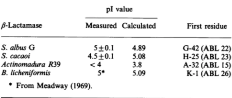
Table 1.plvalues of thefourfl-lactamasesstudied
The measuredpIvaluesarethose of themajor componentseenon thegelfor eachenzyme.The otherpIvalueswerecalculatedusing the amino-acid-sequence data from Dehottay et al. (1987) (S. albusG), Lenzinietal. (1988) (S. cacaoi),Houba etal.(1989) (Actinomadura R39) andAmbler & Meadway (1969) (B. licheni-formis).Ineachcase,theN-terminal residue isindicated (usingthe
one-letternotation).
plvalue
fi-Lactamase
Measured Calculated FirstresidueS.albus G 5+0.1 4.89 G-42(ABL22)
S.cacaoi 4.5+0.1 5.08 H-25(ABL23)
Actinomadura R39 <4 3.8 A-32(ABL15)
B. licheniformis 5* 5.09 K-1(ABL26)
* FromMeadway (1969).
catalytically
active and distinct fractions bychromatofocusing
on MonoP. As discussed
below,
with the Actinomadura R39 enzyme, anothermethod hadtobeused, owingtoitsparticularly
low
pI.
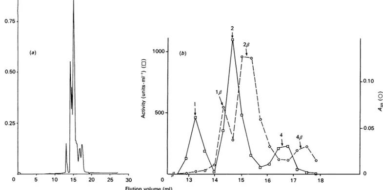
With theB.licheniformis
,-lactamase preparation, four activefractionswereobtained,whichwereelutedrespectivelyatpH
values of5.12, 5.0, 4.95 and 4.8 (Fig. la).Alternatively,
distinct activefractionswerealsoobtained with thesameenzyme
by
chromatography on MonoS atpH3.0 (results notshown),
confirmingthechargeheterogeneity.
The pure preparation of the S. albus G ,J-lactamase was resolved into threemajor peaks,whichwereeluted
respectively
atpHvalues of4.64,4.52 and4.49(Fig. lb).Similar
chromato-focusing
experiments wereperformedwitha crudepreparation
ofthe same enzyme. After reaching the maximum of enzyme production,asampleofculturemedium,freed from thebacterial myceliumbycentrifugation, wasdirectly appliedtothe column. The measurement of enzymic activity in thecollected fractions indicated the presence of several peaks, demonstrating that charge heterogeneity of the enzyme was already present in the culturemedium andwasthusnotduetomodificationsoccurring duringthepurificationoruponlong-term storage of thepurified enzyme. The major peaks were present in both the crude and pure preparations, but the relative quantities were somewhat modified, probably reflecting the fact that fractions with lower specific activities were sometimes discarded during the
purifi-cation. Variations in the culture conditions also altered the chromatographic patterns of the crude supernatant. This was probably duetomodifications inprocessing of theexoenzyme,a
phenomenon which is known to be very sensitive to the exact
compositionandpHofthegrowthmedium(Mezesetal.,
1985).
With the S. cacaoi enzyme, chromatofocusing resolved the pure preparation into at least three activefractions, elutedatpH values of4.35, 4.2 and 4.15 (Fig. lc). With both
Streptomyces
enzymes, several activepeaks were also separated byapplyingan NaCl gradientto theMonoQ column.
Chromatofocusing failed to separate distinct fractions of the Actinomadura R39 /?-lactamase. Under standard conditions(pH gradientfrom 5.7to4), theenzyme was not eluted.Adjustment of thePolybuffer74topH 3 yieldedaregularpH gradient down tothatpH, but failedtoelute theenzyme. The ionicstrength of the eluent was then increased in an attempt to decrease the electrostatic interactions between the enzyme and the ion-exchanger. At a constant pH of 4.5, the enzyme could be eluted from the MonoP column by adding 650 mM-NaCl to the Polybuffer. However, anNaCl concentration as low as 100mm 504
©2
0.75 0.50 0.25 0 5 10 15 20 25 30 35 I 5.2 - 4.9 4.6 I a - 4.3 4.0 35 -800 600 E -400 3 z 200 0 0 10 20 30 40 Elution volume(ml)Fig.1.Resolution of the variouspurifiedenzymes intodistinct
catalytically
activefractions(a),(b)and(c)ChromatofocusingonMonoPof theenzymesfromB.licheniformis(0.7mg,a);S.albus G(2mg,b)and S. cacaoi(1mg, c). The pHgradient is represented bythediagonalline.(d) Elution of the Actinomadura R39 enzyme (0.5 mg) from the MonoQ column byanNaCl gradient (thetwodiagonal lines). Ringednumbers refertofractions described in the Tables.
grossly perturbed the formation of the pHgradient, makingit enzymefrom theMonoQ (500mmi impossibletoutilizeacombination ofpHgradientandhighsalt and
150
mmrespectively
for theSconcentration toelute theenzyme.
Finally,
ananion-exchanger
lactamases)
andits retentionon N (MonoQ HR5/5)wasusedatpH7.2,and elutionwasperformed allagreed
with the very low value with ashallow salt gradient. Thepreparation thusyielded
two R39,6-lactamase.
active fractions (Fig. ld, peaks 1 and 2). The
u.v.-absorbing
material eluted before these two peaks consisted of inactive Structuralanalysis of the variants protein; in fact they weredegradation products ofthe enzyme The
heterogeneity
ofthe pure (see below). The high salt concentration needed to elute thealready
beenshowntobe dueto a-NaClas
against only
200mm.cacaoiand the S. albus G
,8-v4onoP
atverylowpH
(pH
3)
of the
pl
oftheActinomaduraS. albus G
f8-lactamase
hadA.Matagne and others Table2.N-terminalaminoacid sequencesofthevarious B.licheniformis749/Cfl-lactamasespecies
Fraction numberingrefers to Fig. l(a). Theverticalarrowatthe bottomindicates the beginning of theal-helix(Moews etal., 1990). Observed
Fraction Sequence Calculatedpl elutionpH
2 3 4 ABLnumbering... + 10- 15 KTEMKDDFAK 20 MKDDFAKLEE - + 10- 15 EKTEMKDDFA _ 20 EMKDDFAKLE I s + - + SQPAEKNEKT + 10- 15 KTEMKDDFAKL 20 MKDDFAKLEE - + 10- 15 EKTEM KDDFA 20 EMKDDFAKLE 1 5 + _+ SQPAEKNEKT 20 25
34
Table3. N-Terminal amino acid sequences of the various Actinomadura R39ilactamasespecies
Fractionnumbering refers toFig. l(d).Thearrows indicatethepositionswhere thea1-helixbeginsin the enzymes of S. albusG (A; Dideberg etal., 1987) andB. licheniformis (B; Moewsetal., 1990).
Fraction Sequence Calculated pl
Shortestand 35
longest forms AVD 3.84
observedin the initial 1 5 10 15 20 25 30 preparation AEAEPASAEV(TAEDLSGEFERLESEFDARLGVY) 3.80 3.81 EPASAEVTAEDLS SAEVTAEDLSGEF VTAEDLSGEFERL TA EDLS G EFE R L E 10 15 20 VTAEDLSGEFERL TAEDLSGEFERLE AEDLSGEFERLE DLSGEFERLE 2 3.82 3.83 3.83 3.83 3.83 3.83 3.84 ABLnumbering... 15 20 25 t 30 A B 35 40 45 50
et at., 1987). In that case the enzyme had been produced by
Streptomyceslividans PD6.Upon production byS.albusG,asin thepresentstudy, asimilar pattern, with similarpl values,was
observed. It was thus quite likely that the new preparation
contained a family of variants similarto that observed before.
Indeed, thelossofhistidine-41 (ABL 21), observed by Dehottay
et al. (1987) nicely explained the ApH (0.12) observed by us
betweenpeaks 1 and 2(Fig. lb). Inconsequence, thesequences
of the S. albus G variants werenotfurtheranalysed. Similarly, Lenzinietal. (1988)hadalready obtained various activespecies
5.12 5.0 4.95 4.8 5.09 5.08 5.00 Asn-273 4.98 5.01 4.99 4.98 4.91 Asp-273 4.90 4.93 35 506 I
of the S. cacaoi/J-lactamase whenproduced byS. lividans MLl and shown that the loss of histidine-25(ABL 23)was oneof the contributingfactors. Inthecaseof both Streptomycesenzymes, it thus appeared that ragged N-termini were obtained whether the hostproducingcells were S. lividansor S. albus G.
Thedeterminationof N-terminal sequences of the four active fractions obtained by chromatofocusing of the B.
licheniformis
enzyme (Fig. la) indicated that each fraction was still hetero-geneous and that the mature ,3-lactamase consisted ofat least five differentmolecularspecies (Table 2). This N-terminal hetero-geneity could not completely account for the charge hetero-geneity of the B. Iicheniformis 8-lactamase. Indeed, fractions 1and 3, which weredistant by morethan 0.1 pHunit, contained variantsexhibiting the same N-terminal sequences(Table2). The sameobservation was madefor fractions2and4.Moreover, the distance (inretention volume)between fractions 1 and 3wasthe sameas that between fractions 2and 4. It was thuslikely that thesamefactor wasresponsible for theseparation of molecular species with the same N-termini into two distinct groups. The moreacidicproperties of theenzymesinfractions 3 and4could beexplained by the presence ofoneadditional negativecharge, whichmight originatefromthe spontaneous deamidation of the antepenultimate asparagine residue, a position that was shown by Ambler (1979) to be particularly sensitive to that type of modification.
The effects of the asparagine-.aspartic acid modification
onthe pI values of the various formswerecomputed (Table2) and foundtoexplain nicely the elution pattern from the MonoP column. Moreover,the pl values calculatedforthetenmolecular variants were ingood agreement with their distribution into the different fractions separated by chromatofocusing.
With the Actinomadura R39 enzyme, the situation was somewhat different. Upon completion of the purification, two major N-terminal residues werefound, alanine-32 (ABL 15) and alanine-65 (ABL 48), accompanied by three minor forms:
valine-41, phenylalanine-57 and aspartic acid-58. At that stage, chromatography of the preparation on the MonoQ column yielded no indication of the presence of inactive protein peaks. Thepeakofactive enzyme wasnotsymmetrical,whichsuggested apossible heterogeneity. That all the molecular species present in the preparation represented active enzyme was, however, demonstrated by the homogeneous behaviour on the phenyl-boronate-agarose affinity column(Piron-Fraipont etal., 1989). The enzyme was stored at 4 °C in 50mM-sodium phosphate, pH 7.0, or 100mM-Tris/HCl, pH 7.2, and a slow decrease of activity wasobserved. After 4 monthsonly 53% of theoriginal activity wasretained. When thepartially inactivated enzymewas analysed on MonoQ (Fig. ld), several u.v.-absorbing peaks appearedwhichwere notpresentwhen thesameexperiment had been performed just after completion of the purification. The specific activityofthese newpeakswasless than 0.5% of that of the newly purified enzyme, whereas thetwo peaks numbered 1 and 2 on Fig. l(d) also exhibited the same maximum specific activity. The proteincontentof theinactivefractions, determined onthe basis of the A280 value, corresponded to 46% ofthe total, a valueagreeing well with the proportion of activity lost upon storage.
Sequencing of the active proteins corresponding to peaks 1 and2(Table 3) yieldedatleast six differentN-terminal residues: glutamic acid-36 (ABL 19), serine-38, valine-41, threonine-42, alanine-43 and aspartic acid-45. As shown by the calculated values in Table 3, the loss of one negative charge had a very limited effect on the pl value, probably due to the very acidic properties of the protein.
As Table 3 shows, the pattern of N-terminal residues had changed during the4 months after the purification. In the case of the Actinomadura R39 ,-lactamase, the heterogeneity of the preparation was, at least partially, due to the presence of a proteinase which modified the enzyme upon storage. One of the remarkable features was the disappearance, after 4 months, of
Table4. B.licheniformis
6-lactamase:
kinetic parametersof the starting mixture and of the various fractionsFraction 1 Fraction 2 Fraction4
Mixture
Relative Relative Relative
Km kcat 103Xkcat./Km kcat./Km Km kcat 10-3Xkat./Km kat./Km Km kcat 10 3xk,,t./K. kat./Km Km keat. 10
3xk,1t.1Km
Substrate CUM) (s ') (M S') (%)* (M) (s ) (M S ) (%)* M) (S ) (M *S) (%)* UM) (s ') (M' S )
Benzylpenicillin 72 1510 21100 57 74 2470 32900 88 75 2890 37200 100 71 2710 38200
Ampicillin 93 1130 12200 56 91 1850 20300 93 94 2060 21800 100 86 1810 21300
Nitrocefin 50 690 13 600 76 49 880 17800 100 48 760 15900 89 42 930 22200
Cephaloridine 89 510 5700 79 92 590 6400 89 89 640 7200 100 89 680 7600
* Foreach substrate, the maximum value ofkcat
/Km
is arbitrarily set as 100%0;
the values weredetermined with an S.D. of+1000.Table 5.S. albus
/I-lactamase:
kinetic parameters of the starting mixture and of the various fractionsFraction I Fraction 2 Fraction 3
Mixture Relative Relative Relative
Km kcat. 10 3Xkcat./Km keat./Km Km kcat. 0-3xkCat./Km kcat./Km Km kCat l0o- Xkcat./Km kcat./Km Km keat 10-3Xkct. /m Substrate (aM) (S -) (M *'S) (oS)* '(UM) (S-) (M-1 - ) ()* UM) (S 1) M X 1) (%)* (M) (S ') (M- .S-)
Benzylpenicillin 1100 860 760 33 1300 3000 2320 100 1300 2800 2150 93
Ampicillin 510 980 2020 36 610 3100 5130 91 560 3100 5610 100
Penicillin V 890 880 1030 36 1100 3100 2890 100 1100 3100 2840 98
Cephaloridine 400 50 121 26 320 150 464 100 330 150 458 99
Cephalothin 830 70 80 25 800 250 310 97 820 260 320 100
* Foreachsubstrate,the maximum value ofkcat /Km isarbitrarilyset as100%;the values weredeterminedwithan S.D.Of+10%.
1000 2800 650 3900 1000 2800 320 200 720 260 2800 6100 2900 620 370
A. Matagne and others Table 6.S.cacaoiIi-lactamase:kinetic parameters of thestarting mixtureand of thevarious fractions
Fraction I Fraction2 Fraction 3
Mixture
Relative Relative Relative
Km
kcat.
103xkcat./Km kcat./Km
Kmk,at
103xkcat./Km kcat./Km
Kmkcat
10-3 xkcat./Km
kcat./Km
Km kat. 103xk,,t./KmSubstrate (UM) (S') (M'.S-') (%)* (UM) (S-') (M'* S-) (%)* (/M) (S-') (M1. s') (%)* (jM) (S-') (Mf sI)
Benzylpenicillin 132 1150 8400 78 141 1530 10800 100 137 1270 9300 86 96 1050 11000
Ampicillin 56 310 5500 79 53 380 7000 100 52 280 5400 77 52 310 5700
PenicillinV 79 840 10800 78 85 1160 13900 100 79 760 9600 69 50 770 15000
Nitrocefin - - 775 74 - - 1040 100 - - 755 72 1300 1050 800
Cephaloridine 1080 220 205 69 1110 330 295 100 1160 230 195 66 1050 260 250
* For eachsubstrate, the maximumvalueof
kcat./Km
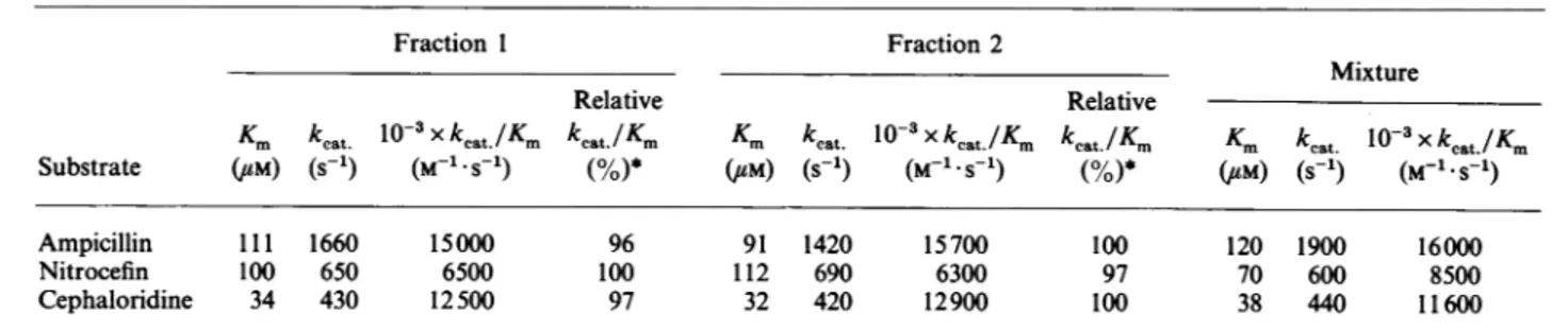
isarbitrarily setas 100%; the valueswere determined withanS.D.of+10%.Table 7.ActinomaduraR39I)-lactamase:kinetic parameters of thestartingmixture andofthe variousfractions
Fraction 1 Fraction 2
Mixture
Relative Relative
Km
keat
10-3xkcat./Km kcat./Km
K.
kcat
1o-3xkcat./Km
kcat./Km
Km
kcat
1o-3Xkcat./Km
Substrate (UM) (S-1) (M-1 S'1) (%)* CUM) (S 1) (M-1.S1) (%)* (UM) (S-1) (M-1.S_1)
Ampicillin 111 1660 15000 96 91 1420 15700 100 120 1900 16000
Nitrocefin 100 650 6500 100 112 690 6300 97 70 600 8500
Cephaloridine 34 430 12500 97 32 420 12900 100 38 440 11600
* Foreachsubstrate, themaximum value of
kcat./Km
isarbitrarilyset as 100%; the valuesweredetermined withanS.D. of+10%.Table 8.Thermalinactivationof the/lactamasesfrom B.lcheniformis(a) andS.albus G(b):comparisonof the fractions
SeeFigs.1(a)and1(b).k" and kl-arethefirst-order inactivationrate constantsat66°C and45OCrespectively.
(a)B. licheniformis
Heterogeneous
Parameter preparation Fraction 1 Fraction2 Fraction 4
103xk66(s-1) 17.3 17.8 19.4 18.2
ti(s) 40 39 36 38
(b)S. albus G
Heterogeneous
Parameter preparation Fraction 1 Fraction 2 Fraction3
103xk45(s-') 0.21 0.195 0.225 0.215
ti(min) 55 59 51 54
theshorter forms of the enzyme
(phenylalanine-57,
aspartic
acid-58 andalanine-65),
suggesting
that those forms wereprobably
more susceptible todigestion
by
theendogenous proteinase.
Kinetic propertiesandthermal
stability
The kinetic propertiesand thermal
stability
of the individual fractionswerecomparedwith those of theoriginalmixtures. The results aresummarized in Tables 4-8.With the B.
licheniformis
enzyme, theKm
values were very similarfor each fraction.Catalytic-centre
activitieswereidenticalfor
peaks
2and4,
butweresignificantly
lower forpeak
1. Table8 also shows that the thermostabilities ofthe various fractions did notdiffer from that of the mixture. It was thus difficultto decide whether the lower specific activity of peak 1 was an intrinsic characteristic of that molecular variant or if it could be due to a partial inactivation during the chromatofocusing experiment. Nevertheless, Table 4 also shows that the smaller activity of peak 1 didnotsignificantly influence the
k,8t
of the mixture.Asimilarsituationprevailed for the S.albus G enzymes. The minor fraction 1 exhibited decreased
kcat
values, but a similar thermostability. Again,thepropertiesof that minorfractiondid not appear to have a major impact on those of the mixture (Tables5 and 8).With the S. cacaoi and Actinomadura R39 enzymes, the properties of the differentfractionswere notsignificantlydifferent fromthose of themixture (Tables 6 and 7).
Interaction with
fi-iodopenicillanate
6-fl-Iodopenicillanate
is a mechanism-based inactivator of classA, class C and classD,-lactamases;
itinactivates stoichio-metrically the,8-lactamase
of B.licheniformis(De Meester et al., 1986). Theinactivation is characterized by the rearrangement of thepenicilloyl moietyoftheacyl-enzyme into a dihydrothiazine chromophore.Afterpartial(about50
%)
inactivation of theB.licheniformis ,-lactamase by6-,J-iodopenicillanate,the enzyme wassubmitted tochromatofocusing (Fig. 2). The attachment of the inactivator totheactive site of the enzyme addedonenegativechargetothe protein. Thisnewnegativecharge displacedthe whole chromato-graphic profile towards the lowerpH values. Fig. 2 showsthat each peak was split into an active fraction (high pH) and an inactive fraction(low pH). This experimentconfirms the form-5080.75. 0.50 0.25 (a)
+
0 la U) 0 5 10 15 20 25 500* 30 0 13 (b) 4 4, 14 15 16 17 18 Elution volume (ml)Fig.2.Chromatofocusing onMonoP of the B.licheniformis
II-lactamase
after partial inactivation by/I-iodopenicilianate
Pureenzyme(0.7 mg, 23 nmol) was allowed to react with 10 nmol off-iodopenicillanatein atotal volume of 700
#1
ofbuffer A for 10min at 30 'C. The samplewastheninjected into the column.(a)Proteinprofile. (b) Activity and labelling profiles; 350,1 fractions were collected and analysed asfollows. A325allows one to detect theinactivatedenzyme,whereas catalytic activity, measured with a 'good' substrate (nitrocefin), reveals the intactenzyme(active enzyme). Thearrowsshowthe relationshipbetweentheintact and inactivated forms of variants.ation ofthe irreversibly inactivated dihydrothiazine-containing rearrangementproduct.Itespecially underlinesthesensitivity of themethodused, asitclearlyappearsthattheappearanceofone
singlenewcharge,eveninthe active site of theprotein,couldbe
easily detectedbychromatofocusing. DISCUSSION
Chromatofocusing was found to be a method of choice for
the separation of protein variants originating from post-transcriptional modifications. The technique only failed in the
case of the Actinomadura R39 ,6-lactamase, which exhibits an
exceptionallylowpI. Separationof the molecularvariants of that
enzyme was only possible by simple ion-exchange chromato-graphy with the help of a very shallow gradient. Even under thoseconditions, theresolution remainedpoor. Fortheproteins withpIvalues of about5,the addition ofonenegatively charged ortheremoval ofonepositively chargedgroupdecreased thepl by at least 0.1 unit, allowing a rather easy separation. By contrast, with the R39 enzyme (calculated pl3.8), the loss of threeglutamicacid residuesonlymodified thecalculatedplvalue (3.8) by 0.03 unit. That value could not be experimentally confirmed.Inotherstudies(A.Matagne, unpublished work),the techniquewasalso foundveryusefultopurifysmallquantitiesof modifiedenzymesobtainedbysite-directedmutagenesis.Indeed, recording similar specific activities in different active peaks supplied good indication ofpurity.
The kinetic parameters of the variants were generally not significantly different from each other. The only exceptionwas
fraction 1 of the S. albus Genzyme, whichexhibited only 30%
of the activity of the other fractions. However, since the Km valuesweresimilarfor allvariants,itwasquite possiblethatthe
lower
kcat
value wasdue to partial inactivation of that variant duringtheseparation process.Anotherobservation in favour ofthathypothesiswasthat, wheneveralower activitywasmeasured,
it was similarly decreased for all tested substrates. The most
important observation was that the kinetic parameters deter-mined on the mixture were not significantly different from those of the major individual species, indicating that the
con-formation of all the molecules was probably the same. This was also confirmed by the similar thermostabilities. For the B. licheniformisand S. albus Genzymes, it isalsoimportantto stressthefact thatgoodcrystals, allowing the collectionof high-resolutiondata, havebeenobtainedfrom themixtures(Dideberg
et al., 1987; Moewset al., 1990), clearly indicating thatall the variants presented identical conformations. Interestingly,
how-ever, the crystallographic studies have shown that the first N-terminal residues of thoseproteins have no rigid structure but
seem to be freely floating in the solvent. The first well-defined residues are asparticacid-31 (ABL) in the B.
licheniformis
andserine-27(ABL)inthe S.albus Genzymes. Inbothcases,those residues are at the beginning ofthe
al-helix,
and the shortest forms which have been found start at methionine-29 for the former and serine-27 for the latter. It thusseemsthat the residuespreceding the N-terminus of the a1-helix are expendable. In agreement with those results, the major form of the S. cacaoi
enzymestarts athistidine-23(ABL)and the shortest formfound after storing the Actinomadura R39 ,?-lactamase for 4months starts at aspartic acid-28 (ABL). In both cases, the al-helix is certainly notdrastically shortened. However, sequencingofthe fresh Actinomadura R39 ,J-lactamase preparation indicated an
important proportionofmoremarkedlyshortenedforms, start-ing mainlyatalanine-48(ABL) and,inmuchsmallerproportions, phenylalanine-40andasparticacid-41. Inallthese variants,the
.0.10
-0.05
0
A. Matagne and others al-helix would be absent and, in the first one, aportion of the
,f-i
strand would also be deleted. This could result in a much decreased stability of these proteins, making them more susceptible to the proteinase(s) seemingly present in the Acti-nomadura R39 f6-lactamase preparation. It should be rem-embered that theinitial preparation lost about45%activityafter 4months ofstorage,but that therateof activity decrease became much lower thereafter.A few indications have been obtained on the phenomena which are responsible for the appearance of those variants. As stated above for the Actinomadura R39
,/-lactamase,
the presence of aproteinase in thefinalpreparation is extremely likely,since different N-terminal residues are found before and after long-term storage of the solutions (those solutions cannotbe frozen without important losses of enzyme activity). The activity of proteinases can also probably explain the results of Brive etal. (1977), who observed the appearance of new satellite bands correlated to a significant loss ofenzyme activity during con-servation ofGram-negative cells at 37'C. With the B.licheni-formis
enzyme, deamidation of theantepenultimate asparagine residue, which had been observed previously (Ambler, 1979), nicely explains the separation of molecules exhibiting the same N-termini into fractionsdiffering bymorethan 0.1pH unit after chromatofocusing. Butthenumerousvariations observed inthe N-terminal part of the enzymes must be explained by a lack of specificity of the signal peptidase(s) orby the presence ofone or more proteinases in the culture supernatant. Erpicum et al. (1990) have reported low, but significant, proteinase activities in the culture supernatants of Streptomyces lividans TK24 and S. albus G. On the other hand,manyofthe hydrolysissitesdo not appear to reflect the usual 'consensus' sequences charac-teristic ofsignalpeptidases.We believe thatoneof theimportant conclusions of the presentwork is that thekineticproperties of the mixtures are not significantly different from those of the isolated forms. With the R39 enzyme, where more extensive degradation occurred, the enzymic activity seemed to be either totally conserved orcompletely lost.This work was supported in part by the Fonds de la Recherche Scientifique Medicale (contractn°3.4537.88),anActionconcerteewith theBelgianGovernment(convention86/91-90),the Fonds de Recherche de laFacultedeMedecineULgandaConvention tripartite betweenthe Region wallonne, SmithKline Beecham, U.K., and the University of Liege. A.M. is a fellow and B.J. a chercheur qualifie of the Fonds
Nationalde laRechercheScientifique,Brussels. The sequence work was supported bythe BelgianNational Incentive program on fundamental research in Life Sciences initiated by the Belgian Science Policy Programing Department(contract Bio 22).
REFERENCES
Ambler, R. P. (1979) in fl-Lactamases (Hamilton-Miller, J. M. T. & Smith, J.T., eds.), pp. 99-125, Academic Press, New York and London
Ambler,R. P.&Meadway, R.J. (1969) Nature (London) 222, 24-26 Brive,C.,Barthelemy,M., Bouanchaud, D. H. &Labia, R. (1977) Ann.
Microbiol. (Paris) 128B, 309-317
Dehottay, P., Dusart, J., De Meester, F., Joris, B., Van Beeumen, J., Erpicum, T., Frere, J. M. &Ghuysen, J. M.(1987)Eur. J. Biochem. 166,345-350
De Meester, F., Joris, B., Reckinger, G., Bellefroid-Bourguignon, C., Frere, J. M. &Waley,S. G.(1987) Biochem. Pharmacol.36,2393-2403 DeMeester,F.,Frere, J. M.,Waley, S. G.,Cartwright, S. J., Virden,R.
&Lindberg, F.(1986)Biochem. J.239,575-580
Devereux,J.,Haeberli,P.&Smithies,0.(1984)Nucleic Acids Res.12, 387-395
Dideberg,O.,Charlier,P., Wery, J. P.,Dehottay, P., Dusart, J., Erpicum, T., Frere, J. M. &Ghuysen,J. M.(1987) Biochem.J.245, 911-913 Erpicum, T., Granier, B., Delcour, M., Lenzini, M. V., Ngugen-Disteche,
M., Dusart, J. & Frere, J. M. (1990) Biotechnol. Bioeng. 35, 719-726 Houba, S., Willem, S., Duez, C., Molitor, C., Dusart, J., Frere, J. M. &
Ghuysen,J. M.(1989) FEMS. Microbiol.Lett.65, 241-246 Izui, K., Nielsen, J. B.K., Caulfield, M.P. & Lampen, J.0. (1980)
Biochemistry 19, 1882-1886
Joris, B.,DeMeester,F.,Galleni, M., Reckinger, G., Coyette, J.,Frere, J. M.&Van Beeumen,J.(1985) Biochem.J.228, 241-248
Lenzini,M.V.,Nojima,S., Dusart, J.,Ogawara, H., Dehottay,P., Frere, J. M.&Ghuysen, J.M. (1987)J. Gen. Microbiol. 133,2915-2920 Lenzini, M.V., Ishihara, H., Dusart, J., Ogawara, H., Joris, B., Van
Beeumen,J.,Frere,J. M. &Ghuysen,J. M.(1988)FEMS Microbiol. Lett.49, 371-376
Matagne,A.,Misselyn-Bauduin,A. M.,Joris,B.,Erpicum,T.,Granier, B.&Frere, J. M. (1990)Biochem. J.265, 131-146
Meadway, R. J.(1969) Ph.D. Thesis, University ofEdinburgh Mezes,P.S.F., Blacher, R. W. & Lampen, J.0.(1985) J. Biol. Chem.
260, 1218-1223
Moews, P.C., Knox, J. R., Dideberg, O., Charlier, P. & Frere, J. M. (1990) ProteinStruct. Funct. Genet.7, 156-171
Piron-Fraipont, C., Duez, C., Matagne, A., Molitor, C., Dusart, J., Frere,J. M.&Ghuysen,J. M.(1989) Biochem.J.262,849-854 Simons, K., Sarvas, M., Garoff,H.&Helenius,A.(1978)J.Mol. Biol.
126, 673-690
Thatcher,D. R.(1975a) Biochem.J.147, 313-326 Thatcher,D.R.(1975b)MethodsEnzymol.43, 653-664
Received 26 June 1990;accepted 10August 1990 510