ACADEMIE UNIVERSITAIRE WALLONIE-EUROPE UNIVERSITE DE LIEGE
FACULTE DE MEDECINE VETERINAIRE
DEPARTEMENT DES MALADIES PARASITAIRES ET INFECTIEUSES.
SERVICE D’EPIDEMIOLOGIE ET ANALYSE DES RISQUES APPLIQUÉS AUX SCIENCES VETERINAIRES
CONTRIBUTION TO THE STUDY OF AFRICAN SWINE FEVER IN
THE DEMOCRATIC REPUBLIC OF CONGO: EPIDEMIOLOGICAL
AND VIROLOGICAL APPROACHES
CONTRIBUTION A L’ETUDE DE LA PESTE PORCINE AFRICAINE
EN REPUBLIQUE DEMOCRATIQUE DU CONGO : APPROCHES
EPIDEMIOLOGIQUES ET VIROLOGIQUES
MULUMBA-MFUMU KAZADI Léopold
THESE PRESENTEE EN VUE DE L’OBTENTION DU GRADE DE DOCTEUR EN SCIENCES VETERINAIRES
“ Je ramasserai toutes les pierres avec lesquelles j’ai été lapidé pour me bâtir une cabane ”
Acknowledgements
I have to pay gratitude to Professor Claude Saegerman, not only for having accepted to mentor this research work, but also for friendly advices dealing with our career as scientist, teacher and administrative. I was frequently encouraged by Claude, given my double occupation, which obliged me to be shared between Kinshasa in the Democratic Republic of Congo and Liege in Belgium. I also took advantage of Claude’s excellency and consistency; in fact, as a team man, Claude allowed me to work side by side with joyful and interactive young scientists who also contributed largely at the successful end of this challenge.
I have also to be very grateful to Professor Etienne Thiry, co-mentor of this work, who was the first to welcome me at the Ulg Veterinary Medicine Faculty, and thanks to whom I met Claude. Etienne accepted to come along with me not only as scientific leader, but also as a family friend. I really enjoyed to be squeezed between these two internationally recognized scientists and professors during the guidance of this thesis.
Professor Paul Pierre Pastoret, ‘may God bless his soul’, and Professor Pascal Leroy, played an important role by stimulating me to rejoin the Ulg, for state-of-the art completion of this postgraduate degree, I am taking advantage of this opportunity to express my warm thanks to all of them.
My thesis committee, as an entity, is invited to find here my profound gratitude and my high consideration, I am just promising that; this is just the beginning of long term collaboration in tropical veterinary sciences between North and South.
I also feel obliged to stress that, without some friendship links, within such a stressful and competitive environment, my daily life would not be easy; thus, this sheet gives me an opportunity to renew my friendship with Dr. Nicolas Antoine Mousseaux, the Vice Chancellor Professor Freddy Coignoul, Professor Jean-Luc Hornnick, and Professor Daniel Desmecht to whom I have to address as well my warm thanks.
Last but not the least are my UREAR colleagues, Drs. Marie-France Humblet, Fabiana Dal Pozzo and Ludovic Martinelle, I won’t forget your free of charge inputs. In fact, I am not able
to pay back your worth expertise and social assistance, please find here the expression of my gratitude.
Rosette Mushiya Mulumba, my wife, can you please accept my apologies and thousands thanks, I was obliged to leave you alone for weeks and even for months with our turbulent kids, can you please believe in this trophy I am bringing back to our family, as an outcome of our pain.
You too, my children, Mireille Tshibola Mulumba, Olivier Mutshima-Kola Mulumba, Dr. Christian Busakayi-Yombo Mulumba, Jean Marie Katumba-Mulumba, Sabine Tshialamba - Mulumba, Jules Cesar Bitetela Katumba Mulumba, Belinda Tshiswaka-Mulumba, Gregoire Tshimanga-Mulumba and, Leon Abel Kalambayi-Mulumba, you were missing me during months, can you please find here a life pathway to follow.
My sisters Theresia Katundundu, Marie Tshiswaka and, Mujinga wa ba Mulumba, you filled the gap early left by our lovely mother Tshialamba Kayombo wa Musenga who was called back by God just when she was very needed by us, as I am not able to repay your social support and sacrifices, can you please find here a gift for you, your children and grandchildren.
My brothers Felicien Bidinkuna - Mutshima Kola, Jean Michel Lumbayi Tshitenge, Juge Yvon Mwamba-Muleba and Pasteur Lukalu - Kalambayi, here is the price of our shared hopes and union as taught by our father Mutshima-Kola wa Kapambala.
To all of my nieces and nephews, especially the youngest of them, Benedict Mutshima-Kola Ndongolola, Stella Tshialamba-Kayombo and Davena, I know you are still praying a lot for me, please find here a template for your life.
I have to confess that the achievement of this thesis would not be possible without assistance of some of our technical staff at the Veterinary Laboratory in Kinshasa; I have to pay gratitude to all of them, notably Dr. Georges Tshilenge, Louison Balowe Katshiayi, Charlotte Tshinguta Lonji, Gaspard Nsome Kumo, Jean Pierre Matondo-Lusala, Dr. Boniface Lombe, Dr. Stanislas Kayimbi and Jules Mande Madika.
I am also saying thank to our administrative team, especially to Crispin Ngwanda Eloba, Bernadette Bavwazana Lukombo, Yoland Belesi and Dieudonné Lumpungu; these collaborators were several times asked to download, to print, to photocopy many of my dataset; without that contribution, achievement of this research work could not be possible.
Apart from my parents, my easy life during my youth and even after, was facilitated with diverse types of social assistance in terms of money, gifts, and encouragement from some sponsors, among whom I have to cite Abbé Gregoire Marie Tshimanyika, Abbé Pierre Mukendi, Papa Nsenga Robert, Mr. Jean Van Lancker (may their souls stay in peace) and Abbe Joseph Nkwasa Bupele, Professor Albert Mutamba, Mr. Yves Wissocq and Dr Linda Dixon; these people were like my angels positioned by God on my way; they finally deserve my thousands of thanks for their worth help and sponsorship.
This list of gratitude cannot be ended without recognizing psychological and financial support from many of my friends among whom, Professors Dr. Jean Marie Kayembe, the Dean, Faculty of Medical Sciences / University of Kinshasa; Professor Gerrit Viljoen at the IAEA in Vienna, Professor Mark Rweyemamu at the Royal Veterinary College in the UK, Professor Georges Mvumbi Lelo; Dr. Baptiste Dungu Kimbenga, GALVmed, Drs Darmon Inger, Darin Carol and Jeff Doty, CD Atlanta, Dr Leon Abel Kalambayi, Mr. Patrick Huges, and Sir Edwardo Arghasi Arghasi; they are also very very thanked on this sheet.
Acronyms and abbreviations List
9RL: specific designation of coding gene in one of the two variable genomic regions of African swine fever virus
ABC: avidin biotin complex Abs: antibodies
ABTS: 2, 2’-azino-bis (3-ethylbenzothiazoline-6-sulphonic acid ADN: acide désoxyribonucléique
AITVM: association of institutions for tropical veterinary medicine AP: apparent prevalence
ASF: African swine fever
ASFV: African swine fever virus AUC: area under the curve
BAD: Banque Africaine de Développement Bacp30: protein 30 baculovirus vector
BMC Vet Res: biomedical central veterinary research BSA: Bovine Serum Albumin
Buffer AL: specific designation of a buffer used in the QIAGEN extraction kit, lysing the cells and exposing DNA
CD: cluster of differentiation
CDC: Center for Disease Control and prevention Chi2: Chi square test
CI: confidence interval
CISA: centro de investigacion en Sanidad animal (Center for Animal Health Research, Madrid, Spain)
CSF: classical swine fever
CVL: Central Veterinary Laboratory CVR: central variable region
DAB: 3, 3-diaminobenzidine ddH2O: double distilled water DNA: deoxyribonucleic acid
dNTPs: dexyriboNucleotide Tri-Phosphate (exclusively including adenine, cytosine, guanine thymine)
DP: domestic pig
DR: Democratic Republic dUTPase: dUTP diphosphatase E. coli: Escherichia coli
EDTA: Ethylene Diamin Tetra Acid EFSA: European Food Safety Authority
ELISA: Enzyme Linked Immunosorbent Assay EP: enzootic pneumonia
Epidem Infect: Epidemiology and Infection (Journal)
ESACWA: Europe, South America, Caribbean and West Africa FAO: Food and Agriculture Organisation
FAOSTAT: Food and Agriculture Organisation Statistic
FASTA: DNA and protein sequence alignment software package GPS: global positioning system
HAD: haemadsorption
HPMPA: a broad spectrum antiviral HRP: horseradish peroxidase
IAEA: International Atomic Energy Agency IAH: Institute for Animal Health
IAP: Intracisternal A-type particle IC: intervalle de confidence ID: identity
ICCN: Institut Congolais pour la Conservation de la Nature iELISA: indirect ELISA
IFAD: International Fund for Agricultural Development IFN: Interferon
Ig G: immunoglobin G
INIA: Instituto Nacional de Investigacion y Technologia Agraria y Alimentaria ISBN: International Standard Book Number
ISSN: International Standard Serial Number IU: international unit
Kb: kilobase
Kbp: kilo base - pair KDa: kilodalton
LW: Large White
mAb: monoclonal antibody
MEGA: Molecular Evolutionary Genetics Analysis MGF: multigene family
mRNAs: messenger ribonucleic acid rRNA: ribosomial ribonucleic acid
MWG: specific designation of "eurofins ǀ mwg operon" which is a factory specialised in DNA and RNA oligonucleotides supplying
NK: natural killer cells NT: non tested
O: Ornithodoros OD: optical density
OIE: Office International des Epizooties (Organisation Mondiale de la Santé Animale) OPD: Orthophenylenediamine
ORF: open reading frame
ORF9RL: open reading frame 9RL
OURT: Ourique (Portuguese village) ticks P: prevalence
p30: protein 30 p72: protein 72
PAGE: PolyAcrylamyl Gel Electrophoresis pAR3038: plasmid AR3038
PBMM: pig bone marrow macrophages PBS: phosphate buffer saline
PCR: Polymerase Chain Reaction PPA: Peste Porcine Africaine pSG72a: plasmid SG72a 5
QIAamp: QIAGEN amplification system (specific designation from the manufacturer) QMS: quality management system
qPCR: quantitative PCR R.: Rhipicephalus
RDC: République Démocratique du Congo RNA: ribonucleic acid
rp30: recombinant protein 30 rp72: recombinant protein 72 rpm: round per minute
RSA: Republic of South Africa rtPCR: real time PCR
SDS: Sodium Dodecyl Sulphate Se: sensitivity
SEM: Scanning Electron Microscope SOP: standard operating procedure Sp: Specificity
SSA: sub-Saharan Africa
T7 (bacteriophage): specific identification of T7 infecting the majority E. coli serotypes TAE: Tris, Acetate, EDTA buffer
TaqMan: acronym from the factory, to be understood as thermo - aquaticus enzyme acting as a man, i.e. hydrolysing probe designed to increase qPCR specificity
TBED: Transboundary and Emerging Disease (Journal) TCID: tissue culture infecting dosis
TDA: Transvaal Department of Agriculture Tet-type: amino acid tetramers type
TMB: tetramethylbenzidine TNF-E: tumor necrosis factor E TP: true prevalence
TRs: tandem repeats UK: United Kingdom
UREAR-Ulg: Unité de recherche en épidémiologie et analyse de risques appliquées aux sciences vétérinaires-Université de Liège
USA: United States of America USD: United States Dollars UV: Ultraviolet
VP54: Virus Protein 54 VP72: Virus Protein 72
VPPA : virus de la Peste Porcine Africaine WB: World Bank
Content
Résumé 11
Summary 18
General Preamble 24
Part 1 - General Introduction
Chapter 1: General Background 30
Chapter 2: African Swine Fever: an update 39
Chapter 3: African swine fever virus serodiagnosis: a general review with a focus on the analyses of African serum samples.
83
Part 2 - Objectives
Chapter 4: Objectives 93
Chapter 5: Research strategies and methods 95
Part 3 - Experimental work
Chapter 6: Molecular characterisation of African Swine Fever Virus isolates involved in infection persistence in Central Africa: Democratic Republic of Congo
107
Chapter 7: Estimation of the prevalence of African swine fever in the Democratic Republic of Congo and assessment of sensitivity and specificity of the ELISA tests in field conditions.
120
Chapter 8: Genetic assessment of African swine fever isolates involved in outbreaks in the Democratic Republic of Congo between 2005 and 2012 reveals circulation of p72 genotypes I, IX and XIV and 13 variants.
Chapter 9 : Immunisation of African indigenous pigs with attenuated Genotype I African swine fever virus OURT 88/3 induces protection against challenge with virulent strains of the same genotype
175
Part 4 - General discussion, conclusions and perspectives
Chapter 11: General discussion 182
Chapter 12: Conclusions, recommendations and perspectives 206
Part 5 - Bibliographical references 211
APPENDICES
APPENDIX 1 237
Résumé
Contexte et justification de la recherche
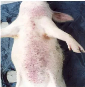
A partir de la fin des années 1980 et pendant les années 1990, une maladie fatale, mais insidieuse apparut dans les exploitations porcines des zones annexes de la Ville de Kinshasa et ses environs qui dépendent des provinces voisines du Bas-Congo et de Bandundu. Cette maladie qui affectait surtout les porcs à partir de l’âge de 5 mois, tuait entre un et cinq sujets par semaine et ce, jusqu’à l’extermination de tout l’effectif d’une porcherie. Cliniquement, la toux, la dyspnée, la fièvre, l’anorexie, de discrets piquetés hémorragiques qui pouvaient être confondus avec de la saleté cutanée avec parfois la présence de vésicules sur l’abdomen autour des glandes mammaires étaient observés. En plus de ces observations épidémiologiques et cliniques, à l’Est de Kinshasa, il avait été constaté que les exploitations porcines d’où partaient ces alertes étaient pour la plupart situées le long de la rivière Ndjili, petit affluent du fleuve Congo, qui tire sa source dans la province voisine du Bas-Congo. Vers avril et mai 1984, les informations reçues des mêmes zones, notamment du village Kingantoko, avaient fait état de l’occurrence d’une maladie qui, après avoir tué, tous les porcs dans une ferme porcine commerciale d’une vingtaine des porcs se serait propagée dans tout le village d’où elle avait frappé même les porcs indigènes errants, ne laissant que de rares survivants. Officiellement, y compris même par le passé, aucune déclaration de foyer de la peste porcine africaine n’avait été faite dans cette partie de la RDC (Figure 1). Dans le même temps, des alertes similaires provenaient de l’Ouest de Kinshasa, plus précisément de Lutendele, une vallée abritant plusieurs autres exploitations porcines, non loin de la rive gauche du fleuve Congo (Figure 2).
L’augmentation des pertes associées au syndrome décrit ci-dessus, le malentendu et la confusion entretenus par les professionnels de l’élevage sur le terrain autour du diagnostic, en pointant du doigt la maladie du Rouget, malgré l’échec du traitement avec des antibiotiques à large spectre et, le manque d’une unité spécifique de diagnostic au sein du laboratoire national de référence constituent des faits qui ont justifié cette recherche. Le but de ce travail était de permettre une maitrise de la situation de la peste porcine Africaine en RDC à travers des enquêtes de terrain et des analyses de laboratoire.
Ainsi, six études de terrain ont été menées pouvant être regroupées en quatre thématiques : la confirmation ou infirmation des cas suspects de peste porcine Africaine dans les zones
à problèmes (une étude) ;
l’étude de la prévalence, la vérification de l’état d’endémicité et l’évaluation de tests de diagnostic utilisés sur terrain (trois études) ;
la caractérisation moléculaire des souches du virus de la peste porcine africaine et l’évaluation de leur distribution (une étude) ;
le testage de l’efficacité d’un candidat vaccin pour la protection des porcs natifs de l’Afrique (une étude).
Stratégie et méthodologie
Pour les enquêtes de terrain, les zones concernées par les investigations ont été choisies en fonction de l’information épidémiologique disponible, leur lien commercial avec la ville ou province de Kinshasa, leur importance en cheptel porcin, la présence d’une interface entre porcs domestiques et porcs sauvages (interactions) ainsi que leur accessibilité. C’est ainsi que les provinces de Bas-Congo, Equateur, Katanga, Orientale, Kasaï Oriental et Maniema ont été sélectionnées. Parmi ces provinces, celles qui ont été choisies en raison de la présence d’une interface entre les porcs domestiques et porcs sauvages étaient : le Bas-Congo, l’Equateur et le Kasaï Oriental.
Pour des raisons logistiques, les données épidémiologiques et les prélèvements des échantillons ont été effectués en deux périodes (1997 à 2006 et de 2005 à 2012). Les données de terrain et les prélèvements étaient enregistrés sur un fichier Excel renseignant l’origine, le lieu de chaque prélèvement, la date et la nature du prélèvement, les coordonnées géographiques (longitude et latitude), les facteurs de risque et les résultats de laboratoire. S’agissant des facteurs de risque, les principaux étaient les suivants : l’espèce (porc domestique ou sauvage), l’âge, le poids, la race, le sexe, le système d’élevage, la présence et le type d’ectoparasites trouvés sur la peau, les lésions hémorragiques, l’état de nutrition et la distance avec la ferme la plus proche. D’autres observations étaient notées telles que, par exemple, la température pendant le transport vers le laboratoire. Les porcs adultes inclus dans l’étude étaient de toute race et âgés de plus de cinq mois. Au niveau de laboratoire, les
méthodes suivantes ont été mises en œuvre : l’identification des tiques, la détection des anticorps dirigés contre le virus de la peste porcine Africaine (PPA), la détection des antigènes et la détection de l’ADN du virus de la PPA, le séquençage et la construction des phylogrammes. En outre, un candidat vaccin atténué a également été testé et les outils statistiques adaptés ont été utilisés.
Pour la détection des anticorps spécifiques (Ac) dirigés contre le virus de la PPA et l’évaluation des caractéristiques des tests sérologiques les plus utilisés sur terrain, trois formats d’ELISA ont été évalués. C’est ainsi que pour la sensibilisation des plaques en rapport avec lesdits formats, trois types des protéines étaient utilisés comme antigènes (Ag), à savoir:
la protéine totale et cytosoluble p72 (cp72), utilisée dans un test ELISA indirect approuvé comme test de référence par l’Organisation mondiale pour la Santé Animale (OIE) et la FAO ;
la forme recombinante de la même protéine p72 (rp72) utilisée dans un test ELISA de compétition ;
la forme recombinante de la protéine p30 (rp30) du même virus aussi utilisée dans un test ELISA indirect.
Les tests de confirmation des résultats révélés positifs (douteux) consistaient en deux tests d’ « immunoblott » (IB) basés sur la p72 et la p30 et un test d’immunohistochimie (IHC).
Pour l’estimation relative au test de référence de la sensibilité (Se) et de la spécificité (Sp) des tests ELISA basés sur la rp72 et la rp30, quelques outils statistiques ont été utilisés dont le calcul du coefficient Kappa, la courbe ROC (Receiver Operating Characteristic), l’estimation par noyau (Kernel density) et le test de Chi2.
Les tiques prélevées sur les porcs domestiques et sauvages étaient identifiées par scannage grâce à un microscope électronique adapté (SEM, Scanning Electron Microscope) et un microscope stéréoscopique Olympus SZX16. Cette identification a été complétée par l’exploitation d’une clé taxonomique.
Pour la caractérisation moléculaire des souches circulantes, l’ADN viral a été extrait au moyen d’un kit QIAGEN (protocole préconisé pour le sang et les autres tissus). Trois régions cibles du génome du virus de la PPA comprenant les gènes B646L, K183L et B602L
(CVR) ont été amplifiés par PCR (Aguero et al., 2003 ; Bastos et al., 2003, Nix, et al., 2006). Les amplicons ont été purifiés en utilisant le kit Wizard ® SV Gel et le kit PCR Clean Up System, en respectant le protocole préconisé par le fabricant (Promega Corporation, USA). Les arbres phylogéniques ont été construits sur base des logiciels FASTA et MEGA (Tamura et al., 2013). Les séquences des nucléotides de tous les isolats collectés dans cette étude ont été fournies par LGC Genomics (Berlin, Allemagne) chez qui les amplicons purifiés avaient été soumis.
Pour la protection des porcs indigènes contre la PPA, le candidat vaccin utilisé est une souche naturellement atténuée OURT88/3, isolée sur une tique Ornithodoros au Portugal. Huit porcs villageois ont été vaccinés avec cette souche et inoculés successivement par les souches virulentes de génotype I, OURT88/1 provenant du Portugal et DRC085/10, provenant de la RDC. Chaque étape d’infection était séparée de l’autre par 21 jours d’intervalle. Un score clinique était établi et un suivi sérologique était réalisé successivement aux jours 3 (J3), 6, 14 et 21 post-inoculation (pi). La réponse immune ainsi que la charge virale étaient suivies grâce à une PCR quantitative (qPCR). Comme témoins, 7 porcs non vaccinés ont été utilisés lors du premier challenge et, 6 porcs lors du deuxième. Les porcs vaccinés qui ont survécu après les challenges ont été sacrifiés, en adéquation avec les règles éthiques, au 21eme jour après le deuxième challenge, soit au 63eme jour de l’expérimentation. L’essai était autorisé par les Services vétérinaires officiels de la Ville de Kinshasa.
Résultats de la recherche
1. Par rapport aux incertitudes et confusions à propos du diagnostic de la maladie qui était à la base des pertes enregistrées à partir des années 1998-90 dans les exploitations porcines à Kinshasa et dans les environs, au lieu du Rouget, c’est la PPA qui a été confirmée. Les Ac dirigés contre le virus de la PPA ont été détectés par les tests ELISA et confirmés par les tests d’IB et d’IHC. De plus, l’ADN du virus de la PPA a été détecté par PCR dans des échantillons de tissus prélevés dans toutes les localités où les foyers avaient été suspectés. La caractérisation moléculaire des souches impliquées a permis de savoir qu’elles appartenaient toutes au génotype I, dans un même taxon que les souches du Nigeria [Nig. 98/99] et du Ghana [Gha] (Mulumba-Mfumu et al., 2013). Nous avons déduit que la confusion du diagnostic était plus liée à la faible virulence des souches impliquées; en effet, la PPA a toujours été abusivement considérée comme une maladie
exclusivement hémorragique suivie de 100% morbidité et 100% de mortalité. Ce qui n’est pas le cas dans les zones endémiques de l’Afrique sub-Saharienne. Cette croyance empirique a souvent retardé la riposte et le contrôle de la part des autorités vétérinaires et a contribué à la persistance et l’endémicité de la PPA en Afrique.
2. Concernant l’étude de la prévalence, la vérification de l’état d’endémicité et l’évaluation de tests de diagnostic utilisés sur terrain :
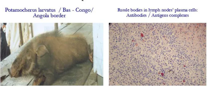
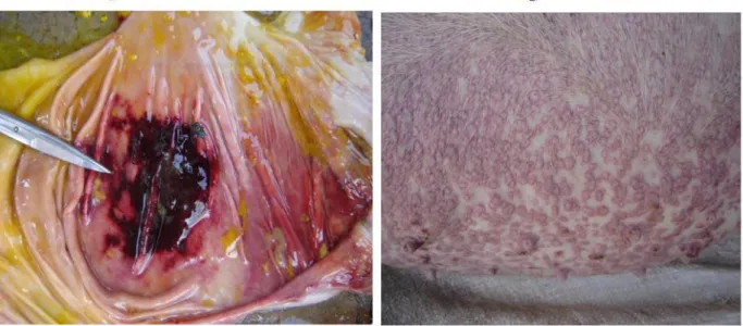
a) L’endémicité de la maladie a été établie, du fait de la détection des anticorps dirigés contre le virus de la PPA et des antigènes de celui-ci, autant chez les porcs domestiques apparemment sains que chez les Potamochères (Figure 5). L’évaluation de la présence des anticorps chez les sujets apparemment sains a révélé une prévalence apparente de 27 %.
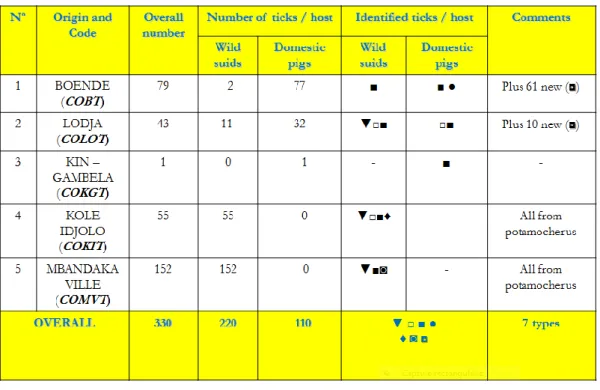
b) Concernant l’épidémiologie de la maladie, deux approches ont été envisagées lors des investigations menées à l’interface où les interactions entre les porcs domestiques et les suidés sauvages étaient possibles, notamment dans les provinces de l’Equateur (Mbandaka et Boende) et du Kasaï-Oriental (Kole). La première approche visait la confirmation de la circulation de la maladie chez les suidés sauvages et la deuxième visait la détection et prélèvement des tiques Ornithodoros dans l’écosystème de la forêt où vivent les potamochères. La première approche a permis la détection d’anticorps dirigés contre le virus de la PPA dans les sérums prélevés de potamochères de la forêt de Lingonda (2o.35’ N, 18 o.55’E), en Province de l’Equateur et la détection des antigènes dans les tissus lymphoïdes de potamochères de la savane en provenance de Kole (3 o.28’S, 22 o.28’E) et de Boma (5o 83 S, 13o07’E), respectivement dans les Provinces du Kasaï Oriental et du Bas-Congo. Par rapport avec la deuxième approche, aucune tique Ornithodoros n’a pu être identifiée. Ceci pourrait être mis en relation avec la méthode utilisée. En effet, plutôt que d’aller chercher les tiques dans les terriers des potamochères (difficiles à repérer dans une forêt inondée), les tiques avaient été prélevées sur les corps des suidés (domestiques et sauvages). Par contre, parmi les tiques prélevées, plusieurs variétés des tiques ont été identifiées telles que les genres Ixodes, Amblyoma, et Rhipicephalus (Table 1). Parmi les Rhipicephalus, une nouvelle espèce a été découverte, non encore décrite dans la littérature et celle-ci a été nommée "Rhipicephalus congolensis" (Apanaskevich et al., 2013). Par ailleurs, nous n’avons pas pu amplifier de l’ADN du
virus de la PPA à partir d’un broyat de toutes les tiques prélevées y compris celles provenant des bêtes séropositives (données non détaillées dans cette thèse).
c) L’évaluation des caractéristiques des tests sérologiques utilisés sur terrain a révélé, dans nos conditions de laboratoire et de terrain, une sensibilité (Se) relative de 93.48 % (95 % IC : 82.10-98.63) et une spécificité (Sp) relative de 87.98 % (95 % IC : 83.62-91.53) pour l’ELISA de compétition utilisant la protéine recombinant p72 (rp72). En outre, sur base des trois études indépendantes menées dans différentes zones et systèmes d’élevage, la prévalence apparente en anticorps a varié de 20 à 27 %. Par ailleurs, en prenant en considération les données de l’étude dans laquelle les autres tests de diagnostic ont été réalisés (3 tests ELISA susmentionnés et test PCR), le nombre de porcs positifs confirmés par au moins un autre test de diagnostic était de 55/328 (17%), ce qui constitue une bonne estimation de la prévalence dans la région et qui est très proche de 16,9 % noté au Sénégal par le passé (Etter et al., 2011).
3. Concernant les souches circulantes du virus de la PPA et leur distribution, une étude a été menée sur une période de sept années (2005 à 2012). Un séquençage basé sur trois gènes choisis comme marqueurs (B646L, K183L, B602L CVR) a permis de mettre en évidence, dans la zone d’étude, la circulation de trois génotypes p72 (I, XI, et XIV) et 19 variants. La co-circulation de différents génotypes en un même endroit (même foyer) a également été révélée. Ceci avait déjà été démontré auparavant (Carmina G. et al., 2010 ; Bastos, 2004). En outre, deux autres observations originales ont pu être faites, à savoir : la co-infection d’un même porc par deux variants du virus de la PPA, au cours d’un même foyer et dans une même ferme ainsi que la présence de répétions en tandems de tétramères d’acides aminés chez des isolats appartenant à différents génotypes. Ce dernier constat fait après analyse des séquences du "locus hypervariable" du génome viral (B602L-CVR) incrimine soit une homoplasie, soit une possible recombinaison génétique de plusieurs souches circulantes dans un foyer ; hypothèse déjà posée dans d’autres études antérieures in vivo (Smith, 1976). Cette étude a dans le même temps aussi démontré que le génotype I était de loin le plus dominant dans la zone d’étude, avec 40 séquences sur 62 (64,5 %), suivi du génotype IX avec 20 séquences sur 62 (32,3%) et enfin le génotype XIV avec 2 séquences sur 62 (3,2%).
candidat vaccin basé sur une souche naturellement atténuée, OURT88/3, isolée sur une tique Ornithodoros au Portugal a été testé. Ce vaccin a été inoculé à 8 porcs natifs de l’Afrique. Les sujets inoculées ont ensuite été challengés deux fois à 21 jours d’intervalle avec deux souches virulentes, OURT88/1 et DRC85/10 /08 provenant respectivement du Portugal et de la RDC. A la fin de cette expérimentation, 50% des porcs soit 4 sur 8 ont été protégés, un porc était mort d’une autre cause et, 3 porcs ont été tués par les souches du virus utilisées pour le challenge. Six porcs sur six, soit 100% et, six sur sept, soit 86%, utilisés respectivement comme animaux témoins dans les challenges 1et 2 sont morts (Mulumba-Mfumu et al., 2015). Les sujets protégés ont présenté des titres élevés en anticorps dirigés contre le virus de la PPA (test ELISA) et étaient négatifs au test de PCR quantitative (qPCR). Les porcs protégés ont été euthanasiés selon les normes éthiques au 21ième jour après le deuxième challenge, soit au 63ième jour de l’expérimentation. Ces résultats indiquent que l’espoir de trouver un vaccin efficace contre le virus de la PPA pourrait venir de souches naturellement atténuées (Sanchez-Vizcaino, 2012).
Summary
Background and justification of the research
By the end of the 1980s and during the 1990s, occurrence of fatal but, insidious disease of swine was reported within a couple of pig exploitations in the Kinshasa’s city hinterlands, including the areas of the neighbouring provinces of Bas-Congo and Bandundu. This disease in which, adult pigs starting from the winning age were the most involved was reported and killing between 1 to 5 animals per week until extermination of the total population of piggeries. Syndromically, cough, dyspnea, pyrexia, loss of food intake, discrete skin petechiae sometimes confused with skin dust, with or without vesicles on abdomen around the mammary glands were the most observed signs. Apart from these epidemiological and clinical particularities, in the East of Kinshasa, it was also observed that the majority of concerned farms were located on the edge of Ndjili river or within the surrounding areas. Ndjili is a tiny affluent of Congo River, with its source located within a hilly area southward within the neighbouring province of Bas-Congo.
Between May and July 1984, alerts received from aforementioned areas, namely from the Kingantoko village, were incriminating a severe disease of swine that killed all the pigs within a commercial farm and that, finally spread to all of the mentioned village where it kills a huge number of free ranging pigs leaving only few survivors. ASF occurrence has never been reported within these areas of DRC (Figure 1). Similar alerts were also received from the Western part of Kinshasa, namely from Lutendele, a farming valley with several piggeries, located on the East bond of Congo River (Figure 2). Finally, progressive increase of losses associated with this syndrome, misunderstanding of diagnosis by field veterinary technicians, incriminating Erysipelas despite failure of treatment with large spectrum antibiotics and, lack of local specific laboratory for ASF diagnosis formed the basis of this research work. A better understanding of ASF situation in the DRC, chiefly within the above mentioned areas was the long term objective of this research work which was carried out through field investigations and laboratory analyses.
In total, six studies grouped in four themes were conducted as follows:
confirmation of circulation of ASF within the suspected areas (one study);
screening tests characteristics (three studies);
genetic assessment of ASFV isolates’ dynamics (one study); vaccine trial for protection of African indigenous pigs (one study).
Methodology
For the field work, the surveyed areas were: peri-urban areas of Kinshasa city, and other provinces selected on the basis of available epidemiological information, their commercial links with Kinshasa, their importance in pigs populations, the presence of interfaces with interactions between wild and domestic suids and, their accessibility. Therefore, the provinces of Bas-Congo, Equateur, Katanga, Orientale, Kasai Oriental and Maniema were selected. Among these provinces those chosen for interfaces with interactions between domestic and wild suids were Bas-Congo, Equateur and Kasai Oriental. Because of logistic constraints epidemiological events and samples were documented during two sequences of time (from 1997 to 2006 and from 2005 to 2012). Field data and samples were recorded in Excel file informing about origin, sampling location and dates, geographical location (latitude and longitude), critical risk factors and laboratory results. Regarding critical risk factors, the most important were the following: species (whether domestic or bush pigs), age, weight, race (breed), sex, farming system, presence of skin ectoparasites, hemorrhagic clinical signs, health status, nearest distance with other pigs’ farms, suspicion of ASF within the area and other remarks such as temperature during transportation of samples, type of preservation of specimens in the laboratory. Adult pigs of any breeds starting from the age of 5 months were targeted.
At the laboratory level, the following methods were used for various types of needed results: ticks identification, anti-ASFV antibodies detection, confirmatory diagnostics, ASFV’s antigens detection, ASFV DNA detection, nucleotides sequencing, phylogenetic trees construction, vaccine trial and statistical analysis. For serological screening of collected sera samples we used 3 ELISAs’ formats for both detection of antibodies (Abs) and assessment of characteristics of the two screening tests exploited for both, active and passive surveillance. Coating Antigens (Ag) for the 3 ELISAs were crude cytosoluble p72 (cp72), recombinant protein p72 (rp72) and recombinant p30 (rp30) respectively. For confirmation of some positive results obtained with serological tests, p30 or p72 immunoblotting (IB) and immunohistochemistry (IHC) were used as confirmatory tests. For assessment and validation
of relative sensitivity (Se) and specificity (Sp) of the two compared screening tests as regard to the reference test (cp72). Some statistical tools were used also like the Kappa coefficient, the Receiver Operating Characteristic (ROC) curve, the Kernel density estimation, and chi-square test. Taxonomy of collected ticks’ specimens was examined using scanning electron microscopy (SEM) and stereoscopic microscope (Olympus SZX16, Olympus Corporation, Tokyo, Japan). ASFV DNA was extracted using QIAGEN kits (blood and tissues protocol). For molecular characterization, three target genomic regions comprising B646L, K183L and B602L (CVR) genes were amplified using PCR (Aguero et al., 2003, Bastos et al., 2003, Nix et al., 2006). Nucleotides sequences from amplicons purified using Wizard ® SV Gel and PCR Clean Up System kit, according to manufacturers’ protocol (Promega Corporation, USA) were provided by LGC Genomics (Berlin, Germany). Phylogenetic trees were generated using FASTA and MEGA software (Tamura et al., 2013). For vaccine trial, eight pigs were inoculated with the naturally attenuated ASFV OURT88/3 strain isolated from Ornithodoros tick in Portugal, and challenged two times at 21 days post-inoculation (pi) interval, using two virulent genotype I strains, OURT88/1 from Portugal and DRC085/10 from DRC. Vaccinated pigs were compared with non-vaccinated ones, used as controls during the assay, comprising six pigs for the first challenge and seven pigs for the second. Clinical score and sera samples were successively collected at each days 3, 6, 14 and 21 pi. Viral charge and immune response were measured using quantitative PCR and rp72 ELISA respectively. More than 50% of vaccinated pigs were protected. One pig was killed by other cause and three pigs were killed by the challenge viruses. Protected pigs were euthanised at the humane end point which was fixed at day 21 post challenge 2, corresponding to day 63 post trial. The overall experimental trial was officially allowed by the chief provincial veterinary authority of the Kinshasa City.
Research results
1. With regard to uncertainty and confusion which have been raised about ASF and Erysipelas or other so-called "red diseases of pigs", ASF occurrence has been confirmed as source of persistent mortalities and heavy losses that were reported in the 1980-90s within the peri-urban areas of Kinshasa and the neighbouring provinces. Anti-ASFV antibodies were detected serologically by ELISAs and confirmed by both, immunoblotting (IB) and immunohistochemistry (IHC) techniques. Furthermore, ASFV DNA was detected in samples collected in pigs from all the suspected areas. The
molecular characterization of involved strains revealed that they were all belonging to p72 genotype I, where they were all clustering in the same taxa with Nig 98/99 from Nigeria and Gha from Ghana (Mulumba-Mfumu et al., 2013). The diagnosis confusion was mainly due to low virulence of involved strains. In fact ASF has been always known by the field professionals as a swine disease that is strictly hemorrhagic and that kills 100% of infected pigs. This empiric knowledge of ASF contributed largely to delays in the implementation of control strategies and to endemicity of this disease.
2. Concerning the prevalence study, the determination of the endemicity status and screening tests characteristics in the field:
a) ASF endemicity was established given the fact that anti-ASFV Abs and ASFV Ags were detected both, in apparently healthy pigs and wild suids (Figure 5). Apparent Abs prevalence in these symptomless animals, revealed by three independent studies ranged between 20 to 27%, with 16 % as the very minimum in terms of reproduced results. (Barbara and Mulumba, AITVM Proceedings, 2007 ; Mulumba-Mfumu et al., 2013; and article in press)
b) Regarding ASF epidemiology, field investigations within interface villages in the Equateur province (Mbandaka and Boende) and Oriental Kasaї province (Kole), two objectives were aimed: the first was the determination of ASF circulation in wild suids and, the second was the identification of Ornithodoros ticks within that forest ecological scenario. The former survey revealed anti-ASFV antibodies in sera samples of two Red river hogs from Mbandaka (Lingonda) (2o.35’ N, 18 o.55’E) and ASFV antigens (p72 protein) in lymph nodes tissues from two Potamocherus porcinus sampled in Kole (3 o. 28’S, 22 o. 28’E) and Boma (5o 83 S, 13o07’E) from the provinces of Kasai Oriental and Bas-Congo respectively. The survey dealing with the second objective failed to detect Ornithodoros ticks, may be owing to collection strategy. Rather than digging borrows or nets (not evident to be identified in such a flooded forest), available tick’s specimens were those found on the bodies of wild suids. Finally, instead of Ornithodoros ticks, other variety of ticks including Ixodes, Amblyoma and Rhipicephalus genera (Table 1) were identified. Amongst Rhipicephalus ticks, new species, never described in existing literature were discovered and named "Rhipicephalus congolensis" (Apanaskevich et al., 2013). In regard to detection of ASFV within these ticks specimens, we failed to amplify ASFV DNA
from a mixture of crushed ticks including those collected from positive wild suids, this finding supported the previous one reported by other workers (Plowright et al, 1994; Owolodun et al., 2010).
c) The assessment of screening tests characteristics, revealed that the rp72 blocking ELISA (Ingenasa) behaved better, with a relative sensitivity (Se) of 93.48 % (95 % CI: 82.10-98.63) and relative specificity (Sp) of 87.98 % (95 % CI: 83.62-91.53) and, with respect to overall prevalence, our third study in which four different tests were combined revealed that 55 out of 328 tested pigs were positive in at least two of the four used tests. This value represents 17% of “objective” apparent prevalence; very closed to 16.9 % previously reported in Senegal upon a similar study (Etter et al., 2011).
3. Genotyping and ASFV isolates dynamics study based on the sequencing of B646L, K183L and B602L CVR genes revealed existence of three p72 genotypes comprising genotypes I, IX, XVI as well as 19 variants of ASFV in the surveyed areas of DRC between 2005 and 2012. It also revealed co-circulation of different genotypes within the same farm and during the same outbreak. This finding has been already reported in previous studies (Carmina Gallardo et al., 2010; Bastos et al., 2004). The same study reveals two other critical events: co-infection of the same pig by two ASFV variants and identity of amino-acids tetramers types in ASFV isolates belonging to different genotypes, which suggests either homoplasy or genetic recombination of co-circulating or co-infecting isolates, as already hypothised in vivo by previous authors (e.g. Smith, 1976). This survey revealed also that, with 40 sequences out of 62 (64.5%), the genotype I (ESACWA genotype) was the most dominant in those surveyed areas of the DRC, which confirmed the findings of previous similar studies on Central Africa (Ekwe, 2000, Bastos, 2003; Lubisi, 2007). Genotype I was followed by genotype IX with 20 sequences out of 62 (32.3 %) and genotype IX was followed by genotype XIV, with 2 sequences out 62 (3.2%).
4. Concerning the prospective study on protection of African indigenous pigs; a naturally attenuated ASFV strain OURT88/3 isolated from Ornithodoros ticks in Portugal was used as vaccine candidate. Eight African native pigs were inoculated and challenged twice at 21 days of interval from each post-inoculation step with two highly virulent isolates, OURT88/1 and DRC085/10 from Portugal and DRC respectively. At the end of
the trial, four pigs out eight (50 %) were protected, one pig died from another cause and three pigs were killed by challenge viruses (Mulumba-Mfumu et al., 2015). Six pigs out of six (100%) used as control for challenge one were all killed by challenge viruses and six pigs out the seven (86%) used as control for challenge two were as well killed by the challenge viruses. Protected pigs showed high titer of anti-ASFV antibodies, by rp72 blocking ELISA with zero copies of ASFV as demonstrated by quantitative PCR (qPCR). Protected pigs were euthanised at day 21 post-challenge two (63th day upon the trial), which was fixed as humane end point. These encouraging results suggest that, the future of protective vaccines against ASF could be expected from naturally attenuated vaccines (Sanchez-Vizcaino, 2012). This trial data were published as original article in Transbound Emerg Dis, 2015, doi10.1111/tbed.12303.
General Preamble
The overall framework of this thesis is a compilation of 13 chapters grouped in 5 parts detailed as follows: - part I, general introduction (4 chapters); - part II, description the objectives (1 chapter); - part III, experimental work (6 chapters); - part IV, general discussion, conclusions and perspectives (2 chapters) and, - part V, bibliographical references.
I. Part I-General Introduction:
1. Chapter 1-General Background: this section is briefly describing some basic data of the DRC including geographical, ecological, and main socio-economic features, husbandry background in general, pig sector and ASF occurrences in particular. The focus of this chapter is to provide the reader with a broad overview that will enable a better understanding of logical links between ecosystems, risk factors, risk drivers and, epidemiological events such as ASF occurrences;
2. Chapter 2-African swine fever, update and literature review: in this chapter I am trying to recall or to provide the reader with basic and update scientific information that deals with various approaches of ASF in general and ASFV infection in susceptible suids and vectors. That is to say, the specific introduction about the disease, its aetiology, epidemiology, pathogenesis, symptomatology, diagnosis, immunology, socio-economic impact, treatment and vaccine prospects will be discussed in this section. 3. Chapter 3: African swine fever virus serodiagnosis: a general review
with focus on the analyses of African serum samples. This chapter is a
specific literature review which is describing the use of ASFV p30 and its exploitation as a coating antigen for detection of anti-ASFV antibodies in routine diagnostic. It is demonstrated in this section that the ELISA based on this protein is suitable detection of the disease in pre-clinical situations or for detection of ASFV signals in the early stages of infection. As a matter of fact, it is demonstrated that ASFV p30 is important for internalisation of virus, just after attachment to infected cells and during resorbative
endocytosis which is allowing it detectable in early stages of infection. This literature review is subject of our co-authored and published article. As outcome of the paper, the diagnostic method was exploited during Eurasia’s ASF outbreaks, where it behaved very well as field investigation tool. In fact, in this situation of lack of vaccine and treatment, rapid and accurate diagnosis of ASF is the only means that can enable early and effective implementation of control strategies.
II. Part II-Objectives
Chapter 4: Objectives. The overall and specific objectives of the research work, as
main pathways to address scientific questions are described in this section.
Chapter 5: Research strategies and methodology. This chapter is depicting, the
investigation strategies and analysis methods that were exploited during our field and laboratory studies. The usefulness of this chapter is that it will enable the reader to detect the strengths and weakness of this research work and if possible to come back to us with constructive remarks that could be applied during future similar work.
III. Part III-Experimental work: this part is devoted to results and outcome of the research work. This comprises six completed articles of which four already published in internationally indexed journals of which two published as first author, two co-authored as main contributor, and two original as first author, under peer reviewing process within internationally recognised scientific journals with good impact factor. The components of this third part are the following:
1. Chapter 6: Molecular characterisation of African Swine Fever Virus
isolates involved in infection persistence in Central Africa: Democratic Republic of Congo. This chapter devoted to a prospective study which not
only confirmed and reported for the first time ASF occurrences in the research areas within the DRC, but also differentiated this epizootic from other so-called "red diseases of pig". This was a pilot study which generated other subsidiary ones. This study was subject of original article (Annales Africaines de Medecine, 2013, 6(3), 1390-1472).
2. Chapter 7: Estimation of the prevalence of African swine fever in the
Democratic Republic of Congo and assessment of sensitivity and specificity of the ELISA tests in field conditions. This chapter is covered
by an original paper which is an outcome of a combined study carried out between 2007 and 2008, reporting on field screening tests characteristics, endemicity and ASF prevalence in the DRC in addition to assessment of relative sensitivity and specificity of two serological tests used for ASF diagnosis. This article has been peer reviewed in "Transboundary and Emerging Infectious Diseases" where it is under publication process.
3. Chapter 8: Genetic assessment of African swine fever isolates involved
in outbreaks in the Democratic Republic of Congo between 2005 and 2012 reveals circulation of p72 genotypes I, IX and XIV and 13 variants. This original paper is dealing with genetic characterisation
dataset of all ASFV isolates recovered during this research work. This was the first extensive study to be conducted on ASFV isolates’ dynamics in the DRC. These data are presented in an original article which is currently submitted to Emerging infectious diseases.
4. Chapter 9: Immunisation of African indigenous pigs with attenuated
Genotype I African swine fever virus OURT 88/3 induces protection against challenge with virulent strains of the same genotype. This
chapter reports novel findings on protection of African indigenous pigs using a naturally attenuated ASFV isolate as vaccine candidate. During this trial, the protection was more than 50 % of vaccinated pigs that were challenged twice with two different strains of the same genotype, while control pigs were killed. The experimental pigs were immunised with a vaccine candidate naturally attenuated in Ornithodoros tick from Portugal where it has been collected. This has been already published in Transboundary and Emerging Infectious Diseases.
IV. Part-IV: General discussion, conclusions and perspectives.
Chapter 10: General discussion. This chapter is devoted to the general discussion
comparing current results with the ones previously published about the same topic.
Chapter 11: Conclusions and recommendations. V. Part-V: Bibliographical references.
VI. PART VI Appendixes
Appendix 1
Rhipicephalus (Acari: Ixodidae), a parasite of Red River Hogs and Domestic Pigs in the Democratic Republic of Congo". This chapter dealing with a survey on the transmission cycle of ASF in wildlife. Rather than identification of Ornithodoros ticks in both, red river hogs and domestic pigs, other variety of ticks including Ixodes, Amblyoma and Rhipicephalus genus (Table 1) were recovered. Among the recovered Rhipicephalus ticks, new species of Rhipicephalus never described in existing literature was found in our research areas. This new species was analysed and named "Rhipicephalus congolensis". This novel investigation was subject of specific article co-authored and published (J. Med. Entomol., 2013, 50(3), 479-484).
Appendix 2
Co – published article about: "The contribution of molecular epidemiology to informing control measures for African swine fever". This chapter is covered by a paper which is dealing with partial data, as outcome of active surveillance conducted between 2005 and 2006, in the same research area. The findings were presented as full article in an international congress and published in related proceedings. Interestingly, prevalence revealed by this partial study was very close to the one revealed by the Chapter 8 paper.
Chapter 1 - General background
1. 1 - Overview of agricultural sector and animal production.
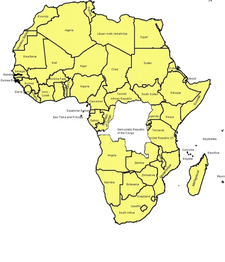
The Democratic Republic of Congo (DRC), with 2,345,409 square kilometres (Km2), is the second largest country of Africa (just after Algeria), the largest in Central Africa and Congo Basin and one of the largest states of the world (Siradou et al., 2012) (Figure 1). Just behind Amazonia basin, the Congo basin is the second largest of the world, representing 13% of the African Continent.
The DRC national border, 9,165 Kms long, is shared with 9 neighbouring countries as follows: Republic of Central Africa and Sudan in the North, Uganda, Rwanda, Burundi and Tanzania in the East, Zambia and Angola in the South and the Republic of Congo and the Cabinda enclave (Angola) in the West. The DRC is characterised by huge, rainy and virgin forest of 135.2 million Km2 representing 21 % of the continental forest and 64.5 % of the overall surface of the country (Kitengie, et al., 2013). With several natural reserves and protected areas of which 8 national parks (Virunga, Kahuzi-Biega, Maiko, Salonga South, Salonga North, Upemba, Kundelungu, and the marine Mangroves Park of Moanda) beside some hunting domains like Swakibula, Bili-Uele, Bombo Lumene and the Elephant domain of Bushimaie, including the specific park for Okapi within the Oriental Province (Kabeya et al., 2010; Siradou et al., 2013), the DRC is internationally recognised for its cornucopia of biodiversity. As a matter of fact, compared to other countries, the DRC has diverse biospheres with other types of ecosystems comprising tropical, mangroves and mountains forests, grasslands savannas and dambos that harbour many wildlife species. Globally, at least 409 mammal species (ICCN, 2010) among which wild suids species have been identified in this country. The Congo river (CR) which is crossing nine provinces including Katanga, Maniema, Oriental, Equateur, Bandundu, Kinshasa, and Bas Congo with its ten effluents of which Mongala, Rubi, Aruwimi, Lomami, Lindi, Lowa, Ulindi, Lukuga, Luvua and Lufira is an important communication means also used for traffic and trade of live animals which sometimes making it involved in the spread of epizootics.
The Congo river’s length is 4,700 Km which is ranking it as second behind Nil in Africa, as the fifth in the world after Nil, Amazon, Mississippi and Yang-Tsé (Siradou, D. et al., 2012). The DRC communication network represents: 16,000 km of navigable rivers, 1,300 km of lakes, 156,000 Km of land roads and 5,138 Km of rail ways, which, in terms of length is ranging
it as the first of Africa and the thirtieth of the world (Kitengie et al., 2013). DRC population is nowadays estimated to 80,000,000 inhabitants (Kitengie et al., 2013) with a growth rate of 3% per year. However, in 2004, this population was estimated to 55,000,000 inhabitants (WOAH/OIE, 2008). Kinshasa, the administrative capital, has currently about 8,000,000 of inhabitants, which is ranking in the second position behind Cairo and then two times more populated than Addis-Ababa. The basic commodities demand in terms of meat, eggs, milk, fish and pork is also increasing. The DRC population is mostly rural, with 75 % of inhabitants basically living in country side. A total of 60% of this population is composed of young persons (WOAH/OIE, 2008).
The DRC agricultural system is basically traditional. This country is harbouring 80% of arable areas of which only 10 % currently exploited for husbandry. The contribution of agriculture to country’s economy was estimated to 60 % in 2007. The husbandry contribution to this economy was estimated to 26 % representing 52 % of the overall agriculture contribution. The DRC’s potential in terms of grasslands is able to support at least 40 million heads of cattle and 54 million of small ruminants. Unfortunately, the local production of meat is based only on 900,000 heads of cattle; 20,000,000 of goats; 967,000 of sheep; 960,000 of pigs and 34,000,000 of poultry.
The national need in meat is complemented with 180,000 tonnes of fishery and aquaculture products per year. Nine per cent of animal production of the country is provided by insects and larvae (Kitengie et al., 2013). Therefore, the animal proteins production is very low representing only 2 % of basic needs and, the relevant deficit is estimated to 130,000 tonnes per year (WOAH/OIE, 2008). In order to address this deficit, about 550,000 tonnes of meat are imported each year. Locally, in order to resorb this gap, intensive husbandry activities consisting in exploitation of short cycle animals such as poultry and pigs have been initiated in peri-urban areas, this notably observed around some important cities of the country like Kinshasa, Lubumbashi, Mbuji-Mayi, Matadi, Mwene-Ditu, Likasi, Kolwezi and Kolwezi (WOAH/OIE, 2008).
Historically, some unfortunate events which negatively impacted on the local production of meat such as the “zaїrianisation” of non-citizens’ properties in the 1970s, the looting of private and State’s properties in the 1990s, the security crisis that followed genocide in the neighbouring Rwanda (1994) and the collapse of the second republic in 1997 (WOAH/OIE, 2008; FAO, 2012b) are still being incriminated among the causes of the aforementioned deficit. In order to boost local production in the agricultural sector, some major initiatives were finally initiated by the government in partnership with some grant organisations like the World Bank (WB), the
FAO, the Belgium government and, the Development African Bank (DAB). The majority of the national programmes were basically taken in compliance with some global and regional development strategies such as the WHO millennium objectives and the African renaissance initiatives in terms of poverty reduction, food security promotion and the employment of vulnerable classes of population. Some of these programmes were national and are still running and comprise: the Multi-sectorial and Urgent Programme for Rehabilitation and Reconstruction (in connection with war episodes, PMURR), the Strategic Document for Growth and Poverty Reduction (DSCRP), the Urgent Programme for Food Auto-sufficiency (PUAA), de Rehabilitation of the Presidential Farm of Nsele (DAIPN) and the implementation of the industrial Park of Bokanga-Lonzo.
1. 2 - Pig sector and African swine fever
Historically, it was not before colonisation, when in the 19th century, European Christian evangelists brought European breeds and introduced another system of pig exploitation in the country (FAO, 2012b). Before that period of time, only local breeds of the mentioned species were raised. With poultry, more than other livestock species, the DRC pig sector is for the moment the most growing and the most promoted, especially in peri-urban areas of growing cities as described above (FAO, 2012b).
Three types of pig exploitation systems are currently remarked in the country (i.e): i) industrial or modern clustered facilities, supplemented with veterinary drugs, mineral premixes, anti-helminthics, well composed feedings and efficient biosafety policy; ii) familial or back yarded exploitation, supplemented with household food waste and iii) traditional or free-ranging farming practice, with pigs scavenging during the day for feeding and returning back home in the evening (FAO, 2012b). Ninety per cent of the DRC population is mostly Christian and, pig husbandry is one of the main sources of animal proteins and income, especially in poor rural populations. Women and young people are the main owners of pigs in the villages. Traditional pig’s production represents 65 % of local production and, industrial system comprising modern premises represents 35 % (FAOSTAT, 2010). Nowadays, estimated at 960, 000 animals (FAOSTAT, 2010), the DRC pig population reached its peak in 1998 when 1,154,000 were counted. Unfortunately, due to abovementioned security crisis, the majority of villages were emptied in their pig populations. The huge movement of internal populations and refugees from Rwanda and Burundi was induced and, this was incriminated as the main risk factor which
population declined tremendously during the wars period that broke out during the 1990s. In terms of biomass, imported pork and by-products represents nowadays, 1000 Tons per year as the local production decreased at 24,000 Tons since 1998 (FAO, 2012b). The average price of a live pig is currently estimated at 4 USD per Kg of live pig in the villages i.e., 5 USD in urban markets. The majority of the pig exploitations of the country are located in 5 Provinces: Equateur, Bas-Congo, Bandundu, Kinshasa and Kasaї-Occidental. Due to the lack of grasslands in the forest areas, pigs and, to some extent goats are the most exploited livestock species in the Province of Equateur. The most raised breeds of pigs in the DRC are improved breeds like Large White, Pietrain (stress negative), Landrace and Duroc; apart from other cross-breeds pigs and African native pigs, so-called indigenous pigs like Ganda breed, the African large black breed (Nsalambi, 1987).
Apart from domestic pigs production, hunting of wild suids is allowed but, it is very often running in government hunting regulations, depending on year’s period or whether the concerned area is protected. In conclusion, we may nowadays confirm that the pig production has become the first of the local production in the DRC, largely before cattle (FAO, 2012b).
1. 3 - ASF within the DRC
ASF has been recognised as endemic in the SSA, an area which largely covers the DRC, by many workers (Haresnape, J. M. et al., 1987; Wilkinson, P. J., 1989). One of the major weaknesses dealing with ASF in the DRC is the lack of alerts, the timely disease reporting and, the lack of records of outbreaks by official veterinarians. According to data collected from different written reports, historical occurrences of ASF in the DRC can be presented as follows: in 1939 (Saliki et al., 1985), 1954 (DeTray, 1963), 1963 and 1967 in Katanga (Boshoff, C. I., et al., 2007), 1977 presented as Zaїre 77 (Anonymous), 1978, 79, 80, 81, 82, 83 84, 86 and 87, ASF was confirmed to be present within the country according to OIE reports (Plowright et al., 1994). During the 1990s and specifically 1997, ASF-like disease was reported in a commercial farm in Ndjili, in the East of Kinshasa, the majority of pigs were naturally dying but, some survivors were observed two weeks or even later in some lodges of the concerned farm.
Some of the pigs showing growth delay with fever were slaughtered, tissues samples were collected from these pigs; ASFV DNA was detected in kidney tissues and, anti-ASFV antibodies were as well detected in the sera samples from some survivors (Mulumba-Mfumu et al., 2013). Since then, in 2001, 2002, 2003, 2005, 2007, 2008, 2009, 2010, 2011, and 2012 more alerts about outbreaks were claimed in many locations in Kinshasa and other provinces of the country.
1. 4 - Risk factors and drivers at the country level
As previously reviewed by several authors about the SSA, a broad range of risk factors that lead to exposure of health pigs to ASF have been characterised. Based on these previous investigations, quantification of exposure pathways as well as ASF transmission models was already developed by some other researchers (Costard et al., 2007). A major outcome from such studies is a confirmation that, risk assessment is a good and efficient tool for identification of ecologically-based risk factors. Then, given the surface and the number of sub-areas of the country, a specific risk assessment framework should be recommended for the Democratic Republic of Congo. A variety of exposure pathways may be gathered:
1) Contact between natural reservoirs of sylvatic cycle (Ornithodoros ticks and wild suids) and domestic pigs;
2) Within domestic pigs, contact between infected and non-infected animals; 3) Contact between domestic pigs and ticks (Jori et al., 2013);
4) Farming practices;
5) Movements of suspected or exposed pigs including derivative products and pork;
6) Risk drivers, involving a wide range of administrative gaps comprising lack of veterinary services on the national borders, weakness of veterinary services, lack of good salaries, lack of data collection, storage and management, lack of strong surveillance systems, lack of diagnostic facilities, etc.
Main risks factors are specifically addressed in the conclusions, recommendations and perspectives chapter.
Figure 2. Map of Kinshasa City Province: ecological profile and commercial pig farms in the
1990s at the beginning of this research study (yellow spots: ASF free farms; red spots: ASF suspected farms; blue spots: pig farms being implemented or under construction)
Chapter 2 - African swine fever: an update
2.1. – Preamble
Between 1921, year of ASF description by Montgomery, and to date, a broad range of relevant articles aiming on important approaches have been published. In order to obtain consistent background as well as accurate scope of disease situation, we have been obliged to consult several libraries and catalogues for further knowledge (EFSA, 2014). As a matter of fact, ASF data about the DRC have been missing (Penrith et al., 2013).
2.2. – The following article is devoted to literature review describing the disease on the basis of field realities.
This section is subject of a review article already submitted.
ARTICLE 1:
Mulumba – Mfumu, L. K.1, 4, Dixon, L. K. 3, Madimba, Y. 1, Kazadi, E.1, Mutalakat, N.T. 1, Thiry E.2 and Saegerman, C.4
African swine fever: an update.
Mulumba – Mfumu, L. K.1, 4, Dixon, L. K. 3,, Madimba, Y. 1, Kazadi, E.1, Mutalakat, N.T. 1, Thiry E.2 and Saegerman, C.4
1
Department of Clinical Sciences, Faculty of Veterinary Medicine, University of Kinshasa
2Veterinary Virology and Animal Viral Diseases, Fundamental and Applied Research for
Animals & Health (FARAH), Faculty of Veterinary Medicine, University of Liege, 4000, Liege, Belgium
3Pirbright Institute, Pirbright, Working, Surrey GU24 ONF, UK.
4Research Unit of Epidemiology and Risk Analysis Applied to Veterinary Sciences (UREAR-
Ulg), Fundamental and Applied Research for Animals & Health (FARAH), Faculty of Veterinary Medicine, University of Liege, 4000, Liege, Belgium.
Abstract
African swine fever virus (ASFV) genome is unique; it is 170 to 190kbp long, both left and right terminal ends of its two DNA strands are covalently cross – linked by hair – loops nucleotides that are not perfectly base paired. Beside the presence of inverted terminal repeats, ASFV genome contains multigene family members, recognised for losing or gaining genomic copies depending on isolate. Instability of ASFV genetic structure has then been established. On the other hand, African swine fever virus (ASF) epidemiology within the majority of sub-Saharan Africa (SSA) countries was recognised as being complex, due to the natural history of the disease which is comprising sylvatic cycle, in addition to domestic cycle and, a wide range of risk factors. Diversity of clinical and pathological pictures of ASF, depending on isolate and host is frequently observed in many endemic ASF areas in the SSA. The acute form of the disease which is highly lethal, with hundred percent of mortality cases within infected pig population is the most stressed and the most frequently reported in published papers. Empirical understanding of ASF has tremendously contributed to persistence of infection, development of carrier status and endemicity of the disease in the SSA. Confusion with the so-called "red diseases of pigs" has as well been a source of delay in implementation of prevention and control strategies. This review is trying to recall some neglected approaches that could be of much help for optimisation of prevention and control strategies. With respect to the lack of neutralising antibodies, description of molecular basis of ASFV evasion to host immune response system is enhanced.