Université de Montréal
PURIFIED PRE- SERTOLI CELLS EXPRESS GENES INVOLVED IN
CELL PROLIFERATION AND CELL SIGNALLING DURING A
CRITICAL WINDOW IN MALE SEX DETERMINATION
par
AR0N TH0MAS C0RY
Département de biomédecine vétérinaire
Faculté de Médecine Vétérinaire
Mémoire présenté à la Faculté des ètudes supérieures
en vue de l’obtention du grade
Maître ès sciences (M.Sc.)
en sciences vétérinairesOctobre 2006
Direction des bibliothèques
AVIS
L’auteur a autorisé l’Université de Montréal à reproduire et diffuser, en totalité ou en partie, pat quelque moyen que ce soit et sur quelque support que ce soit, et exclusivement à des fins non lucratives d’enseignement et de recherche, des copies de ce mémoire ou de cette thèse.
L’auteur et les coauteurs le cas échéant conservent la propriété du droit d’auteur et des droits moraux qui protègent ce document. Ni la thèse ou le mémoire, ni des extraits substantiels de ce document, ne doivent être imprimés ou autrement reproduits sans l’autorisation de l’auteur.
Afin de se conformer à la Loi canadienne sur la protection des renseignements personnels, quelques formulaires secondaires, coordonnées ou signatures intégrées au texte ont pu être enlevés de ce document. Bien que cela ait pu affecter la pagination, il n’y a aucun contenu manquant.
NOTICE
The author of this thesis or dissertation has granted a nonexciusive license allowing Université de Montréal to reproduce and publish the document, in part or in whole, and in any format, solely for noncommercial educational and research purposes.
The author and co-authors if applicable retain copyright ownership and moral rights in this document. Neither the whole thesis or dissertation, nor substantial extracts from it, may be printed or otherwise reproduced without the author’s permission.
In compliance with the Canadian Privacy Act some supporting forms, contact
information or signatures may have been removed from the document. While this may affect the document page count, it does flot represent any loss of content from the document.
Ce mémoire intitulé
PURIFIED PRE- SERTOLI CELLS EXPRESS GENES INVOLVED IN
CELL PROLIFERATION AND CELL SIGNALLING DURING A
CRITICAL WINDOW IN MALE SEX DETERMINATION
présenté par
AR0N TH0MAs
C0RYa été évalué par un jury composé des personnes suivantes
Alan Goff, président-rapporteur
David W Silversides, directeur de recherche
Sex can be defined as a sum of characteristics caused by a network of interacting genes and proteins that give rise to sexual phenotypes within an individual. Sertoli celis drive testis formation and therefore, sex determination in the male. While some of this pathway has been elucidated, a better understanding of the RNA environment within the pre-Sertoli celi will help clarify the mechanisms by which these celis drive the differentiation process. A mouse model expressing Red Fluorescent Protein (RFP) under the control of a hybrid mouse/pig Siy promoter (HybSRYp-RFP) was used to purify e12.0 pre-Sertoli ceils. RNA was extracted and hybridized onto a micro-array (Affymetrix Mouse Genome 430 2.0) to compare the transcriptomes of stage-matched pre-Sertoli celi populations vs. whole female genital ridges. Genes identified as 2.5 fold overexpressed in the male notably consisted of celi signaling and extracellular components. This data represents the earliest microarray expression analysis of purified pre-Sertoli cells available to date. The expression of SoxlO, WntC, Fgfl8, Fgf]3 and $tc2 were characterized by in situ hybridization in the male and female gonads between ell.5 and e13.5. SoxlO, Fgfl8, Fgf]3 and Stc2 were detected in the testis cords while Wnt6 was found in both the male and female gonad. 0f these five genes Stc2, Sox]O and Wnt6 are present just after sex determination and may have a role to play in sex determination. On the other hand Fgf]8 and Fgf]3 are likely involved in the process of sex differentiation. Closer examination of the microarray data provides evidence for the activation of several intercellular signaling pathways and a new model for Sry is proposed.
RÉSUMÉ
Le sexe se définit comme la somme d’une série de caractéristiques émergeant des interactions entre certains gènes et protéines, engendrant le phénotype sexuel chez un individu. Les cellules de Sertoli dirigent la formation des testicules et, du coup, la détermination du sexe chez le mâle. Bien qu’une partie de cette voie ait été élucidée, une meilleure compréhension de l’environnement d’ARNm dans les cellules pré-Sertoli permettra de clarifier les mécanismes par lesquels ces cellules dirigent le
processus de différentiation. Une souris exprimant la protéine fluorescente rouge (RFP) à partir d’un promoteur $ry hybride souris/porc (HybSRYp-RFP) fut utilisée pour purifier des cellules pré-Sertoli à e12,0. L’ARN extrait et hybridé sur micro array (Affymetrix Mouse Genome 430 2.0) permit de comparer les transcriptomes de populations de cellules pré-Sertoli de stages spécifiques à des crêtes génitales femelles entières. Plusieurs gènes impliqués dans la signalisation cellulaire ou dans la formation de composantes extracellulaires comptent parmi ceux identifiés comme étant 2,5 fois plus exprimés chez le mâle. Ces données représentent l’analyse d’expression par micro-array de cellules pré-Sertoli purifiées la plus précoce réalisée à ce jour. L’expression de SoxlO, Wnt6, Fgfl8, Fgf]3 et $tc2 fut étudiée par hybridisation in situ dans les gonades mâles et femelles entre ell,5 et e14,5. $ox]O, Fgfl8, Fgfl3 et Stc2 furent détectés dans les cordons testiculaires, alors que Wnt6 fut identifié chez les deux sexes. Sox]O, Wnt6 et $tc2, présents immédiatement après la détermination du sexe, pourraient jouer un rôle dans celle-ci, alors queFgf]8 et Fgf]3 sont probablement impliqués dans la différentiation du sexe. Un examen plus poussé du microarray propose l’activation de plusieurs voies de signalisation intracellulaires. Finalement, un nouveau modèle est proposé pour $iy.
Titie Page j
Identification of the Jury ii
Summary in English iii
Summary in French iv Table of Contents y List of Tables ix Listoffigures x List of Abbreviations xi Acknowledgements xvi Dedication xvii INTRODUCTION 1
Hypothesis and Objective 1
1) Literature Review 1
1.1) Definition of Sex 1
1.2) Definition of Sex Determining System 2
1.2.1) Environmental Sex Determination 2
1.2.2) Genetic Sex Determination 2
1.2.3) Honeybee and CDS 3
1.2.4) X:A Sex Determination and Pathway Evolution 4
1.2.4.1) C. etegans 4
1.2.4.2) D. melanogaster 5
1.2.4.3) Networks 6
1.2.4.4) Wilkins’ Theory 7
1.2.5) Sex Chromosomes 8
1.2.5.1) ZWIZZ Sex Chromosomes 9
1.2.5.2) XY/XX Sex Chromosomes 9
1.2.5.3) Platypus 9
2) VERTEBRATE SEX DETERMINATION 10
3) MAMMALIAN SEX DETERMINATION 11
3.1) Alfred Jost and the Testis Determining Factor 11
3.2) Testicles Drive Internai Change 11
3.3) An Indifferent Embryo and Bi-potential Gonads 12
4) CELLS AND STRUCTURES 0F THE GONAD 13
4.1) Germ Celis 13
4.2) The Ovary 14
4.3) The Testicle 15
4.3.1) Celi Proliferation and Differentiation of Sertoli celis 16
4.3.2) Migrating Ceils 16
4.3.3) Vasculature 17
4.3.4) Leydig celis 1$
5) GENES IN MAMMALIAN SEX DETERMINATION 19
5.1) Methods used to Identify Genes in Sex Determination and Differentiation 19
5.2) Transcription Factors 19
5.2.1) SRY Function and Expression 20
5.2.1.1) Siy the Transcription Factor 20
5.2.1.2) Sry the Spiicing Factor 21
5.2.1.3) Sry and higher chromatin order 21
5.2.1.4) Repressor of a repressor 22
5.2.1.5) Odsexmice 22
5.2.2) SRY-related HMG box 9 (Sox9) 23
5.2.2.1) Redundancy of E box Sox Family and Sry 24
5.2.3) Chromobox Homolog 2 (M33) 25
5.2.4) Empty Spiracles 2 (EMX2) 25
5.2.5) Odd-Skipped Related 1 (Oddl) 26
5.2.6) Podocyte-Expressed 1 (Podi) 26
5.2.7) LIM Homeobox gene 1 (LIM1) 26
5.2.8) LIM Homeobox gene 9 (LHX9) 27
5.2.9) Wilms’ Tumor 1 (WT1) 27
5.2.10.1) Steroidogenic Factor 1 (SF1) 2$
5.2.10.2) Liver Receptor Homolog-1 (LRH]; NR5A2) 29
5.2.11) NROBFamily 30
5.2.11.1) Dosage sensitive sex reversal-Adrenal hypoplasia
congenita critical region on the X chromosome (DAX1) 30
5.2.11.2) Small Heterodimer Protein (SHP) 31
5.2.12) GATA Proteins 32
5.2.12.1) Gata4 33
5.2.12.2) GATA4 Function 33
5.2.12.3) GATA4 and Friend of GATA (FOG) 33
5.2.13) Forkhead Transcription Factor Foxl2 (Foxl2) 34
5.2.14) Aristalles-Related Homeobox Gene (Arx) 34
5.3) Extra-cellular Signais 35
5.3.1) Retinoic Acid (RA) 35
5.3.2) Prostaglandin (PDG2) 36
5.3.3) Desert Hedgehog (DHH) 36
5.3.4) Wingiess-Type MMTV Integration Site Famiiy, Member 4 (WNT4)
37
5.3.4.1) FGF/WNT baiance 38
5.3.5) Fibroblast Growth Factors (FGF) 38
5.3.6) Piatelet Derived Growth Factors (PDGF) 40
5.3.7) Neurotropins (NT) 41
5.3.8) Hepatocyte Growth Factor (HGF) 42
5.3.9) Insulin Receptor Famiiy 42
5.3.10) MAPK and P13K Transduction Pathways 43
5.3.11) Anti Mifilerian Hormone (AMH) 44
5.3.12) Inhibin
E3
and Follistatin (FST) 455.4) Activation of $RY 45
5.4.2) Prostaglandin, Sry, Sox9, Importin
f3
and Calmodulin Work Together 476) MATERIAL$ AND METHODS 48
6.1) Mouse Line, Ceil Collection, Celi Purification 48
6.2) Generation of Micro Array List 48
6.3) Generation of Riboprobes 49
6.4) Whole mount in Situ Hybridization (WISH) 49
7) ARTICLE 51
8) CONCLUSION 92
8.1) Contributions to Sex Determination and Differentiation 92
8.1.2) Other Interpretations of the MicroarrayData 94
8.1.3) Sry as a Canonical Transcription Factor 95
8.2) Analysis of the Microarray Technique 96
8.2.1) Limitations of the Microarrays 97
8.2.2) Potential of the Microarray 98
8.3) Defining Sex 100
Table2 82
Table3 83
Figure 2 88
Figure 3 89
Figure 4 90
Figure 5 92
Percent Microgram
pi Microlitre
Micromolar
OC Degrees Celcius
Aldhla2 Aldehyde dehydrogenase 1 family, member A2
AHC Adrenal hypoplasia congenita
AMH Anti Mtillerian Hormone
ARX Aristalles-Related Homeobox gene
ASE Autosomal signal elements
BCW 5-bromo-4-chloro-3-indoyl phosphate
BMP Bone morphogenic protein
bp Base pair
BPES Blepharophimosis-Ptosis-Epicanthus-inversus-Syndrome
BrdU 5 ‘-bromo-2’ -deoxyuridine
Ca Calcium
CaMKII Ca2/calmodulin-dependent protein kinase II
CD Campomelic dysplasia
CDS Complementary sex determination
cDNA Complementary deoxyribonucleic acid
Cyp Cytochrome
Da Daughterless
Dax 1 Dosage sensitive sex reversal-Adrenal hypoplasia expression critical region on the X chromosome
Dct Dopachrome tautomerase
DEPC Diethylpyrocarbonate
DHH Desert Hedgehog
Dmcl Disrupted meiotic cDNAY
DMRT1 Doublesex and mab-3 related transcription factor 1
DNA Deoxyribonucleic acid
Dpc Days post-coitum
Dpn Deadpan
Dpy Dosage compensation dumpy
Dscrl Down syndrome critical region homolog 1
e or E Embryonic day
Emc Extramacrochaetae
Ems Empty spiracle
Emx2 Empty Spiracles 2
ESD Environmental sex determination
FACs Fluorescent assisted ccli sorting
FGF Fibroblast growth factor
FGFr Fibroblast growth factor receptor
FigΠFactor in the germiine alpha
FOG Friend of GATA
Foxl2 Forkhead box L2
Fst Follistatin
GATA Transcription factors which bind to the concensus sequence WGATAR
GFP Green fluorescent protein
G1i3 Gli-Kruppel family member 3
Gro Groucho
Grg6 Groucho related gene 6
GSD Genetic sex determination
11cr- 1 Hermaphrodite-1
HDAC Histone deacetylase
HGF Hepatocyte growth factor
HMG High mobility group
ic Intercellular
Igfr Insulïn like growth factor 1 receptor
Inhb Inhibin
Insl3 Insulin-like peptide 3
fr Insulin receptor
kr Insulin receptor-related receptor
Ix Intersex
Kap-1 Krab-associating protein 1
Kb Idiobase
KO Knockout
Krab-O Kruppel-associated box only gene
LacZ Lactose operon
Lef 1 Lymphoid enhancer factor 1
LH Luteinizing hormone
LHX LIM homeobox domain
LUVI Transcription factor homeobox domain family
M33 Chromobox homolog 2
MAP Mitogen activated protein
MAPK Mitogen activated protein kinase
Mb Megabase
MIS Mifilerian inhibiting substance
mRNA Messenger ribonucleic acid
Msl-2 Male specific lethal-2
NLS Nuclear localization signal
NR Nuclear receptor
Nt Neurotropin
Oddl Odd-skipped related 1 gene
ORF Open reading frame
P Postnatal day
Pax Paired box
PGD2 Prostaglandin D2
Pdgf Platelet-derived growth factor
Pdgfr Platelet-derived growth factor receptor
PFA Paraformaldehyde
Pfoxic Promoter FOXL2 inverse complementary
PGC Primordial germ celi
pH relative hydrogen proton (H+) concentration
PI3K Phosphatidylinositol 3-kinase
Pis Polledlintersex syndrome
PKA Protein kinase A
PLC phospholipase C
Podi Podocyte-expressed Ï
PSR Paternal-Sex-Ratio
Ptch Patched receptor
RA Retinoic acid
RAR Retinoic acid receptor
RARE Retinoic acid-responsive element
RFP Red fluorescent protein
RNA Ribonucleic acid
RNAse Ribonuclease
RT Reverse transcriptase
RT-PCR Reverse transcriptase- Polymerase chain reaction
Run Runtgene
Scp3 synaptonemal complex protein 3
Sdc sex and dosage compensation
Sf1 Steroidogenic factor 1
SIS Sisterless
SHP Small Heterodimer Protein
sl-CDS single locus-complimentary sex determination
Smad Mothers against DPP homolog 3
SRY Sex-determining region-Y chromosomè
SSC Side chain cleavage
SSH Suppression subtractive hybrïdization
StAR Steroidogenic acute regulatory protein Stra$ Stimulated by retinoic acid $
Sxl Sex lethal
Tcf T-cell transcription factor
TDF Testis determining factor
TGF Transforming growth factor
Tra Transformer
Trk Tyrosine kinase receptor family
ts Tau somites
TSD Temperature-dependent sex determination
WARG Wilms’ tumor-anirida-genitourinary anomalies mental retardation
syndrome
WISH Whole mount in situhybridization
WNT Wingless-Type MMTV Integration Site Family, Member
WT1 Wilms’ tumor gene 1
Xoll XO lethal protein 1
XSE X signal element
YFP Yellow fluorescent protein
Acknowledgements
I would like to thank Dr. David W. Silversides who gave me thïs chance. I came to Quebec as a cowboy with high hopes and a story. I told him my story and he gave me ajob. I imagine I have flot been his best student and we have flot aiways seen eye to eye. Regardless of this he gave me this chance when nobody else would and for that I will aiways be greatful.
Dr. Nicolas Pilon and Alexandre Boyer thanks to them both for their friendship and guidance. Both of them have been like brothers. They have taught me to survive in a lab, looked out for me when I needed it and brought me to heel when I deserved it.
Céline Forget and Diana Raiwet thank you for everything. Thank you for your support in the beginning without which I would have been lost. Thank you for keeping my experiments running by working at a million things behind the scenes.
To Isabelle and Manon thank you for the million other things. Having both of you in the lab is like sunshine through a window, warm, bright and welcoming.
To my friends at the University of Montreal, thank you for being my friends even though I am flot aiways the best friend to have.
Thanks to my family for believing in me when flot even I did. Thank you for keeping me going financially and spiritually.
Thank you, Stéphanie Bolduc-Beaulieu.
Sincerely,
This Memoire is dedicated to my father, Dr. Neil Cory, for his good advice.
“Just carry on Aron. Just carry on.”
The main objective of Dr. David W Silverside’s laboratory has been to study sex determination and differentiation in mammals. We hypothesize that the pre Sertoli celis drive male sex differentiation and that the genes involved are largeiy undefined. To further characterize genes differentially expressed by pre-Sertoli ceils at the beginning of sex differentiation we have made use of a mouse model expressing Red Fluorescent Protein (RFP) under the control of a hybrid mouse/pig Sîy promoter (HybSRYp-RFP) (Boyer et al., 2006). This mouse une was used to isolate and purify pre-Sertoli ceils after the peak of S’y expression but before histological testis cord formation. RNA from purified e 12.0 pre-Sertoli celis or stage matched whole female genitai ridges was hybridized onto microarrays to study the transcriptomes of pre-Sertoli celis.
Literature Review 1.1) Definition of Sex
Throughout the animal ldngdom the existence of two sexes, which contain ceils that undergo meiosis and reproduce sexually, is aimost universal. The word “sex” cornes from the Latin word sexus, meaning division (frorn secare, to cut or separate). fronically, sex (being male or female) is flot a clear-cut bïological entity. In several species sex is immaterial, with animais changing from male to female several tirnes throughout their lives or being hermaphrodites, containing both sexes within one individual (Dimijian, 2005). Even within species with obvious maie or femaie groups, masculinity and femininity wiii vary among individuals in a population. This means that populations are composed of individuais differing aiong a spectrum of peculiarities of structure and function that define maies or females. Sex couid be better defined as a sum of characteristics caused by a network of interacting genes ai-id proteins that give risc to sexual phenotypes within an individual.
1.2) Definition of Sex Determining System
A sex determining system is a biologicai system that determines the development of sexual characteristics in an organism. Sexual reproduction requires a balance of two sexes in a population in order to function efficiently. if a male and a female are required for reproduction, having ail male or ail female offspring in the population can ultimately iead to extinction (Miller et al., 2004). In order to avoid this unfortunate quirk several sex determining systems have arisen to balance the ratio of maies and females in a population. These strategies can be divided into two main categories: environmental sex determination (ESD) and genetic sex determination (GSD).
1.2.1) Environmentai Sex Determination (ESD)
Severai species of fish and reptiles rely on environmentai dues to determine the sex of individuals. The sex of offspring can be determined by pH, environmental temperature or even the existing male/femaie ratio in a population (Haqq and Donahoe, 199$). For example, the marine turtle L. otivacea is ciassified as having temperature-dependent sex determination (TSD). Turtle eggs incubated at temperatures of 26°C will ail deveiop as maies whiie eggs incubated at temperatures of 33°C will produce femaies (Moreno-Mendoza et al., 2001). Environmental sex determination is taken one step further in some fish species. In C. citrineÏlum sexuai identity is conferred to an individuai through social signais such as size and dominance (Francis and Barlow, 1993).
1.2.2) Genetic Sex Determination (GSD)
In birds, mammais and severai types of insects sex is determined at conception by one or more genes. Much like ESD there are severai strategies contained within this category. Within Hymenoptera insects, which include ants, wasps and honeybees, the sex of the individuai is determined by the total chromosome number. DrosophiÏae metanogaster and Caenorhabditis etegans measure relative amounts of inherited sex chromosomes. Within birds, inheritance of sex Z or W chromosomes defines sexual identity. Within humans, and mammals in
general, sex is thought to be assigned at fertilïzation with the inlieritance of either an X or Y chromosome. In general these animais are bom either male or female and develop testis or ovaries to produce sperm or ovum respectively. While populations often follow these defined mechanisms it is flot the rule in every case.
1.2.3) Honeybee and Complementary Sex Determination (CDS)
The reason for sexual reproduction in general has been explained by several theories. The two main and opposing theories are Muller’s ratchet (Muller, 1964) and the Red Queen (Ridley, 1993). Muller’s ratchet basically states that sex acts as a way to purge harmful mutations and genetic disease from the gene pool while the Red Queen states that sexual reproduction is a means to increase genetic diversity to offset evolving parasites and a changing environment. An excellent example of these two theories working in tandem can be seen in the insect order Hymenoptera. This species comprises over 200,000 species of ants, bees, wasps and sawfties (Bull, 1983; White, 1973) Ail members have haplodiploid sex determination: diploid individuals derived from fertilized eggs develop as females and haploid individual derived from unfertilized eggs develop as males.
One mode of sexual determination in Hymenoptera is termed single locus complimentary sex determination (sl-CDS). In 2003 it was determined that the primary gene regulating sexual development was CDS (Beye et al., 2003). Honeybees heterozygous at this allele develop as females while hemizygous haploid individuals are male. Occasionally, crosses that produce homozygote individuals for the sex determining genes can result in diploid maies as well. Within honeybee populations homozygous diploid males are usually sterile and in most cases are killed and eaten by the workers (van Wilgenburg et al., 2006). This negative selection against homozygosity in diploid individuals ensures that neutral or beneficially rare alleles have a higher transmission rate as postulated in the Red Queen theory. At the same time haploid males with detrimental mutations will die because a functional gene does not mask the mutated gene. As stated in Muller’s Ratchet hypothesis this gene will flot be passed on and it will be purged from the gene pool.
1.2.4) X:A sex determination and pathway evolution
Two well-studied organisms highlight how different sex determining pathways can develop and differentiate. The nematode Caenorhabditis etegans and the fty Drosophilae metanogaster dispiay sex determining mechanisms based on a ratio of sex chromosomes to autosomes (reviewed by Cime and Meyer, 1996). In both animais sex is determined by a set of dosage sensitive genes on the X chromosomes. These X signal elements (XSEs) transmit information relative to the amount of X chromosomes to autosomes in order to determine sex. In this manner a double dose of XSEs (two X chromosomes) wiil specify a female fate whule a single dose (one X chromosome) specifies a male fate. In C. etegans the primary target of XSEs is the repression of XO lethal -l (xol-1), whule in D. melanogaster XSEs activate sex tethat (sxl). Both xol-] and sxl have dual roies: first to control gene dosage compensation and second to initiate sexual differentiation cascades through the gene transformer. Even though both proteins have similar functions the functional strategies differ greatly.
1.2.4.1) C. etegans
C. etcgans uses a sex determining system that measures the ratio of X chromosomes to autosomes. The sexual determination mechanism of C. etcgans has been studied in great detail (reviewed by Stothard and Piigrim, 2003; Cime and Meyer, 1996). Worms with an X:A ratio of 0.75 or above wiii be hermaphrodites, producing both sperm and eggs, while worms with an X:A ratio of 0.67 or below wiii be males, producing oniy sperm (Madi and Herman, 1979). The major factor influenced by this ratio is the expression of the transformer-] gene (TRA-1). The suppression of this gene leads to the deveiopment of sperm and male phenotypes whereas the expression of this gene causes the development of oocytes and female sexual characteristics. Rather than being a short cascade, the X:A ratio does not directiy control the expression of TRA- 1.
The initiai signais in this cascade consist of repressive XSEs and up reguiating autosomai signal elements (ASE). The balance of repressive XSEs to positive ASEs determines if xoi- 1 is active in sufficient amounts to promote maie deveiopment
(Poweli et al., 2005). if xol-1 is inhibited (X:A=1) a group of sex and dosage compensation (sdc-1, sdc-2 and sdc-3) and dosage compensation dumpy (dpy) genes are transcribed. The SDC proteins are thought to act together as complexes with DPY proteins to hypo-activate both X chromosomes and suppress the expression of hermaphrodite-1 (her-]). In the absence of her-] the intraceliular tau of TRA-2 is cleaved by TRA-3. This creates an intraceliular TRA-2 (TRA-2ic) ligand that binds and inhibits the FEM proteins. Inactive FEM allows TRA-1 to repress genes such as egi-] and mab-3 that, in tum, promote male development.
In the presence of xol-1 (X:A=0.5), SDC-2 protein production is suppressed ailowing the activation ofher- 1. When active, lier- 1 produces a ligand that binds and inhibits the cleavage of the transmembrane protein TRA-2. TRA-2ic production is suppressed thus FEMs are not inhïbited. Within the male nematode FEM proteins suppress TRA-1 action that allows egt-] and mab-] to be expressed. For a more in depth review the reader is directed to a review by Goodwin and Eliis (2002) and Cline and Meyer (1996).
1.2.4.2) D. melanogaster
D. metanogaster also uses an X:A sex determining system. Again the major downstream gene affected by this ratio is Tra. However rather than using negative feedback Ïoops D. meÏanogaster achieves Tra activation through titration and gene splicing (reviewed by SchUtt and N5thiger, 2000; Cline and Meyer, 1996).
The initial control ofSxl requires maternai factors, XSEs and ASEs. Maternai factors are those products passed from the mother to the embryo via the oocyte and include the product of genes such as daughtertess (da), hermaphrodite (her), groucho (gro) and extramacrochaetae (emc). The known XSEs are three sisterless (sisA, sisB and sisC) genes and the gene runt (run) while the oniy known ASE is deadpan (dpn). In this system DPN works to prevent SIS proteins from forming heterodimers with DA. if there are more SIS proteins than DPN proteins, as in femaies, a DA-SIS heterodimer is formed which binds and activates an establishment promoter of sxl
(SXlpe) which in turn causes the production of a functional SXL protein. In males DPN
In the absence of SXL the gene mate spectflc Ïethat-2 (mst-2) is expressed and works with three other msl genes to hyper activate the single X chromosome in the male. Within the female there is also some evidence that SXL directly represses the expression of several genes including mst-2. Regardless of the protein involved X chromosome gene dosage is baianced in males and females.
Slightly after the titration step sxÏ is activated in both sexes through a maintenance promoter. This promoter transcribes a non-functional mRNA, which needs posttranscriptional splicing to occur in order to produce a functional protein. In the female, the functionai SXL produced earlier, by activation ofSXtpe, will promote
the proper spiicing of sxl mRNA leading to more functional SXL protein. These functional SXL proteins will regulate the proper production of more SXL proteins. In males the absence of functional SXL wiil lead to a non-functional SXL protein and sxt gene expression will be lost.
InD. melanogaster, tra is active in both sexes and produces a pre-mRNA that again needs to be spliced. In females, the functional SXL protein will bind to and help splice tra mRNA in order to give a functional protein. TRA along with TRA2 will bind to a sub optimal 3’ site on thedoubtesex (dsx) gene promoter which causes a female form of DSX protein to be produced, DSXF. Along with her and intersex (ix), DSXF works to up-regulate female specific genes while repressing genes involved in male differentiation. In the absence of SXL, and therefore functional TRA, a different 3’ binding site activates dsx and leads to the production of a male form of DSX (DSXM or doubtesex male protein). This gene promotes the activation of male genes and represses female genes.
1.2.4.3) Networks
The SXL protein is well conserved between severai species of fly including Musca, Chiysomya, Megasetia, Ceratitis and Drosophita but SXL does flot seem to determine sex in any other species other than Drosophila. On the other hand, homologues of DSXM and DSXF do appear to follow a sex specific expression pattern in ail of these species (Schtitt and Nithiger, 2000). Several experiments in Drosophita could possibly illustrate that this genetic network can be rearranged to
give several different sex determining systems (reviewed by Cime and Meyer, 1996; Schûtt and N5thiger, 2000).
XX embryos from da mutant mothers do flot activate SXL and therefore die resulting in 100% male offspring from these mothers (Parkhurst et al., 1990). if the production of DA became dependent on nutritional cues this would allow the mother to preferentially produce male offspring in certain environmental conditions and female offspring in others. When msl-2 is ectopically expressed in heterozygotic msl 1 knockouts in Drosophila, male files will flot readjust X chromosome gene dosage in response to ectopically expressed sxt. Therefore msÏ-2 becomes the genetic switch for dosage compensation freeing the fiy from sex specific lethality. In this case a functional sxt product will act as a dominant female determining factor regardless of the X:A ratio. There are also several examples in the literature showing circumvention of the sxt sex determination within Drosophila due to interruption or improper expression of tra-] or tra-2. These range from the gain of function causing male to female sex reversal (McKeon et al., 1988), loss of function causing female to male sex reversai (Sturtevant, 1945) and even a temperature sensitive allele that causes female to male sex reversal when larvae are raised in temperatures at or above 29°C (Belote and Baker, 1982). While none of these examples are shown to be actual sex determining mechanisms in nature they do demonstrate that sex is flot a result of one gene but of genes working together as a network. Furthermore genes that function in a certain way in one network may have a very different role in another network. This is something that should be kept in mmd when studying the vertebrate sex determining systems.
1.2.4.4) Wilkins’ Theory
The information gathered from the study of sexual determination in Caenorhabditis elegans bas led to the theory that sex determining pathways evolve in a reverse stepwise fashion driven by frequency-dependent selection for the mlnority sex (Wilkins, 1995). The theory assumes that a primordial sex determination pathway would have been very simple, depending on one or two genes that in turn responded to environmentai cues. These environmentally responsive elements would have
allowed for an optimal sex ratio to be achieved in the population. Either through diseases, which skew sex determination, such as PSR or Wolbachia (reviewed by Normark, 2002; Werren and Stouthamer, 2003), or because of mutations in key genes this balanced sex ratio, would have become skewed to favour one sex (i.e. females). In an attempt to re-equilibrate the sex ratio, alleles that promote the minority sex (males) would have been under positive selection pressure.
One strategy may be to suppress the gene causing the unbalanced phenotype, as seen in Caenorhabditis etegans. A separate strategy may be flot to suppress the disease but mask it by increasing gene expression, as seen in Drosophila melanogaster. if these alleles aggressively promoted the minority sex they would themselves push the population towards the sex originally suppressed by the disease (male) causing positive selection for genes promoting the new minority sex (female). In either case this seesaw sex ratio would continue until an equilibrium mechanism is eventually achieved or the species goes extinct. The stepwise evolution of superimposed sex determining mechanisms may be a major confounding factor in unraveling the genetic cascade behind vertebrate sex determination systems and sex differentiation.
1.2.5) Sex Chromosomes
In birds, mammals and several other species sex is genetically determined at the time of fertilization through the inheritance of sex chromosomes. The sex chromosomes are distinct from autosomes in that they differ in size, number, staining characteristics and gene content when the two sexes are compared. Ohno’s law asserts that heteromorphic sex chromosomes originated from an ancestral autosomal chromosome, which obtained a mutation that confened a sexual advantage (Ohno, 1967). It has been hypothesized that chromosomal rearrangements, such as inversions of specific sex related genes and operons, suppressed recombination and fostered the accumulation of sex specific genes within the sex chromosomes. The differences in size often observed between the sex chromosomes have been caused by subsequent deletions. This degeneration has been demonstrated in the W chromosome of
Neognathan birds and the Y chromosome of mammals (Ansari et al., 1988; Pigozzi and Solari, 1997).
1.2.5.1) ZW/ZZ sex chromosomes
The ZW/ZZ sex determination system is found in birds and some insects. In the ZW system females are the heterogametic sex (ZW) while the males are the homogametic sex (ZZ).Bombyx mon, the commercial silk worm, is known to have a dominant female determining factor residing on the W chromosome and therefore the inheritance of the W chromosome drives female development (Goldsmith et al., 2005). On the other hand, within Lepidoptera (butterfiies and moths) examples of ZO, ZZW and ZZWW females have been found suggesting that the W chromosome is flot essential for female development in some species. Within birds the presence of a female determining gene on the W chromosome has not been found. Currently the most accepted hypothesis stipulates that a double dose of an unknown gene on the Z chromosome is needed for male development (reviewed by Clinton and Haines, 2001).
1.2.5.2) XYIXX sex chromosomes
Most mammals use the XX/XY sex determination system. In the XX/XY system sex is conferred through the inheritance of either homogametic chromosomes (XX) creating females or heterogametic chromosomes (XY) creating males. The existence of Klineffelter’s males, in which two or more X chromosomes and one Y chromosome are inherited, show that it is the presence of the Y chromosome and not the dosage of the X chromosome that determines the entry into the male developmental pathway. In 1990 the Sex —Determining Region of the Y chromosome (SRY) gene was identified as a dominant gene found on the Y chromosome, which drives testes determination (Sinclair et al., 1990).
1.2.5.3) Platypus
Comparative mapping of genes from the vertebrate Z and X chromosomes showed that the two share no homology, implying that they are derived from different
autosomes (Nanda et al., 1999; Shetty et al., 1999). According to a review by Gruetzner et al. (2006) the ZW/ZZ and XY/XX sex chromosomes may be reiated. This review points to studies of the piatypus, which contain ten sex chromosomes, five X and five Y. These sex chromosomes construct multivalent chains in male meiosis creating X1X2X3X4X5 gametes and Y1Y2Y3Y4Y5 gametes. Gene mapping experiments have revealed that X5 at one end contains homologues of bird Z chromosome genes and the X1 shows homologues of mammalian X chromosome genes. These observations have iead to severai theories which suggest that both sex determination system may have evoived and diverged from a common source.
2) VERTEBRATE SEX DETERMINATION
2.1) Common Goals in Vertebrate Sex Determination
As mentioned earlier reptiles, bird and mammals have differing sex
determination strategies including ESD, ZZ/ZW chromosomai and XXIXY
chromosomai sex determination respectively. Despite these differing strategies there is a close anatomical relationship between the development of the genital ridge and the excretory system during early ontogeny of ail vertebrates. In reptiles, birds and mammals, a mesodermal layer ventral to the somites differentiates into structures involved in excretion and reproduction. Gonadai organogenesis begins with the thickening of the coelomic epitheiium on the medial aspect of the mesonephros in which the germ ceiis wili eventually settie (Merchant-Larois et ai., 1984). At this point it is impossible to morphologicaily distinguish males from femaies. The embryo and the genital ridge are said to be bi-potential, possessing structures and celi types that can differentiate into male or female phenotypes (Ito et al., 2006). In response to signais from the ovary or testis, the femaie reproductive tract system deveiops primarily from the paramesonephros (Mifilerian) duct, and the maie reproductive tract forms from the pronephros (Woiffian) duct, respectiveiy (Grobstein, 1955). There are differences among species in how closeiy connected the structures are in regard to sharing ducts for secretion, but amongst teleost fish, reptiles, birds and mammals the mature testes contain Sertoli and Leydig cells in addition to germ celis, and the ovaries consist of thecai and granuiosa ceils surrounding the ovum (reviewed by
Blum, 1984). There is evidence of some conservation of genes involved in sexual differentiation among ail of these creatures. While sex determination of fish, birds and reptiles has been and is cunently being studied the largest amount of information has been found in mammals.
3) MAMMALIAN SEX DETERMINATION 3.1) Alfred Jost and the Testis Determining Factor
The studïes donc in the 1940’s by Alfred Jost were the first to point towards a Testis Determining Factor and the Female Default Pathway in mammals. Jost showed evidence that rabbits castrated in utero before the time of sex determination developed female reproductive systems (Jost, 1947). Through this study Jost showed that the testicle in mammals drives development of the male reproductive tract. This study also became the biological basis behind the theory of the female default pathway. The female default pathway stipulated ovaries and female reproductive organs would develop only in the absence of a Y chromosome male determining gene(s). This in turn lcd to the hypothesis of the Testis Determining Factor (TDF), a dominant gene that actively drives testis development. This gene remained hypothetical until 1990, when Sex —Determining Region of the Y chromosome (Sry) was identified (Sinclair et al., 1990).
3.2) Testicles Drive Internai Change
Early in embryonic development it is impossible to distinguish male embryos from female embryos. In effect the embryo is bi-potential containing both Miillerian ducts as well as Wolffian ducts. Two hormones secreted by the testicle drive male development of the Wolffian ducts. Sertoli cells produce the hormone Mifilerian Inhibiting Substance (MIS) also known as Anti-Miillerian Hormone (AMH) and Leydig cells produce testosterone. MIS is responsible for the regression of the Mifilerian ducts while testosterone maintains and differentiates the Wolffian ducts as well as the extemal male genitalia (reviewed by Viger et al., 2005). Due to the action of testosterone and its derivatives the Wolfftan ducts differentiate into the epididymis, vas deference and seminal vesicle of the male reproductive tract. As well,
testosterone heips to deveiop the penis and scrotum. In the absence of MIS and testosterone the Miillerian ducts are maintained while the Wolffian ducts degenerate. The effector causing the differentiation of the Mifilerian ducts is not weii known. It has been suggested that the estrogens produced by the placenta, the fetal ovaries or even that from maternai circuiation plays a role in Mifilerian duct differentiation (Langmand and Sadier, 1996). As weii, the morphogen Wnt4 has been impiicated in the development of the Mûilerian ducts and the repression of testosterone production (Vaino et al., 1999). In either case, the Mûilerian ducts contribute a iarge portion of
the female reproductive tract, differentiating into the oviduct, uterus, cervix and upper part of the vagina. While these processes are themselves accompanied by several other changes throughout the body they are au dependent upon the determination of sex and the subsequent interaction of the primordiai germ ceiis, support ceiis, steroidogenic ceils and connective tissue celis.
3.3) An Indifferent Embryo and Bi-potentiai Gonads.
During vertebrate embryogenesis, the urogenitai system derives from the intermediate mesoderm of the gastruia. The male reproductive tract forms from the pronephros (Wolffian) duct and the femaie reproductive tract system deveiops primarily from the paramesonephrotic (Mifilerian) duct. In the mouse, the Wolffian duct is first formed from the intermediate mesoderm by embryonic day 9 (e9). Before the Woiffian duct reaches the cloaca, the mesonephros forms. A thickening of the coelomic epithelium on the ventral medial surface of the mesonephros is the first indication of the genital ridge (Grobstein, 1955; Brennan and Chapei, 2004; Viger et ai., 2005). At or near the same time, germ ceiis migrate from the hindgut to begin to populate the genital ridge (McLaren, 2000). Shortly after the arrivai of the germ ceils in the genital ridge at elO.5, the celis of the coelomic epitheuium of the genital ridge begin to actively divide and invade the underiying mesenchyme.
The Mtiilerian duct starts to form by invagination of the surface epithelium of the anterïor mesonephros around ell.5 in the deveioping urogenital ridge. This epitheliai invagination extends caudally along the Woiffian duct, lateraily and then mediaily towards the cloaca (Gruenwaid, 1941). Thus regardless of their genetic sex,
the embryos have both male and female reproductive tract precursors, before morphological signs of sexual differentiation occur.
In the mouse, around e 12.0, the epithelial celis of the genital ridge multiply to form primitive sex cords. The primitive sex cords actively surround the germ celis while stiil staying in contact with the surface epithelium. At this stage it is stiil morphologically impossible to distinguish male from female gonads (Langmand and Sadler, 1996). The gonads now contain the four main precursor cell types needed to form either the testicle or ovary: The primordial germ cells (PGC), the support cells, the steroidogenic ceils and the connective tissue celis. The PGCs migrate into the gonad surrounded by the bi-potential support celis (Karl and Capel, 1998). The mesenchymal cells will eventually differentiate into the connective ceils and steroidogenic cells (Buehr et al., 1993; Martineau et al., 1997; Ito et al., 2006). Within the male these celi types will differentiate into pro-spermatogonia, Sertoli cells, peritubular myoid cells, endothelial cells and Leydig celis. Within the female they form ovocytes, granulosa cells, stromal cells and theca cells.
4) CELLS AND STRUCTURES 0F THE GONAD 4.1) Germ Cells
Primordial germ cells (PGCs) are first detectable at e7.0 as a group of approximately 50 cells clustered at the posterior of the primitive streak in the extra embryonic region. As the embryo develops to the pre-somite stage, the germ celis become a distinct group of cells distributed along the base of the vitelline vesicle (Hogan et al., 1994).
By e8.0 the PGCs begin to actively migrate through the hindgut to the genital ridge (Hogan et al., 1994). Around elO, germ celis begin to enter the genital ridge (McLaren, 2000) and continue to do so until ell.0 — ell.5 (Hogan et al,. 1994).
Those that do not enter the genital ridge by this time are lost and eventually eliminated (Molyneaux et al., 2001). During this migration and colonization of the genital ridge, the germ ceils will divide mitotically once every 16 hours resulting in close to 25 000 PGCs colonizing each gonad by e13.0. Within the male genital ridge the PGCs will be requisitioned to the testis cords which consist of pre-Sertoli celis
surrounded by peritubular myoid celis. Through interactions with Sertoli celis the PGCs will stop dividing at phase Gi of the mitotic celi cycle at about e12.5 (McLaren, 2000). Though they are essential for fertility they play no role in the development of the testis (McLaren, 2000; Adams and McLaren, 2002). Within the female genital ridge the PGCs enter meiosis in an anterior-to-posterior wave and are theorized to drive differentiation of the ovary (McLaren, 1984; Yao et al., 2003; Bullejos and Koopman, 2004).
4.2) The Ovary
In the absence of Sry, the bi-potential gonads enter the female differentiation pathway. In the mouse, the first recognizable morphological difference in the ovary, the entrance of germ celis into meiosis, is seen at e13.5 (Nordqvist, 1995). In the ovaries, the primitive sexual cords segment themselves into irregular cellular masses containing germ cells. At this point germ cells are termed gonocytes. These gonocyte nests are situated in the medullar region of the ovary and are later replaced by vascular stroma (Langmand and Sadler, 1996). In contrast to the testicle, the superficial epithelium of the ovary thickens and continues to proliferate finally creating a second generation of sexual cords, the cortical sex cords. These cords will penetrate into the underlying mesenchyme without separating from the coelomic epithelium (Langmand and Sadier, 1996). The cortical cords themselves segment into cellular masses containing one or more gonocytes that subsequently transform into ovogonia and drive the differentiation and organization of the follicular ceils and structures (Langmand and Sadier, 1996).
In the absence of Sertoli celis, gonocytyes enter meiosis at e13.5 and stop development at prophase I. Most of these ceils will recommence their meiotic division at the beginning of the ovarian cycle at puberty (McLaren, 2000). The meiotic germ cells antagonize certain pro-testicular events. Meiotic gonocytes are essential to follicular formation as cell migration and testicular cord formation can be induced in XX gonads before gonocytes enter meiosis but not afterwards. Once meiosis begins XX gonads loose their plasticity and are committed to an ovarian pathway (Tilmann and Cape!, 1999; Yao et al., 2003). Gonocytes in the ovary enter
into meiosis in an anterior to posterior wave, parallel to genetic markers that define follicular ceils suggesting an influence of meiotic germ ceils on the support celi population (Bullejos and Koopman, 2004).
Littie biologie or genetic information is available to explain ovarian development. The PGCs are needed for the differentiation of the follicular ceils. In the absence of PGCs follicles fail to develop in the ovary (Huang et al., 1993). One gene expressed by the ovocytes, Figa, is necessary for the initial stages of folliculogenesis; the recruitment of the granulosa cells around the primordial follicle (Soyal et al., 2000). At the level of the somatic cells, FoxÏ2 is necessary for the differentiation of the granulosa cells. Altliough Foxl2 is not essential for ovary formation, granulosa ceils of FoxÏ2 KO mice are unable to become squamous cuboid ceils and there is an arrest of folliculogenesis (Schmidt et al., 2004; Uda et al., 2004). For a long time Daxi was considered to be the ovary determining factor but the creation of a Daxi KO mouse demonstrated it was flot essential to ovary formation (Yu et al., 1998a). A second gene, Wnt4 lias been shown to restrict migration of endotlielial and steroidogenic celis in the XX gonad and also inhibits tlie formation of the coelomic vesicle in male gonads when expressed ectopically. One last gene, Follistatin (Fst), is necessary to prevent XY specific vascularization and also permits survival of meiotic germ celis in tlie ovary (Yao et al., 2004).
4.3) The Testicle
There is stili no visible difference between the male and female gonads by day 11.0 even tliough Sry starts being expressed around elO.5. Not until e12.5, two days after the initial expression of $iy, do tlie testicular cords become evident in the male. By e13.5 tlie male gonad lias doubled its size compared to the ovary of the same age and is morphologically far more complex. Four processes rely directly or indirectly on the expression of Siy and are specific to male testicular development: 1) cellular proliferation and formation of the testicular cords; 2) cellular migration; 3) testis specific vascularization; and 4) differentiation of the Leydig celis.
4.3.1) Celi Proliferation and Differentiation of Sertoli Cells
Quantitative studies on the mammalian embryo have shown that faster growth of the male gonad compared with that of the female can be detected before any histological differences are apparent (Lindh, 1961; Mittwoch et al., 1969). This has led to the suggestion that testicular differentiation is dependent on an accelerated rate of celi proliferation (Mittwoch et al., 1969; Schmahl and Capel, 2003; Hunt and Mittwoch, 1987). During the early phase of proliferation both Sertoli celis and interstitial celis (which include Fibroblast and Leydig cells) are thought to originate from a population of celis expressing Steroidogenic factor 1 in the coelomic epithelium (Karl and Capel, 1998). Schmahl et al. (2000) investigated Siy’s role in the initiation of gonad size increase using 5’-bromo-2’-deoxyuridine (BrdU) incorporation into dividing cells. They reported that celi proliferation in the coelomic epithelium increases in the XY gonad by ell.2 and these cells migrate into the gonad. Later studies showed that as these ceils migrate, Steroidogenic factor 1 (Sf1)
expression is eventually lost from the coelomic epithelium (Sekido et al., 2004). Sekido et al. (2004) theorized that after the coelomic epithelial celis migrate into the gonad, there is a decision to become either interstitial or supporting celis based on the expression level ofSiy. Relative levels of this gene in these celis may determine their fate as Sertoli cells or interstitial celis.
Pre-Sertoli cells are considered to be the organizers of testis differentiation. Early studies by Burgoyne et al. (1988) using XX/XY chimeric mice showed that over 80% of the Sertoli cells are XY, whereas other lineages are composed more evenly of XX and XY cells. This shows a high bias for the Y chromosome and Siy in Sertoli celis, but the incorporation of roughly 10% XX containing cells indicate the existence of a paracrine signaling system that is able to recruit ceils lacking Sry expression to a Sertoli cell fate (Palmer and Burgoyne, 1991).
4.3.2) Migrating Cells
In 1993, Buehr, Gu and McLaren performed an in virto tissue incubation experiment, which placed a membrane between the mesonephros and male gonad. While this filter allowed cell signals to pass through, cell migration was effectively
stopped. This fiiter effectiveiy blocked the formation of the testis cords demonstrating the importance of migrating ceils for the formation of the testicie. Later studies found that Siy initiates these migration signais. Studies with XY gonads that do not express Sry showed no ceil migration, whule XX gonads expressing Siy do migrate (Capei et aL, 1999). Several morphogens have been shown to aid in this migration process including TGFs, NT3 and HGf (Cupp et al., 2000; 2003; Ricci et al., 1999, 2002; Ross et al., 2003). The only inhibitor of migration seems to be caused by meiotic germ celis (Tiimann and Capel, 1999). The role of meïotic germ ceils in blocidng celi migration is shown in PGC abiated XX genitai ridges. Studied have shown that ceii migration can be observed in these genital ridges (Yao et ai., 2003).
Migration of mesonephric ceils occurs in the XY gonad between e 11.5-e 16.5 (Martineau et al., 1997). 1h 1997, Martineau et ai. characterized the celis migrating into the testis cords and conciuded through morphology that they consist of peritubuiar myoid ceiis, endotheiial celis and perivascular ceiis. In 2001, Nishino et al., managed to separate and characterize the ceils migrating into the testis. The migrating ceils contained three celi forms: a sharp ceil form, a cluster-forming ceil and a round celi form. Celi Culture studies showed that the cluster-forming ceiis readiiy differentiated into the round ceii form and expressed 313-HSD. From these observations it was predicted that the sharp celi form were precursor ceiis for peritubuiar myoid celis and the other two cell types corresponded to Leydig ceii precusors. Brennan et ai. (2003) used a marker to iabei the coeiomic epithelium of the gonads between el 1.5 — e12.0 and found that many of the migrating ceils were Sf1
negative (85%) while only 3% exhibited high levels of 5f] expression. They conciuded that this wouid flot represent a major source for the fetal Leydig ceii lineage. While the origins of fetal Leydig ceils is stili under debate the importance of migrating celis to the testicuiar vascuiature is flot.
4.3.3) Vasculature
As with other processes in the embryo, initialiy the vasculature of the gonad is identical in both sexes. Differences in the maie vascuiature begin to arise around
el 1.0 after the expression of L’y and the initiation of celi migration into the testes. Within the ovary vasculature will be created using ceils already present in the organ. On the other hand, vasculature of the testes is dependent upon migration of cells from the mesonephros. In fact, the coelomic vessel, a large blood vessel situated on the surface of the testicle, is comprised almost exclusively of epithelial ceils that have migrated into the testis. The early growth of the coelomic vessel is important for the masculinization of the embryo. Rather than simply bringing nutrients to a rapidly growing organ the vessel is also indispensable for delivering testosterone and other masculinization signais to the rest of the embryo (Brennan and Capel, 2004).
4.3.4) Leydig ceils
In the mouse, the Leydig celis appear in the interstitial tissue around e 12.5 through the differentiation of mesenchymal-like stem cells (Byskov, 1986). While adult Leydig ceils do produce testosterone, fetal Leydig ceils lack the enzyme
1713-hydroxysteroïd dehydrogenase type 3 (HSD17f33) and therefore secrete
androstenedione which is in tum converted to testosterone by the pre-Sertoli celis in the testis cords which express HSD17f33 (O’Shaughnessy et al., 2000). This testosterone ensures the masculinization of embryo. Several androgens as well as Insulin-like growth factor 3 (Insl3) are secreted by fetal Leydig ceils that are
important for the decent of the testis in some mammals (Kitamura et al., 2001).
The origins of fetal Leydig celis remain uncertain but it has been proposed that the steroid-secreting celis of the gonad and adrenal gland share a common progenitor. Both of these ceil populations are thought to arise from Sf1 expressing celis found at the cranial end of the mesonephros (Hatano et al., 1996). Several studies have suggested that Leydig celis arise from the migratory cell population (Nishino et al., 2001; Merchant-Larios and Moreno-Mendoza, 1998) but other studies dispute these origins. Studies by Merchant-Larios et al. (1993) have shown that if the mesonephros is the origin of fetal Leydig cells they must enter the gonad before el 1.5 as removing the mesonephros after that time does not effect fetal Leydig development. Another study using the Desert Hedgehog inhibitor forskolin raised the
possibility that the Leydig ceil population does flot originate from the migratory ccli population (Yao and Capel, 2002). This study showed that while migration of mesonephric celis was blocked by forskolin, fetal Leydig celi differentiation could be ectopicaliy activated. The authors predicted that fetai Leydig ceils originate from other sources such as the coelomic epithelium of the gonad or that fetal Leydig cell precursors migrate into the gonad before cl 1.25.
5) GENES N MAMMALIAN SEX DETERMINATION
5.1) Methods used to Identify Genes in Sex Determination and Differentiation
Studying human patients or transgenic mouse knockout models displaying sex inversion or gonadai irregularities have permitted the discovery of severai genes important in sex determination. These include transcription factors (Si-y, 5f], Wt], Pod], Lhx9, Gata4, M33, $ox8 and Sox9) and cdl signaling molecuies (Wnt4, TFGfis, Fgf9, Dhh, Fdgf FDG2, Ir, Irr) (reviewed by McLaren, 2000; Brennan and Capei, 2004; Ross and Capel, 2005). Severai iaboratories have employed subtractive screens or microarrays to search for sexually dimorphic gene expression between whole maie and female embryonic mouse gonads (Bowies et ai., 2000a; Grimmond et ai., 2000; Wertz and Herrmann, 2000; Menke and Page, 2002; Smith et ai., 2003; McCiive et aL, 2003; Smaii et ai., 2005). Recentiy, a few groups have developed mice expressing fluorescent protein driven by various lengths of the
8f]
or Si-y promoter which have been used to compare gene expression of ccii types in the genitai ridge around the time of sex determination (Boyer et ai., 2004; Nef et aL, 2005; Beverdam and Koopman, 2006; Bouma et ai., 2006). We wiii now review sex determination genes starting with transcription factors.5.2) Transcription Factors
Transcription factors are proteins that regulate the activation of transcription in the eukaryotic nucieus. This is accomplished either through direct binding to specific promoter and enhancer sequence elements in the DNA or by creating complexes with other DNA bound proteins. The reguiation of gene expression by
transcription factors is a highly complex process that often involves several transcription factors and specific DNA sequences, DNA structure and higher chromatin order.
5.2.1) SRY function and Expression
With few exceptions, Sry is the testis-determining factor in mammals. The demonstration in 1991 that the addition of S’y, as a transgene, could cause XX mice to develop into males confirmed the invoivement of this gene in male sex determination (Koopman et al., 1991). Further support for Siy being the sex determining factor in humans came after the identification of several human XY sex reversais involving mutations of this gene (Schafer and Goodfellow, 1996). Since the discovery of Sry, over fifteen years ago, the mechanism of its action has been studied but remains largely unknown and somewhat debated.
RegardÏess of its action, Sry is transiently activated around e 10.0 in the supporting celi lineage (Selddo et ai., 2004; Aibrecht and Eicher, 2001; Wilhelm et al., 2005; Hacker et al., 1995; Bullejo and Koopman, 2001). S’y is first seen in a small population in the center of the genital ridge. Either due to paracrine signaiing or ceii proiiferation the number of cells expressing S’y increases, expanding from the center towards the poles untii the most of the Sertoli cells express Sry (Bullejos and Koopman, 2001; Wilhelm et ai., 2005). Within the mouse this will be foiiowed by the reduction and ioss of S’y expression by e12.5. In this regard, the mouse may be the exception as other reports note continued expression in the pig, sheep and human (Daneau et ai., 1996; Payen et al., 1996; Hanley et al., 2000).
5.2.1.1) S’y the Transcription Factor
Structurally S’y is a single, or in some marsupials a double, exon gene containing a conserved DNA binding high mobility group (HMG) box (O’Neill and O’ Neill, 1999). Several genes, including Sry-like HMG-box protein 9 (Sox9), display possible S’y binding sites (Sekido et al., 2004). Sox9 has been shown to co-localize with S’y in the nucleus of Sertoli celi precursors as early as el 1.5 (Sekido et al., 2004) consistent with the hypothesis that Sox9 is a direct target of S’y. Though
several other putative SRY binding sites have been found in vitro, features that characterize active SRY binding sites in vivo are unknown. This may be further frustrated as Siy may have very few direct targets in vivo. It lias been suggested that the male determining cascade may grow tlirough tlie action of one or more secondary genes (Canning and Loveli-Badge, 2002).
5.2.1.2) Sry the RNASpiicing Factor
One study done by Olie et al. (2002) lias suggested a role for Sry as an RNA
spiicing factor. A spiicing factor is a class of molecules usually, RNA or proteins, which modify mRNA post transcriptionally. When Sox6 was immunodepleted from HeLa nuclear extracts there was a significant reduction in spiicing activity at the first step of the spiicing reaction. When recombinant GST-SOX6, GST-SRY or GST SOX9 HMG were added to SOX6 depleted HeLa celi nuclear extracts spiicing activity was restored. Furthermore this study showed evidence that both SOX6 and SRY arefunctionally co-localized with spiicing factors SC-35 in the nucleus of HeLa and NT2/D1 liuman embryonic carcinoma ceils.
One hypothesis publislied by Lalli et al. (2003) lias suggested tliat Sry may influence sex determination through sex specific spiicing of mRNA similar to the action of sxl in Drosophila. Within this hypothesis similarities between SRY HMG domain and tlie RNA binding domain of hepatitis delta small antigen is pointed out by Veretnik and Gribskov (1999). As well this study mentions unpublished data that confirms SRY and SOX6 can bind RNA.
5.2.1.3)Sry and Higher Chromatin Order
Siy has been theorized to act as a chromatin remodeling protein. In vitro identification and characterization of the Kruppel-associated box only (Krab-O) protein as an Siy-interacting protein has provided experimental evidence supporting a model for Siy functioning as a chromatin-remodeling agent. Sry may recruit tlie Krab-Kapl (Krab-associating protein 1) complex as a cliromatin modulator (Oh and
Lau, 2006). It was shown that the mouse Sry-Krab-Kap] complexes with
Kapl-Hp] organized transcriptional regulatory complex to regulate downstream target genes through histone modification (Oh et al., 2005). At least one study debates the role of Sïy as a chromatin-remodeling agent. Mizukami et al. (2004) incubated e 11.5 genital ridges in Trichostatin A, a histone deacetylase inhibitor, at several different concentrations for three days to see the effect of inhibiting chromatin remodeling during sexual determination. This study found that Trichostatin A affects the development of germ celis, but it did flot affect sex determination (Mizukami et al., 2004).
5.2.1.4) Repressor of a Repressor
The “Z” gene hypothesis postulates that an as yet unidentified gene is expressed in the XX gonad that represses the testis pathway and allows ovarian development to occur (McElreavey et al., 1993). Alternatively this hypothesis states that Siy, the male determining gene acts as an inhibitor of an inhibitor by repressing this gene. When mutated or inactivated the “Z” gene would result in the masculinization of the XX gonad with female to male sex reversai. Support for this theory is seen in the XX sex reversal in goats with Polledlintersex syndrome (Pis) (Vaiman et al., 1996). In accordance with the double-inhibition model, the Fis mutation affects primarily the support celis of the gonads. Deletions in the Pis region effected the transcription of the protein Foxl2 and two non-coding genes, Pisrti and Ffoxic (Pailhoux et al., 2001; Pannetier et al., 2005). Though studies on Pisrti have shown it is unlikely to be involved in sex determination both Foxl2 and PJoxic are
thought to play a role in the female pathway, as will be discussed below (Loffler et al., 2005; Pannetier et al., 2003).
5.2.1.5) Odsex Mice
Information involving Odsex (Ods) mice lends credence to the idea that $ry antagonizes a repressor protein. When mice carry a 150-kb deletion and transgene insertional mutation, approximately 1 Mb upstream of Sox9, XX mice develop as sterile XX males in the absence Siy. It is thought that this section of DNA represents a long-range, gonad specific repressor regulatory element that down regulates Sox9 in
the ovary (Bishop et al., 2000) This was brought into question when data coilected later by the same group supported the idea that the Dct promoter in the transgene couid be modifying $ox9 expression over a distance of 980 kb in Ods mice (Qin et al., 2004).
5.2.2) SRY-reiated HMG box 9 (Sox9)
The most studied of the Sox genes in sex determination is Sox9. During gonadogenesis of the mouse, Sox9 is initiaiiy expressed in both sexes, with expression decreasing in the deveioping ovary and strongiy increasing in the developing testis, concomitant with the peak of Siy expression at ell.5. As mentioned earlier Sry disappears after e 12.5 but Sox9 on the other hand is continuously expressed (Morais da Silva et al., 1996; Kent et al., 1996). Regulation of $ox9 is thought to be initiated by Sry, but new data shows that continued expression is dependent on Wtl action (Canning and Loveli-Badge, 2002; Gao et ai., 2006). In vivo and in vitro studies have shown Sox9 is essential for the expression of severai genes including Arnh, Sf1, and Vnnl (de Santa Barbara et ai., 1998; Arango et ai.,
1999; Shen and Ingraham, 2002).
The genitai ridge is sensitive to the level of Sox9 expression. In mice and humans, extra copies of the Sox9 gene have been associated with XX sex reversai (Vidai et ai., 2001; Huang et al., 1999). Even biocking nuciear export of Sox9 in normaiiy functioning XX genitai ridges with leptomycin B can activate Arnh and cause sex reversai (Gasca et ai., 2002). On the other hand, hapioinsufficiency (the ioss of one of two functioning aiieies) of Sox9, in humans, causes a sever bone disease, campomelic dyspiasia (CD), that is associated witli female deveiopment of XY individuals in 75% of the cases (Houston et al., 1983). Whiie many of the genetic iesions that cause this disease have been identified in the open reading frame of Sox9, several breakpoints found between 50kb to 950kb upstream of Sox9 have also been associated with CD XY sex reversai cases (Pfeifer et ai., 1999). One study lias shown that mice have a higher tolerance to Sox9 hapioinsufficiency compared to liumans, as they do not deveiop abnormal phenotypes. The authors attribute this to the overiapping function ofSox8 in mice (Chaboissier et ai., 2004). On the fartliest end of
the spectrum, tissue specific knockouts of Sox9 in the gonads of mice do not show any signs of male differentiation. These mice lack expression of testis-specific markers, such as AMH and steroidogenic side chain cleavage (5cc) genes, and instead express the female-specific markers Wnt 4, Bmp2, Fst and Foxl2 even in the presence of Sîy expression (Barrionuevo et al., 2006; Chaboissier et al., 2004). furthermore Sox9 may need to be expressed in a threshoÏd number of ceils within the testes. If prolïferation of the coelomic epithelium is inhibited during an eight-hour period around e 11.0, Sox9 expression will be blocked and testis cords will fail to develop (Schmahl and Capel, 2003).
5.2.2.1) Redundancy of E box Sox Family and Sry
Si-y, Sox8, Sox9 and Sox]O are ail present in the genital ridge during sex determination in a testis specific manner (Sock et al., 2001; Chaboisser et al., 2004; Barrionuevo et al., 2006; Beverdam and Koopman, 2006; Nef et al., 2005). Members of the E Sox proteins, Sox8, Sox9 and SoxlO, are thought to have evolved from a common ancestry (Bowies et al., 2000b; Wegner, 1999) and studies have shown significant functional redundancy between them (Bylund et al., 2003; Cheung and Brïscoe, 2003; Graham et al., 2003; Taylor and LaBonne, 2005; Kellerer et al., 2006). Sox8 and Sox9 may be directly up regulated by Sry (Sekido et al., 2004) to influence male sex determination and Sertoli celi differentiation (Barrionuevo et al., 2006; Chaboisser et al., 2004; Sock et al., 2001; Takada and Koopman, 2003). $ox9 has also been shown to completely replace the function of Siy in Ods mice (Qin and Bishop, 2005) and the HMG motif of Sox9 can functionally substitute for Siy when expressed as an Sry/Sox9 transgene (Bergstrom et al., 2000). Several studies also show that Sox$ and SoxlO have limited functional redundancy (Kellerer et al., 2006; Maka et al., 2005; Stolt et al., 2004) and ectopic expression of Sox9 will induce endogenous SoxlO expression in some ceÏÏ types (Chueng and Briscoe, 2003). While Sox8 and SoxÏO have not been shown to be essential to sex determination they may aid in tipping the balance towards male determination through their redundant action (Chaboissier et al., 2004).
Before the expression of SRY, loss-of-function mutations in a number of other transcription factors resuit in the degeneration of gonads before sex determination, suggesting that these proteins are required for the specification and maintenance of the gonadal expression. M33, Envc2, Oddl, Podi, Lhx], Lhx9, Wt], and Sf1 have been shown to play important roles both in the development of other organs and in several stages of gonadal differentiation. Mutations in these genes cause gonadal dysgenesis (or agenesis), and embryos develop a female phenotype regardless of chromosomal sex.
5.2.3) Chromobox homolog 2 (M33)
Poiycomb genes in DrosophiÏa maintain the repressed state of certain regulated genes by mediating changes in higher-order chromatin structure. Knockout mice for M33, a mouse homologue of Polycomb, were created and many exhibited male-to-female sex reversai though development was deiayed in both XX and XY gonads. It was theorized that M33 caused sex reversais by inteffering with steps upstream of Sîy (Katoh-Fukui et al., 199$).
5.2.4) Empty Spiracles 2 (Emx2)
The gene Ernpty Spiractes 2 (Emx2) is the mammaiian homologue of Drosophila gene empty spiracles (ems). Emx2 nuii (-I-) mouse embryos appear normai untii e 11.5, when abnormal degeneration of the Wolffian ducts commences and the ceolomic epithelium fails to thicken where the gonadal ridge should form. By e13.5, Mifilerian ducts also fail to develop. Finaiiy, in Emx2 mutant mice, the lddneys, ureters, gonads and genital tracts are compietely absent, while the biadder and adrenal glands remain (Miyamoto et al., 1997).
Emx2 is first expressed in the urogenitai system around e9.0 in the pronephric primordium. This is thought to be the earliest stage of urogenitai differentiation from the intermediate mesoderm. Ernx2 is subsequently expressed in the mesonephros and the coelomic epitheiium covering the genital ridges by elO.0 Expression is seen within the bi-potential gonad by e 11.0 and persists in both XX and XY gonads after sex determination (Peiiegrini et al., 1997). The Emx2 promoter has binding sites for
G1i3, TcJ and Smad proteins that are responsive to Brnp and Wnt signaling pathways (Theil et al., 2002).
5.2.5) Odd-skipped related 1 (Oddl)
Oddl is first transcribed in the nascent intermediate mesoderm shortly after its migration from the primitive streak at the midgastrulation stage (e7.5). Knock out mice suggest that Oddl is essential for the differentiation of the intermediate mesoderm. Homologous Odd] nuli mice exhibit complete agenesis of the adrenai glands, metanephric kidneys and gonads via massive apoptosis by e 10.5 (Wang et ai, 2005). Oddl is proposed to be a regulator of several genes including Lhx], Fax2, and Wtl (Wang et al., 2005).
5.2.6) Podocyte-Expressed 1 (Podi)
Using a lacZ reporter knocked into the Podocyte-Expressed 1
(Podl/Tcf2l/capsulin) gene, putative expression patterns have been deduced for ell.5 and e12.5 mouse embryos. At ell.5, Podi expression is observed in both XY and XX urogenital ridges localized primarily to the ceolomic epithelium of the gonad, and to the boundary region between the gonad and mesonephros. This same expression pattern was seen in e 12.5 embryos with slightiy increased expression in the maie. Homozygote embryos, which are functional knockouts of this gene displayed an XY sex reversai, being phenotypically female. By el$.5 testes not only failed to descend but most were fused to the adrenals. Testes were significantly smailer and had an irregular shape. Ovaries on the left side were aiso often fused to the adrenals and had an irregular shape. Evidence aiso showed that Sf1 was up regulated leading to ectopic steroidogenic ceils in both the maie and female genitai ridges (Cui et al., 2004).
5.2.7) LIM homeobox gene Ï (Liml)
LIM homeobox gene 1 (Lim] or Lhxl) is expressed in the intermediate mesoderm and lacZ mouse knockin models have shown expression as eariy as e7.5. Later on Lim] expression is also detected in the fetal gonad (Nagamine et al., 1999;

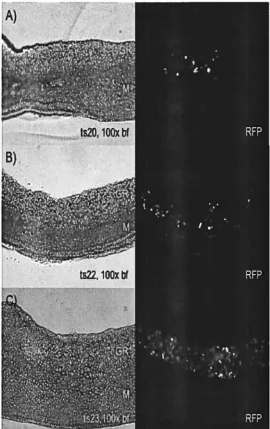

![Figure 4. WISH expression patterns for SoxlO, Stc2, Fgf]8, Fgf]3, and Wnt6 genes in mouse fetal gonads at different deveiopmentai time points](https://thumb-eu.123doks.com/thumbv2/123doknet/12234174.318644/109.918.212.623.127.874/figure-expression-patterns-soxlo-gonads-different-deveiopmentai-points.webp)