Université de Montréal
Masses kystiques latérales du cou : une analyse comparative des approches diagnostiques
Par Paul Tabet MD
Faculté de Médecine
Mémoire présenté à la Faculté des études supérieures et postdoctorales en vue de l’obtention du grade de
Maîtrise en Sciences biomédicales, option médecine expérimentale
Août 2019-08-15
Université de Montréal
Faculté des études supérieures et post-doctorales
Ce mémoire intitulé :
Masses kystiques latérales du cou : une analyse comparative des approches diagnostiques
Présenté par : Paul Tabet, MD
évalué par un jury composé des personnes suivantes :
Monsieur Olivier Abboud MD, FRCSC, président-rapporteur
Monsieur Apostolos Christopoulos MD, MSc, FRCSC, directeur de recherche Monsieur Nader Khaouam MD, FRCP(C) membre du jury
Résumé (Français)
Les masses kystiques latérales du cou (MKLC) bénignes et malignes sont difficiles à différencier cliniquement. L’utilité des modalités d’imagerie et de prélèvement doit être clarifiée.
Une revue rétrospective de cas entre 2010 et 2016. Les données d’imagerie ont été récoltées et plusieurs variables propres à la masse furent analysées. Les rapports de cytoponction à l’aiguille fine (CAAF), de la biopsie au trocart (BT) et des examens extemporanés (EE) ont été analysés. La sensibilité, la spécificité, la valeur prédictive positive (VPP) et la valeur prédictive négative (VPN) pour prédire la malignité ont été calculées pour toutes les variables comparées entre les masses kystiques bénignes et malignes.
Aucune variable d’imagerie n’a pu différencier les masses kystiques bénignes de malignes. La sensibilité de la CAAF est plus basse que celle de la BT (59% vs 83%; p=0.036) et de l’EE (59% vs 93%; p=0.01). L’EE a une meilleure VPN que la CAAF (92% vs 40%; p<0.001) et que la BT (92% vs 50%; p=0.062). La VPP et la spécificité étaient similaires dans tous les groupes.
Les cliniciens ne peuvent pas se fier uniquement à l’imagerie pour différencier les masses bénignes des masses malignes. Vu sa VPP adéquate (92%), la CAAF devrait être utilisée initialement pour tous les patients avec une MKLC. Si la CAAF s’avère négative, la BT devrait être utilisée vu sa meilleure sensibilité. Un examen extemporané devrait toujours suivre une BT négative vu la faible VPN de la BT. Un résultat positif à l’une des trois modalités de prélèvement indique la présence de malignité.
Mots-clés : Masse kystique latérale du cou, échographie, tomodensitométrie, tomodensitométrie par émission de positron, cytoponction à l’aiguille fine, biopsie au trocart, examen extemporané.
Résumé (Anglais)
Benign and malignant lateral cystic neck masses (LCNM) are difficult to distinguish clinically. The usefulness of imaging and sampling modalities in clarifying the diagnosis remains unclear. Retrospective review of cases between 2010 and 2016. Imaging data was reviewed and the variables pertaining to the mass were assessed including the following: size, nodal level, fat stranding, extracapsular spread, calcifications, vascularity, necrosis and standardized uptake value. Sampling reports of fine-needle aspiration (FNA), core-needle biopsy (CNB) and frozen section (FS) were also assessed. Sensitivity, specificity, positive predictive value (PPV) and negative predictive value (NPV) for predicting malignancy were calculated for all variables and compared between benign and malignant cystic neck masses.
Ultrasound was used in 47.2% and CT-Scan in 90.5% of patients. No variables on imaging could definitely differentiate benign from malignant LCNM. FNA had a lower sensitivity then CNB (59% vs 83%; p=0.036) and FS (59% vs 93%; p=0.01). FS had a better NPV when compared to FNA (92% vs 40%; p<0.001) and CNB (92% vs 50%; p=0.062). Specificities and PPV were similar among all groups.
Clinicians cannot rely on imaging to differentiate benign from malignant LCNM. Given its adequate PPV (92%), FNA should be used initially on lateral cystic neck masses. Because of its high sensitivity, CNB should be considered if FNA is not diagnostic of malignancy. FS should always follow a CNB not indicative of malignancy, because of the low NPV. Any result diagnostic of malignancy on either FNA, CNB or FS strongly indicates presence of malignancy.
Keywords : Lateral cystic neck mass, ultrasound, computed tomography, positron emission tomography, fine-needle aspiration, core-needle biopsy, frozen section.
Tables des matières
Résumé (Français) ... 3
Résumé (Anglais) ... 4
Table des matières ... 5
Liste des tableaux ... 7
Liste des figures ... 8
Liste des annexes ... 9
Liste des sigles et abréviations ... 10
1. Introduction ... 13
2. Recension de la littérature et de méthodologie ... 15
2.1. Diagnostic différentiel de masses kystiques latérales du cou ... 15
2.1.1. Kystes branchiaux ... 15
2.1.2. Métastases régionales de carcinomes épidermoïdes ... 15
2.1.3. Métastases régionales de carcinomes papillaires de la thyroïde ... 16
2.2. Modalités d’investigation ... 16
2.2.1. Les modalités d’imagerie ... 17
2.2.2. Les modalités de prélèvement ... 17
3. Article 1: Lateral cystic neck masses: diagnostic value of ultrasound, computerized tomography and positron emission tomography for predicting malignancy ... 18
3.1. Abbreviations ... 19
3.2. Abstract ... 20
3.3. Introduction ... 22
3.4. Method ... 23
3.4.1. Patient selection ... 23
3.4.2. Data collection and outcomes measures ... 23
3.4.3. Statistical analysis ... 24
3.5. Results ... 25
3.6. Discussion ... 30
4. Article 2: Cystic masses of the lateral neck: diagnostic value comparison between
fine-needle aspiration, core-fine-needle biopsy and frozen section ... 34
4.1. Abbreviations ... 35
4.2. Abstract ... 36
4.3. Introduction ... 37
4.4. Methods ... 38
4.4.1. Patient selection ... 38
4.4.2. Data collection and statistical analysis ... 38
4.4.3. FNA, CNB and FS characteristics ... 39
4.5. Results ... 40 4.6. Discussion ... 48 4.6.1. Limitations ... 52 4.7. Conclusion ... 54 5. Discussion générale ... 55 6. Conclusion ... 57 7. Références bibliographiques ... 58 8. Annexes ... 66
Liste des tableaux
Table 1: Demographic factor differences between patients presenting with malignant and benign cystic neck masses ... 25 Table 2: Demographic features, size, proportion of unilateral and single masses among all
patients presenting with lateral cystic neck masses ... 43 Table 3: Sensitivity, specificity, PPV and NPV of FNA, CNB and FS in detecting the presence or
absence of malignancy in the cystic masses of the lateral neck ... 43 Table 4: Sensitivity for detecting malignancy according to specific diagnosis made on final
pathology ... 44 Table 5: FNA parameters for detecting malignancy according to the presence of either
squamous cell carcinoma or papillary thyroid carcinoma on final pathology ... 45 Table 6: FNA sensitivity in detecting malignancy in cystic masses of the lateral neck ... 49
Liste des figures
Figure 1: Differential diagnosis of cystic masses of the lateral neck based on excision biopsy or neck dissection pathology. ... 26 Figure 2: Differences in size between cystic malignant and cystic benign masses of the lateral
neck. ... 27 Figure 3: Differences in lymph node drainage level between cystic malignant and cystic benign
masses of the lateral neck ... 27 Figure 4: Differences in the presence of multiple adenopathy between cystic malignant and
cystic benign masses of the lateral neck. ... 28 Figure 5: Differential diagnosis of cystic masses of the lateral neck based on excision biopsy or
neck dissection pathology. ... 40 Figure 6: Diagnostic tool sensitivity in detecting malignancy in solid and cystic masses of the
lateral neck. ... 46 Figure 7: FNA sensitivity in detecting malignancy in solid SCC and cystic mass subgroups
diagnosed on final pathology. ... 46 Figure 8: Proposed diagnostic algorithm for the management of cystic masses of the lateral
Liste des annexes
Annexe 1: Preuve d'approbation du Département de Santé Publique (DSP) ... 66 Annexe 2: Preuve d’approbation du Comité d’Éthique de Recherche (CÉR) du CHUM ... 68 Annexe 3: Demande d'autorisation de rédaction de mémoire ou de thèse par articles ... 70 Annexe 4: Autorisation des co-auteurs - Cystic masses of the lateral neck: diagnostic value
comparison between fine-needle aspiration, core-needle biopsy and frozen section ... 72 Annexe 5: Autorisation des co-auteurs - Cystic masses of the lateral neck: diagnostic value of
ultrasound, computed tomography and positron emission tomography for predicting malignancy ... 74
Liste des sigles et abréviations Français:
BT = Biopsie au trocart
CAAF = Cytoponction à l’aiguille fine
CHUM = Centre Hospitalier de l’Université de Montréal
EE = Examen extemporané
MKLC = Masse kystique latérale du cou
ORL-CCF = Oto-rhino-laryngologiste et chirurgien cervico-facial VPH = Virus du papillome humain
VPN = Valeur prédictive négative VPP = Valeur prédictive positive Anglais:
AP = Anterior-posterior dimension BCC = Branchial cleft cyst
CC = Cranial-caudal dimension CNB = Core-neede biopsy
CT = Computed tomography
ECS = Extracapsular spread H&N = Head and neck
HNSCC = Head and neck squamous cell carcinoma HPV = Human papillomavirus
FNA = Fine-needle aspiration
FS = Frozen section
LAT = Lateral-lateral dimension LCNM = Lateral cystic neck mass NPV = Negative predictive value PET-CT = Positron-emission tomography PPV = Positive predictive value
11 SCC = Squamous cell carcinoma SUV = Standardized uptake value
1. Introduction
Tous les oto-rhino-laryngologistes et chirurgiens cervico-faciaux (ORL-CCF) doivent posséder une compréhension approfondie du diagnostic différentiel des masses de la tête et du cou. Plusieurs patients sont référés aux ORL-CCF par des omnipraticiens qui cherchent un plan de traitement définitif des patients avec des masses cervicales. Afin d’accomplir ceci, l’ORL-CCF doit déterminer le diagnostic le plus probable. À l’aide de l’histoire, de l’examen physique et de diverses modalités diagnostiques telles que l’imagerie et la biopsie, l’ORL-CCF peut accomplir cette tâche.
Les différentes masses du cou sont typiquement catégorisées en trois types : les anomalies congénitales, les étiologies infectieuses ou inflammatoires et les néoplasies. Les anomalies congénitales sont sous-catégorisées en lésions vasculaires (hémangiomes, malformations artérioveineuses, malformations lymphatiques) et en lésions non vasculaires (kystes thyréoglosses, kystes branchiaux, ranules plongeantes, tératomes. Les étiologies infectieuses et inflammatoires incluent les sialadénites, les adénopathies bactériennes et virales et multiples maladies granulomateuses telles que la sarcoïdose, la griffe du chat et la tuberculose. Finalement, les néoplasies incluent ceux de la thyroïde, des glandes salivaires et les métastases régionales de tous les cancers de la tête et du cou.
Les masses du cou sont aussi typiquement catégorisées selon leur localisation au niveau du cou. Les masses peuvent aussi être catégorisées selon leur apparence à l’imagerie initiale, soit des masses solides ou des masses kystiques. Le sujet de ce projet porte plus spécifiquement sur les masses kystiques présentes dans les zones 2, 3 et 4 du cou. Ces trois zones ont comme limite latérale le muscle sterno-cléido-mastoïdien. La zone 2 est délimitée supérieurement par la base du crâne, médialement par le ventre postérieur du muscle digastrique et inférieurement par le niveau de l’os hyoïde. La zone 3 est limitée supérieurement par le niveau de l’os hyoïde, médialement par les muscles infrahyoïdiens et inférieurement par le niveau du cartilage cricoïde. Finalement la zone 4 est délimitée supérieurement par le niveau du cartilage cricoïde, médialement par les muscles infrahyoïdiens et inférieurement par la clavicule.
Le diagnostic le plus probable d’une masse kystique latérale du cou a longtemps été le kyste branchial. Par contre, l’incidence croissante des cancers de la tête et du cou lié au virus du papillome humain (VPH) a mené à une augmentation de métastases régionales kystiques dans les zones 2, 3 et 4 du cou. Les tumeurs primaires de ces cancers se retrouvent fréquemment dans l’oropharynx et dans la thyroïde. Par contre, il est possible que la seule manifestation du cancer soit une masse kystique latérale du cou sans évidence de tumeur primaire (primaire occulte). Ainsi, il y a maintenant une importance grandissante de reconnaître et de différencier ces métastases régionales de cancer de la tête et du cou avec primaire occulte de masses bénignes telles que les kystes branchiaux. En effet, la prise en charge de masses bénignes et malignes diffère énormément et une erreur de traitement chez des patients porteurs de cancer peut mener à des conséquences sérieuses pour le patient.
2. Recension de la littérature et de méthodologie
2.1. Diagnostic différentiel de masses kystiques latérales du cou 2.1.1. Kystes branchiaux
Les kystes branchiaux sont formés de résidus des tissus branchiaux embryonnaires et leur localisation dans la tête et le cou est dépendante de l’arc branchial originaire. Dans 95% des cas, le deuxième arc branchial est affecté. 1, 2 Ceci mène à la formation d’une masse kystique antérieure au muscle sterno-cléido-mastoïdien qui est souvent accompagnée d’un tractus de drainage latéral à l’artère carotide interne se terminant ultimement dans la loge amygdalienne. Habituellement ces kystes sont diagnostiqués à l’aide de l’histoire, de l’examen physique et d’une modalité d’imagerie telle que l’échographie ou la tomodensitométrie. Vu le potentiel de surinfection, ces masses sont traitées par une exérèse chirurgicale.
2.1.2. Métastases régionales de carcinomes épidermoïdes
Le carcinome épidermoïde est le sous-type de cancer de la tête et du cou le plus fréquent. Ces cancers peuvent se manifester au niveau du nasopharynx, de la cavité orale, de l’oropharynx, de l’hypopharynx et du larynx. Les cellules cancéreuses de ces tumeurs ont la capacité de migrer vers les ganglions lymphatiques du cou et se multiplient. Ceci entraîne la formation d’une masse au niveau du cou. Il est donc nécessaire d’effectuer un examen physique minutieux de toutes les régions de la tête et du cou pour déceler la présence d’une tumeur primaire. Par contre, les métastases cervicales de carcinomes épidermoïdes se présentent parfois sans la présence d’une tumeur primaire. L’incidence de cancer à « primaire inconnu » se situe entre 2% et 9% parmi tous les cancers de la tête et du cou. 3, 4
Récemment, le virus du papillome humain (VPH) a été identifié comme une cause de carcinomes épidermoïdes de la tête et du cou, spécifiquement de l’oropharynx et de ceux à primaire inconnu. Aux États-Unis, la prévalence de tumeur de l’oropharynx causé par le VPH serait de l’ordre de 60% et serait en augmentation. 5-10 Radiologiquement, les tumeurs de l’oropharynx présentent
parfois des métastases cervicales kystiques souvent faussement diagnostiquées comme des kystes branchiaux.11-16 De plus, les carcinomes épidermoïdes liés au VPH se présentent plus fréquemment comme un primaire inconnu lorsque comparés à ceux non liés au VPH.17, 18 La localisation de la masse cervicale au niveau du cou peut orienter vers le site primaire de la tumeur. Par exemple, les métastases régionales des cancers de la cavité orale se retrouvent dans les zones 1, 2 et 3 tandis que celles des cancers l’oropharynx, de l’hypopharynx et du larynx se situe préférentiellement dans les zones 2, 3, 4. 19, 20 Habituellement, le site de la tumeur primaire est identifié lors de l’examen physique de la tête et du cou ou de la rhino-pharyngo-laryngoscopie. Si une tumeur primaire est identifiée, des biopsies sont prélevées afin de confirmer le site et l’histologie de la tumeur primaire. Si le primaire n’est pas identifié, des imageries telles que la tomodensitométrie (TDM) et la tomodensitométrie par émission de positron (TEP) peuvent être utilisées pour aider la localisation de celui-ci. Par contre, la sensibilité de ces imageries est variable.21, 22 Les cancers à primaire inconnu peuvent être traité chirurgicalement (résection de la base de langue, amygdalectomie, évidement cervicale) ou par radiothérapie avec une irradiation de tous les sites potentiels de tumeur primaire et du cou. 2.1.3. Métastases régionales de carcinomes papillaires de la thyroïde
Le carcinome papillaire représente 60% à 70% des cancers de la thyroïde.23, 24 La lésion primaire est souvent confinée à la glande thyroïde, par contre, jusqu’à 30% des patients peuvent se présenter avec des métastases régionales au niveau du cou central et latéral.25, 26 Ces métastases régionales se présentent le plus souvent dans les zones 2 à 6.27
2.2. Modalités d’investigation
Vu l’aspect clinique similaire et la prise en charge différente entre les kystes branchiaux et les carcinomes épidermoïdes à primaire inconnu, le clinicien doit se fier à diverses modalités d’investigation pour différencier ces deux entités cliniques. Ces modalités peuvent être séparées en deux catégories : les imageries et les prélèvements. Les imageries utilisées couramment dans ces situations sont l’échographie, la tomodensitométrie et la tomodensitométrie par émission de
17 trocart et l’examen extemporané.
2.2.1. Les modalités d’imagerie
Les modalités d’imagerie sont cruciales à la prise en charge des patients avec des masses kystiques au niveau du cou. Par contre, il peut être parfois difficile de différencier des métastases cervicales de cancers de la tête et du cou des masses cervicales bénignes. 28, 29 En effet, selon plusieurs études, les kystes branchiaux infectés sont souvent confondus pour des métastases régionales de carcinomes épidermoïdes de la tête et du cou, notamment à l’échographie et à la tomodensitométrie.30, 31 La tomodensitométrie par émission de positron a également été démontrée utile pour localiser les tumeurs primaires chez des patients souffrant de carcinomes épidermoïdes à primaire inconnu avec une sensibilité de 62% et une spécificité de 81.9%.32 Par contre, cet outil s’est montré peu fiable pour différencier des masses bénignes de masses malignes.33
2.2.2. Les modalités de prélèvement
La valeur diagnostique de la cytoponction à l’aiguille fine a été étudiée à plusieurs reprises et a démontré des résultats différents de sensibilité pour détecter la malignité.34-39 Par contre, la cytoponction de masses kystiques spécifiquement a démontré des résultats inférieurs en termes de sensibilité.40, 41 La biopsie au trocart a aussi été démontrée comme un outil utile pour diagnostiquer les cancers de la tête et du cou malgré que son utilité pour diagnostiquer des masses kystiques demeure controversé.42-44 Finalement, l’examen extemporané est souvent proposé dans les algorithmes de prise en charge des masses kystiques du cou. 39, 45 Une étude a démontré une sensibilité de 100% pour détecter les kystes branchiaux.46 Toutefois, aucune étude n’a spécifiquement étudié la sensibilité de l’examen extemporané pour détecter le cancer.
3. Article 1: Lateral cystic neck masses: diagnostic value of ultrasound, computerized tomography and positron emission tomography for predicting malignancy
Paul Tabet, MD1,2; Nadim Saydy, MD1; Laurent Létourneau, MD,1,2; Olga Gologan, MD1,2; Éric Bissada, MD1,2; Tareck Ayad, MD1,2; Jean-Claude Tabet, MD1,2; Louis Guertin, MD1,2; Phuc Félix Nguyent-Tan, MD1,2; Apostolos Christopoulos, MD1,2
1Centre de Recherche du Centre Hospitalier de l’Université de Montréal, Montreal, QC, Canada; 2Centre Hospitalier de l’Université de Montréal (CHUM), Montreal, PQ, Canada
*Institution where the work was done: Université de Montréal, Montreal, Quebec, Canada **Manuscript region of origin: Montreal, Quebec, Canada
Corresponding Author:
Apostolos Christopoulos, MD, MSc, FRCSC
Associate Professor, Division of Otolaryngology Head & Neck Surgery, Centre Hospitalier Universitaire de Montréal (CHUM)
Canada
Key words: Lateral cystic neck masses, computed tomography, ultrasound, positron emission tomography, squamous cell carcinoma, lymph node metastasis
Short title: Diagnosis of lateral cystic neck masses
There are no conflicts of interest for all authors
19 3.1. Abbreviations
BCC : branchial cleft cyst CT : computerized tomography H&N : head & neck
HPV : human papillomavirus LCNM : lateral cystic neck mass
PET-CT : positron-emission tomography SCC : squamous cell carcinoma
SUV : standardized uptake value U/S : ultrasound imaging
3.2. Abstract
The clinical presentation of lateral neck metastases from head & neck cancer is often indistinguishable from benign lesions such as branchial cleft cysts. That said, treatment guidelines and prognosis differ widely. Thus, proper and early identification of malignancy is a cornerstone of oncological management. We aimed to study different radiologic modalities’ performance for identifying features of malignancy that could suggest a more urgent need for obtaining a histological specimen.
In this retrospective case control study, a review of cases from a single tertiary center was undertaken between November 2010 and December 2016. Masses were considered malignant if one of two conditions were met: (1) malignant on final pathology after excision or (2) primary lesion subsequently identified and confirmed with biopsy. Imagery reports were reviewed, and key features were reported. Lesions were measured along three dimensions: the latero-lateral (LAT), antero-posterior (AP), and cranio-caudal (CC) axes. Size, nodal level, extracapsular spread (ECS), fat stranding and necrosis were evaluated with CT-scan; vascularity was measured with U/S. PET-CT was used to assess the SUV of the dominant node and to determine the ability to correctly identify a primary tumor prior to pathologic confirmation.
In total, 127 patients presenting with LCNM were included in our study, of which 89 were found to be malignant masses (70%). U/S was used in 60/127 (47.2%) and CT-Scan in 115/127 (90.5%) of patients. Vascularity was higher in cystic malignancy (7/13; 54%) when compared to benign disease (2/12; 17%) (p = 0.03). There was a statistically significant difference between calcifications for cystic SCCs and PTCs (p = 0.02). No difference in average SUV was noted between cystic malignancies (SUV=9.76) and benign disease (SUV=6.63) (p = 0.5).
Within our cohort, level in the neck, incidence of multiple adenopathy and size (apart from cranio-caudal dimension) appeared similar between malignant and benign lesions. Increased vascularity on U/S with intra-lesional calcifications was highly suggestive of a PTC. Measurement of SUV on nuclear imaging was not statistically different between groups, although sample size
21
for benign pathology was small (n=6). Negative predictive value for imaging was low (62%). Thus, an absence of malignant features should not slow down the process of obtaining a biopsy.
3.3. Introduction
Solitary lateral cystic neck masses (LCNM) are often benign, especially in young patients.47 In fact, second branchial cleft cysts (BCC) are the most common congenital etiology for a cervical mass. 48 That said, a high index of suspicion for malignancy must be preserved since metastases from head and neck squamous cell carcinoma (HNSCC) and papillary thyroid carcinoma (PTC) may have a similar clinical presentation.
Due to the human papilloma virus (HPV) epidemic, incidence of p16-positive HNSCC is increasing. 49 There is a known association between the latter and cystic neck lesions, which makes radiologic assessment more difficult, especially when presenting in lymph node drainage levels II through IV 11. Although imaging is essential to the work-up of LCNMs, some cystic metastases of HNSCCs may be radiologically undistinguishable from BCC. 28, 29 In fact, infected BCC have been shown to be confused with metastatic HNSCC on ultrasound (U/S) and computed tomography scan (CT-Scan). 30, 31 Positron-emission tomography (PET-CT) has been proposed as a tool to differentiate these pathologies, but the only study assessing PET-CT suggests it is unreliable in identifying malignant lesions. 33. The limits of these diagnostic modalities lead to the erroneous diagnosis of certain masses as benign. Studies have shown rates as high as 24% of lesions initially thought to be BCC on imaging subsequently found to be malignant. 35, 37-39, 45, 50
Our aim was to further characterize the usefulness of ultrasound, CT-Scan, and PET-Scan in differentiating malignant from benign LCNMs. This is important because management varies greatly between a benign and malignant lesions thus proper identification is paramount.
23 3.4. Method
3.4.1. Patient selection
A retrospective review of cases from a single tertiary center was undertaken between November 2010 and December 2016. All patients were initially evaluated by a fellowship-trained otolaryngologists or radiation oncologists with an expertise in head and neck oncology. All patients presenting with a LCNM without evidence of a primary tumor on initial history, physical exam, flexible laryngoscopy or initial imaging were included. The pediatric population was excluded. The mass had to be within the boundaries of cervical lymph node drainage levels II through IV as shown on either U/S or CT-Scan.
Purely cystic masses and cyst-like masses (i.e. partially cystic, cystic component, necrotic, liquid content) were included. Masses were benign when considered as such by a pathologist after mass excision (through neck dissection or excisional biopsy). Masses were considered malignant if one of two conditions were met: (1) malignant on final pathology after excision or (2) primary lesion subsequently identified and confirmed with biopsy.
All procedures were in accordance with the ethical standards of the Helsinki Declaration of 1975 as revised in 1983 and were approved by the committee on human experimentation of our institution.
3.4.2. Data collection and outcomes measures
Each imaging study was reviewed by a fellowship-trained neuro-radiologist. Lesions were measured along three dimensions: the latero-lateral (LAT), antero-posterior (AP), and cranio-caudal (CC) axes. CT-scan or U/S measurements were used to assess LCNM. Size, nodal level, extracapsular spread (ECS), fat stranding and necrosis were evaluated with CT-scan; vascularity was measured with U/S. PET-CT was used to assess the SUV of the dominant node and to determine the ability to correctly identify a primary tumor prior to pathologic confirmation.
3.4.3. Statistical analysis
Data were analyzed using the Microsoft Excel software. Chi-squared test and Fisher’s exact test were used to analyze categorical variables and Student’s T-test was used for continuous variables. A p-value of <0.05 was considered statistically significant.
25 3.5. Results
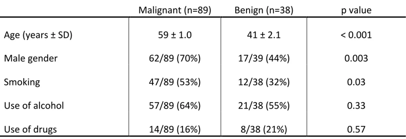
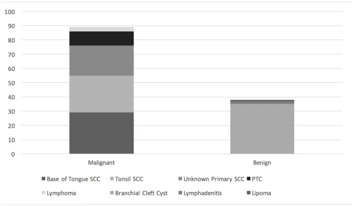
In total, 127 patients presenting with LCNM were included in our study, of which 89 were found to be malignant masses (70%). Ultrasound was used in 60/127 (47.2%) and CT-Scan in 115/127 (90.5%) of patients. Patient demographic factors are presented in Table 1 and final pathology of all cystic masses are presented in Figure 1. Results for size, nodal level, and multiple adenopathy are all shown in Figures 2 through 4. Of note, a high proportion of regional metastasis of PTC (8/10; 80%) presented in the level IV. These results were statistically different from the rate of level IV regional metastasis in cystic SCC (80% vs 2.7%; p<0.001).
Table 1: Demographic factor differences between patients presenting with malignant and benign cystic neck masses
Malignant (n=89) Benign (n=38) p value
Age (years ± SD) 59 ± 1.0 41 ± 2.1 < 0.001
Male gender 62/89 (70%) 17/39 (44%) 0.003
Smoking 47/89 (53%) 12/38 (32%) 0.03
Use of alcohol 57/89 (64%) 21/38 (55%) 0.33
Use of drugs 14/89 (16%) 8/38 (21%) 0.57
Figure 1: Differential diagnosis of cystic masses of the lateral neck based on excision biopsy or neck dissection pathology.
27
Figure 2: Differences in size between cystic malignant and cystic benign masses of the lateral neck.
AP: Antero-posterior dimension; LAT: latero-lateral dimension; CC: cranio-caudal dimension
Figure 3: Differences in lymph node drainage level between cystic malignant and cystic benign masses of the lateral neck
Figure 4: Differences in the presence of multiple adenopathy between cystic malignant and cystic benign masses of the lateral neck.
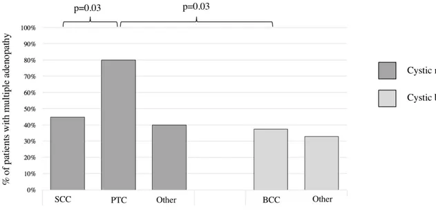
SCC: squamous cell carcinoma; PTC: papillary thyroid carcinoma; BCC: branchial cleft cyst. Only statistically significant differences are shown.
Vascularity was higher in cystic malignancy (7/13; 54%) when compared to benign disease (2/12; 17%) (p = 0.03). Calcifications were reported in 1/5 (20%) cystic SCC, 6/6 (100%) cystic PTCs and 0/3 (0%) BCCs. There was a statistically significant difference in the incidence of calcifications between cystic SCCs and PTCs (p = 0.02), and a trend towards statistical significance between malignant and benign cystic lesions (p = 0.06).
Fat stranding and ECS were only assessed using CT-Scan. Fat stranding was present 10/19 (52%) of cystic malignancies and 4/10 (40%) of benign disease. A trend towards statistical significant was found between these two groups (p=0.07). Finally, 2/2 (100%) malignant cystic lesions demonstrated ECS whereas presence or absence of ECS was never described among benign masses.
29
Positron emission tomography was ordered for 54/89 (60.7%) patients with cystic malignancies and in 6 patients (15.8%) with benign disease. No difference in average SUV was noted between cystic malignancies (SUV=9.76) and benign disease (SUV=6.63) (p = 0.5). A primary tumor was suspected in 20/54 (37%) of patients with cystic malignancies and no primary (0/6) was suspected in PET-CTs of patients with benign masses (p=0.07).
The term “necrosis” was used to describe lesions in 12 U/S reports and 76 CT-Scans. Its sensitivity for detecting malignancy was 38% with U/S and 74% with CT-Scan. The radiologist’s initial presumptive diagnosis based on imaging had a sensitivity of 85%, a specificity of 69%, a positive predictive value of 89% and a negative predictive value of 62%.
3.6. Discussion
Older age, male gender and smoking were all significantly more prevalent among the malignant cystic neck group. Therefore these three parameters must be carefully taken into account when evaluating a patient with a cystic neck mass and should raise the suspicion for a malignant disease. The marked difference between the incidence of level IV masses of non-PTC masses and PTC cystic masses is not surprising given a known elevated incidence of level IV lymph node metastasis in PTC. In fact, ipsilateral level IV have been shown to be the second most common location of PTC lymph node metastasis after pretracheal metastasis.51 Cystic PTC lesions were also significantly more prevalent in level IV when compared with cystic benign masses (77% vs 5%; p<0.001) Therefore, we encourage an ultrasound for all level IV cystic neck masses to rule out the presence of a suspect thyroid nodule and regional metastasis of a possible thyroid malignancy.
When comparing the size of the different mass types, the only difference was noted in the cranio-caudal dimension average between cystic SCC and BCC (32 mm vs 39 mm; p=0.016). This difference is in fact corroborated in the article by Goyal et al. which also found a larger CC dimension for branchial cleft cysts.52 Malignant adenopathy are well known for their rounder shape when compared to benign adenopathy which may also explain their morphological difference with branchial cleft cysts.
When comparing cystic SCC and cystic PTC, the former showed fewer multiple nodes (44% vs 80%; p=0.03). Furthermore, cystic PTC showed a significantly higher rate of multiple adenopathy when compared to cystic BCC (80% vs 37%; p=0.03). In fact, as previously stated, PTC initially spreads to level VI before spreading to level IV which would explain the higher likelihood of multiple adenopathy is this subgroup. 51 Finally, no difference exists between the cystic SCC and BCC with regards to multiple node status which may be explained by the tendency of BCC to get infected which consequently is accompanied by reactive lymphadenopathy. Therefore, we encourage clinicians to not rely on multiple nodes for clinical suspicion of malignancy when faced with a lateral cystic neck mass.
31
Vascularity was shown to be more frequent in cystic malignancies when compared to benign masses (p=0.03), namely because of the high rate of vascularity among the cystic PTC masses. In fact, PTC nodal metastasis have often been regarded as highly vascular lesions. 53 When cystic SCC was isolated and compared to BCC, vascularity only trended toward a statistical difference (p=0.09).
Given the near statistically significant difference in calcification status between malignant and benign cystic lesions, this feature may be considered as an adequate marker for detecting cancerous lesions, specifically PTC. However, similarly to the published literature, calcification does not differentiate cystic SCC from cystic BCC. 52 Without surprise, calcifications were more frequent in PTC regional metastasis when compared to SCC; this difference in calcification status was previously demonstrated by Aruja and colleagues. 54
Both fat stranding and ECS are known as indicators of malignant disease. Our findings only revealed a trend toward significantly a higher incidence of fat stranding in malignant cystic versus benign cystic disease. This, combined with presence of fat stranding in both malignant and benign disease as described by Goyal and colleagues52, puts into question its use in detecting malignancy. Unfortunately, only two patient reports mentioned the presence of ECS among cystic masses, it is therefore difficult to conclude the utility of this imaging parameter within our study. However, the absence of report of this finding among benign masses and the fact that it represents a known marker for poor prognosis, leads us to still consider ECS as a feature of malignancy. 55
No difference was found between all groups in terms of SUV of the neck mass however PET-Scan was scarcely used to evaluate benign disease and therefore these results should be interpreted with caution. However, based on this finding, we conclude that SUV may not reliably differentiate benign cystic from malignant cystic neck lesions. A high SUV in the benign cystic mass group may also be due to an active infection or ongoing inflammatory process. Also, most PET-CTs are ordered after diagnostic sampling of the mass (e.g. fine-needle aspiration) which has been shown to further elevate the SUV and affect its diagnostic accuracy. 56, 57 However, suspicion of primary tumor on PET-CT is a finding that may indicate the presence of a malignant node (p=0.07).
No universal diagnostic definition exists regarding necrosis on imaging. Some have been proposed but are not systematically utilized by radiologist. In fact, some authors define “necrosis” on imaging as either “nodes with thicker solid walls and irregular, complex central low attenuation” or a “masses that tends to have undulating irregular walls”. 11 In sum, the definition needs to be further clarified to be used objectively on imaging analysis. In our study, we found that the term was more often used with CT-scans compared to U/S. When used on CT-Scan, its sensitivity was much higher than when used on U/S (74% vs 38%, respectively). This leads us to encourage clinicians to disregard the term when used on U/S and to rely more on the above-cited U/S parameters. Regardless of the modest sensitivity on CT-Scan, further studies should be undertaken to ensure inter-evaluator reliability for the term to not confuse an infected BCC with a necrotic regional lymph node metastasis.
Overall, the sensitivity of all imaging modalities to detect malignancy was high, however, considering the low specificity and NPV of these same diagnostic tools, we caution the clinician to always be wary of a reassuring diagnosis of a lateral cystic neck mass based solely on imaging.
33 3.7. Conclusion
In summary, malignant and benign cystic neck lesions are difficult to distinguish based on imaging. Cystic SCC and BCC have a similar location in the neck and incidence of multiple adenopathy. Apart from a difference in cranio-caudal dimensions, size appears to be similar as well. A level IV cystic mass or one with high vascularity and calcifications is highly suspicion of PTC and, in such circumstances, a primary within the thyroid gland must be ruled-out. Also, SUV may not help differentiate malignant from benign cystic masses. Finally, a benign presumptive diagnosis on imaging is often inaccurate (NPV=62%) and clinicians must interpret these reports with caution.
4. Article 2: Cystic masses of the lateral neck: diagnostic value comparison between fine-needle aspiration, core-needle biopsy and frozen section
Authors and affiliations:
Paul Tabet MD 1,2; Nadim Saydy MD 1,2; Laurent Létourneau MD 1,2; Olga Gologan MD 1,2; Éric Bissada MD 1,2; Tareck Ayad MD 1,2; Jean-Claude Tabet MD 1,2; Louis Guertin MD,1,2; Phuc Félix Nguyen-Tan MD 1,2; Apostolos Christopoulos MD 1,2.
1Centre de Recherche du Centre Hospitalier de l’Université de Montréal (CRCHUM), Montreal, QC, Canada; 2Centre Hospitalier de l’Université de Montréal (CHUM), Montreal, PQ, Canada
Corresponding author : Apostolos Christopoulos, MD, FRCSC
Division of Otolaryngology-Head and Neck Surgery Centre Hospitalier de l'Université de Montréal (CHUM) 3840 rue St Urbain
Montréal, Québec H2W 1T8
Brief running title: Diagnosis of lateral cystic neck masses
Keywords: Lateral cystic neck masses, fine-needle aspiration, core-needle biopsy, frozen section, squamous cell carcinoma
Funding source: No funding source
35 4.1. Abbreviations
CNB = Core-needle biopsy HPV = Human papillomavirus
HNSCC = Head and neck squamous cell carcinoma FNA = Fine needle aspiration
FS = Frozen section
PPV = Positive predictive value NPV = Negative predictive value
4.2. Abstract
The usefulness of fine-needle aspiration (FNA), core-needle biopsy (CNB) and frozen section (FS) for assessing lateral cystic neck masses remains unclear.
A retrospective review of patients presenting with a lateral cystic neck mass was undertaken. In total, 135 patients were included. FNA had a lower sensitivity then CNB (59% vs 83%; p=0.036) and FS (59% vs 93%; p=0.01). FS had a better negative predictive value (NPV) when compared to FNA (92% vs 40%; p<0.001) and CNB (92% vs 50%; p=0.062). Positive predictive values (PPV) and specificities were similar among all groups.
Given its adequate PPV (92%), FNA should be used initially on lateral cystic neck masses. Because of its high sensitivity, CNB should be considered if FNA is not diagnostic of malignancy. FS should always follow a CNB not indicative of malignancy, because of the low NPV. Any result diagnostic of malignancy on either FNA, CNB or FS strongly indicates presence of malignancy.
37 4.3. Introduction
Lateral cystic neck masses are often misdiagnosed as benign lesions such as branchial cleft cysts (BCC) when a large proportion of these are malignant.38 Regional metastases of oropharyngeal squamous cell carcinoma (SCC) and papillary thyroid cancer (PTC) are among the different types of malignancy found in lateral cervical cysts.36, 58 Thus, several authors have proposed specific treatment algorithms for this disease variant which includes among other diagnostic tools fine-needle aspiration cytology (FNA), core-fine-needle biopsy (CNB) and frozen section (FS).39, 45
FNA’s diagnostic value for lateral cervical cysts has been studied numerous times and has yielded different results when assessing sensitivity for detecting malignancy.34-39 However, when compared to FNA results of all head and neck lesions regardless of the presence of a cystic component, FNA of cysts demonstrate a much lower sensitivity.40, 41 Authors have suggested that this may be due to the hypocellularity secondary to the cystic fluid’s dilutional effect as well as to the presence of inflammatory cells and cellular debris.36 In fact, this phenomenon is not isolated to the head and neck since cystic lesions throughout the body have shown to be a significant source of FNA diagnostic error.59 CNB is also regarded as a useful tool for diagnosing malignancy in head and neck masses; their use however remains controversial when addressing masses of a cystic nature.42-44 FS is another diagnostic tool often proposed in management algorithms of cystic neck lesions.39, 45 One study showed that FS had a 100% sensitivity for detecting branchial cleft cysts.46 No report of FS’s sensitivity for detecting malignancy in lateral cervical cysts was found.
The objective of this study is therefore to offer a detailed description of the diagnostic value of each of these tools in differentiating benign from malignant cystic lesions of the lateral neck. Also, we wish to compare these values with those found on solid malignant masses to possibly further isolate lateral neck cysts as a unique clinical scenario with specific management considerations.
4.4. Methods
4.4.1. Patient selection
A retrospective review of cases from a single tertiary center was undertaken between November 2010 and December 2016. All adult patients presenting with a lateral cystic neck mass without evidence of a primary tumor on initial history, physical exam or flexible laryngoscopy were included. The mass had to be within the boundaries of cervical lymph node drainage levels 2 through 4 as shown on either ultrasound (U/S) or computed tomography scan (CT-Scan) interpreted by a neuroradiologist.
Solid masses, purely cystic masses and cyst-like masses (i.e. partially cystic, cystic component, necrotic, liquid content) were included. For the solid mass group, only malignant SCC on final pathology were included. All masses had to have undergone either FNA, CNB or FS. All masses had to have a definitive diagnosis of either benign disease or malignancy. Masses were benign when considered as such by a pathologist after mass excision (through neck dissection or excisional biopsy). Masses were considered malignant if one of two conditions were met: (1) malignant on final pathology after excision or (2) primary lesion subsequently identified and confirmed with biopsy.
All procedures were in accordance with the ethical standards of the Helsinki Declaration of 1975 as revised in 1983 and were approved by the committee on human experimentation of our institution.
4.4.2. Data collection and statistical analysis
FNA, CNB and FS results were categorized as inadequate, benign and suspicious or diagnostic of malignancy. Sensitivity, specificity, positive predictive value (PPV) and negative predictive values (NPV) were calculated for cystic masses based on definitive diagnosis. Sensitivity results for solid masses were compared with those for cystic masses. Only results with stated absence of malignancy were considered negative; non-diagnostic results were excluded from calculations.
39
Complications rates were also assessed. Chi-squared test and Fisher’s exact test were used to analyze categorical data. A p-value of <0.05 was considered statistically significant.
4.4.3. FNA, CNB and FS characteristics
Either a surgeon, a radiologist or a cytopathologist performed FNA. Needle passes and ranges were available in 51/101 (50.5%) FNA reports. The number of passes ranged from 1 to 5; both the median and the mode were 3. These were done with either a 18, 22 or 25 gauge needle; the mode was 25G. Aspirates were alcohol-fixed and smears were Pap stained. Cell blocks stained with HE were prepared but no ancillary studies were performed on these. Biopsies were considered non-diagnostic when samples were too small, when there was absence of viable epithelial cells or presence of air-dried cells, degenerated cells or cells obscured by blood, inflammatory exudates, necrosis and debris. All these features, in the presence of viable cells, can also be considered obscuring features that can lead to false-negative results in all biopsy modalities. Specifically, for FS, obscuring features include the latter as well as freezing artifact. Cellularity ranged from hypocellular (but satisfactory) to hypercellular. Most specimens were low to moderately cellular. Frozen section was performed in patients with FNA and/or CNB which were not diagnostic of malignancy despite high clinical suspicion of malignancy. Frozen sections were performed directly in patients when risk of malignancy was assessed as low, for example in suspected branchial cleft cysts. This was mostly the case for patients who underwent work-up and surgery before referral to our tertiary care center. Because of the oncological population referred to our healthcare center, FNA is generally performed first, followed by CNB, and ultimately FS. We assure that it is common practice at our center to send the whole mass for pathological diagnosis. Anatomo-pathologists examines between 1 and 2 slides prepared for FS and the rest of the mass is sent for final pathological diagnosis in formaldehyde.
4.5. Results
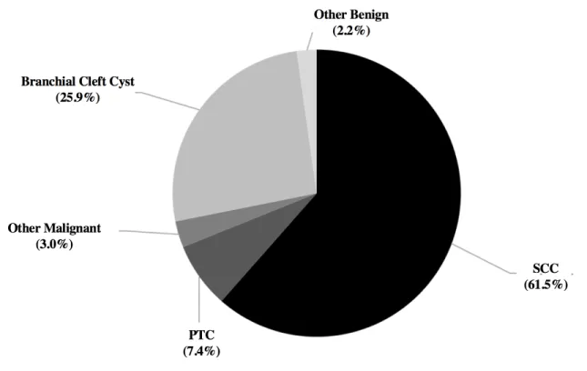
In total, 135 patients with cystic masses were included. Definitive diagnosis of each of these masses as well as their relative frequencies are shown in Figure 5. Ninety-eight patients with solid masses were included.
Figure 5: Differential diagnosis of cystic masses of the lateral neck based on excision biopsy or neck dissection pathology.
SCC: squamous cell carcinoma; PTC: papillary thyroid carcinoma
Within the cystic mass group, 7/135 (5%) samples were considered inadequate to make a diagnosis compared to 8/98 (8%) within the solid group. All the inadequate material came from FNA sampling. No statistical difference existed between cystic and solid groups regarding inadequate sampling (p=0.14).
41
Detection of p16 gene overexpression was undergone on either the cervical mass of the primary tumor in 43% patients with cystic cervical SCC and 51% patients with solid cervical SCC. Overexpression of p16 was found in 97% of patients in the cystic group and 90% in the solid group; no significant difference was found between these two groups (p=0.51). Of note, no ancillary studies were performed on either FNA, CNB or FS, including p16 immunohistochemistry. In total, 101 FNA were undergone on both benign and malignant masses, of these 90 (89.1%) were done without concurrent CNB. Of the 20 CNB performed, 9 (45%) were not done concurrently with FNA. The remaining 11 patients in both groups all had FNA and CNB done concurrently. Of these, 5 (46%) presented false-negative FNA when concurrent CNB were diagnostic of malignancy. The remaining 6 patients (55%) showed results diagnostic of malignancy on both FNA and CNB. Among the FNA done on cystic masses, 21 (20%) were performed by surgeons, 58 (57%) by radiologists, 3 (3%) by cytopathologists and 18 (18%) where data is missing.
All FNA and CNB final reports were reviewed. For FNA, 58/101 (57%) were definitely performed under U/S and 3/101 (3%) were definitely not performed under U/S. In all other 40/101 (40%) FNA results, use of U/S was not confirmed or infirmed by official report. Thus, rigorous comparison of image-guided vs no-guidance for FNA is therefore not feasible; this has been included in the limitations of this study. For CNB, U/S was definitely used in all but 2 (90%) patients, where data is lacking.
Out of the 25 patients having undergone FS, 14 (56%) had prior FNA and 3 (12%) had prior CNB. Incisional biopsies were submitted for FS in at least 2 (8%) patients before they were referred to our center. Of the 25 patients who underwent FS, 16 (64%) were done in our center.
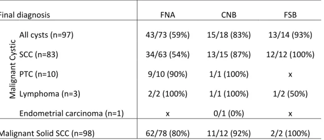
Table 2 presents demographic features, size, proportion of unilateral and single masses among all patients presenting with lateral cystic neck masses. Sensitivity, specificity, PPV, NPV of FNA, CNB and FS for cystic masses are shown in Table 3 and sensitivity for detecting malignancy for each specific pathological diagnosis is shown in Table 4. Statistically significant differences were shown when comparing the following: FNA sensitivity (59%) versus CNB sensitivity (83%), p=0.03;
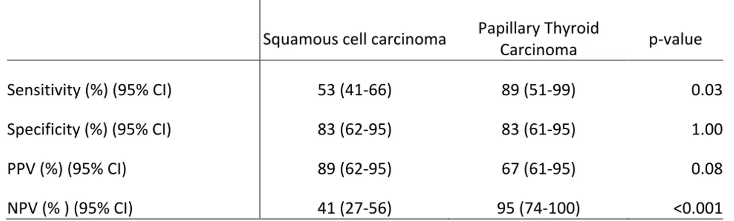
FNA sensitivity (59%) versus FS sensitivity (93%; p=0.01); FNA NPV (40%) versus FS NPV (92%; p<0.001). A trend towards a statistical difference was shown when comparing CNB NPV (50%) versus FS NPV (92%; p=0.07). No statistical differences were found between specificities and PPVs and no differences were found between sensitivities of CNB and FS as well as between NPV of FNA and CNB. FNA results of cystic masses diagnosed as SCC compared to those diagnosed as PTC are shown in Table 5, with corresponding p-values.
43
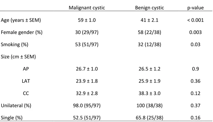
Table 2: Demographic features, size, proportion of unilateral and single masses among all patients presenting with lateral cystic neck masses
Malignant cystic Benign cystic p-value
Age (years ± SEM) 59 ± 1.0 41 ± 2.1 < 0.001
Female gender (%) 30 (29/97) 58 (22/38) 0.003 Smoking (%) 53 (51/97) 32 (12/38) 0.03 Size (cm ± SEM) AP 26.7 ± 1.0 26.5 ± 1.2 0.9 LAT 23.9 ± 1.8 25.9 ± 1.9 0.36 CC 32.9 ± 2.8 38.3 ± 3.0 0.12 Unilateral (%) 98.0 (95/97) 100 (38/38) 0.37 Single (%) 52.5 (51/97) 65.8 (25/38) 0.16
SEM: Standard error of the mean; AP: antero-posterior dimension; LAT: lateral-lateral dimension; CC: cranio-caudal dimension
Table 3: Sensitivity, specificity, PPV and NPV of FNA, CNB and FS in detecting the presence or absence of malignancy in the cystic masses of the lateral neck
FNA CNB FS
Sensitivity (%) (95% CI) 59 (47-70) 83 (63-96) 93 (32-100)
Specificity (%) (95% CI) 83 (62-95) 100 (40-100) 92 (62-100)
PPV (%) (95% CI) 92 (78-97) 100 (75-100) 93 (64-100)
NPV (%) (95% CI) 40 (27-55) 50 (14-86) 92 (62-100)
CI: Confidence interval; FNA: fine-needle aspiration; CNB: core-needle biopsy; FS: frozen section; PPV: positive predictive value; NPV: negative predictive value
Table 4: Sensitivity for detecting malignancy according to specific diagnosis made on final pathology
Final diagnosis FNA CNB FSB
Mal ig nan t C ys tic All cysts (n=97) 43/73 (59%) 15/18 (83%) 13/14 (93%) SCC (n=83) 34/63 (54%) 13/15 (87%) 12/12 (100%) PTC (n=10) 9/10 (90%) 1/1 (100%) x Lymphoma (n=3) 2/2 (100%) 1/1 (100%) 1/2 (50%) Endometrial carcinoma (n=1) x 0/1 (0%) x Malignant Solid SCC (n=98) 62/78 (80%) 11/12 (92%) 2/2 (100%) FNA: fine-needle aspiration; CNB: core-needle biopsy; FS: frozen section; SCC: squamous cell carcinoma; PTC: papillary thyroid carcinoma
45
Table 5: FNA parameters for detecting malignancy according to the presence of either squamous cell carcinoma or papillary thyroid carcinoma on final pathology
Squamous cell carcinoma Papillary Thyroid Carcinoma p-value
Sensitivity (%) (95% CI) 53 (41-66) 89 (51-99) 0.03
Specificity (%) (95% CI) 83 (62-95) 83 (61-95) 1.00
PPV (%) (95% CI) 89 (62-95) 67 (61-95) 0.08
NPV (% ) (95% CI) 41 (27-56) 95 (74-100) <0.001
CI: Confidence Interval; PPV: positive predictive value; NPV: negative predictive value.
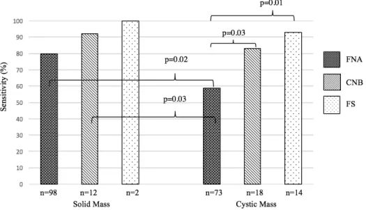
Sensitivity results of FNA, CNB and FS between solid masses and cystic masses are illustrated in Figure 6. FNA and CNB sensitivity for solid masses (80% and 92% respectively) were significantly better than FNA sensitivity for cystic masses (59%). FNA sensitivities per definitive diagnosis of solid and cystic masses are shown in Figure 7. FNA, CNB and FS of solid masses did not show significantly different results when compared among them and when compared to CNB and FS in cystic masses. No difference existed when comparing FNA sensitivity of solid SCC, cystic PTC and cystic lymphoma.
Figure 6: Diagnostic tool sensitivity in detecting malignancy in solid and cystic masses of the lateral neck.
Only statistically significant p-values are shown. FNA: fine-needle aspiration; CNB: core-needle biopsy; FS: frozen section.
47
Only statiscally significant p-values are shown. FNA: fine-needle aspiration; SCC: squamous cell carcinoma; PTC: papillary thyroid carcinoma
Three patients were ultimately diagnosed with lymphoma. Of these, 2 underwent FNA before surgical excision and 1 underwent CNB. All three neck masses were subsequently excised and analyzed. Two (1 FNA and 1 CNB) were analyzed by flow cytometry, and lymphoma was diagnosed. On the other cystic mass with previous FNA, southern blot and immunohistochemical analysis was performed and showed Bcl-2+ and Bcl-6+, which was suggestive of low-grade follicular lymphoma. There was however a lack of CD10 co-expression, which was atypical. Thus, a PCR analysis was undertaken and showed presence of 14:18 translocation (Bcl-2:IgG).
Of the 97 patients with cystic masses diagnosed with malignant disease, 56 (58%) were diagnosed by direct specimen pathological analysis and 41 (43%) by subsequent discovery of primary tumor. FNA sensitivity for detecting malignancy for both these groups was similar (56% vs 50%; p=0.65).
4.6. Discussion
Among all cystic neck masses, 72% were found to be malignant masses and 85% of these were SCC. Our findings indicate a much higher proportion of malignancy than those found in the literature which ranges from 10% to 24%. 35, 36, 38, 39 This discrepancy in the incidence of malignant disease may be due to the following reasons. First, there is no mention that partially cystic and necrotic masses were included in these articles which, if excluded, may greatly diminish the incidence of malignancy. Masses described as «necrotic» were included in our study because of the presence of a hypodense center as well as the well-known difficulty in radiologically differentiating an infected branchial cleft cyst with a necrotic metastatic node.30, 31 In sum, we thought it necessary to include these types of masses given their similar susceptibility to diagnostic errors, notably by FNA and CNB. Second, patients included in our study were treated either in a tertiary center specialized in oncologic head and neck surgery and included referrals from primary and secondary centers which may have treated a large proportion of benign diseases without our knowledge. Finally, a recent study has shown an association between cystic neck masses and HPV11 and it is known that the incidence of HPV-related oropharyngeal cancer is increasing at a dramatic rate.60 Therefore, it is possible that malignant cystic neck masses are much more prevalent today than previously recognized. Articles cited above were in fact published no less than 15 years ago. This is also reflected in our results which showed a high p16 positivity in the cystic and solid neck groups (97% and 91% respectively). This high positivity, however, may be overestimated given the low proportion of samples tested for p16 within our study group (43% for the cystic group and 51% for solid group).
PTC has long been considered a cause of cystic metastasis to the lateral neck with an incidence as high as 11%58 among all lateral cystic neck masses, a rate similar to our results (7%). Cystic lymphoma, however, is a much rarer diagnosis in the head and neck, described only in certain isolated cases.61-64 Of note, the term “lymphoproliferative disorder” in either FNA or CNB reports was considered an adequate prediction of the definitive diagnosis of lymphoma. Finally, without surprise, branchial cleft cysts were the most prevalent benign cystic lesions in the neck.31, 65
49
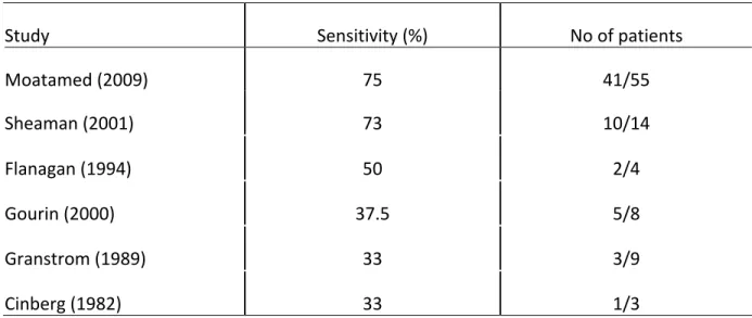
As mentioned above, FNA sensitivity results vary widely in the literature (33-75%) (Table 5). Our results are therefore comparable to these. As for specificity and PPV, results are more promising in our assessment (83% and 92% respectively) as well as in the literature reaching as high as 100% for both specificity and PPV.34 NPV on the other hand was much lower in our group (40%) when compared to rates in literature which were at 92% in a study which included, however, midline cysts and therefore had a higher probability of benign disease (e.g. thyroglossal cysts).34 FNA sensitivity for detecting malignancy was significantly better in solid masses (80% vs 59%; p=0.02). This value is slightly inferior to the ones found in the literature which range from 87-95%, further illustrating the difference of FNA usefulness between solid and cystic masses. 40, 41, 66
Table 6: FNA sensitivity in detecting malignancy in cystic masses of the lateral neck
Study Sensitivity (%) No of patients
Moatamed (2009) 75 41/55 Sheaman (2001) 73 10/14 Flanagan (1994) 50 2/4 Gourin (2000) 37.5 5/8 Granstrom (1989) 33 3/9 Cinberg (1982) 33 1/3
With regards to all cervical masses as shown in a large meta-analysis of 554 cervical masses, CNB had an overall accuracy of 96% in detecting malignancy.42 However, when considering only cystic neck masses, no results in the literature were found for comparative purposes. Furthermore, controversy regarding their use in cystic masses exists, notably because of the supposed higher risk of inadequate material extraction.43, 67 These conclusions were, however, contrasted by Karstrup et al. who demonstrated sufficient material in 86% of cystic thyroid lesions when using
CNB.68 Interestingly, inadequate sample rates showed no statistical difference when comparing FNA samples from solid masses to those of cystic masses. Furthermore, no CNB samples in either solid masses or cystic masses were considered inadequate. This further refutes the presumption that cystic masses are not amenable to CNB because of high rates of inadequate sampling.43 In our study, CNB sensitivity for cystic masses presented no difference with sensitivity results for solid masses (83 vs 92%; p=0.43). This latter value is in fact similar to the one Kraft et al. found of 97% when assessing head and neck lesions with CNB.67
Finally, FS results in cystic lesions were also difficult to compare with literature given their scarcity. FS is frequently cited as having 100% specificity in different neck lesions, including in the detection of branchial cleft cysts.46, 69, 70 Interestingly, among our patients, one was diagnosed with a suspicion of SCC on FS with a final diagnosis of branchial cleft cyst after undergoing a selective neck dissection. However, its sensitivity remains high (92%) and still represents a crucial tool for confirming malignancy in lateral cystic neck masses.
Cystic metastasis of PTC had a better sensitivity (p=0.03) and NPV (p<0.001) with FNA when compared to SCC cystic lesions. This is possibly due to the more easily discernable and distinctive features on PTC FNA smears such as multinucleated giant cells, psammoma bodies and foamy macrophages. 71, 72
In the present series, no complications were noted for any of the diagnostic interventions. FNA and CNB are in fact renowned for having very few procedural complications. 73-78
Given all these results, we propose the following management for cystic neck lesions with regards to the use of FNA, CNB and FS (See Figure 8). Given its minimal invasiveness, safety, wide availability as well as adequate PPV in cystic lesions (92%), we suggest the initial use of FNA in patients with both solid and cystic masses. However, in the advent of a FNA not indicative of malignancy, CNB should then be used in assessing cystic masses since it has a significantly higher sensitivity than FNA (83% vs 59%, p=0.03) for detecting malignancy. If CNB is not diagnostic of malignancy, it should always be followed by FS to confirm the absence of malignancy since this
51
FNA, CNB and FS in cystic masses, which all have a high PPV (92%, 100%, 93% respectively), foreshadows the presence of malignancy and an extensive search for primary tumor should be initiated.
FNA sensitivity for detecting malignancy in solid masses is sufficiently high for detecting malignancy and has no significant difference with CNB (80 vs 92%; p=0.22). Consequently, a patient with FNA of a solid mass not indicative of malignancy could either be observed or have a repeat FNA depending on clinical suspicion of malignancy.
Figure 8: Proposed diagnostic algorithm for the management of cystic masses of the lateral neck.
U/S: ultrasound; CT-Scan: computed tomography scan; FNA: fine-needle aspiration; CNB: core-needle biopsy; FS: frozen section; SCC: squamous cell carcinoma; PTC: papillary thyroid
4.6.1. Limitations
Firstly, at our tertiary oncological center, almost all patients undergoing FNA for oncological indications are referred to expert interventional radiologists for U/S-guided FNA or CNB. That said, many patients included in this study were referred after having already undergone FNA or CNB. Therefore, it was not always clear who had undergone image-guided vs no-guidance in patients with an initial work-up outside our healthcare center. Also, comparison of image-guided vs non-guided FNA was not feasible given the low number of confirmed non-guided FNA (n=3) and high percentage of patients (40%) with lack of data. Additionally, information was missing with regards to how many cases who underwent FNA had rapid on-site evaluation. Secondly, we are aware that the inclusion of patients with primary lesions subsequently identified and confirmed with biopsy creates an issue calculating sensitivity, specificity, PPV, and NPV of all diagnostic modalities since the mass has not been proven to be malignant. However, all the patients who presented with neck masses had results diagnostic of malignancy on either FNA, CNB or FS. Given the presence of results indicating malignancy on all biopsy modalities and the presence of a primary tumor with pathological confirmation of malignancy, we consider it highly improbable that the mass in question would be benign. Although we cannot be absolutely certain of this, in most centers, if radiotherapy is planned, it is not indicated to excise the mass for pathological diagnosis before undertaking radiation treatment to the primary site and neck. For example, if a patient would present at onset with a T1 squamous cell carcinoma of the tonsil with a partially cystic neck mass, the mass would not be biopsied or excised. Most would agree that a biopsy of the tonsil would suffice to treat the neck by radiotherapy. Therefore, given the high proportion of patients without an excised cervical mass in this patient population (43%), we consider their inclusion to be invaluable to the power of our study and to the clinically relevant of these diagnostic procedures. Furthermore, excluding the patients presenting with cystic neck masses treated with radiotherapy would create a selection bias in our study population. In fact, most patients undergoing surgery in this study group are patients with a low suspicion of malignancy. Therefore, only including these patients would create a tendency toward selecting patients with benign disease and would not be representative of the true lateral cystic mass
53
it impossible to have a uniform treatment plan for every one of these cases. This therefore limits the ability to fully respect the definition of sensitivity, specificity, PPV and NPV with regards to the need for a histologic confirmation as gold standard.
4.7. Conclusion
In our study, 72% of cystic neck masses were malignant. After evaluating FNA, CNB and FS results, we observed that sensitivity for detecting malignancy was significantly better when using CNB and FS when compared to FNA. FNA and CNB had similar negative predictive values, both being significantly less that FS’s. Furthermore, FNA sensitivity for malignancy in solid masses was also shown to be significantly better compared to sensitivity in cystic masses. All tests, however, had a high PPV and only FNA showed inadequate specimen sampling, similarly in solid and cystic masses.
Overall, FNA and CNB should both be regarded as safe and useful tools for the diagnosing cystic neck masses but one should still be wary of a non-diagnostic result or the mention of absence of malignancy given relatively low NPV. Novel techniques such as image cytometry DNA-analysis can also be used to augment the sensitivity of these tests and, if systematically used, could further help in reducing the use of non-essential surgical interventions. 79
5. Discussion générale
Les modalités d’imagerie démontrent des limites évidentes. La présence de multiples adénopathies à l’échographie ou à la tomodensitométrie ne semble pas prédire la malignité d’une masse kystique. En effet, la tendance des kystes branchiaux à se surinfecter peut entrainer la présence d’adénopathies réactives au pourtour du kyste. À l’échographie, la vascularité et la présence de calcifications ne permettent pas non plus de différencier définitivement les kystes de carcinomes épidermoïdes des kystes branchiaux. À la tomodensitométrie, l’infiltration des graisses aussi ne semble pas différencier avec certitude les deux entités cliniques. Également, au niveau de la tomodensitométrie à l’émission de positron, le SUV de la masse kystique ne permet de pas de clarifier le diagnostic. Le seul paramètre ayant démontrer une certaine différence est la dimension cranio-caudale. En effet, les kystes branchiaux ont démontré une dimension cranial-caudale significativement plus élevée lorsque comparés aux carcinomes épidermoïdes (39 mm vs 32 mm; p=0.016). Malgré ceci, la combinaison de toutes les variables d’imagerie a permis aux radiologistes d’établir un diagnostic de malignité adéquat la majorité du temps. Par contre, la spécificité et la valeur prédictive négative étaient plutôt faibles. Ceci nous mène à suggérer aux cliniciens de maintenir une suspicion élevée de malignité malgré un diagnostic rassurant du radiologiste.
La sensibilité de la cytoponction à l’aiguille fine était significativement meilleure pour les masses solides lorsque comparée aux masses kystiques (80% vs 59%; p=0.02). La sensibilité de la biopsie au trocart était par contre comparable entre les masses solides et kystiques. Il est important également de noter que l’examen extemporané a démontré une sensibilité élevée (92%) pour les masses kystiques du cou. En vue de ses résultats, nous proposons l’algorithme de prise en charge suivant. Vu la qualité peu invasive, l’accessibilité et la haute valeur prédictive positive de la cytoponction à l’aiguille fine, nous suggérons son utilisation dans la prise en charge initiale des patients ayant des masses solides ou kystiques. Toutefois, si la cytoponction n’indique pas la présence de malignité, la biopsie au trocart devrait être tentée vu la sensibilité plus élevée lorsque comparée à la cytoponction (83% vs 59%; p=0.03). Si la biopsie au trocart est négative pour la présence de malignité, un examen extemporané devrait suivre pour confirmer l’absence