Selection for
Staphylococcus aureus small-colony
variants due to growth in the presence of
Pseudomonas aeruginosa
Lucas R. Hoffman*, Eric De´ziel†, David A. D’Argenio‡, Franc¸ois Le´pine†, Julia Emerson*, Sharon McNamara*, Ronald L. Gibson*, Bonnie W. Ramsey*, and Samuel I. Miller‡§¶储
Departments of *Pediatrics,‡Microbiology,§Medicine, and¶Genome Sciences, University of Washington, Seattle, WA 98195; and†Institut National de la
Recherche Scientifique–Institut Armand-Frappier, Universite´ du Que´bec, 531 Boulevard des Prairies, Laval, QC, Canada H7V 1B7
Edited by Emil C. Gotschlich, The Rockefeller University, New York, NY, and approved November 9, 2006 (received for review August 4, 2006) Opportunistic infections are often polymicrobial. Two of the most
important bacterial opportunistic pathogens of humans, Pseudo-monas aeruginosa and Staphylococcus aureus, frequently are coisolated from infections of catheters, endotracheal tubes, skin, eyes, and the respiratory tract, including the airways of people with cystic fibrosis (CF). Here, we show that suppression of S. aureus respiration by a P. aeruginosa exoproduct, 4-hydroxy-2-heptylquinoline-N-oxide (HQNO), protects S. aureus during cocul-ture from killing by commonly used aminoglycoside antibiotics such as tobramycin. Furthermore, prolonged growth of S. aureus with either P. aeruginosa or with physiological concentrations of pure HQNO selects for typical S. aureus small-colony variants (SCVs), well known for stable aminoglycoside resistance and per-sistence in chronic infections, including those found in CF. We detected HQNO in the sputum of CF patients infected with P. aeruginosa, but not in uninfected patients, suggesting that this HQNO-mediated interspecies interaction occurs in CF airways. Thus, in all coinfections with P. aeruginosa, S. aureus may be underappreciated as a pathogen because of the formation of antibiotic-resistant and difficult to detect small-colony variants. Interspecies microbial interactions, analogous to those mediated by HQNO, commonly may alter not only the course of disease and the response to therapy, but also the population structure of bacterial communities that promote the health of host animals, plants, and ecosystems.
4-hydroxy-2-heptylquinoline-N-oxide兩 antibiotic resistance 兩 interspecies 兩 polymicrobial兩 tobramycin
B
acterial communities consisting of multiple species exist in the soil and water and on animal and plant surfaces. These polymicrobial populations can be important determinants of host organism health. Commensal communities of animal flora contribute to normal digestion, intestinal development, and disease resistance, whereas imbalances in these communities can lead to pathogenic states such as colitis and periodontal disease. Bacteria infecting human tissues also commonly comprise mixed communities, particularly when mucosal barriers have been compromised (1). Relatively little is known regarding microbial interactions within such communities compared with what is known of individual bacterial species living in isolation (2–6).Infection with mixed communities of bacterial pathogens is common in hospitalized and immunosuppressed patients (1). One of the best studied pathogenic bacterial communities occupies the airways of people with the disease cystic fibrosis (CF). The Gram-positive bacterium Staphylococcus aureus is the pathogen most commonly cultured from young children with CF (7), and in the preantibiotic era, many CF patients succumbed to S. aureus infection (8). CF patients infected only with S. aureus have airway inflammation (9), and treatment of these patients with antistaphylococcal antibiotics often leads to clinical im-provement (9). Of the multiple opportunistic bacteria that may infect these patients, however, the Gram-negative bacterium
Pseudomonas aeruginosa is considered to be the most significant pathogen (10). As patients acquire P. aeruginosa, S. aureus is cultured less frequently (7, 11), although both organisms are commonly coisolated from CF respiratory cultures [20–25% of cultures in one CF clinic population; supporting information (SI) Table 1]. Although this shift could result from antibiotic treat-ments, an alternative explanation is that P. aeruginosa competes with S. aureus within CF airways.
P. aeruginosa produces an antistaphylococcal substance,
4-hydroxy-2-heptylquinoline-N-oxide (HQNO), that sup-presses the growth of many Gram-positive bacteria (11, 12). HQNO also allows some Gram-positive bacteria to grow, albeit slowly, in the presence of the first-generation aminoglycoside antibiotics streptomycin and dihydrostreptomycin (13). After these initial discoveries, the apparent paradox of how HQNO both could inhibit growth and protect against aminoglycosides remained unresolved, and the potential implications for bac-terial mixed species infections never were pursued (12). Given that more recent research showed that electron transport is required for aminoglycoside uptake (14), we reasoned that HQNO protects S. aureus from killing by aminoglycosides by inhibiting electron transport. The newer aminoglycoside to-bramycin commonly is used in treating P. aeruginosa infections such as those in CF (7, 10), and in vitro testing indicates that
S. aureus and P. aeruginosa isolates from CF patients are
usually tobramycin-susceptible (15, 16). We therefore hypoth-esized that S. aureus density within CF airways ref lects a balance between the suppressive effects of antibiotics and HQNO, and HQNO-mediated protection from aminoglyco-sides (which would escape detection by routine susceptibility testing). To provide support for this hypothesis, we examined the effects of HQNO on aminoglycoside susceptibility of S.
aureus under conditions that were clinically and physiologically
relevant, and that had been previously unexamined. We used HQNO at concentrations normally produced by P. aeruginosa, together with aminoglycosides that are in clinical use (includ-ing tobramycin), and with both brief and extended bacterial incubations, so as to mimic conditions characteristic of acute and chronic infections, respectively.
Author contributions: L.R.H., E.D., D.A.D., R.L.G., B.W.R., and S.I.M. designed research; L.R.H., E.D., and F.L. performed research; L.R.H., E.D., and S.I.M. contributed new reagents/ analytic tools; L.R.H., E.D., D.A.D., F.L., J.E., R.L.G., B.W.R., and S.I.M. analyzed data; and L.R.H., E.D., D.A.D., F.L., J.E., R.L.G., B.W.R., and S.I.M. wrote the paper.
The authors declare no conflict of interest. This article is a PNAS direct submission.
Abbreviations: CF, cystic fibrosis; HQNO, 4-hydroxy-2-heptylquinoline-N-oxide; MH, Mueller–Hinton; MIC, minimal inhibitory concentration; SCV, small-colony variant.
储To whom correspondence should be addressed. E-mail: [email protected].
This article contains supporting information online at www.pnas.org/cgi/content/full/ 0606756104/DC1.
Results
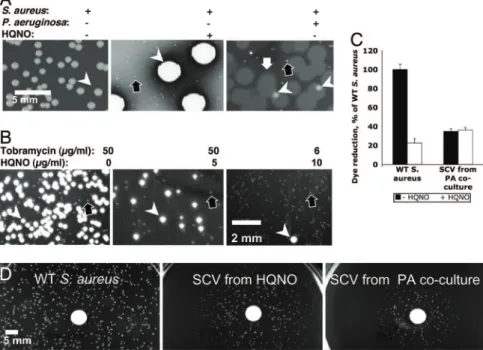
HQNO at Physiological Concentrations Has Short-Term Effects onS. aureus.The short-term effects on S. aureus of HQNO produced by P. aeruginosa were examined by coculturing the two organisms overnight in the presence of various antibiotics. Significantly, S. aureus grew in the presence of tobramycin levels above the minimal inhibitory concentration (MIC; 0.4 g/ml) in a zone surrounding P. aeruginosa colonies (Fig. 1A). P. aeruginosa conferred protection against additional aminoglycosides (gen-tamicin, amikacin, and kanamycin) but not against any other antibiotic classes tested: -lactams (carbenicillin and ceftazi-dime), macrolides (azithromycin), and chloramphenicol (data not shown). Colonies of P. aeruginosa mutants unable to produce HQNO (pqsA and pqsL mutants; refs. 17 and 18) did not protect S. aureus from tobramycin (Fig. 1 A and B), whereas pure HQNO did (Fig. 1B). These results indicate that HQNO production by P. aeruginosa colonies protects S. aureus from the antibiotic effect of tobramycin.
In the absence of tobramycin, wild-type P. aeruginosa colonies, but not pqsA (Fig. 1A) or pqsL (Fig. 1C) mutant colonies, visibly suppressed S. aureus growth, as demonstrated in refs. 11, 13, 17, and 19. We found that both wild-type P. aeruginosa culture supernatants (data not shown) and pure HQNO (Fig. 1C) suppressed S. aureus growth to a degree similar to P. aeruginosa colonies. Furthermore, S. aureus formed smaller colonies when near an HQNO source than when distant from such a source (Fig. 1D). Cells from the smaller colonies, when transferred to media without HQNO, formed colonies of wild-type size,
indi-cating that growth limitation by HQNO was reversible. When either HQNO or P. aeruginosa was added to S. aureus liquid cultures, neither resulted in a decrease in the number of viable S. aureus cells (data not shown). Reversible, incomplete growth suppression by HQNO is consistent with the fact that S. aureus could grow adjacent to P. aeruginosa colonies in the presence (Fig. 1 A) or absence (data not shown) of aminoglycosides and adjacent to a source of pure HQNO (Fig. 1D).
Long-Term Exposure ofS. aureus to HQNO Selects for Small-Colony Variants (SCVs).The two phenotypes associated with S. aureus grown in the presence of HQNO, aminoglycoside resistance and small colony formation, are also characteristic of genetically stable S. aureus SCVs (20). S. aureus SCVs are observed in diverse infections, including the airways of as many as 50% of CF patients (21). Such SCVs are selected by aminoglycoside expo-sure (20, 22), frequently arise because of mutations that impair electron transport (20, 21), and are resistant to aminoglycosides because of diminished electron transport-mediated drug uptake (23). Given the similarities between SCVs and the transient, HQNO-induced S. aureus phenotypes (Fig. 1), we explored whether prolonged growth in the presence of HQNO ultimately selects genetically stable SCVs.
Growth of S. aureus for 5 days in the presence of 10 g/ml HQNO (a concentration typically found in stationary-phase P. aeruginosa cultures; ref. 17) yielded a high proportion of SCVs (Fig. 2A). Similarly, after 5 days in coculture with wild-type P. aeruginosa, SCVs represented the majority of the S. aureus population (Fig. 2 A). In contrast, cocultures with HQNO-deficient P. aeruginosa strains (pqsA or pqsL mutants, data not shown) and S. aureus monocultures (Fig. 2 A) yielded no SCVs. A significant number of S. aureus SCVs were obtained after a shorter incubation period (overnight growth) when 50 g/ml tobramycin was added to the growth medium (Fig. 2B), as reported for the aminoglycoside gentamicin (24). Although overnight growth in the presence of either 6–12g/ml tobra-mycin or 10 g/ml HQNO yielded rare SCVs (Fig. 3), the combination of these conditions led to a population composed nearly entirely of SCVs (Figs. 2B and 3). Therefore, the effect of the two compounds acting together is greater than the sum of their individual effects, consistent with synergy between amino-glycosides and HQNO during selection for S. aureus SCVs. Such synergy is likely to be clinically relevant, because the two compounds would be predicted to be found together during treatment of many clinical coinfections with S. aureus and P. aeruginosa.
S. aureus SCVs Derived from Selection with HQNO Have the Charac-teristics of Clinical SCVs.To determine whether the S. aureus SCVs selected in vitro had the reduced respiration characteristic of clinical SCVs, we used the redox-sensitive dye Alamar blue (ref. 25; Fig. 2C). HQNO (10 g/ml) inhibited dye reduction by wild-type cells by 80%, consistent with the known ability of HQNO to inhibit S. aureus cytochrome activity (12). Using this assay, the respiratory activity of an SCV selected by coculture with P. aeruginosa was 40% of that of wild-type cells, and it was not further inhibited by HQNO (Fig. 2C). Clinical SCVs com-monly have respiratory defects that can be complemented by hemin or menadione (vitamin K3), which are required for biosynthesis of electron transport chain components, although the mutations responsible for these defects have not been described for clinical isolates (20, 21). Menadione restored wild-type colony size to S. aureus SCVs selected in vitro when grown with either chemically defined medium (data not shown) or with rich medium (Fig. 2D), suggesting that they are mena-dione auxotrophs (20). Menamena-dione-auxotrophic S. aureus SCVs are known to accumulate aminoglycosides more slowly than wild-type cells, and they have been isolated from clinical
infec-Fig. 1. HQNO produced by P. aeruginosa simultaneously suppresses the growth of S. aureus and protects it from tobramycin killing. (A) Colonies of P.
aeruginosa grown on a lawn of S. aureus on LB agar plates with tobramycin
added as indicated. WT, wild-type P. aeruginosa PAO1. The pqsA mutant is defective for HQNO production. The MIC of tobramycin for these P.
aerugi-nosa strains is 1g/ml. White arrow, zone of S. aureus growth. S. aureus grows slowly in the zone surrounding the wild-type (WT) P. aeruginosa colony, most evident when the S. aureus lawn is suppressed by tobramycin concentrations ⬎0.3g/ml, but similar S. aureus densities are present in the zones under each condition (data not shown). (B) A paper disk (containing 15g of pure HQNO) and colonies of P. aeruginosa on a lawn of S. aureus cells on agar media containing 0.6g/ml tobramycin. WT, wild-type P. aeruginosa PA14. The pqsL mutant is defective for HQNO production. White arrow, zone of S. aureus growth. (C) Experiment performed as in B, except without tobramycin. (D) S.
aureus colonies (of cells from a diluted culture) growing on LB agar near disks
containing HQNO or methanol solvent alone. For all P. aeruginosa experi-ments shown, equivalent results were obtained by using P. aeruginosa strains PAO1 and PA14 and their derived pqsA and pqsL mutants.
tions, including those in CF airways (20, 23, 26). Similarly, the S. aureus SCVs selected in vitro had increased resistance to tobramycin (Fig. 4 A and B), and susceptibility to tobramycin was restored to wild-type levels in the presence of menadione (Fig. 4C).
Given that laboratory growth of clinical S. aureus SCVs can yield revertants with wild-type colony morphology (20), we examined the genetic stability of SCVs selected in vitro. Wild-type revertants appeared at a rate of⬇0.5% per hour of culture in the absence of HQNO or P. aeruginosa, whereas no such revertants were observed after culture in the presence of tobramycin (data not shown). In environments where P. aeruginosa and S. aureus coexist, there is likely to be a dynamic interaction among the number of P. aeruginosa cells, the proportion of S. aureus SCVs in the bacterial population, and the response of each pathogen to tobramycin. Adding further
complexity, subinhibitory levels of aminoglycosides induce P. aeruginosa biofilm formation, a form of bacterial growth characterized by high cell density (27). Because HQNO bio-synthesis is up-regulated by the cell density-dependent quorum sensing signaling system (17), HQNO production would be predicted to be increased in biofilms, increasing the selection pressure for S. aureus SCVs. The recent identification in the CF lung of SCVs of P. aeruginosa, which have an increased capacity for biofilm formation (28) and also have been selected by aminoglycoside exposure (29), would be predicted to fur-ther augment these effects.
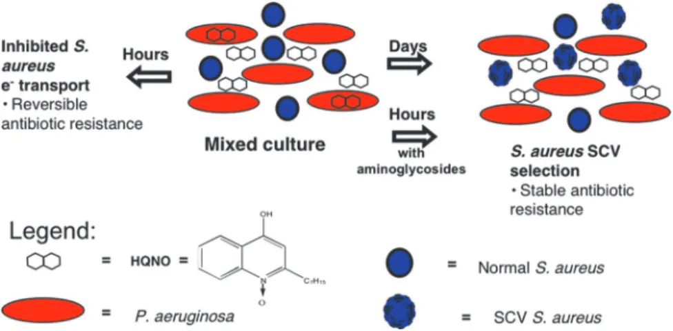
HQNO Is Present in Sputum from CF Patients Infected withP. aerugi-nosa.The biosynthetic pathway that produces HQNO generates several additional 4-hydroxy-2-alkylquinolines (17), two of which have been found in CF respiratory secretions (11, 30). We therefore sought to detect HQNO in sputum from CF patients. The reducing agents used in previous studies to liquify these highly viscous samples (11) maximize solvent extraction but destroy HQNO, and we therefore performed these extractions without liquification. Despite these limitations, we detected HQNO and only in samples from patients infected with P. aeruginosa (Table 1). These results suggest that P. aeruginosa in the CF airway produces HQNO and affects S. aureus as it does in vitro. Indeed, HQNO produced by P. aeruginosa could select for S. aureus SCVs not only in the CF airway, but wherever these two opportunistic pathogens occur together (Fig. 5).
Discussion
S. aureus SCVs have been observed in infections for decades, but our current understanding of their clinical importance is limited (20, 26). S. aureus SCVs are of particular clinical concern
Fig. 2. HQNO inhibits S. aureus electron transport, ultimately selecting for SCVs. (A) S. aureus grown for 5 days in static cultures alone, with P. aeruginosa PAO1, or with HQNO as indicated, before plating on selective media to distinguish species and morphotypes. Results shown are on sheep’s blood agar. Cells from the culture with HQNO were incubated longer to better display SCVs. Results are representative of three separate experiments; equivalent results were obtained with P. aeruginosa strains PAO1 and PA14 and with S. aureus clinical isolates from five separate CF patients. White arrowheads, normal S. aureus. Black arrows, SCV S. aureus. White arrow, P. aeruginosa. (B) Colonies of cells from S. aureus cultures grown overnight in the presence of the indicated tobramycin and/or HQNO concentrations. Results are representative of two separate experiments. (C) Alamar blue, a redox-sensitive dye, was used to quantify reduction potential (as a measure of electron transport) of the S. aureus strains indicated in the presence and absence of HQNO. Results shown are the average of triplicates⫾ SD and are representative of three separate experiments. (D) Cells of S. aureus SCVs isolated after HQNO exposure form colonies of wild-type size when growing in close proximity to a disk containing 1.5g of menadione and with colony size diminishing with distance from the disk. No such colony changes were observed with disks containing thymidine or hemin. Results are representative of three separate experiments.
Fig. 3. Synergy between HQNO and tobramycin for S. aureus SCV formation. Quantitative results of the experiment are described in Fig. 2B. The relative proportions of SCVs were determined among 100 –300 total colony-forming units in overnight cultures of S. aureus grown in HQNO and tobramycin. Growth in the presence of either 10g/ml HQNO (Left) or 12 g/ml tobramycin alone (Right) yieldedⱕ5% SCVs, whereas in combination, they yielded nearly 100% SCVs, consistent with synergy. Results are representative of two sepa-rate experiments, each with at least two technical replicates.
because they are resistant to aminoglycosides and, perhaps, to other antibiotics (20). S. aureus SCVs are persistent, infecting CF patients for many years (20), and are capable of intracellular growth (20), which may facilitate their persistence in chronic infections (Fig. 5). SCVs also may be missed by clinical labora-tories because of their slow growth and atypical phenotype relative to other bacteria in mixed cultures (20, 21). We propose that the observation that S. aureus is cultured less frequently from CF patients after P. aeruginosa is acquired (7, 11) in part is attributable to HQNO-mediated selection for persistent, yet poorly detectable, SCVs. Furthermore, S. aureus may be un-derappreciated as a pathogen not only in CF, but in all coinfec-tions with P. aeruginosa, including hospital-acquired pneumonia and infections associated with burns, catheters, and endotracheal tubes. Hospitalized patients are frequently colonized or infected with multiple pathogens, including S. aureus and P. aeruginosa (6, 31). Our results argue for routine investigation into potential
undiagnosed roles of S. aureus SCVs in hospital-acquired infec-tions when P. aeruginosa is present, and the detection and antibiotic treatment of S. aureus SCVs may improve patient outcomes.
In considering the physiologic role for P. aeruginosa produc-tion of HQNO, a molecule that inhibits Gram-positive bacterial respiration, it is important to note that P. aeruginosa dwells predominantly in environmental niches, such as soil and water, rather than mammalian tissues. Only in the setting of compro-mised mucosal barriers or prolonged antibiotic therapy does P. aeruginosa opportunistically infect humans. P. aeruginosa com-monly coinhabits soil with antibiotic-producing Gram-positive bacteria such as Streptomyces tenebrarius, the source of tobra-mycin (32). Therefore, HQNO production could have evolved, at least in part, to metabolically restrain environmental bacteria that produce antipseudomonal compounds. In support of this idea, we found that wild-type P. aeruginosa suppressed the growth of at least two antibiotic-producing, soil-dwelling Gram-positive bacteria: S. tenebrarius and Paenibacillus polymyxa (data not shown). Such microbial competition has been proposed to contribute to the evolution of P. aeruginosa acute virulence factors (4).
Given the complexity of microbial communities in the environment, P. aeruginosa exoproducts are likely to have effects that extend beyond straightforward restraint or elimi-nation of competitors. The majority of antibiotics that revo-lutionized medicine in the mid-20th century are simply mi-crobial products deliberately administered at supraphysiologic concentrations, although these products can have distinctly different effects at naturally occurring concentrations (27, 33). Thus, under natural conditions, these compounds could main-tain equilibrium between microbial community members rather than eradicating them. For example, HQNO produced by P. aeruginosa at physiologic levels impairs S. aureus growth, perhaps allowing P. aeruginosa to benefit from useful S. aureus metabolites (19). Community interactions also may aid the bacterial colonization of animal mucosal surfaces by facilitat-ing resistance to host defenses, includfacilitat-ing the activities of phagocytes (20, 22). Such interactions may have been impor-tant in the development of the commensal bacterial commu-nities that colonize multicellular organisms.
We have shown that P. aeruginosa simultaneously suppresses the growth and enhances the aminoglycoside resistance of S. aureus by producing HQNO, a compound found in the sputum of infected CF patients. Prolonged exposure to HQNO selects for aminoglycoside-resistant genetic variant (SCV) S. aureus, which are difficult to detect. Our results highlight the poten-tially important roles for bacterial interspecies interactions in virulence and response to therapy. In any large and complex bacterial community, including polymicrobial infections and commensal bacterial populations, diverse interspecies inter-actions ranging from antagonism to cooperation are likely to exist and to significantly impact the health of host plants and animals.
Materials and Methods
Bacteria, Chemicals, and Growth Conditions.P. aeruginosa wild-type and mutant strains derived from PAO1 (Iglewski strain), including pqsA mutants, were gifts from L. Gallagher and C. Manoil (University of Washington) (34), except for the pqsL mutant, which was described by D.A.D. in ref. 35. Equivalent strains in the PA14 background were reported in refs. 17 and 18. S. aureus Newman strain was a gift from C. Rubens (University of Washington). Five CF clinical isolates of S. aureus with a normal colony size were provided by the University of Washington CF Core Microbiology Laboratory, directed by J. Burns. HQNO, menadione, and tobramycin were from Sigma (St. Louis, MO); Alamar blue was from Trek
Fig. 4. HQNO selects for S. aureus SCVs that are resistant to tobramycin. (A) Tobramycin susceptibility of wild-type S. aureus and of an SCV isolated after HQNO exposure. A disk containing 1.5g of tobramycin was placed on a S.
aureus lawn on LB agar. (B) The MIC of tobramycin for wild-type S. aureus
growing in the absence and in the presence of HQNO, and for SCVs derived as indicated. Results shown are averages of technical duplicates⫾ SD and are representative of three separate experiments. (C) The MIC of tobramycin, with and without menadione, for wild-type S. aureus and SCVs derived as indi-cated. Menadione restored sensitivity to tobramycin to S. aureus SCVs (but menadione itself was toxic to wild-type S. aureus at 10g/ml); results shown are representative of two separate experiments.
Table 1. P. aeruginosa and S. aureus cfu vs. HQNO content extractable with ethyl acetate from unliquified, frozen sputum samples from six CF patients
Sputum specimen no. S. aureus, cfu兾ml* P. aeruginosa, cfu兾ml Extractable HQNO, g兾g of dry weight sputum 1 ⬎105 0 0 3 0 0 0 4 1,400 105 1.24 6 ⬎105 0 0 7 80 4,000 0.39 11 70 0 0
*The presence or absence of S. aureus SCVs was not noted.
Diagnostics (Cleveland, OH); and hemin and thymidine were from Fluka Biochemica (Buchs, Switzerland). Mueller–Hinton (MH) broth, tryptic soy broth, and Luria–Bertani (LB) broth were from GIBCO (Grand Island, NY). Mannitol salt and sheep’s blood agar plates were from Fisher (Pittsburgh, PA). M9 minimal medium plates were made as described in ref. 36, except using UltraPure Agarose (Invitrogen, Carlsbad, CA) at 1.5% and with the addition of all 20 aa plus hydroxy-L-proline at 20g/ml, cystine at 10 g/ml, and biotin at 0.2 g/ml. All incubations were performed at 37°C unless otherwise indi-cated. Sputum from CF patients was obtained from a collec-tion (stored frozen) that was maintained by the Cystic Fibrosis Research Development Program at Children’s Hospital and Regional Medical Center (CHRMC), Seattle, WA. Access to and the use of these samples, and the use of clinical S. aureus isolates, were approved by the CHRMC Human Subjects Institutional Review Board.
Bacterial Susceptibility Assays.Antibiotic susceptibility was de-termined by standard broth microdilution MIC assays in 96-well microtiter plates in MH broth by using 105cells in 100 l per well.
Assays withS. aureus on Agar Surfaces.S. aureus cultures grown overnight with LB broth were diluted 1:10 and applied to the surface of MH agar by using a cotton swab, followed by the addition of P. aeruginosa cells or test compounds, as described below, and incubation overnight at 37°C. To test for inhibition by P. aeruginosa colonies, 5l of P. aeruginosa cultures, grown overnight with LB broth and diluted 1:10, was added as spots to the S. aureus lawn. To test for inhibition by P. aeruginosa culture supernatants, P. aeruginosa overnight cultures grown with LB broth were centrifuged to pellet cells, the supernatants were filtered through a 0.22-m filter, and 20 l of the supernatant was placed in a well in the MH agar made by removing a plug of agar. For disk assays, the indicated chemicals (in methanol or water, as appropriate) were added to sterilized 9-mm-diameter Whatman filter paper disks, which were allowed to dry before being placed on the agar surface. Chemical complementation experiments were performed on M9 minimal medium/agarose (and in parallel on MH agar for improved photographic images), using disks containing thy-midine, menadione, or hemin to examine for auxotrophy as described in ref. 37.
Alamar Blue Reduction Assay.S. aureus cells were resuspended from agar surfaces into PBS (to minimize SCV reversion to wild-type phenotype) to an OD600of 1.0. The resulting suspen-sion was diluted 1:10 in MH broth for a final volume of 1 ml. HQNO (3.3l of 3 mg/ml methanol stock) or methanol alone was added to each sample, which was mixed before and after addition of 50l of Alamar blue. The samples were incubated for 1 h at 22°C and centrifuged to pellet cells, and supernatant was removed for analysis. HQNO in methanol (or methanol alone as control) was added so that each sample contained a final concentration of 10g/ml HQNO (to control for direct effects of HQNO on dye reduction). Percent reduction of Alamar blue was calculated as described in ref. 25.
S. aureus SCV Selection.S. aureus Newman strain and P. aerugi-nosa wild-type PAO1 or pqsL mutant strains were grown overnight with MH broth and diluted 1:1,000 with MH broth with and without HQNO. These cultures, alone or combined as indicated, were incubated statically for 120 h at 37°C, then vortexed and serially diluted with PBS before being added onto four types of agar media (sheep’s blood, mannitol salts, LB, and LB with ampicillin) to discriminate between and enumer-ate the two different species and normal versus SCV S. aureus. Alternatively, S. aureus Newman strain was grown statically in tryptic soy broth at 37°C with and without 10g/ml HQNO and subcultured daily into equivalent conditions for 10 days. Each day, an aliquot of each culture was spread on tryptic soy agar to screen for SCVs. SCVs for this analysis were defined as colonies at least 10-fold smaller in size than normal colonies (20, 38). For all S. aureus strains isolated for further study, the presence of catalase and coagulase activities was confirmed by using commercially available tests (BBL, Cockeysville, MD) per manufacturer’s instructions.
Analysis of CF Sputum.The sputum samples tested were selected as the first six from a collection of 20 to have both an adequate volume for the analysis and culture positivity for only S. aureus and/or P. aeruginosa, or neither. For these samples,⬇500l of thawed sputum was diluted 1:1 with distilled water, vortexed in the presence of 10 2-mm glass beads, then extracted three times with 900 l of ethyl acetate. The solvent was removed from pooled extract, and the residue was redissolved in 20l of 98% acetonitrile:2% acetic acid before LC/MS analysis and HQNO
Fig. 5. Model for interspecies interactions between P. aeruginosa and S. aureus. After short-term exposure (hours), S. aureus grown in the presence of P.
aeruginosa producing HQNO becomes transiently resistant to aminoglycosides and, possibly, other antibiotics (20, 21). After long-term exposure (days), S. aureus
becomes stably resistant because of selection for S. aureus SCVs. Such SCVs can persist intracellularly in nonprofessional phagocytes and are difficult to detect (20). The presence of tobramycin also can select for S. aureus SCVs either independently (20) or synergistically with HQNO (Fig. 3). The production of HQNO and, thus, selection pressure for S. aureus SCVs, could be augmented by increased biofilm formation, either due to reversible induction by aminoglycosides (27) or associated with hyperadherent P. aeruginosa SCVs (28).
quantitation by using an internal standard as described in refs. 18 and 39.
We thank C. Rubens, J. Burns, and J. Foster (University of Washington) for supplying S. aureus laboratory strains and clinical isolates; J. Stapp and X. Qin for laboratory assistance; and E. Fetsch, H. Kulasekara, S.
Selgrade, and M. Parsek for editorial assistance. This work was sup-ported by funding from the National Institutes of Health (to D.A.D., S.I.M., and L.R.H.), the Canadian Institutes of Health Research (to E.D.), the North American Cystic Fibrosis Foundation (to L.R.H.), and the Cystic Fibrosis Foundation Research Development Program (to B.W.R., J.E., R.L.G., S.M., and S.I.M.).
1. Brogden KA, Guthmiller JM, Taylor CE (2005) Lancet 365:253–255. 2. An D, Danhorn T, Fuqua C, Parsek MR (2006) Proc Natl Acad Sci USA
103:3828–3833.
3. Straight PD, Willey JM, Kolter R (2006) J Bacteriol 188:4918–4925. 4. Hogan DA, Kolter R (2002) Science 296:2229–2232.
5. Bik EM, Eckburg PB, Gill SR, Nelson KE, Purdom EA, Francois F, Perez-Perez G, Blaser MJ, Relman DA (2006) Proc Natl Acad Sci USA 103:732–737. 6. Chastre J, Fagon JY (2002) Am J Respir Crit Care Med 165:867–903. 7. Cystic Fibrosis Foundation (2005) in Cystic Fibrosis Foundation Patient Registry
(Cystic Fibrosis Found, Bethesda, MD).
8. Do¨ring G (1997) Pediatr Pulmonol Suppl 16:235–236. 9. Smyth A (2005) Pediatr Pulmonol 40:471–476.
10. Gibson RL, Burns JL, Ramsey BW (2003) Am J Respir Crit Care Med 168:918–951.
11. Machan ZA, Taylor GW, Pitt TL, Cole PJ, Wilson R (1992) J Antimicrob
Chemother 30:615–623.
12. Lightbown JW, Jackson FL (1956) Biochem J 63:130–137. 13. Lightbown JW (1954) J Gen Microbiol 11:477–492.
14. Bryan LE, Van Den Elzen HM (1977) Antimicrob Agents Chemother 12:163– 177.
15. Horrevorts AM, Mouton JW (1991) Eur J Clin Microbiol Infect Dis 10:785–786. 16. Burns JL, Emerson J, Stapp JR, Yim DL, Krzewinski J, Louden L, Ramsey
BW, Clausen CR (1998) Clin Infect Dis 27:158–163.
17. De´ziel E, Le´pine F, Milot S, He J, Mindrinos MN, Tompkins RG, Rahme LG (2004) Proc Natl Acad Sci USA 101:1339–1344.
18. Le´pine F, Milot S, De´ziel E, He J, Rahme LG (2004) J Am Soc Mass Spectrom 15:862–869.
19. Mashburn LM, Jett AM, Akins DR, Whiteley M (2005) J Bacteriol 187:554– 566.
20. Proctor RA, von Eiff C, Kahl BC, Becker K, McNamara P, Herrmann M, Peters G (2006) Nat Rev Microbiol 4:295–305.
21. Looney WJ (2000) Br J Biomed Sci 57:317–322.
22. Balwit JM, van Langevelde P, Vann JM, Proctor RA (1994) J Infect Dis 170:1033–1037.
23. Miller MH, Edberg SC, Mandel LJ, Behar CF, Steigbigel NH (1980) Antimicrob
Agents Chemother 18:722–729.
24. Pelletier LL, Jr, Richardson M, Feist M (1979) J Lab Clin Med 94:324–334. 25. Byth HA, McHunu BI, Dubery IA, Bornman L (2001) Phytochem Anal
12:340–346.
26. Kahl B, Herrmann M, Everding AS, Koch HG, Becker K, Harms E, Proctor RA, Peters G (1998) J Infect Dis 177:1023–1029.
27. Hoffman LR, D’Argenio DA, MacCoss MJ, Zhang Z, Jones RA, Miller SI (2005) Nature 436:1171–1175.
28. Ha¨ussler S (2004) Environ Microbiol 6:546–551.
29. Gerber AU, Craig WA (1982) J Lab Clin Med 100:671–681.
30. Collier DN, Anderson L, McKnight SL, Noah TL, Knowles M, Boucher R, Schwab U, Gilligan P, Pesci EC (2002) FEMS Microbiol Lett 215:41–46. 31. Harlid R, Andersson G, Frostell CG, Jorbeck HJ, Ortqvist AB (1996) Am J
Respir Crit Care Med 154:124–129.
32. Kharel MK, Basnet DB, Lee HC, Liou K, Woo JS, Kim BG, Sohng JK (2004)
FEMS Microbiol Lett 230:185–190.
33. Yim G, Wang HH, Davies J (2006) Int J Med Microbiol 296:163–170. 34. Gallagher LA, McKnight SL, Kuznetsova MS, Pesci EC, Manoil C (2002) J
Bacteriol 184:6472–6480.
35. D’Argenio DA, Calfee MW, Rainey PB, Pesci EC (2002) J Bacteriol 184:6481– 6489.
36. Washburn RS, Marra A, Bryant AP, Rosenberg M, Gentry DR (2001)
Antimicrob Agents Chemother 45:1099–1103.
37. von Eiff C, Proctor RA, Peters G (2000) Int J Clin Pract Suppl, 44–49. 38. von Eiff C, Peters G, Becker K (2006) Injury 37(Suppl 2):S26–S33. 39. Le´pine F, De´ziel E, Milot S, Rahme LG (2003) Biochim Biophys Acta
1622:36–41.