Direction des bibliothèques
AVIS
Ce document a été numérisé par la Division de la gestion des documents et des archives de l’Université de Montréal.
L’auteur a autorisé l’Université de Montréal à reproduire et diffuser, en totalité ou en partie, par quelque moyen que ce soit et sur quelque support que ce soit, et exclusivement à des fins non lucratives d’enseignement et de recherche, des copies de ce mémoire ou de cette thèse.
L’auteur et les coauteurs le cas échéant conservent la propriété du droit d’auteur et des droits moraux qui protègent ce document. Ni la thèse ou le mémoire, ni des extraits substantiels de ce document, ne doivent être imprimés ou autrement reproduits sans l’autorisation de l’auteur.
Afin de se conformer à la Loi canadienne sur la protection des renseignements personnels, quelques formulaires secondaires, coordonnées ou signatures intégrées au texte ont pu être enlevés de ce document. Bien que cela ait pu affecter la pagination, il n’y a aucun contenu manquant.
NOTICE
This document was digitized by the Records Management & Archives Division of Université de Montréal.
The author of this thesis or dissertation has granted a nonexclusive license allowing Université de Montréal to reproduce and publish the document, in part or in whole, and in any format, solely for noncommercial educational and research purposes.
The author and co-authors if applicable retain copyright ownership and moral rights in this document. Neither the whole thesis or dissertation, nor substantial extracts from it, may be printed or otherwise reproduced without the author’s permission.
In compliance with the Canadian Privacy Act some supporting forms, contact information or signatures may have been removed from the document. While this may affect the document page count, it does not represent any loss of content from the document.
l,
Université de Montréal
Role of nucleotides on lung epithelial cells :
mechanism of release and development of a side-view microscopic chamber to study nucleotide-dependent airway surface liquid
height
par Sabina Tatur
Institut de génie biomédical Faculté de médecine
Thèse présentée à la Faculté des études supérieures en vue de l'obtention du grade de Doctor of Philosophy (Ph.D.)
en génie biomédical
Décembre 2007
Z008 MARS ,1 3
© Sabina Tatur, 2007
Université de Montréal
Faculté des études supérieures
Cette thèse intitulée:
Role of nucleotides on lung epithelial cells :
mechanism of release and development of a side-view microscopie chamber to study nucleotide-dependent airway surface liquid
height
présentée par Sabina Tatur
a été évaluée par un jury composé des personnes suivantes:
Aimé Robert LeBlanc, Ph.D.
président-rapporteur Ryszard Grygorczyk, Ph.D . . . . 'O . . . , . , . .. ,. 5,..,..' ••• ,. ... ,.,.,.,. . . . ,. ... . directeur de recherche Yves Berthiaume, Ph.D. codirecteur Jean-Louis Schwartz, Ph.D. membre du jury John Hanrahan, Ph.D. examinateur externe Josette Noël, Ph.D.
La signalisation extracellulaire par les nucléotides régule d'importants pro-cessus physiologiques via les récepteurs purinergiques. Dans les poumons, les nucléotides extracellulaires sont impliqués dans la défense primaire en facili-tant la clairance mucociliaire et en augmenfacili-tant la sécrétion du surfacfacili-tant. Cet ouvrage comprend deux parties: la première traite de l'étude du mécanisme de la libération des nucléotides, particulièrement de l'ATP, par les cellules A549 (une lignée utilisée comme système modèle des cellules alvéolaires); et la seconde partie est consacrée à une méthode d'imagerie que nous avons développée pour pouvoir éventuellement étudier l'implication des nucléotides extracellulaires dans le contrôle de la hauteur du liquide de surface (L8).
1. Pour la première partie, des cellules A549 ont été insérées dans une chambre à perfusion maintenue à 37 0 C et la sécrétion des nucléotides a
été induite par un choc hypotonique de 50%. La concentration de l'ATP extracellulaire a été évaluée à l'aide d'un essai luciférine-luciférase. Nous avons testé des agents connus pour intervenir dans l'exocytose et dans les voies de signalisation calcique et purinergique. L'inhibition de l'exo-cytose a réduit significativement la sécrétion de l'ATP et ce résultat indique qu'une exocytose Ca2+ -dépendante est le principal mécanisme de sécrétion de l'ATP par les cellules A549. De plus, l'étude exhaustive du signal calcique a révélé que la sécrétion de l'ATP dépend entière-ment de la mobilisation du Ca2+ des sources intracellulaires et qu'il provient essentiellement du reticulum endoplasmique, une source sen-sible à la thapsigargine. Toutefois, une petite fraction du signal calcique provient d'une source non identifiée, insensible à la thapsigargine et af-fectée par le pH intracellulaire. Les concentrations extracellulaires des divers nucléotides ont également été évaluées par HPLC. Nos données indiquent que les nucléotides sécrétés lors d'un choc hypotonique aug-mentent la sécrétion de l'ATP par un phénomène de feedback positif autocrine/ paracrine.
2. Le montage expérimental conçu pour l'étude du LS comprend une chambre d'observation équipée d'un système de contrôle de la tem-pérature et de l'humidité pour prévenir l'évaporation, et montée sur un microscope à épifluorescence. Les cellules sont cultivées sur filtre dans des conditions air-liquide et le filtre est ensuite inséré dans la chambre d'observation de façon à ce que la mono couche soient orientée perpen-diculairement à l'objectif. Ce dispositif permet d'avoir une vue de profil du LS marqué avec une sonde fluorescente et de suivre avec une haute résolution temporelle les variations de sa hauteur.
Cette étude aura contribué à une meilleure compréhension des mécanismes physiologiques impliquées dans la défense primaire des poumons par les nu-cléotides extracellulaires et que cette connaissance pourra éventuellement servir au développement des nouvelles thérapies contre des maladies liées à des problèmes de clairance mucociliaire.
Mot clés:
signalisation purinergique, ATP extracellulaire, signalisation calcique, méca-nisme de la sécrétion, exocytose, défense pulmonaire, liquide de surface pul-monaire, système optique, chambre à observation latéral, micrOScopie d'épi-fluorescence
Abstract
Extracellular nucleotides, acting via purinoreceptors, regulate important physiological pro cesses in most body tissues. In the airways, extracellular nucleotides are implicated in the primary defence mechanism by potentiat-ing mucociliary clearance and surfactant secretion. Two different approaches for studying extracellular nucleotides on the lung epithelium are presented and divide this thesis into two parts. The first part focuses on the physio-logical aspect of nucleotide release, especially ATP, from the alveolar type II cellline A549 and the second on the development of a side-viewing technique for the observation of ATP-dependent airway surface liquid (ASL) height variations:
1. While previous results showed that cell-swelling induced ATP secre-tion, and that this secretion was tightly correlated with an intracellu-lar Ca2+ elevation, the present research was designed to establish the mechanism which is responsible for nucleotide release from A549 cells. Moreover, the source and the progress of the intracellular Ca2+ sig-nal were thoroughly asig-nalyzed. In this study, nucleotide release from A549 cells was induced by a 50% hypotonie shock in a flow-through chamber at 370
C.
Agents, known to interfere with the exocytotic process, intracellular Ca2+ signalling and purinergic signalling, were applied, and the ATP concentration in the perfusate aliquots was de-termined with a luciferase-Iuciferin assay. The individual concentra-tion of aIl nucleotides was determined by HPLC. The inhibiconcentra-tion of the exocytotic pro cess significantly reduced ATP secretion and indi-cated a Ca2+ -dependent exocytosis as the principal release mechanism. The study of the Ca2+ signal revealed that ATP release depended en-tirely on Ca2+ mobilization from intracellular stores, primarily from the thapsigargin-sensitive endoplasmic reticulum and to a lesser ex-tent from an unidentified thapsigargin-insensitive source, which was, however, sensitive to intracellular pHalterations. FinaIly, ceIl-swellinginduced, Ca2+ -dependent ATP release from A549 ce Us was shown to be amplified by a positive autocrinejparacrine feedback of co-released nucleotides.
2. The side-viewing imaging technique was designed and constructed for a potential time-resolved study of ASL height alterations upon stimula-tion with extraceUular ATP. A custom-designed side-viewing chamber was mounted on the microscope stage and was equipped with a tem-peraturejhumidity control system to keep an in vitro ceU culture in a physiological environment and to prevent the ASL from insensible water loss. The chamber was comprised of a housing including a perforated knob, which accommodated a filter insert with an air jliquid grown cell monolayer and aUowed perfusion of the basal side. The housing posi-tioned the filter insert perpendicular to the microscope objective and enabled a direct observation of fiuorescently labeUed ASL through an epifiuorescence microscope.
These two areas of study aim to contribute to a better understanding of the physiological mechanisms involved in the primary lung defence via extracel-lular nucleotides. This knowledge is the basis for the development of novel therapies especiaUy against diseases of impaited mucociliary clearance.
Keywords:
purinergic signalling, extracellular ATP, Ca2+ -signalling, release mechanism, exocytosis, airway defence, airway surface liquid, optical system, side-viewing microscopy, epifiuorescence microscopy
l ,
Contents
List of Tables List of Figures List of Abbreviations Acknowledgments Introduction1 Mechanisrn of ATP release
1 Physiological background
1.1 Extracellular ATP . . .
1.1.1 Introduction . . . . 1.1.2 Purinergic signalling in the lung epithelium . 1.1.3 Release mechanisms of ATP
1.2 lntracellular calcium . . . . 1. 2.1 Overview . . . . 1.2.2 Basics of calcium signalling
2 Theory of the experimental methods
2.1 Luminescence . . . . 2.1.1 Overview . . . . . 2.1.2 Photoluminescence 2.1.3 Bioluminescence ..
2.2 ATP release chamber and ATP evaluation
v vi x xvi 1
5
6 6 6 7 8 11 1114
24 24 2426
28
30
,
CONTENTS 11
2.2.1 Evaluation of ATP . . . 30 2.2.2 Chamber for hypotonically induced ATP release 34
2.3 Ratiometric imaging . . 36 2.3.1 Basic principles . 36 2.3.2 2.3.3 Calcium imaging Intracellular pH .
3 Articles: Mechanism of ATP release
3.1 ARTICLE 1: Ca2+-dependent Nucleotide Release 3.1.1 Preamble . . . .
3.1.2 A r t i c l e . . . 3.1.3 Further unpublished results . . . 3.2 ARTICLE 2: Nucleotide Autocrine Signalling
3.2.1 Preamble . . . . 3.2.2 A r t i c l e . . . 3.2.3 Further unpublished results
II Development of a side-viewing technique
4 Airway surface liquid4.1 Characteristics of the ASL 4.2 Regulation of the ASL
5 Optical microscopy 5.1 Introduction . . .
5.1.1 Development of the microscope 5.1.2 Components of a microscope .. 5.2 Sorne principles of optics . . . . 5.2.1 Refraction and reflection of light . 5.2.2 Interference of light . .
5.2.3 Diffraction of light . . 5.3 Magnification and resolution . 5.4 Lenses . . . .
5.4.1 Basic properties of lenses . 5.4.2 Optical defects . . . . 38 39 40
40
40
41 73 81 81 81 · 134141
142 · 142 · 143 146 · 146 · 146 · 147 · 149 · 149 · 151 · 152 · 153 · 157 · 157 · 1615.4.3 Objectives · 163
5.4.4 Eyepiece . · 167
5.4.5 Condenser · 167
5.5 Light path in a compound microscope. · 168
5.6 Specialized methods · 170
5.6.1 Brightfield . . . · 170
5.6.2 Phase contrast
...
· 1715.6.3 Fluorescence microscopy · 171
5.7 Photomicrography
. . .
· 1735.7.1 Image formation on the sensor . · 173 5.7.2 Image resolution
...
· 174 .5.7.3 Requirements for digital photography · 1756
Article 3: Side-viewing microscopy178
6.1 Preamble · 178
6.2 Article . . . · 178
7
Conclusions and future directions212
References
216
ApPENDICES249
A Materials250
A.1 Celllines ..
,...
· 250 A.l.l 16HBE14o- · 250 A.1.2 A549 . . . . · 253 A.2 Solutions . . . · 255A.2.1 Growth media . .255
A.2.2 Balanced salt solutions · 257
A.2.3 Hypotonie solution · 260
A.3 Chemicals and active agents · 261
B Microscope components
284
CONTENTS IV
C Technical drawings 287
Col Side-view microscopy set-up - Essential elements 0 287
Co2 Stage ring 0 0 0 0 0 0 290
Co3 Side-view chamber 0 0 0 292
Co301 Assembly 0 0 0 0 292
Co302 Chamber frame 0 294
Co303 Filter holder 0 0 0 296
C.4 Cell culture filter inserts 0 298
C.4.1 Modification of the Millicell® culture plate inserts 0 0 298
C.402 Preparation of the MCPI for cell culture 0 299
Co5 Incubator 0 0 0 0 0 0 0 0 301
Co501 Assembly 0 0 0 0 301
Co502 Main chamber 0 0 303
Co503 Cover 0 0 0 305
Co6 Valve 0 0 0 0 0 0 0 0 0 0 0 307
Co601 Assembly 0 0 0 0 307
Co602 Main valve tube 0 0 309
1 Various types of luminescence . . . .. 26
II Peak and total release of ATP after interfering with the secre-tory pathway .. . . 75
III Objective designations for common optical corrections. . 164 IV Optical specifications and requirements for digital photography 176
•
List of Figures
1.1 Potential ATP release mechanisms .. 1.2 Sources of intracellular Ca2+ elevation 2.1 Jablonski diagram . . . . 2.2 Luciferase-Iuciferin bioluminescence 2.3 ATP release chamber . . . .
3.1 Selected steps of the secretory pathway
10 15 25 32 34 73 3.2 ATP Release after interfering with the secretory pathway 77 3.3 Intracellular pH and Ca2+ alteration upon hypotonie shock . 134 3.4 Intracellular pH and Ca2
+ alteration induced by ionomycin . 134 3.5 Intracellular pH and Ca2+ alterations induced by intracellular
alkalization and acidification . . . 135
4.1 Dual signalling system of ASL height control .
5.1 Reflection and refraction of light. . 5.2 Dispersion of light by a glass prism 5.3 Two-wave interference . . . . . 5.4 Diffraction of light . . . . 5.5 Rayleigh criterion for resolution 5.6 Numerical aperture . . . . 5.7 Common types of lenses .. . . 5.8 Image formed by a converging lens 5.9 Spherical aberration . . . . 5.10 Chromatic aberration . . . . 5.11 Geometrical optics of the light microscope
· 144 · 149 · 150 · 151 · 153 · 154 · 155 · 158 · 159 · 162 · 162 · 169
5.12 Schematic illustration of epifluorescence microscopy 172 5.13 Field of view - monitor versus eyepiece . . . 174
7.1 Signalling pathway of Ca2+-dependent ATP release . 213
A.1 Phase contrast picture of the cellline 16HBE14o- . 250 A.2 Phase contrast picture of the cell line A549 . . 253
A.3 ADP . . . .261
A.4 A3P5PS . . . . 261
A.5 Alexa Fluor® 488 . 262
A.6 2-APB . 262
A.7 ARL . . . .264
A.8 ATP . . . .264
A.9 Bafilomycin Al . 265
A.lO BAPTA. . . 265
A.11 BCECF, free acid . . 266
A.12 Brefeldin A . . . 266
A.13 Caffeine. . . 267
A.14 Cytochalasin D . . . 267
A.15 Dapoxyl® sulfonic acid, sodium salt. . 268
A.16 DMsO . . . 268
A.17 EGTA . . . . 269
A.18 Ethidium bromide . 269
A.19 FCCP . . . 270
A.20 Fura 2-AM . . . 270
A.21 GPN . . . 271 A.225-Hexadecanoylaminofluorescein . 271 A.23 Ionomycin . . . 272 A.24 Jasplakinolide . 273 A.25 Latrunculin A . 273 A.262-MeSADP . 274 A.27 Monensin . . 274 A.28 NEM . . . . 275 A.29 Nigericin . . 275 A.30 Nocodazole . 276
LIST OF FIGURES
A.31 NPPB . . . A.32 Oligomycin A. . A.33 Pluronic® F-127 A.34 PMA .. . A.35 PPADS . . . . . A.36 Probenecid . . . A.37 Ruthenium Red A.38 Ryanodine . A.39 Suramin .. A.40 Thapsigargin A.41 UDP . . . . A.42 UDP-glucose A.43 UTP . . . . A.44 Xestospongin C .B.1 Right-hand view of the microscope TE300 B.2 Left-hand view of the microscope TE300
C.1 Entire stage set-up - assembled .. C.2 Entire stage set-up - cross-section . C.3 Entire stage set-up exploded. C.4 Stage ring 3D view . . . . C.5 Stage ring cross-section . . . . C.6 Stage ring - 2D engineering drawing C.7 Side-view chamber assembled . C.8 Side-view chamber cross-section C.9 Side-view chamber - exploded . C.lO Chamber frame - 3D view . . . . C.11 Chamber frame cross-section ..
C.12 Chamber frame 2D engineering drawing C.13 Filter holder - 3D view . . . . C.14 Filter holder cross-section . . . . C.15 Filter holder - 2D engineering drawing C.16 Original MCPI C.17 Modified MCPI . . . . Vlll .276 .277 · 277 · 278 .278 .279 .279 .279 .280 · 281 · 281 · 282 · 282 .283 · 285 · 286 .287 .288 · 289 .290 .290 · 291 · 292 · 292 · 293 .294 .294 · 295 · 296 .296 .297 · 298 .298
C.18 Dimensions of the modified MCPI . . . . . C.19 Design of the MCPI holding tube . . . . .
C.20 Inserting the modified MCPI into the tube
C.21 3D view of modified MC PI in the tube
C.22 Incubator assembled .
C.23 Incubator cross-section...
C.24 Incubator - exploded . . . . . .
C.25 Main incubator chamber - 3D view
C.26 Main incubator chamber - cross-section.
C.27 Main incubator chamber - 2D engineering drawing
C.28 Chamber cover - 3D view . . . .
C.29 Chamber cover - cross-section . . . .
C.30 Cham ber cover - 2D engineering drawing .
C.31 Valve - assembled ..
C.32 Valve - cross-Section . . ..
C.33 Valve exploded . . . . ..
C.34 Main valve tube - 3D view .
C.35 Main valve tube - cross-section
C.36 Main valve tube - 2D engineering drawing
C.37 Reducing tube - 3D view . . . . .
C.38 Dimensions of the reducing tube .
C.39 Valve shutter - 3D view . . . .
CAO Dimensions of the valve shutter .
.298 .299 · 299 .300 · 301 · 301 .302 .303 .303 .304 · 305 · 305 .306 .307 .307 .308 .309 · 309 .310 .311 .311 .311 .311
List of Abbreviations
A ADO ADP 2-APB AM AMP ARL 67156 ASL AT II ATP B adenosine adenosine 5'-diphosphate 2-aminoethoxy-diphenylborate acetoxymethy lester adenosine 5' -monophosphate6- N, N -diethy 1-(3, -y-dibromomethy lene-D-adenosine-5' -tri-phosphate
airway surface liquid alveolar type II cells adenosine 5'-triphosphate
BAPTA 1,2-bis(2-aminophenoxy)ethane-N,N,N',N'-tetraacetic acid BAPTA-AM 1,2-bis(2-aminophenoxy)ethane-N,N,N',N'-tetraacetic acid
tetrakis( acetoxymethyl ester)
BCECF 2',7' -bis-(2-carboxyethyl )-5,6-carboxyfluorescein BFA brefeldin A
BSS balanced salt solution
C
cADPR CaCC CaM cAMP cyclic ADP-ribose Ca2+ -activated Cl--channel calmodulinCCD CF CFI CFTR CICR ChS CM CNT CR D DAG DMEM DMSO E EDTA EGTA ELWD ENaC EO ER EtBr EtOH F FCCP FN FoVS F.W. G
charge-cou pied device cystic fibrosis
chromatic aberration-free infini ty-corrected
cystic fibrosis transmembrane conductance regulator Ca2+ -induced Ca2+ release
CCD sensor chip size
relay optics coupler magnification concentrative nucleotide transporter correction coIlar
diacyl glycerol
Dulbecco's Modified Eagle Medium dimethylsulfoxide
ethylenediaminetetraacetic acid
ethy leneglycol-bis(,8-aminoethylether)-N, N, N ',N' -tetraacetic acid
extra long working distance epithelial sodium channel ethylene oxide
endoplasmic reticulum ethidium bromide ethyl alcohol, ethanol
Carbonylcyanide-4-( trifluoromethoxy )phenylhydrazone field number
field of view size on the monitor formula weight
LIST
OF
ABBREVIATIONS XIlH
HAF HEPES HPLC-F HS 1 IICR Imdiag INM INO IP3 IP3R IS M MALDI-MS MCPI MEM MM M.W. N NA NAADP NaOAc NEM NPPB NSF 5-hexadecanoylaminofl uorescein N-(2-Hydroxyethyl)piperazine-N'-(2-ethanesulfonic acid) high performance liquid chromatography combined with fluorescence detectionhypotonie solution
inositol-l,4,5-trisphosphate indueed Ca2+ release digital image diagonal dimension
inner nuclear membrane inosine
inositol-l,4,5-trisphosphate
inositol-l,4,5-trisphosphate receptor isotonie solution
Matrix-assisted laser desorption/ionization mass speetrometer Millicell® Culture Plate Inserts
Minimum Essential Medium monitor magnifieation moleeular weight
numerical aperture
nicotinic acid adenine dinuleotide phosphate sodium acetate
N -ethylmaleimide
5-nitro-2-(3-phenylpropylamino)benzoic acid
o
OM
ONM
P
PBS PCL pHi pixmax PIP2 PKC PMA PMCA PO PPADS PS PTPR
ROS RS RyR S SERCA SOC SPCAT
TG
TEER TM objective magnification outer nuclear membranephosphate buffered saline periciliary liquid layer intracellular pH maximum pixel size
phosphatidylinositol bisphosphate protein kinase C
phorbol 12-myristate 13-acetate plasma membrane Ca2+ ATPase propylene oxide
pyridoxal phosphate-6-azo(benzene-2,4-disulfonic acid) physiological solution
permeability transition pore
reactive oxygen species ringer solution
ryanodine receptor
sarcojendoplasmic reticulum Ca2+ ATPase store-operated Ca2+ channel
secretory pathway Ca2+ ATPase
thapsigargin
transepithelial electrical resistance total magnification
LIST OF ABBREVIATIONS U UDP UDP-glc UTP Uridine 5'-diphosphate Uridine 5'-diphosphoglucose Uridine 5'-triphosphate xiv
Ta my family
Acknow ledgments
Being enrolled in a PhD program in a French university was as mu ch an ex-citing experience as a formidable challenge. Thankfully, the late Dr. Ramesh Gulrajani, former Director of the "Institut de génie biomédical", enabled a smooth st art for me through his steadfast support. In the course of the last four years, my director, Dr. Ryszard Grygorczyk, provided me with the op-portunity to explore the world of surface lung physiology through microscopy - a field which opened for me new insights into interdisciplinary research. My co-director, Dr. Yves Berthiaume, with his in-depth knowledge of the work-ings of the "Université de Montréal" was of inestimable value to me during these four years. 1 could al ways count on his pertinent assistance for which 1 owe him many thanks.
1 will always remember the warm welcome to the lab and the ambience of the "CHUM - centre de recherche" which made my work not only captivating but also engaging. It was a pleasure to work with Abdel, André, Camille, Cé-cile, Cedric, Chantal, Chloé, Emanuelle, Émilie, Florent, Francis, François-Xavier, Grégory, Jaques-Aurélien, Jean-Charles, J~drzej, Johannes, Juanjo, Laura, Marie-Claude, Nicolas, Sergei, Stan, Thu Ngan, Vicky and Viviane. Especially, to Hélène Chabot, without whose assistance many times 1 would have felt lost in the labyrinth of our research centre as weIl as of the French language, 1 am profoundly grateful. It would have been a great loss for me to have missed our enriching discussions.
1 have long depended on the financial support of the Canadian Cystic Fi-brosis Foundation which made my studies at the "Université de Montréal" possible. And for that, 1 owe a special debt of gratitude to them. Blackwell
and Springer Publishing were kind enough to give me permission to repro-duce my two published articles in this thesis. Furthermore, l would like to thank Dr. Christopher Rammond for his permission to reproduce an impor-tant figure.
There is no way to express how fortunate l am to have had Dr. Geneviève Patterson's generous encouragement, open ears and wise advice in times of need. l owe special debt to my teacher Dr. Ekkehard Seitz for his infectious enthusiasm for chemistry which instilled in me a love of science. Marcia Goldberg and Susan Kazenel, who painstakingly read through my script, of-fered meticulous attention to detail and incisive comments.
Finally, my family has accompanied me "through thick and thin" over the last four years. l am fortunate to have had the opportunity to have known my late Uncle Jerry, who - as he described himself - was indeed "A man offew words but many (wise) paragraphs." l count myself lucky to have shared the last years of my grandmother's life with her in Montréal. Rer life-long thirst for knowledge until her very last years was inspirational. My father's enthusiasm for this country's natural beauty during his visit opened my eyes even more to the magnificence of this part of the world. l greatly treasure my Aunt Marie's hospitality, which went beyond sharing breakfast and dinner. She, above all, was generous with her time and thoughts. With Marco, l argued, debated and ended up usually following his advice - but most importantly, he made me laugh. My mother not only gave me the independence and freedom to implement my ideas and dreams but also participated and supported them in every possible way. To my family and my friends, my thanks and appreciation knows no bounds.
Introduction
In the lungs, on a surface of 140 m2, the blood is separated from the air by a tissue barrier of only 0.5 - 30 /-Lm
[11.
This huge surface, which is almost directly exposed to the environment, risks being damaged resulting from contamination by bacteria, viruses and pollutants. To protect the lung from these harmful airborne insults, the respiratory tract has a range of powerful and effective defence mechanisms. Dysfunction of any of these mechanisms can result in respiratory diseases, among which cystic fibrosis (CF) is one of the most severe[21.
Sneezing and coughing are reflex defence mechanisms, providing the most instantaneous protection against airway irritants. The non-reflex mechanisms are independent units, and consist of the following processes:
• Physical defence:
1. Air-conditioning is based on cooling, warming and humidification of the inhaled air through the evaporation of water from the airway surface liquid (ASL), which lines the air-conductive part of the respiratory tract [3].
2. Air-filtration is based on the elimination and deposition of inhaled particles on the ASL-covered ciliated epithelium. Its efficiency is augmented by the anatomic structure of the lung with its bronchial tree [1] .
• Chemical defence is based on the ability of the ASL to buffer and dilute inhaled toxic substances [4, 5].
• Cellular def~nce consists of the innate immunity response1 to an invad-ing microbe. The innate responses use phagocytic cells (neutrophils, monocytes, macrophages), cells that release infiammatory mediators (basophils, mast cells, and eosinophils), and natural killer cells [6, 7]. An important stage in cellular defence is the adhesion of the invading microbe to the airway fiuids, i.e. the ASL in the air-conductive part and the alveolar fiuid in the gas-exchange alveoli [8] .
• Humoral defence is based on the secretion and effectivity of diverse biomolecules in the airway fiuids. These biomolecules can be classified as: antimicrobial proteins, immunoglobulins, cytokines and infiamma-tory mediators, as weIl as antioxidants [9]. They can be released either continuously as a constant constituent of the airway fiuids (e.g. an-timicrobial surfactant proteins in the alveoli [10]) or temporary during an innate immunity response (e.g. cytokines, infiammatory mediators
[9]).
In aIl of these non-refiex defence mechanisms, the airway fiuids, which line the entire respiratory tract, play a crucial role in the successful maintenance of a sterile environment:
The ASL provides through its well-regulated periciliary fiuid and mucus layer the best conditions for an effective mucociliary clearance. In this clearance process, particles bigger than 2 - 3 /-Lm adhere on the mucus and are transported out of the airways by synchronous beats of cilia on epithelial cells.
The alveolar fluid contains surfactant proteins, among other bioactive mol-ecules, that are synthesized and secreted by alveolar type II cells (AT II), one of the major cell types in the alveolar epithelium. Surfactant pro-teins, apart from their role in the reduction of surface tension in the lung, enhance phagocytosis by opsonizing2 bacteria and other parti-cles [10, 12].
IThe innate immune system consists of the immune defence that lacks immunologie memory [61.
20psonization is the process whereby particles become coated with molecules that allow them to bind to receptors on phagocytes [11].
INTRODUCTION 3
Surfactant secretion from AT II ceUs [13], mucus secretion from goblet ce Us [14], and ciliary beat frequency on ciliated ceUs [15, 16] are aIl stimulated by extracellular ATP through purinergic receptors. Airway fluid homeostasis is likewise controlled by extraceUular ATP through the regulation of transep-ithelial Cl- transport
117].
This dissertation focuses on the significance of ATP and other nucleotides on the lung epithelium, which act as extracellular signalling molecules via purinergic receptors. Two different approaches for studying the involvement of extraceUular nucleotides in lung defence are presented here, and therefore divide this Ph.D. thesis into two parts.
Part l, which comprises article 1 and article 2 (see sections 3.1 and 3.2,
re-spectively), deals with the investigation of the mechanism of Ca 2+ -dependent
secretion of nucleotides and adenosine from A549 ceUs. A general introduc-tion to extracellular ATP as a signalIing molecule is given in secintroduc-tion 1.1.
Section 2.2 describes our set-up system for ATP release and the analytical method for its quantitative evaluation via bioluminescence. The A549 celI line is a model system for the study of AT II cell function and is briefly char-acterized in appendix A.1.2. Various types of mechanical stress, including membrane stretch and shear stress, provoke ATP-dependent surfactant se-cretion from AT II ceUs [18, 19, and references therein]. For the investigation of the associated ATP and nucleotide secretion, we chose hypotonie shock as an in vitro example of mechanical stress. This choice was motivated by the
following reasons:
• Transient reorganization of actin cytoskeleton induced by hypotonie stress is quite similar to that induced by shear stress [19-21].
• Reported hypotonie stress-induced responses share sorne common char-acteristics with those that are shear stress-induced: both of these stresses induce ATP release in the endothelium [22, 23].
• Shear activated chloride current is similar to hypotonic stress-activated volume-regulated anion channel (VRAC) current [24, 25].
The investigation of hypotonie stress-induced ATP-release provides, there-fore, significant information about its mechano-sensitive release mechanism. In addition, hypotonie stress can be easily applied on liquid-covered cell-cultures (see appendix A.1), and does not require any sophisticated set-up, unlike mechanical stress. The magnitude of hypotonie stress can be easily controlled by the degree of solvent dilution.
As previously reported, a hypotonie shock induces a Ca2+ signal that is tightly involved in ATP [26] and surfactant release [27] from A549 cells. Sec-tion 1.2 off ers a general physiological insight into Ca2+ signalling, whereas section 2.3 addresses ratiometric imaging as a measuring technique for intra-cellular Ca2+ alteration. For the extensive study of the ATP release mecha-nism and the Ca2+ signal, pharmacological agents were used to interfere with the exocytotic process, the Ca2+ signal and purinergic signalling. A complete list of these agents with their description is provided in appendix A.3.
Part II of this Ph.D. thesis concerns the design and construction of a side-viewing method based on epifluorescence microscopy. The aim of this method is to achieve a time-resolved ('" 100 ms) observation of ASL height on in vitro
cell cultures, which will potentially provide a possibility to study ASL height variations upon stimulation of ATP secretion. The technique is described in article 3, chapter 6. Basic properties of the ASL and its regulation via ex-tracellular ATP are described in chapter 4. The fundamentals of microscopy, which were necessary for the development of this technique, are explained in chapter 5. AlI technical drawings of the side-viewing system are reproduced in appendix C.
Every analytical method applied in the course of this Ph.D. project, Le. bioluminescent ATP evaluation, fluorescent intracellular Ca2+ imaging and fluorescent ASL height measurement, is based on the principles of lumines-cence. Section 2.1 describes the princip les responsible for the similarities and differences between these methods. The conclusions and the outlook for both parts of this thesis are given jointly at the end of part II in chapter 7.
Part 1
Physiological background
1.1
Extracellular ATP
1.1.1
Introduction
In 1929 Drury and Szent-Gyorgyi were the first to describe potential actions of extracellular adenosine 5'-monophosphate (AMP) and adenosine on the mammalian heart muscle [28]. Fundamental studies of the effect of adenine nucleotides on cardiovascular physiology followed and were reviewed in a book by Green and Stoner in 1950 [29]. A few years later, adenosine S'-triphosphate (ATP) was shown to be released from the adrenal medulla and sensory nerves following stimulation [30, 31]. In 1963, Robert M. Berne published a paper proposing a possible role for adenosine in the control of blood fiow to the heart [32]. This work set the direction for the following years in the research field of extracellular signalling by adenosine. Ensuing studies on the transduction mechanisms triggered by extracellular adenosine were carried out by Sattin and RaIl [33].
In 1972 Burnstock postulated that ATP was a neurotransmitter involved in non-adrenergic, non-cholinergie responses of smooth muscles in the gastroin-testinal tract and the bladder, and introduced the term "purinergic" [34]. With his comment entitled liDo sorne nerves release more than one trallsmit-ter?" [35], he challenged the hitherto accepted Dale's principle, which stated that each neuron releases only one synaptic transmitter, and substantially
CHAPTER 1. PHYSIOLOGICAL BACKGROUND 7 infiuenced the development of the concept of purinergic signalling. However, given the fact that ATP was initialIy recognized by Lipmann as the universal energy carrier of alIliving celIs in the early 1940s [36], Burnstock's work ex-perienced resistance to this new concept. Only in the early 1990s was ATP finally recognized as an important signalling mole cule that mediates diverse biological effects via celI surface receptors - termed purinergic receptors [37]. To date, these receptors have been found in almost all organs, tissues and celIs in the body [38].
In 1978 Burnstock provided a basis for distinguishing two types of purinergic receptor families, named Pl (adenosine) and P2 (ATP, ADP and UTP, UDP) [39]. Since then, four subtypes of Pl receptors have been cloned: Al, A2A' A2B and A3 [40]. AlI Pl adenosine receptors are typical G protein-coupled (metabotropic) receptors (see page 21).
The P2 receptors were further subdivided into a P2X family of ligand-gated ion channel (ionotropic) receptors and a P2Y family of metabotropic recep-tors. Currently, seven subtypes of the P2X family and eight subtypes for the P2Y family have been cloned and functionalIy characterized [38].
1.1.2 Purinergic signalling in the lung epithelium
P2 receptors are widely distributed throughout the lung. P2Y2, P2Y4 , and P2Y 6 are the predominant and the most relevant receptor types in airway epithelial cells. Several P2X receptor subtypes have also been identified, primarily the P2X4 receptor [38, 41].
A549 celIs, an in vitro model of the alveolar type II (AT II) epithelium (see appendix A.1.2), express the P2Y2 receptor, which responds equally to ATP and UTP, as well as the P2Y6 receptor, whose agonist is UDP and to a much smaller extent UTP and ATP [38,42]. Recently, they have been found to also express the UDP-glucose specifie Gi protein-coupled P2Y 14 receptor [43].
16HBE14o- celIs, a human bronchial epithelial celIline (see appendix A.1.1), express the P2X4 and P2Xs receptors, both activated through ATP, and the P2Y4 receptor, which responds to UTP and ATP [44,45].
Purinergic receptors transfer various signaIs and cause diverse effects in air-way epithelial cells. In AT II cells, surfactant release is stimulated and reg-ulated via P2Y receptors [46, 47]. The most potent endogenous stimulus for surfactant secretion is extracellular ATP. Several second messengers are activated in AT II cells following agonist binding: intracellular Ca2+ is mo-bilized, prostaglandin levels increase, and PKC is activated [13, 47].
ATP also exerts various effects on bronchial cells. Ciliated epithelial cells show an increase in ciliary activity in response to ATP enhancing mucocil-iary transport [48, 49]. Goblet cells are induced to synthesize and secrete mucins in response to extracellular ATP [50, 51]. Clara, serous and ciliated ce Ils are stimulated by ATP and UTP to secrete Cl- and HCO; [52, 53]. AlI these effects are involved in the regulation of the airway surface liquid (ASL) volume. The ASL volume in turn is crucially involved in the efficient removal of noxious materials from the airway surfaces, and constitutes the lung's powerful primary defence mechanism (see chapter 4).
Airway epithelial cells release nucleotides under basal conditions and, in am-plified form, after stimulation (e.g. shear stress, hypotonie stress, stretch) [54-56]. These released nucleotides are metabolized by surface-attached ecto-nucleotidases by sequential dephosphorylation (e.g. into ADP, AMP, and adenosine) [57]. This pro cess terminates the large transient effects mediated by ATP and UTP and produces a sustained P2Y6 receptor-mediated event
[58, 59].
1.1.3
Release mechanisms of
ATP
The cytoplasmic level of ATP under normal metabolic conditions is 5 - 10 mmol· 1-1• This concentration is essential for maintaining the
mem-brane potential and a low intracellular Ca2+ level, for assuring the synthesis of GTP and the activity of the G-proteins, for the signal transduction through various protein kinases, and for certain steps of exocytosis [60].
CHAPTER 1. PHYSIOLOGICAL BACKGROUND 9
especially in the ER and Golgi, where ATP is required for the phosphoryla-tion of transmembrane and secretory proteins and for other energy-requiring processes, such as the dissociation of complexes between chaperones and cor-rectly folded and assembled proteins, disulfide formation and protein poly-merization [61, 62]. Since ATP and other nucleotides are heavily charged molecules, they cannot pass the membranes by simple diffusion. Therefore, specifie transporters are assumed to facilitate their uptake [61-64].
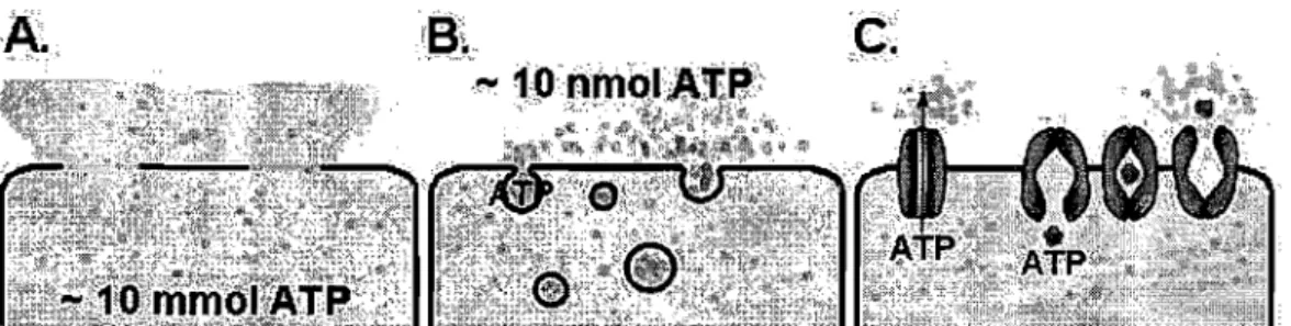
At first, it was assumed that the only source of extracellular ATP acting on purinoceptors was damaged or dying cells. But it is now recognized that ATP release following stimulation does not occur only through ceIllysis but primarily through a weIl-controlled physiological mechanism [65]. There is an active debate, however, about the precise transport mechanism(s) involved. There are at least three general mechanisms by which ATP release may occur [65, 66] (see Fig. 1.1 on page 10):
Cytolysis during cell membrane damage or death - contributing to patho-physiological mechanisms.
Exocytosis of ATP-filled vesicles or granules.
Facilitated diffusion through ATP release channels or through ATP trans-porters (e.g. ABC proteins).
For a long time, exocytosis was considered as the common release mechanism of neurotransmitters [67]. As a neurotransmitter, ATP is also believed to be released through exocytosis; however, non-exocytotic release has not been dismissed yet [37, 60, 68]. The evidence of Ca2+ dependence in the process of ATP release is typically consistent with regulated exocytosis [69] (see also section 1.2.2).
For ATP release from non-neuronal ceIls, various transport mechanisms have been proposed, including ATP-binding cassette (ABC) transporters [70-72], connexin or pannexin hemichannels [73-75], plasmalemmal voltage-dependent anion channels (VDAC) [76], as weIl as vesicular exocytosis [26, 77, 78].
Figure 1.1: Potential ATP release mechanisms:
(A) cytolysisj (B) exocytosisj (C) facilitated diffusion - ATP channel, carrier protein ( transporter)
The biggest controversy arose when the cystic fibrosis transmembrane con-ductance regulator (CFTR), one of the most common ABC proteins in eu-cariotic organisms, was suggested to conduct ATP [71]. Since then, various papers have described the observation of ATP transport by ABC proteins [79]. On the other hand, many other studies have convincingly determined the inability of ABC proteins to transport ATP
[80-83].
Various efforts were made to explain earlier daims to the role of ABC proteins as ATP channels, such as differences in experimental set up or conditions [79].Other possibilities for an implication of CFTR on ATP release were also pro-posed, such as the regulation of an associated ATP channel
[84]
or even the regulation of exocytosis [79].To conc1ude, exocytosis appears to predominate as release mechanism, al-though sorne cell types may also release ATP by several parallel mechanisms, such as by exocytosis and through the activation of VDAC-like channels. Whichever way ATP and other nucleotides are released from the cells, their extracellular importance is finally weIl established.
CHAPTER 1. PHYSIOLOGICAL BACKGROUND 11
1.2 Intracellular calcium
1.2.1
Overview
Ca2+ in the organism
Ca2+ is the most abundant metal ion in the body. An average adult body contains in total around 1 kg of calcium, 99% of which is a component of the skeleton in form of calcium phosphate and the remaining 1 % is present in the extracellular fluid3 and the cellular protoplasm4 [85].
The free Ca2+ level in the extracellular fluid is he Id close to 1 mM, which allows the body to manipulate the precipitation of phosphates by slightly modifying phosphate and Ca2+ concentrations and, in this way, to build up bones and teeth. In the cytosol of eukaryotic cells, the resting level of free Ca2+ is closely controlled to around 100 nM. The large gradient from extra-cellular fluid to cytosol of about 10 000 is held up by actively pumping Ca2+
out of the cytosol, either into the extracellular space or into intracellular or-ganelles, such as the endoplasmic reticulum (ER), which accumulates Ca2+ close to a concentration of 1 mM and serves as an intracellular Ca2+ store. The intracellular and extracellular Ca2+ level is further buffered by certain Ca2+ -binding proteins, such as parvalbumin, calmodulin and calbindin [86]. With the toolkit of intracellular Ca2+ stores, channels, pumps and Ca2 +-binding proteins of various +-binding affinity and kinetics, a cell is capable of precisely regulating alterations of the intracellular Ca2+ level both tem-porally and locally. Accordingly, specific triggers can increase the cytosolic Ca2+ level up to 500 - 1000 nM in different spatial and temporal domains by opening channels in the ER and/or plasma membrane. These Ca2+ signaIs, in turn, can controllocalized pro cesses (e.g. exocytosis) and global responses (e.g. myocyte contraction) as weIl as responses of extremely wide time scale, from microseconds (e.g. activation of ion channels) to many hours, weeks, months or even years (e.g. synaptic plasticity, memory, long-term
adapta-3Extracellular fluid encompasses the blood plasma, the intersitial fluid and the lymph [Il].
4The protoplasm is the substance within a cell enclosed by the cell membrane. It
tion, neuronal ageing) [87]. Almost every cellular process - from fertilization to apoptosis - includes a Ca2+ -dependent step, making the Ca2+ ion the most versatile and universal signalling agent in advanced biological systems [88].
Properties of the Ca2+ ion
The following properties make the Ca2+ ion suitable for this unique role: Ca2+ is a large divalent cation with a radius of 0.95 Â. It is widely available since it is abundant in most natural waters in the presence of common anions such as CI-, RCOi and NOi [86]. The charge-to-size ratio distinguishes the Ca2+ ion from ions of similar size, such as Na+, and from ions of equal charge, such as Mg2+ and Zn2+ [86]. It also provides the Ca2+ ion with a particularity in structure and thermodynamic affinity for certain classes of ligands, as well as in reaction rates, which are summarized under the following three points:
• Structure
Ions with a radius r
2:
0.75A,
such as Ca2+, allow irregular coor-dination geometry, bond angle, bond distance, and high coorcoor-dination numbers (7-10) in their complexes. This flexibility enables them to eas-ily adjust to steric crowding5. Ca2+ differs in this respect strongly fromMg2+
(r
= 0.6 A) - an ion equally abundant in the body and essential for numerous biochemical pro cesses - which is strictly octahedral in symmetry, and from Zn2+(r
= 0.65 A) which is strictly tetrahedral in symmetry. Only conformational changes induced by binding to a Ca2+ ion will lead, therefore, to activation of Ca2+ -sensitive proteins. Based on the high coordination number, Ca2+ is, furthermore, an ideal cross-linking agent for proteins and biopolymers [86] .• Kinetics
Ca2+ has a much lower tendency to form complexes than Mg2+, whose complexes with monodentate ligands, such as water, are relatively sta-ble. Ca2+, in contrast, exchanges water very rapidly, with a rate close
5Steric crowding occurs when a large number of functional groups belonging to one or several mole cules accumulate inside a restricted area - usually around an ion. It very often results in steric hindrance of the individual groups.
CHAPTER 1. PHYSIOLOGICAL BACKGROUND 13
to the collisional diffusion limit of 1010 S-l. The activation speed of a
protein through complexation of Ca2+ is, therefore, diffusion-limited, whereas the extent and duration of activation by a single Ca2+ ion is limited by the stability of the protein-Ca2+ complex [86] .
• Binding
In contrast to monodentate ligands, multidentate ligands can form very stable chelation complexes with Ca2+, whose stability constants exceed, in this case, those for Mg2+. Within a multidentate ligand, Ca2+ is even able to interact with neutral oxygen donors such as carbonyls and ethers. This increase in stability and affinity can be attributed to steric factors, which favour the bigger ion to accommodate the donor atoms in a more convenient way [86].
Discovery of Ca2+ as signalling agent
The importance of Ca2+ for signal transduction was discovered by Sydney Ringer at the end of the 19th century. He showed that Ca2+ ions were indis-pensable for fish survival, development of fertilized eggs, muscle contraction, and ceIl adhesion [89-93]. Shortly after, Locke [94], and then Overton [95], found that Ca2+ is necessary for signal transmission between nerve and mus-cle. But only half a century later, a general theory of Ca2+ as a universal second messenger was promulgated by Lewis Victor Heilbrunn in his book
An Outline of General Physiology [96]. There he wrote: "The reaction ofthis
calcium with the protoplasm inside the ceil is the most basic of ail
protoplas-mic reactions." This theory was ignored for another couple of decades, until
the invention of the patch clamp technique and the synthesis of fluorescent Ca2+ indicators paved the way for the establishment of Ca2+ signalling as the most ubiquitous and pluripotent signalling system involved in almost aIl known cellular pro cesses [97].
Nowadays, a vast number of research articles and reviews, dedicated to Ca2+ signalling, is being published every year. Many aspects of Ca2+ signalling (e.g. sources of Ca 2+, Ca 2+ channels and pumps, local and global signalling, and trigger and responses) in physiology and pathophysiology of diverse cell
systems are being addressed and investigated.
The following two sections will introduce sorne of the principles of Ca2+ signalling by focusing this broad topic on the aspects that are relevant to this Ph.D. thesis, Le. the variety of intracellular Ca2+ stores, the types of
Ca2+ events, the variety of stimuli that can trigger Ca2+ signalling, and, finally, exocytosis as an example for Ca2+ -regulated processes.
1.2.2 Basics of calcium signalling
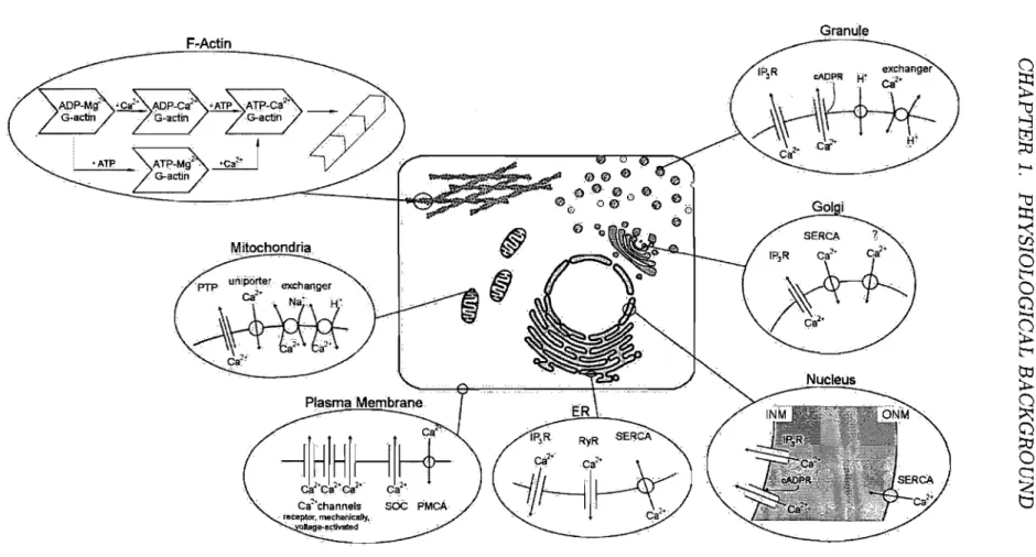
Intracellular calcium storesAs mentioned above, the concentration of free Ca2+ -ions in the cytosol is precisely regulated, and can rapidly increase in response to various types of stimuli. A rise in the cytosolic Ca2+ concentration can be caused by the opening of Ca2+ channels in the plasma membrane and/or by the release of Ca2+ from internaI stores. The major intracellular pool of Ca2+ is the ER with two subsets of Ca2+ release channels: the Ca2+ -gated Ca2+ release chan-nel, generally known as ryanodine receptor (RyR) , and the IP3~gated Ca2+ release channel, referred to as IP3-receptor (IP3R) [98]. Almost aIl other or-ganelles of an eukaryotic cell, such as the mitochondria, the nucleus, the Golgi apparatus, lysosomes and acidic secretory granules were also found to play a significant role in the shaping of cytosolic Ca2+ signalling in various cells [99-102]. AlI of these organelles have both specifie Ca2
+ release channels and Ca2+ uptake mechanisms. A more recent concept of Ca2+ signalling involves even the cytoskeleton of the cell. Ca2+ storage at high-affinity binding sites of F-actin subunits were shown to be involved in the store-operated Ca2+ influx pathway, in Ca2+ spiking and oscillations, as well as in Ca2+ induced Ca2+ release (CICR) [103]. Figure 1.2 on page 15 illustrates the potential Ca2+ stores within a cell with the major transport pathways responsible for Ca2+ movemertt. Each of these stores will be briefly characterized in the following descriptions.
Figure 1.2: Schematic drawing of a cell showing parts of its structure responsible for controlled intracellular Ca2+ movement.
PTP, permeability transition pore; SOC, store-operated Ca2+ channel; PMCA, plasma membrane Ca2+ ATPase; ER, endoplasmic reticulum; IPsR, inositol-l,4,5-triphosphate receptor; RyR, ryanodine receptor; SERCA, sarcojendoplasmic reticulum Ca2+ ATPasej INM, inner nuclear membrane; ONM, outer nuclear membrane; cADPR, cyclic ADP-ribose
It forms a complex continuous network system of endomembranes which can be structurally divided into rough ER, smooth ER and nuclear membrane. The entire ER controls a wide range of cellular pro cesses such as synthesis and st orage of transmembrane and secretory proteins, lipids, steroids and other biomolecules [11]. Mainly the smooth ER also plays a central role in signalling processes by producing local or global Ca2+ fluctuations. It releases Ca2+ into the cytosol through RyR Ca2+ channels via CI CR and cyclic ADP-ribose (cADPR) activation [87], or through IP3R Ca2+ channels via IP3-induced Ca2+ release (II CR) [104]. The role of the ER as an internaI Ca2+ store and signalling organelle is supported by several families of proteins localized in the endomembrane as weIl as within the ER lumen: especially by the Ca2+ pumps, which belong to the sarcojendoplasmic reticulum Ca2+ ATPase (SERCA) type, three types of IP3R and three types of RyR Ca2+ release channels, as weIl as intraluminal Ca2+ -binding proteins most of which also serve as Ca2+ -regulated enzymes. It is, furthermore, supported by the high degree of organization with specialized regions that are localized close to areas where they can generate specific Ca2+ signaIs [104]. Since the ER has a finite capacity, its signalling function depends on the abundance of Ca2+ in its lumen. To prevent Ca2+ store depletion, the cell employs a store-operated entry mechanism to ensure a sufficient replenishment of the ER with Ca2+. The entry of Ca2+ takes place through store-operated Ca2+ channels (SOC) in the plasma membrane after their stimulation by an empty ER. The nature of the signal emanating from the ER and the identity of the SOCs were a matter of debate for 20 years. Only recently were two major players in the signalling and permeation mechanism discovered: an ER Ca2+ -sensor, called STIM16, was found to be involved in the activation of SOCs by triggering Orai proteins, which constitute subunits of SOCs, to form a pore [105, 106].
The Mitochondria are functionally closely connected with the Ca2+ signalling function of the ER. They cooperate in generating Ca2+ signaIs of precise shape, the ER releasing Ca2+ and the mitochondria assisting in the recovery
6STIMl is a single spanning membrane protein with an unpaired Ca2+ binding EF-hand.
CHAPTER 1. PHYSIOLOGICAL BACKGROUND 17
phase by rapidly sequestering it. After the recovery phase, the mitochondria return the sequestered Ca2+ back to the ER [107]. At equilibrium, most of the Ca2+ resides within the lumen of the ER. During a Ca2+ signal, Ca2+ moves from the ER lumen into the mitochondria, where it indu ces the gen-eration of reactive oxygen species (ROS) [107]. ROS can feedback to the ER and sensitize its Ca2+ release channels. This ROS-dependent sensitization of the Ca2+ release channels is particularly important wh en the cell has to generate repetitive Ca2+ spikes [104, 108]. A prolonged shift of Ca2+ from the ER to the mitochondria induces a number of stress signaIs (including ROS) which triggers the onset of apoptosis by activating the formation of a permeability transition pore (PTP) [108].
The Nucleus is surrounded by a double membrane, the nuclear envelope, that separates it from the cytoplasm. Both of the membranes have characteristics of the ER, the outer nuclear membrane (ONM) containing a SERCA and the inner nuclear membrane (INM) containing IP3R and RyR Ca2+ channels [102]. The nuclear envelope contains large nuclear pores with a large ion conductance which is drastically reduced either after accumulation of Ca2+
inside the nuclear envelope or by transport of macromolecules through nu-clear pore complexes [102]. Upon stimulation, many cells exhibit Ca2+ signaIs both in the cytosol and the nucleus. The following ways of nuclear Ca2+ sig-nal generation exist: • Ca2+ may reach the nucleus directly from the cytosol through the nuclear pores [102]. • Nuclear Ca2+ signaIs can be triggered independently of cytosolic Ca2
+ signaIs by direct release of Ca2+ from the nuclear envelope. IP3 , cADPR and nicotinic acid adenine dinucleotide
phos-phate (NAADP) were observed to indu ce such a release [102] .• Ca2+ cou Id be released to the nucleoplasm from Ca2+ containing microvesicles which re-side in the nucleoplasm [102]. In contrast to the mitochondria and the ER, the nucleus does not act as a Ca2+ sink. Rather, the nuclear envelope is destined to slow down the cytosolic Ca2+ wave to the nucleus since a rise in nuclear Ca2+ controls specific nuclear pro cesses such as gene transcription, development, protein transport into the nucleus and cell growth [109].
The Golgi Apparatus primarily pro cesses, sorts and packs macromolecules synthesized by the cell for secretion (exocytosis) or for use wi thin the cell. Changes in Ca2+ concentration either within the Golgi lumen or in the ad-jacent cytosol regulate Golgi function. In addition, the Golgi apparatus contains a release and sequestration apparatus for Ca2+ which accumulates it to a concentration of around 300 J.LM [110]. The generation of IP3 stim-ulates Ca2+ release from the Golgi through the IP3R release channel which
is the only Ca2+ release channel found in the Golgi membrane. The Golgi apparatus contains, furthermore, two types of Ca2+ ATPases, one of which is a thapsigargin-sensitive SERCA and the other a thapsigargin-insensitive secretory pathway Ca2+ ATPase (SPCA) [110]. The Golgi collaborates with the ER - with different kinetics - in elevating cytosolic Ca2+ in response to IP3 stimulation and modulates the duration and pattern of cytosolic Ca2+ signaIs (1101.
Lysosomes and Acidic Secretory Granules were found to be responsible for
nicotinic acid adenine dinucIeotide phosphate (NAADP) induced Ca2+ re-lease [111]. NAADP-sensitive Ca2+ channels are located on lysosome-related acidic organelles. These organelles are thapsigargin-insensitive and are loaded with Ca2+ by a H+ jCa2+ exchanger [111]. There has been a consider-able amount of controversy regarding the ability of secretory granules to actively participate in the regulation of Ca2+ concentration in their environ-ment. Since, however, exocytosis requires high local Ca2+ concentrations, it strongly suggests that Ca2+ release from secret ory granules is an appropriate signal [99].
The theory of F-Actin 7 -dependent Ca2
+ signalling is based on the in vitro observation that F-actin is able to store and release Ca2+ within the physi-ological concentration range of 20 - 1000 nM Ca2+ [103]. Ca2+ is bound to a high-affinity binding site of G-actin8 in a reversible manner. In the poly-merized F-actin form, the Ca2+ binding site is inaccessibly hidden within the
7F-actin or filamentous actin is a polymer of G-actin subunits [111.
8G-actin or globular actin is the actin monomer which polymerizes above a critical concentration to F-actin [UI.
CHAPTER 1. PHYSIOLOGICAL BACKGROUND
19
filament structure [103]. The Ca2+ -storing actin system has the following characteristics: • Monomeric ATP-G-actin is the high-affinity Ca2+ -binding species of the store exhibiting a pH-dependent K~a2+ = 2 8 nM [103] . • Monomeric ADP-G-actin is the low-affinity Ca2+ -binding species of the store with a 100-foid lower affinity for Ca2+ (K~a2+ 400 nM) compared to the high-affinity monomeric ATP-G-actin [103]. • Ca2+ bound to the high-affinity Ca2+ -binding site of F-actin can only be released upon depoly-merization to G-actin monomers. The Ca2+ /Mg2+ exchange rate, k~xa2+, on F-actin is about 4 OOO-fold lower than on G-actin [103].
Spatiotemporal modulation of Ca2+ signaIs
Since intracellular Ca2+ controls a large number of different pro cesses in the same cell, the spatial and temporal organization of cytosolic Ca2+ signaIs is of crucial importance. Stimulation of cells can evoke Ca2+ signaIs that are either local or global, depending on the agonist concentration and the length of the stimulation period
[112].
Moreover, the following features of the Ca2+ signalling machinery control the localization of high-Ca2+ concentration:• Although the Ca2+ ion is very mobile in water, it diffuses only slowly in the cytosol due to the many high-affinity binding sites on relatively im-mobile proteins, such as actin. The combination of the rapidly diffusible messenger IP3 with the slowly diffusible Ca2+ in succession enables the
restriction of Ca2+ signalling events to a local area [86].
• Many effects of Ca2+ are initiated through binding to calmodulin (CaM). In these cases, the final response to the Ca2+ signal depends on both the local Ca2+ and CaM concentrations
[113].
• Upon activation, elementary Ca2+ -releasing events are produced by the brief opening of a single or a small group of Ca2+ channels located in either the plasma membrane or in intracellular Ca2+ -storing organelles. These elementary events appear as localized plumes of Ca2+ around the channels and have been labelled with different names depending on the channel responsible for their formation: 1) A sparklet is formed as a
result of the brief opening of voltage-operated channels. 2) A spark is formed by the opening of RyR Ca2+ channels.
3)
A puff results from the release of Ca2+ from IP3R Ca2+ channels [114].• Of particular significance with regard to the spatiotemporal aspects of Ca2+ signalling is the pro cess of CI CR from both types of intracellular receptor channels, the IP3R and RyR. This Ca2+ sensitivity enables the Ca2+ released from one receptor to stimulate Ca2+ release from its neighbours, thereby causing a regenerative wave of Ca2+ release throughout the entire cytosol. EspeciaIly, puffs are the building blocks of intracellular Ca2+ waves in cells and are likely to be responsible for global Ca2+ signaIs [112].
• A close juxtaposition of mitochondria with a cluster of release channels can form a rapid and transient high-Ca2+ microdomain in the perimi-tochondrial space (see page 16).
• Intracellular pH is known to modulate IP3-induced Ca2+ release by altering binding of IP3 and Ca2+ to the receptor site [115, 116]. In addition, the activity of several membrane proteins, including channels, exchangers and pumps are pH sensitive. A change in intracellular pH may control, therefore, intracellular Ca2+ homeostasis [117-119].
As a result of the different Ca2+ influx pathways (see page 14) and the feature of the Ca2+ signalling machinery, the cell is able to precisely manipulate the location, size, shape and duration of intracellular Ca2+ elevation, enhancing greatly the versatility, accuracy and efficiency of Ca2+ signalling.
Stimuli provoking calcium signalling
Inside the ceIl, the Ca2+ ion acts as a secondary messenger, relaying signaIs received on the outer cell surface into the cell interior and amplifying their strength.
The main stimuli that can trigger intracellular Ca2+ elevation are:
CHAPTER 1. PHYSIOLOGICAL BACKGROUND 21
1. Hormones - chemical messengers, usually peptides or steroids, prod uced and released into the bloodstream by one tissue and affecting another [120].
2. Neurotransmitters - chemical messengers, such as acetylcholine, dopamine, etc., that transmit nerve impulses across a synapse [120].
3. Extracellular matrix components - including fibronectin, laminin, collagen, etc., that are involved in the transmission of mechanical stimuli via membrane-attached integrins [1211.
4. Cytokines - regulatory proteins and peptides, e.g. interleukines and interferon, produced by cells of the immune system [122].
5. Extracellular nucleotides and adenosine - messengers involved in purinergic signalling (see chapter 1.1).
• Environmental stimuli, such as:
1. Physical stimuli (e.g. light [123], change in electrical potential difference [124], shear stress [125], osmotic stress [26, 126]).
2. Chemical stimuli (e.g. extracellular oxidative stress [123], taste [123], odour [127]).
AH molecular messengers bind to receptors on the outer side of the plasma membrane, which can be classified as:
• Ionotropic receptors, which are ligand-gated ion channels that open or close in response to agonist binding [1281·
• Metabotropic receptors, which are G-protein9 coupled receptors that dissociate into


![Figure 4 400 50% Hypotonie shoek 300 )--200 100 V I- I -1 Il \1-1 -.-control -.-100jJM ARL -2 -1 o 2 3 Time [min]](https://thumb-eu.123doks.com/thumbv2/123doknet/2158601.9554/152.915.269.739.149.622/figure-hypotonie-shoek-il-control-jjm-arl-time.webp)
