HAL Id: dumas-01347959
https://dumas.ccsd.cnrs.fr/dumas-01347959
Submitted on 22 Jul 2016HAL is a multi-disciplinary open access archive for the deposit and dissemination of sci-entific research documents, whether they are pub-lished or not. The documents may come from teaching and research institutions in France or abroad, or from public or private research centers.
L’archive ouverte pluridisciplinaire HAL, est destinée au dépôt et à la diffusion de documents scientifiques de niveau recherche, publiés ou non, émanant des établissements d’enseignement et de recherche français ou étrangers, des laboratoires publics ou privés.
Myocardial function at the early phase of traumatic
brain injury : a prospective controlled study
Adrien Cuisinier
To cite this version:
Adrien Cuisinier. Myocardial function at the early phase of traumatic brain injury : a prospective controlled study. Human health and pathology. 2016. �dumas-01347959�
AVERTISSEMENT
Ce document est le fruit d'un long travail approuvé par le
jury de soutenance et mis à disposition de l'ensemble de la
communauté universitaire élargie.
Il n’a pas été réévalué depuis la date de soutenance.
Il est soumis à la propriété intellectuelle de l'auteur. Ceci
implique une obligation de citation et de référencement
lors de l’utilisation de ce document.
D’autre part, toute contrefaçon, plagiat, reproduction illicite
encourt une poursuite pénale.
Contact au SID de Grenoble :
thesebum@ujf-grenoble.fr
LIENS
LIENS
Code de la Propriété Intellectuelle. articles L 122. 4
Code de la Propriété Intellectuelle. articles L 335.2- L 335.10
http://www.cfcopies.com/juridique/droit-auteurUNIVERSITE GRENOBLE ALPES
FACULTE DE MEDECINE DE GRENOBLE
Année 2016
N°
Myocardial function at the early phase of traumatic
brain injury: a prospective controlled study
T
HESE PRESENTEEPOUR L’OBTENTION DU DOCTORAT EN MEDECINE DIPLOME D’ETAT
A
DRIENC
UISINIERT
HESE SOUTENUE PUBLIQUEMENT A LA FACULTE DE MEDECINE DEGRENOBLE*
LE 13 JUILLET 2016
D
EVANT LE JURY COMPOSE DE :P
RESIDENT DU JURY : M. le Professeur Jean-François PAYEND
IRECTEUR DE THESE : M. le Professeur Pierre BOUZATM
EMBRES DU JURY : M. le Professeur Pierre ALBALADEJOM. le Docteur Guillaume WALTHER
*La faculté de Médecine de Grenoble n’entend donner aucune approbation ni improbation aux opinions émises dans les thèses ; ces opinions sont considérées comme propres à leurs auteurs.
REMERCIEMENTS AU JURY
A Monsieur le Professeur Jean-François Payen
Je vous suis reconnaissant au-delà de la formation dont j’ai bénéficié, pour votre ouverture d’esprit et votre écoute concernant les différents projets que j’ai menés au cours de mon internat et qui ont prolongé ce dernier. Je pense notamment à mon année passée à Shanghai ainsi qu’à mon année au sein du laboratoire du GIN. Vous n’avez jamais émis de doute quand à la possibilité de les réaliser.
A Monsieur le Professeur Pierre Albaladejo
Pour m’avoir accompagné et guidé au travers de la simulation et de la pédagogie, je vous dois une part de mon goût et de mon éducation pour la recherche et l’enseignement. Ces activités extra cliniques ont été sources de partage et d’émulation. Pour vous être extasié de l’originalité du projet lorsque je suis venu vous voir pour partir un an en Chine, pour cet enthousiasme que vous avez, je vous remercie.
A Monsieur le Professeur Pierre Bouzat
Mes 7 années à Grenoble t’ont vu passer de ton premier jour de CCA en RPC qui m’a vite valu le surnom de « Ratatouille » (certains s’en souviennent même plus que de mon prénom, il faut croire que l’inspiration était bonne) à ta nomination. Tout cela illustre bien ton sens de l’efficacité, du pragmatisme et de la bonne humeur, il a donc été très agréable, simple et surtout très formateur d’avoir été ton interne et d’avoir avancé sous ta direction dans ce projet.
Au Docteur Guillaume Walther
Merci Guillaume pour avoir accepté de participer à ce jury. Je te remercie ainsi bien sûr que Claire et Stéphane pour avoir très largement contribué à la réalisation de ce travail. Merci de nous avoir permis de bénéficier de votre expertise et ce toujours dans la bonne humeur !
TABLE DES MATIERES
-
A
BBREVIATIONS LIST
8
-
R
ESUME
10
-
A
BSTRACT
12
-
I
NTRODUCTION
13
-
M
ETHODS
15
-
R
ESULTS
21
-
D
ISCUSSION
29
-
C
ONCLUSION
33
-
A
NNEXES
35
-
B
IBLIOGRAPHIE
57
-
S
ERMENT D’HIPPOCRATE
62
-
R
EMERCIEMENTS
63
ABBREVIATIONS LIST
2D 2-dimensional
A atrial (wave)
CVP central venous pressure
E early (wave)
EDD end diastolic diameter
EDPWT end-diastolic posterior wall thickness
EDST end diastolic septum thickness
ESD end systolic diameter
FS fractional shortening
GCS glasgow coma score
GLS global longitudinal strain
ICP intracranial pressure
ICU intensive care unit
IVC inferior cava vena
IVRT isovolumetric relaxation time
LA left atrial
LV left ventricle
LVEF left ventricular ejection fraction
MAC minimal alveolar concentration
MAP mean arterial pressure
RV right ventricle
SR strain rate
STE speckle tracking echocardiography
SVRI systemic vascular resistance index
TAPSE tricuspid annular plane systolic excursion
TBI traumatic brain injury
TCDB traumatic coma data bank
RESUME
Le concept d’interaction cœur-cerveau a été décrit au cours de différents processus pathologiques d’agression cérébrale. Le traumatisme crânien pourrait être responsable d’une dysfonction myocardique mais ceci repose essentiellement sur des données expérimentales ou cliniques rétrospectives.
Nous avons mené une étude prospective cas-témoins dans un trauma center de niveau I. 20 patients adultes consécutifs traumatisés crâniens sévères ont été appariés selon l’âge et le sexe avec 20 patients contrôles. Le groupe contrôle était composé de patients adultes subissant une anesthésie générale pour la réalisation d’une chirurgie traumatologique périphérique. Une échocardiographie en mode conventionnel et en mode speckle tracking a été réalisée dans les 24 heures post traumatiques au sein du groupe traumatisé crânien et en PRE puis PER-opératoire au sein du groupe contrôle. Le critère de jugement principal était la fraction d’éjection ventriculaire gauche mesurée par méthode de Simpson biplan. Les critères de jugement secondaire comprenaient l’analyse de la fonction diastolique gauche en mode conventionnel et l’analyse des fonctions systoliques et diastoliques en mode speckle tracking. Nos résultats mettent en évidence une fraction d’éjection ventriculaire gauche similaire entre le groupe traumatisé crânien et le groupe contrôle PER-opératoire (61% [56-76]) vs. 62% [52-70]). Les paramètres ventriculaires gauches morphologiques et de la fonction systolique étaient également identiques entre les 2 groupes. Concernant la fonction diastolique, le temps de relaxation iso volumétrique était significativement plus élevé dans le groupe traumatisé crânien (125s [84-178] versus 107s [83-141], p=0.04), suggérant une dysfonction diastolique infra clinique. Grâce à l’analyse des paramètres speckle tracking, nous avons observé une tendance avec des valeurs de strain plus élevées dans le groupe traumatisé crânien mais seul le strain circonférentiel au niveau apical et la rotation au niveau basal étaient statistiquement
significatifs. Les paramètres speckle tracking analysant la fonction diastolique montraient une tendance avec des valeurs plus basse dans le groupe traumatisé crânien.
Nous ne retrouvons pas d’altération systématique de la fonction myocardique dans le groupe traumatisé crânien sévère. L’analyse speckle tracking révèle plutôt une adaptation correcte de la fonction systolique gauche tandis que la fonction diastolique semble légèrement altérée.
Mots Clefs : Traumatisme crânien, fonction myocardique, échocardiographie, speckle tracking, interactions coeur-cerveau, syndrome cérébrale cardiaque
ABSTRACT
The concept of brain-heart interaction has been described in several brain injuries. Traumatic brain injury (TBI) may also lead to cardiac dysfunction but evidences are mainly based upon experimental and clinical retrospective studies.
We conducted a prospective case-control study in a level I trauma center. Twenty consecutive adult patients with severe TBI were matched according to age and gender with twenty control patients. Control group included adult patients undergoing general anesthesia for peripheral trauma surgery. Conventional and Speckle Tracking Echocardiography (STE) was performed within the first 24 post-traumatic hours in the TBI group and PRE/PER-operative in the control group. Primary endpoint was left ventricle ejection fraction (LVEF) measured by the Simpson’s method. Secondary endpoints included diastolic function and STE analysis.
We found similar LVEF between the TBI group and the PER-operative control group (61% [56-76]) vs. 62% [52-70]). LV morphological parameters and systolic function were also similar between the two groups. Regarding diastolic function, isovolumic relaxation time was significantly higher in the TBI cohort (125s [84-178] versus 107s [83-141], p=0.04), suggesting subclinical diastolic dysfunction. Using STE parameters, we observed a trend toward higher strains in the TBI group but only apical circumferential strain and basal rotation reached statistical significance. STE-derived parameters of diastolic function tended to be lower in TBI patients.
No systematic myocardial depression was found in a cohort of severe TBI patients. STE rather revealed correct adaptation of left systolic function, while diastolic function slightly impaired.
Key words: Trauma brain injury, myocardial function, echocardiography, speckle tracking, brain-heart interactions, cerebral cardiac syndrome
INTRODUCTION
Myocardial dysfunction has been described after brain injuries, leading to the concept of brain-heart interaction(1). Paroxysmal sympathetic hyperactivity with higher plasma-level of endogenous norepinephrine has been observed after subarachnoid hemorrhage (SAH) and stroke, resulting in cardiac dysfunction(2)(3)(4). Transmural myocardial lesions were also reported after brain death in a series of post mortem examination(5). Experimentally, extreme elevation of intra-cranial pressure by inflating a subdural balloon induced acute cardiac failure related to increased sympathetic activity, electrocardiographic abnormalities and myocardial damage(6)(7). After traumatic brain injury (TBI), neurogenic pulmonary edema has been also reported(8) and high troponin concentration was associated with unfavorable neurologic outcome and mortality(9). Recently, in a retrospective cohort of 139 patients with severe TBI, 12% of the patients had cardiac dysfunction with reduced left ventricular ejection fraction (LVEF) and regional wall motion abnormalities(10). These clinical findings were corroborated by experimental data which reported significant echocardiographic myocardial injury(11), while other authors reported no cardiac dysfunction within the first 24 hours in a rat model of diffuse TBI(12). Hence, myocardial function after TBI remains unclear and its exploration relies upon experimental models and uncontrolled retrospective clinical studies. Possible confounding factors like associated multiple trauma, hemorrhagic shock, cardio-vascular co-morbidities or lung-heart interaction might also hinder the interpretation of the association between cardiac failure and severe TBI.
Assessment of cardiac function in clinical practice is based upon standard two dimensional (2D) echocardiography(13), that provides validated systolic and diastolic indices(14). Since myocardial functionality results from a complex interplay between deformation (longitudinal, radial and circumferential) and twist/untwist mechanics,
conventional echocardiography only partially described cardiac function. Conversely, Speckle Tracking Echocardiography (STE) offers a comprehensive and sensitive evaluation of regional myocardial function. For instance, in pathological conditions like sepsis or heart failure, conventional 2D-echocardiography demonstrated no LVEF impairment whereas speckle tracking echocardiography (STE) detected changes in myocardial function(15)(16).
The present study aimed at evaluating myocardial function at the early phase of TBI using conventional 2D echocardiography and complementary STE analysis. Since TBI patients were under general anesthesia and mechanical ventilation, a matched cohort of non-TBI patients undergoing general anesthesia for peripheral surgery served as control group. We hypothesized that patients with TBI had altered cardiac function compared to the control group.
METHODS
From November 2014 to November 2015, we conducted a prospective case-control single blinded study in a Level-I trauma center (Grenoble University Hospital, France). The regional ethics committee (number: 14-CHUG-34) and the national agency for drugs safety (Number: 2014-A01370-47) approved the study design. This trial was registered on ClinicalTrials.gov, number: NCT02380482. Written informed consent was obtained from the patient or patients’ next of kin.
Patients
In the isolated TBI cohort, we included patients with Glasgow Coma Score (GCS) lower than 9. Patients with GCS from 9 to 13 were also included if mechanically ventilated due to severe cerebral injuries on computed tomography defined by a Marshall score from III to VI following the traumatic coma data bank (TCDB)(17). We did not include patients with expected brain death within 24 hours and multiple trauma patients (Abbreviated Injury Score > 2 in extra-cerebral region). In the control group, patients with peripheral trauma undergoing general anesthesia for emergency surgery were included. Control patients were matched by gender and age ( five years) to the TBI patients. All patients (TBI and controls) were eligible if they were between 18-65 years old without cardiovascular risk factors. Exclusion criteria for both cohorts were: cardiovascular high-risk factors (diabetes, dyslipidemia or hypertension), past medical history of cardio-thoracic surgery or significant cardiovascular events: stroke, myocardial infarction, congestive heart disease, pace maker, atrial fibrillation, coronary artery disease, cardiac valvulopathy or implantable defibrillator. We also excluded patients with traumatic hemorrhagic shock defined by systolic arterial pressure below 90 mmHg, patients under inotrope therapy and elite athletes.
Protocol
Screening and inclusions occurred 24/7 through a dedicated phone procedure within the study period. All patients who met the eligibility criteria were screened. In the TBI cohort, echocardiography was performed at intensive care unit (ICU) admission within the first post-traumatic 24 hours. In the control cohort, two ultrasound examinations were done. The first echocardiography (PRE operative) was performed before general anesthesia under spontaneous ventilation. The second (PER operative) was performed just after the induction of general anesthesia under mechanical ventilation and before the start of surgery.
Echocardiography image acquisition and analysis
Images were acquired by the certified operator (AC) in supine position, using ultrasound equipment (Vivid S5, GE Healthcare Company, USA) with a 2,5Mhz sector scanning electronic transducer. Images were obtained according to the standard and
recommended procedure(14), and based on the average of three cardiac-cycles. Images were
acquired for conventional 2D and strain acquisition in cine loops triggered to the QRS complex and were stored in a cine-loop format of 1 cardiac cycle of uncompressed data with associated electrocardiogram information. Each echocardiography was digitally recorded under a randomized and anonymous number. At the conclusion of the study a subsequent blinded off-line analysis for conventional and STE images was done, using a validated 2D STE tracking software (EchoPac 6.0, GE Healthcare, Horten, Norway). All ultrasound parameters were analyzed blindly to clinical data by AC and CM.
Conventional 2D echocardiography
The LV end-diastolic (EDD) and end-systolic (ESD) diameter, end-diastolic posterior wall thickness (EDPWT), end-diastolic septum thickness (EDST) and left atrial EDD were
obtained in the parasternal long-axis view with M-Mode measurements. The LV fractional shortening (FS) was calculated with the following formula: FS= (LV EDD – LV ESD) / LV EDD * 100). The LV volumes were assessed from the 4- and 2- chamber views. LVEF was calculated from LV volumes measured by the biplane Simpson’s method. Pulsed Doppler LV trans-mitral velocities (Early (E), Atrial (A) waves) recordings were performed from the apical 4-chamber view. The RV basal EDD was assessed from a parasternal long axis. The tricuspid annular plane systolic excursion (TAPSE) was measured to estimate the right ventricle (RV) systolic function by measuring the level of systolic excursion of the lateral tricuspid valve annulus towards the apex in the 4-chamber view. Isovolumic relaxation time (IVRT), stroke volume, heart rate and cardiac output were calculated from an apical 5-chamber view. Cardiac index was calculated as the ratio between cardiac output and body surface area. CVP (Central Venous Pressure) was estimated by measurement of the maximum diameter of the inferior cava vena (IVC). The IVC was imaged in a longitudinal plane with the cardiac transducer in the subxyphoid position. The intrahepatic segment of the IVC was visualized as it entered the right atrium. The maximum anterior-posterior IVC diameter at end-expiration was measured 3 to 4 cm from the junction of the IVC and right atrium. Systemic vascular resistance index (SVRI) was calculated (79.9 x (mean blood pressure-CVP)/cardiac index)(18). Pulmonary artery acceleration time was measured from parasternal short-axis view to estimate systolic pulmonary arterial pressure(19). In order to exclude an unknown cardiomyopathy or valvulopathy, a complete 2D examination was performed for all patients. Color flow Doppler valvular assessment was done.
Tissue Doppler imaging
Wall motion was assessed by pulsed-TDI, with the measurement of the peak myocardial systolic velocity (Sm) and diastolic velocity (Em) at the mitral annulus level on
the lateral wall in apical 4-chamber view. Pulsed-TDI measure of myocardial systolic (S’m) velocity was performed at the tricuspid annulus on the free wall. The E/Em ratio, recorded from the mitral annulus lateral wall, was used as an index of LV filling pressure(20). LV diastolic-dysfunction grading was determined using transmitral flow, and TDI indices, according to documented criteria(20)(21).
Speckle tracking echocardiography (STE)
The STE acquisition and analysis was performed as previously described(22)(23). Global LV longitudinal strain (GLS) were calculated in an 18 segment model from the apical 2-, 3- and 4-chamber views. LV radial and circumferential strains and rotations were obtained in the parasternal short-axis view (at basal and apical levels of the LV). Care was taken to ensure that the basal short-axis plane contained the mitral valve and that the apical plane was acquired distally to the papillary muscle with the LV as circular as possible and proximal to the level with luminal obliteration at end-systole(24). On each cine loop, an optimal frame was selected with the best endocardial border definition. After manually tracing the endocardial border on the end-systolic frame of the 2D sequence, the software automatically tracked myocardial motion. Whenever the software signaled poor tracking efficiency, the observer readjusted the endocardial traceline, the width of the region of interest, or both until a better tracking score could be obtained. Even if the process was approved, the observer checked that the software appropriately tracked the tissue. Net LV peak twist was calculated as the maximal instantaneous difference between basal and apical rotations. The ratio untwisting rate/peak twist was calculated as peak untwist velocity normalized for peak twist to analyze the untwisting efficiency(25). Two-dimensional strain data were processed with specific toolbox (Scilab 4.1, Consortium Scilab, INRIA-ENPC, Paris, France)(22)(23). These specific toolbox were used to adjust all STE imaging data for intersubject differences in heart
rate and frame rate acquisition. The time sequence was normalized to the percentage of systolic duration (i.e., aortic valve closure represented 100% of systolic duration) using interpolations. After normalization, software averaged the data from 3 to 5 cardiac cycles and performed the detection of peak strain events and their timing (expressed as percentage of systolic duration).
Endpoints
The primary endpoint was the measurement of left systolic function within the first post-traumatic 24 hours evaluated by LVEF (Simpson’s method).
Secondary endpoints were: 1) left systolic function with LVEF measured by the Teicholz method, left ventricular shortening fraction, peak velocity Sm, cardiac output, cardiac index and stroke volume; 2) left diastolic dysfunction assessed by peak velocity of waves E, A, Em, ratio E/A, E/Em and IVRT; 3) right function with peak velocity S’m, TAPSE and pulmonary acceleration time; 4) STE variables for the LV evaluation: Global longitudinal strain (GLS) (%), radial and circumferential peak strain (%) at basal and apical level, rotation (degree) at basal and apical level, peak twist (degree), twisting and untwisting velocities (degree/sec). Longitudinal, radial (basal and apical level), circumferential (basal and apical level) peak SR (%/sec), rotational velocity (basal and apical level) (degree/sec) were measured separately for systole and diastole. The ratio between untwisting velocity and peak twist was also calculated.
Data collection
For all patients, demographic data included age, gender, height, weight and body surface. The following hemodynamic and respiratory parameters were obtained during echocardiography examination: non invasive blood pressure (systolic, mean and diastolic)
with the corresponding dose of vasopressor, heart rate, fraction of inspired oxygen, positive end-expiratory pressure, respiratory rate, tidal volume per kilogram, type and doses of anesthetic drugs.
In the TBI cohort, we collected last GCS before sedation, the TCDB classification and signs of elevated intracranial pressure (ICP) such as Cushing reflex, pupillary sizes, osmotherapy and seizure. Neurosurgical interventions were also reported: external ventricular drainage, craniotomy or decompressive craniectomy. ICP value was collected when available. Biological data included troponin and NT pro-BNP. Electrocardiogram interpretation was done. Length of stay in ICU, duration of mechanical ventilation and in-hospital mortality were also reported. In the non-TBI cohort, the reason for emergency surgery was collected.
Sample size calculation
The study population size was calculated considering a LVEF of 65 ( 5) % in the control group, as previously reported(26). Assuming a clinically relevant reduction from 65% to 55% in the TBI group, with a two-sided type 1 error of 0.05 and a power of 80%, 20 patients per group were needed to detect this difference.
Statistical analysis
Data were expressed as median and extreme. Univariate analysis between TBI group and control group was performed with the non parametric Mann-Whitney test for continuous variables and with the chi-square for categorical variables. The statistical analysis was performed with StatView - SAS Institute Inc. 5.0 software. A p value < 0.05 was considered statically significant.
RESULTS
Patients
Out of the 145 consecutive TBI patients screened within the study period, 125 patients were excluded. Flowchart of the study population with reasons for exclusion is represented in
Figure 1 and characteristics of the TBI patients are summarized in Table 1.
Figure 1. Flow chart of the study population.
Not included
- Age under 18y old = 13
- Age over 65y old = 18
- Glasgow score over 13 = 47
- Glasgow score between 9 and 13 with TCDB score I or II = 15 - Past medical history with cardio vascular event = 6
- Traumatic cardiac contusion = 1
- Screening delayed over 24h post trauma = 16
- Moribound patient = 4
- TBI associated with cerebral ischemic event = 2
- Penetrating TBI = 2
Screening of 145 patients with TBI
Excluded
- Non echogenic patient = 1
- Cardiovascular pathology found during echocardiography = 0
20 patients included and analysed - 14 patients with glasgow score < or = 9
- 6 patients with glasgow score between 9 and 13
-1 with cerebral lesion grade III (TCDB classification) - 3 with cerebral lesion grade V (TCDB classification) - 2 with cerebral lesion grade VI (TCDB classification)
Table 1. Characteristics of patients with traumatic brain injury (n=20 patients).
Variable Value
Glasgow coma score 8 [3-13]
Anisocoric pupillary dilatation, n .3.
Bilateral mydriasis, n 1
Cushing reflex, n .3.
Osmotherapy, n 9
Seizure, n 4
External ventricular drainage, n 6
Immediate neurosurgery, n 6 Decompressive craniectomy, n 2 Norepinephrine, µg/kg/min 0.17 [0-0.47] Troponin, µg/l 0 [0-0.42] NT-ProBNP, mg/l 34 [15-1327] ECG abnormalities, n .0.
Diffuse injury (TCDB classification), n
grade III 4
grade IV 1
Focal injury (TCDB Classification), n
Surgical evacuation 6
No surgical evacuation but volume > 25ml 9
ICP value, mmHg 18 [3-50]
Length of stay in ICU, days 21 [3-52]
Duration of mechanical ventilation, days 13 [2-39]
In-hospital mortality, n 1
Values are presented as median [extreme] or number of patients. TCDB: Traumatic Coma Data Bank, ICP: Intracranial pressure, ICU: intensive care unit.
Typical patient was young male suffering from severe TBI. Fourteen patients had a GCS score lower than 9 and only six patients had GCS between 9 and 13. All patients had severe lesions on brain CT scan. Following the TCDB classification, five patients had diffuse injury and fifteen patients had focal injury. Biological parameters were within normal ranges and ECG revealed no abnormality.
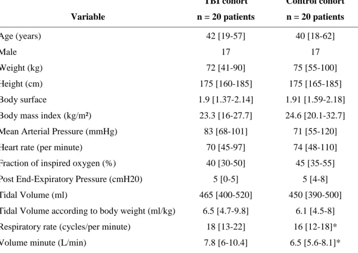
Patients in the control group were admitted for emergency surgery for non-cerebral trauma: 11 patients for orthopedic surgery, 3 patients for maxillofacial surgery and 6 patients for soft tissue surgery. There was no difference between patients with TBI and control patients, except for the respiratory rate and the volume per minute (Table 2). Other ventilation parameters and hemodynamic variables were similar between the TBI cohort and the control cohort. Sedation in the TBI group was maintained through propofol (n=10 patients, dose = 5 [2-6] mg/kg/h) or midazolam (n=10 patients, dose = 126 [84-270] g/kg/h) infusions. All patients were sedated by inhaled anesthesia in the control group (sevoflurane, n=5 patients and desflurane, n=15 patients) with minimal alveolar concentration (MAC) value equal to 1 [0.4-1.2]. All patients received sufentanil for pain management with a median dose equal to 0.48 [0.12-0.96] g/kg/h.
Table 2. Comparisons of main characteristics between Traumatic Brain Injury patients and control patients. Variable TBI cohort n = 20 patients Control cohort n = 20 patients Age (years) 42 [19-57] 40 [18-62] Male 17 17 Weight (kg) 72 [41-90] 75 [55-100] Height (cm) 175 [160-185] 175 [165-185] Body surface 1.9 [1.37-2.14] 1.91 [1.59-2.18]
Body mass index (kg/m²) 23.3 [16-27.7] 24.6 [20.1-32.7]
Mean Arterial Pressure (mmHg) 83 [68-101] 71 [55-120]
Heart rate (per minute) 70 [45-97] 74 [48-110]
Fraction of inspired oxygen (%) 40 [30-50] 45 [35-55]
Post End-Expiratory Pressure (cmH20) 5 [0-5] 5 [4-8]
Tidal Volume (ml) 465 [400-520] 450 [390-500]
Tidal Volume according to body weight (ml/kg) 6.5 [4.7-9.8] 6.1 [4.5-8]
Respiratory rate (cycles/per minute) 18 [13-22] 16 [12-18]*
Volume minute (L/min) 7.8 [6-10.4] 6.5 [5.6-8.1]*
Values are presented as median [extreme]. kg: kilogram, cm: centimeter, Kg/m²: kilogram per square meter, cmH20: centimeter of water, mmHg, millimeter of mercury, ml/kg: milliliter per kilogram, L/min: liter per minute. *p<0.05
Primary outcome
LVEF measured by the Simpson’s method was similar between the TBI cohort (61% [56-76]) and the PER-operative control cohort (62% [52-70], figure 2).
Figure 2. Left ventricle ejection fraction in patients with traumatic brain injury (TBI, n=20
patients) and in control patients (n=20 patients) obtained by conventional cardiac ultrasonography using the Simpson’s method. Echocardiography was performed within the first post-traumatic 24 hours in the TBI group and under general anesthesia/mechanical ventilation in the control group.
Secondary outcomes
Conventional echocardiographic data of TBI cohort and the PER-operative control cohort is presented on Table 3. LV morphological parameters and systolic function were similar between the two groups. IVRT was significantly higher in the TBI cohort. Peak E and E/A tended to be lower in TBI patients. Right ventricular systolic function was similar between the two groups.
50 55 60 65 70 75 80 LV E F (% ) Control Cohort n = 20 patients TBI Cohort n = 20 patients
Table 3. Univariate analysis between patients with TBI and control patients at the
PER-operative phase regarding conventional echocardiography.
Variables
TBI cohort n = 20 patients
PER-operative control n=20 patients
Global systolic function
Fractional shortening ratio (%) 37 [22-54] 38 [26-52]
LVEF (%) (Teicholz method) 65 [50-85] 69 [51-83]
Stroke volume (mL) 67 [41-103] 61 [46-71]
Cardiac output (L/min) 4.4 [2.7-7.3] 4.7 [2.6-6.5]
Cardiac index (L/min/m²) 2.4 [1.5-3.6] 2.4 [1.5-3.6]
Peak Sm (cm/s) 13 [8-20] 12 [7-17]
Global diastolic function
Peak E velocity (cm/s) 69 [44-100] 77 [56-106]
Peak A velocity (cm/s) 59 [29-90] 54 [30-109]
Peak E/A ratio 1.08 [0.67-2.44] 1.38 [0.84-2.40]
Peak Em (cm/s) 13 [6-24] 15 [9-24]
Isovolumic relaxation time (msec) 125 [84-178] 107 [83-141]*
E/Em ratio 4.8 [2.4-8.1] 5.3 [2.8-8.8]
Systemic vascular resistance index (dyn/s/cm5/m2) 2518 [1832-3955] 2341 [1030-3155]
Morphological parameters
Left Atrial diameter (mm) 29 [19-36] 30 [23-36]
Left Ventricular end-diastolic volume (mL) 102 [66-156] 101 [82-132]
Left Ventricular end-systolic volume (mL) 37 [22-67] 39 [27-61]
Left Ventricular end-diastolic diameter (mm) 44 [31-53] 46 [37-52]
Left Ventricular end-systolic diameter (mm) 28 [18-38] 29 [20-37]
Left Ventricular End Diastolic Septal wall thickness (mm) 10 [6-15] 10 [7-15]
Left Ventricular End Diastolic Posterior wall thickness (mm) 11 [7-13] 10 [8-13]
Right ventricular diameter and function
Right Ventricular end-diastolic diameter (mm) 22 [14-29] 23 [21-26]
Peak S'm (cm/s) 14 [8-19] 14 [11-18]
Tricuspid Annular Plane Systolic Excursion (mm) 23 [18-39] 24 [17-29]
Pulmonary acceleration time (msec) 152 [89-188] 138 [94-183]
Values are presented as median [extreme]. TBI: traumatic brain injury, mL: milliliter, L/min: liter per minute, L/min/m²: liter per minute per square meter, cm/s: centimeter per second, msec: millisecond, dyn/s/cm5/m²: dynes per second per centimeter per square meter, mm: millimeter. * p<0.05
STE-derived parameters are presented in Table 4. We observed a trend toward higher strains in the TBI group but only apical circumferential strain and basal rotation reached statistical significance. STE-derived parameters of diastolic function tended to be lower in TBI patients. We found no difference in the control group between the PRE- and PER operative echocardiographic data neither between TBI group and the PRE operative echocardiographic data (data available in the supplemental data: Table 5 to 8).
Table 4. Univariate analysis between TBI patients and control group at the PER-operative
phase regarding Speckle Tracking Echocardiography.
Variables
TBI cohort n = 20 patients
PER operative Control n=20 patients
Systolic function
Global Longitudinal Strain 18 [10.3-23.6] 15.8 [12.4-22.5]
Circumferential Strain peak (%)
basal level 17.2 [9.4-22.1] 16.4 [11.3-27.9]
apical level 26.9 [23.8-32.5] 23.2 [20.1-35.5]*
Radial Strain peak (%)
basal level 14.8 [6-33.3] 13.1 [4.3-24.3]
apical level 16.3 [4.3-30.1] 13.9 [4.9-29.4]
Systolic longitudinal strain rate (%/s) 1.13 [0.73-1.52] 0.98 [0.73-1.33]
Systolic circumferential strain rate (%/s)
basal level 1.24 [0.9-2.47] 1.21 [0.64-2.01]
apical level 1.79 [1.09-2.39] 1.7 [0.83-2.22]
Systolic radial strain rate (%/s)
basal level 1.09 [0.45-2.44] 1.1 [0.5-2.41]
apical level 1.13 [0.42-2.41] 0.9 [0.5-1.59]
Rotation (deg)
basal level 6.3 [2.4-10.8] 5 [1.5-10]*
apical level 7.29 [2.6-15] 8.89 [4.9-13.7]
Peak twist (deg) 12,6 [7.5-20.2] 12.8 [5.1-18.3]
Systolic rotational velocity (deg/s)
basal level 66.3 [43.6-135.6] 64.7 [24.1-83.9]
apical level 62.2 [28.4-126.4] 73.8 [25.1-123.3]
Twisting velocity (deg/s) 77.2 [48.9-156.4] 76.6 [31.4-115]
Diastolic function
Diastolic longitudinal strain rate (%/s) 1.33 [0.59-2] 1.56 [1.11-2.47]
Diastolic circumferential strain rate (%/s)
basal level 1.49 [0.65-2.66] 1.52 [1.06-2.79]
apical level 1,93 [0.38-3.29] 2.12 [0.91-3.71]
Diastolic radial strain rate (%/s)
basal level 1.08 [0.39-2.79] 1.16 [0.5-2.1]
apical level 1.04 [0.24-1.9] 1.16 [0.56-2.46]
Diastolic rotational velocity (deg/s)
basal level 48.8 [22.3-97.5] 53.5 [18.5-156.5]
apical level 65.6 [31.6-109.6] 83.4 [35.6-112]
Untwisting velocity (deg/s) 91.6 [50.8-179.3] 100.4 [50.9-148.2]
Ratio : Untwisting velocity / Peak twist 7.73 [4-11.9] 7.92 [3.4-12.4]
Values are presented as median [extreme]. TBI: trauma brain injury, Deg: degree, Deg/s: degree per second, %/s: percentage per second. *p<0.05.
DISCUSSION
In this prospective controlled study, we did not find systematic major myocardial dysfunction at the early phase of TBI with severe lesions on cerebral CT scan. These findings were obtained from conventional cardiac ultrasonography and speckle tracking analysis. Despite no major change in cardiac function, several STE changes suggested correct adaptation of the left systolic function and a slight impairment of left diastolic function.
We reported the absence of alteration of systolic function after TBI since LVEF was similar between the TBI and the control groups. More interestingly, none of the TBI patients had decreased systolic function and minimum LVEF was 56% using the Simpson’s method. Speckle tracking analyses further confirmed these findings through similar systolic indices. Only circumferential strain peak at the apical level and the rotation at the basal level were higher in the TBI cohort. These findings were in line with a trend toward a global increase of all systolic indices in STE. Taken together, these results rather suggested a slight increase in systolic function after TBI since STE was able to detect early LV functional changes in the setting of systemic diseases with cardiac issue(27). Accordingly, a systemic adrenergic or sympathetic hyperactivity was described after TBI(28)(29). We, thus, assume that these sympathetic and neuro-endocrine adaptations could be more responsible for an activation of systolic myocardial function rather than a global myocardial depression.
Another major result of the present study was the slightly lower, but not clinically relevant, diastolic function. We only reported a significant higher IVRT in the TBI cohort. All STE-derived parameters tended to be lower in TBI patients without reaching statistical significance. Taken together, we cannot conclude to any significant diastolic myocardial dysfunction at early phase TBI. However, the increase in IVRT may be interpreted as a starting diastolic modification in the TBI cohort. A trend in lower diastolic STE indexes
further confirmed this hypothesis. Similarly, diastolic dysfunction was previously described in patients with SAH with concomitant pulmonary edema(30). In a small series of seven patients with TBI, echocardiographic diastolic dysfunction was also reported in patients with neurogenic pulmonary edema(8). Taken together, these findings further corroborated the TBI-induced modifications in left diastolic parameters observed in our cohort.
Our study is inconsistent with several experimental and clinical studies dealing with brain-heart interaction. Several explanations may account for these controversial results. First, we investigated myocardial function at the early phase of TBI, within the first 24 hours. This time point was chosen according to the existing literature that described early myocardial dysfunction(31). Accordingly, in a recent retrospective study, conventional cardiac ultrasonography was mostly performed on day one, revealing acute myocardial depression after TBI(10). Nevertheless, we cannot exclude a delayed myocardial dysfunction occurring within the first post-traumatic days(32). In our study, slight systolic and diastolic STE changes might be undermined by early ultrasound assessment and might be significant in later examinations. Second, we do not deny the concept of acute cardiac failure following brain injuries and hemodynamic evaluation is part of global assessment of severity in critically ill patients(33). Nevertheless, the concept of neurogenic cardiac failure was mainly based on different experimental models(11) such SAH(34) or brain death. In the clinical setting, echocardiography modifications(31) and electrocardiographic abnormalities(35) were also described in SAH patients, epileptic patients(36), stroke patients(32), or brain death patients(37). Myocardial dysfunction after TBI has been extended from these conditions but only few studies showed acute cardiac failure after TBI(10), mainly case report(30) and retrospective study(10). Hence, there is no prospective controlled study comprehensively assessing cardiac function after TBI and possible confounding factors like multiple trauma, associated co-morbidities and hemorrhagic shock might induce cardiac failure independently
of cerebral injury. To our knowledge, we conducted the first prospective controlled trial regarding cardiac dysfunction matching a TBI cohort with a control cohort according to age and gender. The potential sympathetic tonus due to emergency context and traumatic pathology was controlled since both cohorts were trauma patient admitted for emergency treatment. Lung-heart interactions were also controlled since in both cohorts, patients were under mechanical ventilation and general anesthesia. We, thus, believe that our study adds to the existing literature to explore rigorously cardiac consequences of TBI.
We acknowledge several limits to our study. First, only two third of the TBI patients had severe TBI according to the GCS definition. However, all TBI patients had severe injuries on cerebral CT scan. Six patients had a GCS between 9 and 13. Among them, three patients underwent immediate neurosurgery, two patients had cerebral injuries classified TCDB VI and the last one had compromised cerebral blood flow on transcranial Doppler (Diastolic velocity = 26 cm/s and Pulsatility Index = 1,89). Despite relatively high GCS, these patients should be considered as severe from the clinical standpoint and did not explain the lack of difference between the TBI and the control groups. Moreover, median length of stay in ICU was 21 days with 13 days of mechanical ventilation attesting global severity of the included patients. Second, we only tested the hypothesis of a systematic global decrease of cardiac performance after TBI. We cannot conclude that myocardial depression is not possible after TBI. We acknowledge that neurogenic cardiac failure may occur in this context supporting the use of conventional cardiac ultrasonography for hemodynamic management. Since TBI could be associated with preexisting comorbidities or multiple trauma, we even strongly recommend the use of conventional ultrasonography at the bedside to optimize cardiac output in these patients.
Controlling confounding factors for non-cerebral acute cardiac failure, we did not find systematic acute myocardial depression after TBI. STE analysis rather suggested adequate
adaptation of the left systolic function, while diastolic function slightly decreased. All these changes were not clinically relevant and our study further suggested no systematic cardiac consequences of severe traumatic cerebral injuries. These findings did not challenge the usefulness of cardiac ultrasonography for the management of TBI patients.
ANNEXES
-
S
UPPLEMENTAL DATA: TABLE 5 to 8
-
A
VIS FAVORABLE DU COMITE DE PROTECTION DES
PERSONNES
-
A
UTORISATION DE L’ANSM
-
F
ORMULAIRE DE RECUEIL DU CONSENTEMENT ECLAIRE DES
PROCHES DU PATIENT
-
F
ORMULAIRE D’INCLUSION EN URGENCE
-
F
ORMULAIRE DE RECUEIL DU CONSENTEMENT ECLAIRE DU
PATIENT TRAUMATISE CRANIEN OU CONTROLE
-
L
ETTRE D’INFORMATION A DESTINATION DES PROCHES DU
PATIENT
-
L
ETTRE D’INFORMATION POUR LE PATIENT TRAUMATISE
CRANIEN
-
L
ETTRE D’INFORMATION POUR LE PATIENT CONTROLE
Supplemental data: Analysis between TBI and control patients at the PRE-operative phase and between control patients at the PRE and PER operative phase
Table 5. Univariate analysis between patients with TBI and control patients at the
PRE-operative phase regarding conventional echocardiography.
Variables
TBI cohort n = 20 patients
PRE-operative control n=20 patients
Global systolic function
Fractional shortening ratio (%) 37 [22-54] 40 [28-50]
LVEF (%) (Teicholz method) 65 [50-85] 71 [55-81]
Stroke volume (mL) 67 [41-103] 68 [56-85]
Cardiac output (L/min) 4.4 [2.7-7.3] 5 [2.8-8.6]
Cardiac index (L/min/m²) 2.4 [1.5-3.6] 2.6 [1.6-4.2]
Peak Sm (cm/s) 13 [8-20] 12 [8-19]
Global diastolic function
Peak E velocity (cm/s) 69 [44-100] 80 [55-100]*
Peak A velocity (cm/s) 59 [29-90] 63 [40-96]
Peak E/A ratio 1.08 [0.67-2.44] 1.19 [0.88-2.32]
Peak Em (cm/s) 13 [6-24] 16 [9-25]
Isovolumic relaxation time (msec) 125 [84-178] 118 [99-155]
E/Em ratio 4.8 [2.4-8.1] 5 [3.2-11]
Systemic vascular resistance index (dyn/s/cm5/m2) 2518 [1832-3955] 2359 [1370-4250]
Morphological parameters
Left Atrial diameter (mm) 29 [19-36] 34 [26-40]*
Left Ventricular end-diastolic volume (mL) 102 [66-156] 120 [74-155]
Left Ventricular end-systolic volume (mL) 37 [22-67] 43 [23-61]
Left Ventricular end-diastolic diameter (mm) 44 [31-53] 49 [39-54]
Left Ventricular end-systolic diameter (mm) 28 [18-38] 29 [22-35]
Left Ventricular End Diastolic Septal wall thickness (mm) 10 [6-15] 11 [7-17]
Left Ventricular End Diastolic Posterior wall thickness (mm) 11 [7-13] 10 [7-13]
Right ventricular diameter and function
Right Ventricular end-diastolic diameter (mm) 22 [14-29] 23 [20-26]
Peak S'm (cm/s) 14 [8-19] 15 [10-24]
Tricuspid Annular Plane Systolic Excursion (mm) 23 [18-39] 27 [20-33]*
Pulmonary acceleration time (msec) 152 [89-188] 131 [94-173]*
Values are presented as median [extreme]. TBI: traumatic brain injury, mL: milliliter, L/min: liter per minute, L/min/m²: liter per minute per square meter, cm/s: centimeter per second, msec: millisecond, dyn/s/cm5/m²: dynes per second per centimeter per square meter, mm:
Table 6. Univariate analysis between TBI patients and control group at the PRE-operative
phase regarding Speckle Tracking Echocardiography.
Variables
TBI cohort n = 20 patients
PRE operative Control n=20 patients
Systolic function
Global Longitudinal Strain 18 [10.3-23.6] 17.8 [10-23.3]
Circumferential Strain peak (%)
basal level 17.2 [9.4-22.1] 17.7 [10.5-24.6]
apical level 26.9 [23.8-32.5] 25.5 [17.7-33.6]
Radial Strain peak (%)
basal level 14.8 [6-33.3] 16.6 [6.8-34.7]
apical level 16.3 [4.3-30.1] 15.3 [3.5-34.3]
Systolic longitudinal strain rate (%/s) 1.13 [0.73-1.52] 0.97 [0.66-1.49]
Systolic circumferential strain rate (%/s)
basal level 1.24 [0.9-2.47] 1.15 [0.75-2.18]
apical level 1.79 [1.09-2.39] 1.58 [0.94-2.68]*
Systolic radial strain rate (%/s)
basal level 1.09 [0.45-2.44] 1.37 [0.68-2.28]
apical level 1.13 [0.42-2.41] 0.94 [0.49-1.99]
Rotation (deg)
basal level 6.3 [2.4-10.8] 5.1 [3.4-12]
apical level 7.29 [2.6-15] 7.55 [3.36-12.8]
Peak twist (deg) 12,6 [7.5-20.2] 14.2 [7.1-18]
Systolic rotational velocity (deg/s)
basal level 66.3 [43.6-135.6] 66.8 [31.4-140.9]
apical level 62.2 [28.4-126.4] 52.4 [26.7-88.6]
Twisting velocity (deg/s) 77.2 [48.9-156.4] 81.4 [40.5-170.9]
Diastolic function
Diastolic longitudinal strain rate (%/s) 1.33 [0.59-2] 1.47 [0.73-2.12]
Diastolic circumferential strain rate (%/s)
basal level 1.49 [0.65-2.66] 1.81 [0.42-3.15]
apical level 1,93 [0.38-3.29] 2.02 [0.87-2.96]
Diastolic radial strain rate (%/s)
basal level 1.08 [0.39-2.79] 0.78 [0.38-2.4]
apical level 1.04 [0.24-1.9] 1.28 [0.45-3.04]
Diastolic rotational velocity (deg/s)
basal level 48.8 [22.3-97.5] 57.1 [23.8-89.1]
apical level 65.6 [31.6-109.6] 60.7 [32.1-98.6]
Untwisting velocity (deg/s) 91.6 [50.8-179.3] 83.2 [61.9-147.4]
Ratio : Untwisting velocity / Peak twist 7.73 [4-11.9] 6.56 [3.91-11.49]
Values are presented as median [extreme]. TBI: trauma brain injury, Deg: degree, Deg/s: degree per second, %/s: percentage per second. *p<0.05.
Table 7. Univariate analysis between control patients at the PRE-operative phase and control
patients at the PER-operative phase regarding conventional echocardiography.
Variables
PRE-operative control n = 20 patients
PER-operative control n=20 patients
Global systolic function
Fractional shortening ratio (%) 40 [28-50] 38 [26-52]
LVEF (%) (Teicholz method) 71 [55-81] 69 [51-83]
Stroke volume (mL) 68 [56-85] 61 [46-71]*
Cardiac output (L/min) 5 [2.8-8.6] 4.7 [2.6-6.5]
Cardiac index (L/min/m²) 2.6 [1.6-4.2] 2.4 [1.5-3.6]
Peak Sm (cm/s) 12 [8-19] 12 [7-17]
Global diastolic function
Peak E velocity (cm/s) 80 [55-100] 77 [56-106]
Peak A velocity (cm/s) 63 [40-96] 54 [30-109]
Peak E/A ratio 1.19 [0.88-2.32] 1.38 [0.84-2.40]
Peak Em (cm/s) 16 [9-25] 15 [9-24]
Isovolumic relaxation time (msec) 118 [99-155] 107 [83-141]
E/Em ratio 5 [3.2-11] 5.3 [2.8-8.8]
Systemic vascular resistance index (dyn/s/cm5/m2) 2359 [1370-4250] 2341 [1030-3155]
Morphological parameters
Left Atrial diameter (mm) 34 [26-40] 30 [23-36]*
Left Ventricular end-diastolic volume (mL) 120 [74-155] 101 [82-132]
Left Ventricular end-systolic volume (mL) 43 [23-61] 39 [27-61]
Left Ventricular end-diastolic diameter (mm) 49 [39-54] 46 [37-52]*
Left Ventricular end-systolic diameter (mm) 29 [22-35] 29 [20-37]
Left Ventricular End Diastolic Septal wall thickness (mm) 11 [7-17] 10 [7-15]
Left Ventricular End Diastolic Posterior wall thickness (mm) 10 [7-13] 10 [8-13]
Right ventricular diameter and function
Right Ventricular end-diastolic diameter (mm) 23 [20-26] 23 [21-26]
Peak S'm (cm/s) 15 [10-24] 14 [11-18]
Tricuspid Annular Plane Systolic Excursion (mm) 27 [20-33] 24 [17-29]*
Pulmonary acceleration time (msec) 131 [94-173] 138 [94-183]
Values are presented as median [extreme]. TBI: traumatic brain injury, mL: milliliter, L/min: liter per minute, L/min/m²: liter per minute per square meter, cm/s: centimeter per second, msec: millisecond, dyn/s/cm5/m²: dynes per second per centimeter per square meter, mm: millimeter. * p<0.05
Table 8. Univariate analysis between control group at the PRE-operative phase and control
group at the PER-operative phase regarding Speckle Tracking Echocardiography.
Variables
PRE operative Control n = 20 patients
PER operative Control n=20 patients
Systolic function
Global Longitudinal Strain 17.8 [10-23.3] 15.8 [12.4-22.5]
Circumferential Strain peak (%)
basal level 17.7 [10.5-24.6] 16.4 [11.3-27.9]
apical level 25.5 [17.7-33.6] 23.2 [20.1-35.5]
Radial Strain peak (%)
basal level 16.6 [6.8-34.7] 13.1 [4.3-24.3]
apical level 15.3 [3.5-34.3] 13.9 [4.9-29.4]
Systolic longitudinal strain rate (%/s) 0.97 [0.66-1.49] 0.98 [0.73-1.33]
Systolic circumferential strain rate (%/s)
basal level 1.15 [0.75-2.18] 1.21 [0.64-2.01]
apical level 1.58 [0.94-2.68] 1.7 [0.83-2.22]
Systolic radial strain rate (%/s)
basal level 1.37 [0.68-2.28] 1.1 [0.5-2.41]
apical level 0.94 [0.49-1.99] 0.9 [0.5-1.59]
Rotation (deg)
basal level 5.1 [3.4-12] 5 [1.5-10]
apical level 7.55 [3.36-12.8] 8.89 [4.9-13.7]
Peak twist (deg) 14.2 [7.1-18] 12.8 [5.1-18.3]
Systolic rotational velocity (deg/s)
basal level 66.8 [31.4-140.9] 64.7 [24.1-83.9]
apical level 52.4 [26.7-88.6] 73.8 [25.1-123.3]*
Twisting velocity (deg/s) 81.4 [40.5-170.9] 76.6 [31.4-115]
Diastolic function
Diastolic longitudinal strain rate (%/s) 1.47 [0.73-2.12] 1.56 [1.11-2.47]
Diastolic circumferential strain rate (%/s)
basal level 1.81 [0.42-3.15] 1.52 [1.06-2.79]
apical level 2.02 [0.87-2.96] 2.12 [0.91-3.71]
Diastolic radial strain rate (%/s)
basal level 0.78 [0.38-2.4] 1.16 [0.5-2.1]
apical level 1.28 [0.45-3.04] 1.16 [0.56-2.46]
Diastolic rotational velocity (deg/s)
basal level 57.1 [23.8-89.1] 53.5 [18.5-156.5]
apical level 60.7 [32.1-98.6] 83.4 [35.6-112]*
Untwisting velocity (deg/s) 83.2 [61.9-147.4] 100.4 [50.9-148.2]
Ratio : Untwisting velocity / Peak twist 6.56 [3.91-11.49] 7.92 [3.4-12.4]
Values are presented as median [extreme]. TBI: trauma brain injury, Deg: degree, Deg/s: degree per second, %/s: percentage per second. *p<0.05.
FORMULAIRE DE CONSENTEMENT
Proche
Mise en évidence d’une dysfonction myocardique précoce chez les patients traumatisés crâniens graves : Evaluation par échocardiographie transthoracique standard et par
speckles tracking EchoTC
Promoteur de la recherche : Direction de la Recherche Clinique et de l’Innovation, Pavillon Dauphiné CHU de Grenoble CS 10217 38043 GRENOBLE cedex 9
Investigateur principal : Dr Pierre BOUZAT, Pôle Anesthésie Réanimation CHU Grenoble, 04 76 76 92 88, PBouzat@chu-grenoble.fr
Je soussigné(e) ... (nom, prénom) certifie avoir lu et compris la note d’information qui m’a été remise.
J’ai eu la possibilité de poser toutes les questions que je souhaitais au Pr/Dr ... (nom, prénom) qui m’a expliqué la nature, les objectifs, les risques potentiels et les contraintes liées à la participation de mon proche à cette recherche.
Je connais la possibilité qui m’est réservée d’interrompre sa participation à cette recherche à tout moment sans avoir à justifier ma décision et je ferai mon possible pour en informer le médecin qui me suit dans la recherche. Cela ne remettra naturellement pas en cause la qualité des soins ultérieurs.
J’ai eu l’assurance que les décisions qui s’imposent pour la santé de mon proche seront prises à tout moment, conformément à l’état actuel des connaissances médicales.
J’ai pris connaissance que cette recherche a reçu l’avis favorable du Comité de Protection des Personnes Sud-Est V le 12/11/2014 et l’autorisation de l’ANSM le 20/10/2014 et a fait l’objet d’une déclaration à la Commission Nationale Informatique et Libertés (CNIL).
Le promoteur de la recherche (CHU de Grenoble) a souscrit une assurance de responsabilité civile en cas de préjudice auprès de la société SHAM (Contrat N°135.751).
J’accepte que seules les personnes qui collaborent à cette recherche ou qui sont mandatées par le promoteur, ainsi qu’éventuellement le représentant des Autorités de Santé, aient accès à l’information dans le respect le plus strict de la confidentialité.
J’accepte que les données de mon proche enregistrées à l’occasion de cette recherche, puissent faire l’objet d’un traitement informatisé par le promoteur ou pour son compte.
J’ai bien noté que, conformément aux dispositions de la loi relative à l’informatique, aux fichiers et aux libertés, je dispose d’un droit d’accès et de rectification. Je dispose également d’un droit d’opposition à la transmission des données couvertes par le secret professionnel susceptibles d’être utilisées dans le cadre de cette recherche et d’être traitées. Ces droits s’exercent auprès du médecin qui suit mon proche dans le cadre de cette recherche et qui connaît son identité.
Mon consentement ne décharge en rien l’investigateur et le promoteur de la recherche de leurs responsabilités à l’égard de mon proche. Nous conservons tous les droits garantis par la loi.
Les résultats globaux de la recherche seront communiqués directement à moi ou à mon proche, si nous le souhaitons, conformément à l’article L1122-1 du code de la Santé Publique.
Ayant disposé d’un temps de réflexion suffisant avant de prendre ma décision, j’accepte librement et volontairement la participation de mon proche
………. (nom, prénom) à la recherche
EchoTC.
Lien de parenté : ………..
Je pourrai à tout moment demander des informations complémentaires au médecin qui a proposé la participation à cette recherche, 04 76 76 92 88
Fait à ... le Fait à ... le
Signature du proche : Signature du médecin :
- 1er feuillet (original) : à conserver à part par l’investigateur pendant 15 ans dans un lieu sûr fermant à clé
- 2ème feuillet : à remettre au patient/sujet après signatures
DECLARATION DE L’INVESTIGATEUR
Titre de la recherche : Mise en évidence d’une dysfonction myocardique précoce chez les patients traumatisés crâniens graves : Evaluation par échocardiographie transthoracique standard et par speckles tracking -
EchoTC
Investigateur coordonnateur : Dr Pierre BOUZAT, Pôle Anesthésie Réanimation CHU Grenoble, Tél : 04 76 76 92 88 E-mail : PBouzat@chu-grenoble.fr
Promoteur : Direction de la Recherche Clinique et de l’Innovation, Pavillon Dauphiné CHU de Grenoble
CS 10217 38043 GRENOBLE cedex 9
Je soussigné(e), Professeur, Docteur………, médecin investigateur, déclare que l’état du patient et l’absence des membres de sa famille ou d’un proche désigné par le patient ne leur permettent pas de recevoir actuellement une information concernant les objectifs et les modalités de l’étude clinique auquel il est proposé au patient de participer.
Dans l’attente de pouvoir obtenir un consentement signé par le patient ou par un membre de sa famille ou par un proche désigné par le patient, j’inclus le patient dans cette étude.
Dans l’hypothèse d’un refus ultérieur de consentement du patient, je m’engage en tant que médecin investigateur à détruire toutes les données concernant le patient.
Le patient pourra s’opposer à ce que les données recueillies soient utilisées.
Nom et prénom du patient ………
Nom du médecin investigateur ………...
Fait à………...
Le … ./… ./….
FORMULAIRE DE CONSENTEMENT
Patients TC et patients contrôles
Mise en évidence d’une dysfonction myocardique précoce chez les patients traumatisés crâniens graves : Evaluation par échocardiographie transthoracique standard et par
speckles tracking EchoTC
Promoteur de la recherche : Direction de la Recherche Clinique et de l’Innovation, Pavillon Dauphiné CHU de Grenoble CS 10217 38043 GRENOBLE cedex 9
Investigateur principal : Dr Pierre BOUZAT, Pôle Anesthésie Réanimation CHU Grenoble, 04 76 76 92 88, PBouzat@chu-grenoble.fr
Je soussigné(e) ...(nom, prénom) certifie avoir lu et compris la note d’information qui m’a été remise.
J’ai eu la possibilité de poser toutes les questions que je souhaitais au Pr/Dr ... (nom, prénom) qui m’a expliqué la nature, les objectifs, les risques potentiels et les contraintes liées à ma participation à cette recherche.
Je connais la possibilité qui m’est réservée d’interrompre ma participation à cette recherche à tout moment sans avoir à justifier ma décision et je ferai mon possible pour en informer le médecin qui me suit dans la recherche. Cela ne remettra naturellement pas en cause la qualité des soins ultérieurs.
J’ai eu l’assurance que les décisions qui s’imposent pour ma santé seront prises à tout moment, conformément à l’état actuel des connaissances médicales.
J’ai pris connaissance que cette recherche a reçu l’avis favorable du Comité de Protection des Personnes Sud-Est V le 12/11/2014 et l’autorisation de l’ANSM le 20/10/2014 et a fait l’objet d’une déclaration à la Commission Nationale Informatique et Libertés (CNIL).
Le promoteur de la recherche (CHU de Grenoble) a souscrit une assurance de responsabilité civile en cas de préjudice auprès de la société SHAM (Contrat N°135.751).
J’accepte que seules les personnes qui collaborent à cette recherche ou qui sont mandatées par le promoteur, ainsi qu’éventuellement le représentant des Autorités de Santé, aient accès à l’information dans le respect le plus strict de la confidentialité.
J’accepte que les données enregistrées à l’occasion de cette recherche, puissent faire l’objet d’un traitement informatisé par le promoteur ou pour son compte.
J’ai bien noté que, conformément aux dispositions de la loi relative à l’informatique, aux fichiers et aux libertés, je dispose d’un droit d’accès et de rectification. Je dispose également d’un droit d’opposition à la transmission des données couvertes par le secret professionnel susceptibles d’être utilisées dans le cadre de cette recherche et d’être traitées. Ces droits s’exercent auprès du médecin qui me suit dans le cadre de cette recherche et qui connaît mon identité.
Les résultats globaux de la recherche me seront communiqués directement, si je le souhaite, conformément à l’article L1122-1 du code de la Santé Publique.
Ayant disposé d’un temps de réflexion suffisant avant de prendre ma décision, j’accepte librement et volontairement :
de participer à la recherche EchoTC __oui __non
Je pourrai à tout moment demander des informations complémentaires au médecin qui m’a proposé de participer à cette recherche, 04 76 76 92 88
Fait à ... le Fait à ... le
Signature du patient: Signature du médecin :
- 1er feuillet (original) : à conserver à part par l’investigateur pendant 15 ans dans un lieu sûr fermant à clé
- 2ème feuillet : à remettre au patient/sujet après signatures
NOTE D’INFORMATION
Proche d’un traumatisé crânien grave
Mise en évidence d’une dysfonction myocardique précoce chez les patients traumatisés crâniens graves : Evaluation par échocardiographie transthoracique standard et par
speckles tracking EchoTC
Promoteur de la recherche : Direction de la Recherche Clinique et de l’Innovation, Pavillon Dauphiné CHU de Grenoble CS 10217 38043 GRENOBLE cedex 9
Investigateur principal : Dr Pierre BOUZAT, Pôle Anesthésie Réanimation CHU Grenoble, 04 76 76 92 88, PBouzat@chu-grenoble.fr
Madame, Monsieur,
En raison de son état et dans l’urgence, conformément à la loi, c’est à vous que nous demandons l’autorisation de participation de Mme, Mlle, Mr ………. à cette recherche biomédicale dont le CHU de Grenoble est promoteur. Avant de prendre une décision, il est important que vous lisiez attentivement ces pages qui vous apporteront les informations nécessaires concernant les différents aspects de cette recherche. N’hésitez pas à poser toutes les questions que vous jugerez utiles au médecin de votre proche.
Cette participation est entièrement volontaire. Si vous désirez que votre proche ne prenne pas part à cette recherche, il/elle continuera à bénéficier de la meilleure prise en charge médicale possible, conformément aux connaissances actuelles.
Le traumatisme crânien grave est une pathologie fréquente. Il est responsable de séquelles neurologiques potentiellement lourdes. Pour réduire les séquelles chez ces patients, il est important d’améliorer l’irrigation sanguine du cerveau. Cela implique de vérifier le bon fonctionnement du cœur.
L’objectif de cette étude est d’observer le fonctionnement du cœur par échographie chez des patients traumatisés crâniens graves. Nous allons les comparer avec des patients contrôles n’ayant pas de traumatisme crânien. Cela permettra de mettre en évidence ou non une altération du fonctionnement du cœur spécifique aux patients traumatisés crâniens. Les patients contrôles seront des patients intubés, ventilés sous anesthésie générale opérés pour une chirurgie mineure ou modérée.
Nous proposons que votre proche participe en tant que traumatisé crânien grave. Son médecin a fait un examen complet pour vérifier son éligibilité à cette recherche. La participation à cette étude impliquera
• la réalisation d’un électrocardiogramme fait dans les 24 premières heures post-traumatisme, examen fait en routine en réanimation
• et d’une échographie cardiaque à peu près au même moment que l’électrocardiogramme. Votre proche sera ensuite suivi dans le cadre de cette recherche jusqu’à sa sortie d’hospitalisation pour recueillir son statut et les évènements notables pendant son séjour en réanimation. Aucun traitement supplémentaire n’est administré. Les examens réalisés sont non invasifs c'est-à-dire qu’ils ne présentent aucun effet secondaire potentiel et sont indolores.