Comparative effects of neonatal and prepubertal castration
on craniofacial growth in rats
A. Verdonck
a, L. De Ridder
a, G. Verbeke
b, J.P. Bourguignon
c, C. Carels
a,E.R. Kuhn
d, V.
Darras
d, F. de Zegher
e.aSchool of Dentistry, Department of Orthodontics, University Hospital St. Rafael, Capucijnenvoer 7, 3000
Leuven, Belgium
bBiostatistical Centre for Clinical Trials, University of Leuven, Belgium cDepartment of Pediatrics, Faculty of Medicine, University of Liège, Belgium
dDepartment of Biology, Laboratory of Comparative Endocrinology, Faculty of Science, University of Leuven,
Belgium
eDepartment of Pediatrics, Faculty of Medicine, University of Leuven, Belgium
Abbreviations: IGF-I, insulin-like growth factor-I.
ABSTRACT
The role of endogenous testosterone in the craniofacial growth of the young male rat was investigated. First, the effect of neonatal surgical castration was examined in a randomized, cross-sectional study in which male Wistar rats were allocated to be either castrated or sham-operated 4 h after birth. Then, the effect of prepubertal chemical castration was analysed in a second, randomized longitudinal study in which male Wistar rats were randomly allocated either to a control group or to two experimental groups, one injected with triptorelin at day 25 and the other injected on day 25 and on day 45. Every tenth day between 20 and 70 days of age for the first study, and between 30 and 110 days of age for the second, body length and weight were measured, cephalometric X-rays taken, and blood samples obtained. Neonatal and prepubertal castration resulted in decreased plasma concentrations of testosterone and in delayed growth of somatic and craniofacial components. The initiation, duration and magnitude of the effect was dependent on individual bones (cranial base, skull roof) and on the lower incisor, and related to the testosterone concentrations. These results suggest that testosterone effects participate in the process of normal craniofacial growth, particularly during puberty.
1 Introduction
The increased use of hormone therapy in children (de Zegher et al., 1996) requires a detailed understanding of the basic effects of hormones on skeletal growth. Of special interest is testosterone therapy in boys with a constitutional delay in puberty, the purpose being to induce the pubertal growth spurt (Brown et al., 1995). Its effect on linear growth has been investigated (Bourguignon, 1993), but its effect on craniofacial growth is not known. As a number of studies show a relation between the craniofacial growth spurt and the peak height velocity in body length (Grave, 1973; Thompson and Popovich, 1973; Baughan et al., 1979; Fishman, 1982), it can be assumed that an effect will also be reflected in craniofacial growth.
Androgens influence skeletal growth by direct and indirect (i.e., mediated by the growth hormone—IGF-I axis) mechanisms. Independent and additive contributions by gonadal steroid hormones and growth hormone to the adolescent growth spurt are evident (Jansson et al., 1985b; Kerrigan and Rogol, 1992). These findings were demonstrated in vitro (Corvol et al., 1987; Kasperk et al., 1989; 1990), in animals (Schoutens et al., 1984; Jansson et al., 1985a; Turner et al., 1989) and in clinical studies (Tanner et al., 1976; Laron et al., 1980; Attie et al., 1990; Keenan et al., 1993). Recently, a new concept of the effect of androgens in growth regulation via their conversion to oestrogens has been proved (Korach, 1994; Smith et al., 1994; and Morishima et al., 1995). The stimulatory effect of testosterone on body growth in rats by modulating their hypothalamopituitary functions was shown by Jansson et al. (1983). Neonatally secreted testicular androgens imprint on the high-amplitude pulses, characteristic of growth-hormone secretion in adult male rats, which are more favourable for somatic growth than the low-amplitude pulses in female rats (Jansson et al., 1984, 1985b). Further, the much stronger suppression of longitudinal growth by neonatal rather than prepubertal castration indicates the important effect of the amplitude of the growth-hormone pulse on growth (Jansson et al., 1985b).
Very few studies have demonstrated effects of androgens on craniofacial growth (Riesenfeld, 1974; Stutzmann and Petrovic, 1978; Duncker et al., 1988), particularly in the pubertal period. Therefore, the present study was designed to clarify further the role of testosterone in pubertal craniofacial growth. To study the effects of endogenous testosterone secretion on the rat craniofacial complex, experiments were designed around neonatal mechanical and prepubertal chemical castration.
2. Material and methods
2.1. COMPOSITION OF THE DIFFERENT GROUPS
For the first study, pregnant inbred Wistar rats (n = 18) were obtained from the Animal Laboratories of the University of Leuven. After birth, 97 male offspring were taken and randomly divided into two groups, identified by toe amputation at birth (Reitsma, 1963). The experimental group was castrated (n = 52) 4 h after birth and the control group was sham-operated (n = 45).
For the second study, pregnant inbred Wistar rats (n = 14) were similarly obtained. After weaning, male offspring were randomly divided into three groups: two experimental groups, I (n = 20) and II (n = 20), and a control (n = 24).
Animals were pfed a diet adequate for their nutritional needs and were kept in an air-conditioned and light controlled room with an ambient temperature of 238C. The cages each
contained five to six rats. The protocol for animal use was reviewed and approved by the Ethics Committee of the Medical School, University of Leuven.
2.2. DRUG ADMINISTRATION
For the second study, testosterone release was pharmacologically suppressed at the pituitary level by the injection of triptorelin (Decapeptyl-Depot®; Ipsen- Biotech, France). As puberty in the Wistar rat starts around day 25 (Bourguignon et al., 1990), the animals were treated at day 25; both experimental groups were injected intramuscularly with 10 m triptorelin (100- 200 mg/kg body wt).
Group II received a second injection of the drug on day 45, i.e., in the time period (day 40-50) in which the pubertal testosterone rise was observed in the control rats of the first study. The rats of the control group received no injections. With this chemical castration we intended to induce a delayed pubertal testosterone rise to set up a model for constitutional delay in puberty (Stanhope et al., 1985).
2.3. REGISTRATION OF THE RECORDS
For the first study, every tenth day from day 20 until day 70, seven to nine rats were taken from both groups; for the second study, all rats were taken every tenth day from day 30 to 110. A sagittal cephalometric radiograph of the skull was taken under general anaesthesia with a Rontgendevice Model D9 (Ritter A.G., Karlsruhe-Durlach) in a specially designed craniostat using a standardized technique (Jefferys, 1969) with an occlusal film (Agfa Dentus M2 Comfort 2.25/3). The dose of anaesthetic (Nembutal) was adapted to the weight of the rat (25-30 mg/kg). General measures of growth, body length (from the nose tip to the anus), and weight (with an electronic balance) were also registered. Before anaesthesia, blood was collected from the tail around the same time of day, between 7 and 10 a.m. Plasma testosterone, growth hormone and IGF-I were measured by radioimmunoassay.
2.4. CEPHALOMETRIC ANALYSIS
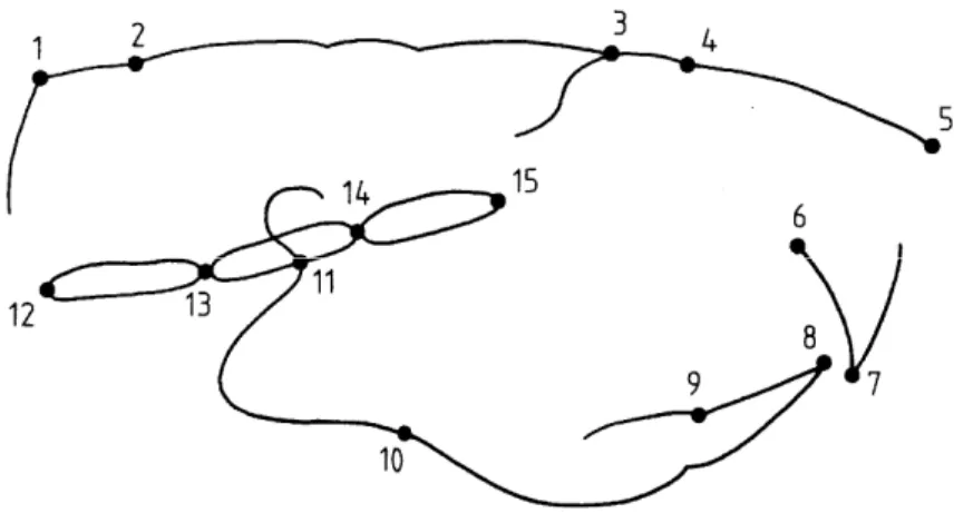
Photographic images were taken from the developed X-ray images and recorded on a Kodak CD-ROM. The craniofacial landmarks were digitized. The linear measurements were extracted from the data-points using a specially made program. The cephalometric technique included linear measurements from the analyses described by Vilmann (1969), Engstrom et al. (1982), Persson et al. (1989) and Kiliaridis et al. (1985). The choice of the craniofacial landmarks was made in relation to the function of different structures represented in the skull. The skull-roof measurements were represented by the distances 1-2, 4-5, 1-3, 1-5; the vertical height by the distance 6-5; the cranial-base bones, including the basi-occipital, the basisphenoid, and the presphenoid, by 12-13, 13-14, 14-15; the mandible by 10-11; and the incisors by 6-7, 8-9
2.5. BLOOD SAMPLING
Plasma testosterone was analysed using kits for testosterone 125I radioimmunoassay (Byk-sangtec
Diagnostica GmbH and Co. KG, von Hevesy-Strasse, D-63128 Dietzenbach). The kit consisted of testosterone antiserum from rabbits, testosterone 125I tracer and testosterone standard dissolved
in human serum and goat antirabbit gammaglobulins in polyethylene glycol solution. The detection limit of testosterone was 0.2 ng/ml. All experimental and control groups were assayed at the same time. The intra-assay coefficient of variation was between 5.8 and 6.3%. The interassay coefficient of variation was between 13.5 and 7.4%.
Radioimmunoassay for growth hormone was done in duplicate on all samples, using kits provided by the National Institute of Diabetes and Digestive and Kidney Diseases (Rockville, MD, U.S.A). The kit consisted of rat GH-I-7 for iodination, standard rat GH- RP-2 and monkey antirat GH-S-5. All treatment and control groups were assayed at the same time. The within-assay coefficient of variation was 1% (Baes and Denef, 1987).
Plasma IGF-I was measured by heterologous radioimmunoassay as described by Renaville et al. (1994). Dilution series of rat plasma with radioimmunoassay buffer showed good parallelism with the standard curve. The intra-assay coefficient of variation was 6.5% and the interassay coefficient was 15% (Hybrechts et al., 1985).
2.6. DATA ANALYSIS
Cross-sectional data from the first study were tested in the Procedure General Linear Model (2-way ANOVA) in SAS. An ANOVA model with two main effects, age and group differences and their interaction, was fitted to the data collected over the entire period. For variables showing a significant interaction effect, the same analysis was performed over the time period before and after physiological puberty (i.e., from day 20 to 40 and from day 40 to 70) (Bourguignon et al., 1991). Also the tenth-day time period with the largest difference between the two groups was examined by the two-sample t-test.
Analysis of the longitudinal data in the second study required a statistical technique for repeated measurements so a linear mixed model was used. The inter- as well as the intra-animal and the group variability were taken into account. An initial check of the data was made with the SAS procedure MEANS. The original variables had a normal distribution. The model used was defined as follows. For each time-point and group (experimental I, II, and control), a mean response was estimated. A deviation of growth in the treated rats (groups I and II) from that of the control group was expected from day 25. Owing to the experimental design, groups I and II were combined for the day 25-45 period. As the effect of the drug may disappear sometime after the injection, it was reasonable to expect a catch-up towards normal growth for group I. Owing to the second injection at day 45, group II might have continued to deviate further from the control group, and also from group I. Sequential F-tests were used to compare the three groups. For the observed differences, we determined the latency as well as the time period during which the deviation was observed. Throughout the statistical analysis a significance level of p < 0.05 was used.
Figure 1. Rat skull in the midsagittal direction with the different anatomical points which are defined in the table.
Table 1. Definition of the anatomical points
No. Name Definition
1 Posterior Lambdoidal suture The midpoint of the external surface of the occipital-interparietal suture 2 Lambda The midpoint of the external surface of the parieto-interparietal suture
3 E The intersection between the frontal bone and the most superior-anterior point of the posterior limit of the ethmoid bone
4 Nasion The midpoint of the external surface of the fronto-nasal suture
5 Rhinion The most anterior margin of the nasal bone
6 The intersection of the palatal bone and the lingual surface of the upper lingual incisors
7 The incisal edge of the maxillary incisor
8 The incisal edge of the mandibular incisor
9 The intersection between the lingual surface of the lower incisors and the most anterior point of
the lingual alveolar bone
10 The deepest point of the antegonial notch curvature
11 Articulare: the intersection between the mandible and the sphenoid bone
12 Basion: the anterior limit of the foramen magnum
13 The midpoint of the endocranial surface of the spheno-occipital synchondrosis
14 The midpoint of the endocranial surface of the intersphenoidal synchondrosis
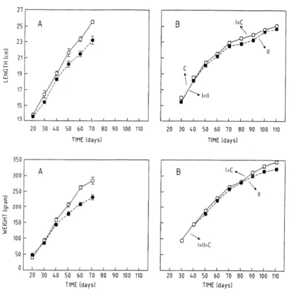
Figure 2. Comparison of the significant results of neonatal castration
(A) (means and standard errors of the mean as a function of rat age n = 7-9; control (
○
) and the castrated (●
) rats) with these of prepubertal castration (B). The estimation of the means and the standard errors of the mean as a function of time for the different groups according to the model. Group I (—), experimental group with one injection (n = 20 day 30; n = 15 day 110); group II (- -), experimental group with two injections (n = 20 day 30; n =15 day 110), and the control group (• • •) (n = 24)) for body length and weight.Figure 3.
(A) Means and standard error of the mean of blood analysis parameters as a function of rat age for the control (
○
) and the neonatally castrated (●
) rats: (i) testosterone level (ng/ml), (ii) growth hormone level (ng/ml) (n = 7-9). (B) The estimations of the means and the standard errors of the mean, for the testosterone level (ng/ml) as a function of time, for the different groups, according to the model. Group I (—), experimental group with one injection (n = 20 day 30; n =15 day 110); Group II (—), experimental group with two injections (n = 20 day 30; n = 15 day 110), and the control group (••••) (n = 24 day 30; n = 15 day 110)3. Results
3.1. GENERAL GROWTH AND BLOOD ANALYSIS
In the surgically castrated rats of the first study, body lengths and weights were significantly lower than in the control group. The effect was more pronounced at days 40-50 (p < 0.05) and days 50-60 (p < 0.05), respectively (Fig. 2A). Testosterone and growth hormone were both significantly lower in
the neonatally castrated rats than in the control group. The reduction was most pronounced at days 40-50 for testosterone ( p < 0.05) and days 50-60 for growth hormone (p < 0.05) (Fig. 3A). For group II (second injection at day 45) of the chemically castrated rats, both total length and weight were affected from day 50 and remained lower than normal for the complete duration of the study (p < 0.005, p < 0.05, respectively) (Fig. 2B). Their blood showed reduced testosterone between day 50 and 80 (p < 0.0005) (Fig. 3B). No significant difference between the groups was found for growth hormone and IGF-I.
3.2. CRANIOFACIAL VARIABLES
The total length of the skull roof (1-5) was significantly shorter in the surgically castrated rats than in the control group; this growth inhibition was most pronounced from day 40 onward (p < 0.0005). Also, a smaller value from day 50 to 90 was found for groupII (second injection at day 45) of the chemically castrated rats (p < 0.001) compared to controls (Fig. 4A/B(a)).
The concatenation of some skull-roof bones (1-3) was significantly shorter (interaction effect) in the surgically castrated rats than in the control group, and this difference was the most pronounced at days 40-70 (p < 0.0005). A smaller length from day 50 to 100 was found in group II of the chemically castrated rats (p < 0.05) (Fig. 4A/B(b)).
The nasal bone (4-5) in the surgically castrated group showed only a significant group difference and no significant interaction. Prepubertal chemical castration did not affect this bone.
For the vertical dimension of the skull, the measurement (6-5) was significantly shorter (interaction effect) in the surgically castrated group than in the control group; this was most evident at days 40-70 (p < 0.001). This measurement was significantly different from day 60 to 90 in the chemically castrated rats given two injections (group II) (with lower and higher values compared to the other groups) (p < 0.005).
For the three bones of the cranial base, only the basisphenoid (13-14) was significantly smaller in the surgically castrated group; this reduction in growth was most pronounced at days 40-70 (p < 0.005) (Fig. 4A(c)). The basi-occipital (12-13) and presphenoid (14-15) bones, and the total craniofacial base (12-15), showed the same tendency, although only a significant group difference not a significant interaction was found. A shorter basisphenoid (13-14) at day 70 and 80 (p < 0.0005)(Fig. 4B(c)) and a shorter presphenoid (14-15) from day 50 to the end of the study (p < 0.0005) were found in the group II chemically castrated rats. The basi-occipital bone in group II was shorter only on day 50 (p < 0.05). The total length of the cranial base in group II was shorter only on day 110 (p < 0.05).
The overall length of the mandible (10-11) was not affected by neonatal surgical or by prepubertal chemical castration.
The lower incisor (8-9) was significantly shorter in the surgically castrated group than in the control group. This length was shorter from day 60 in group II chemically castrated rats (p < 0.05) (Fig. 4A/B(d)). For the upper incisor, no significant difference was found in either group.
4. Discussion
Our experiments with neonatally and prepubertally castrated rats provide evidence for an effect of endogenous testosterone on the craniofacial complex.
4.1. GENERAL GROWTH
In both of our experiments, body length was significantly less in the castrated groups. The growth-inhibition effects observed here were in line with published results on the effects of neonatal castration, and also with those for peri-and postpubertal castrated rats (Jansson et al. 1985a, Turner et al., 1989; Wakley et al., 1991); they all report a decrease of long-bone growth.
In our study, body weight was also significantly reduced in the castrated groups. Schoutens et al. (1984) also reported a decrease in weight with postpubertal castration and Jansson et al. (1985a) found that neonatal castration only resulted in decreased body weight.
4.2. BLOOD
In both of our studies, an increase in testosterone was found in the control groups from day 40-50, which is in agreement with others' findings (Dohler and Wuttke, 1975; Eden et al., 1978). Bourguignon et al. (1991) studied the onset of puberty in rats and found an increase in testicular growth between day 20 and 50, which corresponds to the onset of testosterone release. In our neonatally surgically castrated rats, testosterone never reached the detection level, which indicates the effectiveness of the castration procedure. In our prepubertally chemically castrated rats, testosterone was temporarily reduced from days 50 to 80, which proved that the drug was effective while having only a temporary effect. The age at which the testosterone suppression became effective (day 50) was in the same period in which the pubertal rise in testosterone occurred in control rats (day 40-50).
4.3. EFFECTIVENESS OF TRIPTORELIN
Measurements in group I (drug given at day 25) rarely differed from those of the control group. In contrast, some measurements in group II (drug given at day 25 and day 45) were significantly different from those of the control group. This difference in effect between one and two injections can be explained as follows. The target area for the drug, i.e., the neuroendocrine activity, although promoting the onset of puberty at day 25 in rats (Bourguignon et al., 1990), may not be fully active at the time of the first injection, as testosterone release is not detectable before day 35 (Piascek and Goodspeed, 1978). The drug may therefore be most effective only at the time at which testosterone reaches a maximum in the control group. For the control rats, this pubertal increase in testosterone occurred between days 40 and 50 [in accordance with the age of 37-58 days described by Dohler and Wuttke (1975)]. The effect of the first treatment may have already disappeared before that time, while the second injection was timed in that period (day 45). It should be mentioned that we did not detect an initial increase in serum testosterone in any of the
experimental groups after the administration of triptorelin, as found in some studies (Redding et al., 1984), but not others (Asch et al., 1985).
4.4. INTERPRETATION OF THE CEPHALOMETRIC RESULTS
With neonatal surgical castration, the absence of testosterone effects on craniofacial bones may be responsible for the shorter bones, as the mechanism responsible for a decrease in the craniofacial variables after neonatal castration may be explained by the absence of the neonatal testosterone imprinting on the high-amplitude pulses of growth hormone in 30-day- old rats (Eden, 1979). The significant effect observed for the total skull roof indicates the effect of castration on sutural growth. As the individual bones of the skull roof did not show a significant interaction as compared to the total skull roof and concatenations of individual bones, the effect on the individual constituent pieces was apparently too small to be detected. However, when measurements for the pieces were combined, there was a summation of sutures, bringing the effect on the total skull roof to significance. In a study by Persson et al. (1989), the effect of hormonal manipulation (thyroxine injections) on craniofacial growth was investigated. In analogy with their findings the total length of the skull roof was smaller, indicating that the roof is a target for hormones. In our study, there was a greater effect on the anterior part of the skull roof than on the posterior part. The anterior part includes the frontonasal suture, which has a higher growth rate than the other skull-roof sutures (Cleall et al., 1969). This finding is also consistent with the observation that growth of the anterior part exceeds that of the neurocranium during the adolescent period of the rat (Moss and Baer, 1956; Engstrom et al., 1982).
The effect observed for the vertical dimensions of the rat skull supports the idea that appositional growth is affected by testosterone deprivation. In line with these findings are those of Turner et al. (1989), who demonstrated that castration decreased periosteal growth in the rat tibia.
For the cranial base, only the length of the basisphenoid bone had a significant interaction. The bipolar cartilage growth of the basisphenoid bone in contrast to the unipolar cartilage growth of the two other bones may explain the stronger effect on the basisphenoid. The non-significance of the effect on the measurement of the concatenation of the three bones of the cranial base may thus be explained by the smaller effect on two of the three bones. Alternatively, a possible change in the angle between the bones of the cranial base must also be considered (Vilmann, 1969).
For the growth of the mandible, no significant effect was demonstrated. As the condyle embodies the cartilaginous growth site of the mandible and an effect could be demonstrated in the cranial base, which also grows by a cartilaginous mechanism (synchondrosis), an effect was expected. Imperfect identification of the condylion on the sagittal radiograph, which proved to be a diffcult mark to locate due to the superimposition of the surrounding structures (Killiany et al., 1987), may be responsible for the absence of a significant effect.
For the length of the lower incisor a significant effect was observed, which indicates the effect of testosterone deprivation, and might result from compensatory growth of the tooth. The significant reduction in skullroof growth but not in mandibular growth could reduce the need for
lower-incisor growth required for upper-lower-incisor contact. However, an effect of growth hormone on root formation and enamel mineralization has been demonstrated in dwarf rats (Symons et al., 1995). With prepubertal chemical castration, the total skull roof, concatenations of skull-roof bones, basisphenoid length, presphenoid length, and the lower incisor were significantly shorter. This growth suppression followed the suppression of testosterone (at day 50). The effect of triptorelin on the testosterone was only temporary (from day 50 to 80). In most craniofacial structures with suppressed growth, a catch-up effect was observed related to the normalization of the testosterone. In some structures the growth suppression lasted longer than the duration of the testosterone suppression. The presphenoid bone (19-20) of the cranial base, the lower-incisor length, and the general growth measures of body length and weight, had not caught up, even by the end of the study (day 110). These results are in line with those of Turner et al. (1990), who proved an effect of prepubertal castration on radial growth of the tibia.
Testosterone has an important role in bone growth (Kasperk et al., 1990), which has also been documented in histological studies on sutures, synchondroses and periosteal surfaces of normal rat skulls (Cleall et al., 1969). It may therefore be that the altered growth in the rat skull is primarily caused by a modified bone growth through changed concentrations of testosterone. These craniofacial effects could be possibly related to the absence of the pubertal testosterone surge, to the absence of the neonatal testosterone peak, or to a combination of these effects.
Comparing the effects of neonatal castration with those of prepubertal castration, in both, structures showing periosteal growth (the total skull roof and concatenations of skull-roof bones) as well as structures showing cartilaginous growth (basisphenoid and body length), and also lower-incisor length, were affected. The growth-suppression effect was, however, larger for the neonatally castrated than for the prepubertally castrated rats. In the investigation by Jansson et al. (1985a), a more pronounced effect on long-bone length in neonatally than in prepubertally castrated rats was also found. On the other hand, in the chemically prepubertally castrated rat, the testosterone is reduced, but, at most time-points is still higher than in the neonatally castrated rat. In conclusion, our comparison of the effects of neonatal surgical castration and prepubertal chemical castration indicates that testosterone plays an important part in the normal pubertal growth of the craniofacial complex. No distinction based on growth mechanisms (periosteal or cartilaginous) could be made on basis of this study.
Figure 4.
Comparison of the significant results of neonatal castration (A) (means and standard error of the mean as a function of rat age n = 7-9) (control (
○
) and the castrated (●
) rats) with these of prepubertal castration (B) (The estimation of the means and the standard errors of the mean as a function of time for the different groups according to the model. Group I (—), experimental group with one injection (n = 20 day 30; n = 15 day 110); group II (- -), experimental group with two injections (n = 20 day 30; n =15 day 110), and the control group (••••) (n = 24)) for total skull-roof length (1-5) (a), concatenation of skull roof bones (1-3) (b), basisphenoid bone (13-14) (c) and lower-incisor length (8-9) (d)Acknowledgements
This investigation was supported by the Fund of Scientific Research (FWO: 1.5.693.94N) and by Ipsen Biotec (France), which provided the drug (Decapeptil®). We are indebted to Drs E. Alberga, M. Gaethofs, N. Ucuncu (Dept. of Orthodontics, University of Leuven) and W. Van Ham and F. Voets (Dept. of Animal Endocrinology, University of Leuven) for their practical help in the experiments.
References
Asch, R.H., Rojas, F.J., Andrzej, B., Schally, A.V., Tice, T.R., Klemcke, H.G., Siler-Khodr, Tm, Bray, R.E., Hogan, M.P., 1984. Prolonged suppression of plasma LH levels in male rats after a single injection of an L-RH agonist in poly (DL-lactide-co-glycolide) microcapsules. J. Androl. 6, 83-88.
Attie, K.M., Ramirez, N.R., Conte, F.A., Kaplan, S.L., Grumbach, M.M., 1990. The pubertal growth spurt in eight patients with true precocious puberty and growth hormone deficiency: evidence for a direct role of sex steroids. J. Clin. Endocrinol. Metab. 71, 975-983.
Baes, M., Denef, C., 1987. Evidence that stimulation of growth hormone release by epinephrine and vasoactive intestinal peptide is based on cell-to-cell communication in the pituitary. Endocrinology 120, 280-284.
Baughan, B., Demirjian, A., Levesque, G.Y., La, Palme- Chaput, L., 1979. The pattern of facial growth before and during puberty as shown by French-Canadian girls. Ann. Hum. Biol. 59-76.
Bourguignon, J.P., Gerard, A., Mathieu, J., Mathieu, A., Franchimont, P., 1990. Maturation of the hypothalamic control of pulsatile gonadotropin-releasing hormone secretion at onset of puberty. Increased activation of N- methyl-D-aspartate receptors. Endocrinology 127, 873-881.
Bourguignon, J.P., Gerard, A., Fawe, L., Alvarez-Gonzalez, M.L., Franchimont, P., 1991. Neuroendocrine control of the onset of puberty: secretion of gonadotrophin-releasing hormone from rat hypothalamic explants. Acta Paediatr. Scand. (Suppl. 372), 19-25.
Bourguignon, J.P., 1993. Delayed puberty and hypogonadism. In: J. Bertrand, R. Rappaport, P. Sizonenko (Ed.). Pediatric Endocrinology. Physiology, Pathophysiology and Clinical Aspects. Williams and Wilkins, Baltimore, pp. 404- 419.
Brown, B.C., Butler, G.E., Kelnar, C.J., 1995. A double blind, placebo controlled study of the effects of low dose testosterone undecanoate on the growth of small for age, prepubertal boys. Arch. Dis. Child. 73, 131-135.
Cleall, J.F., Wilson, G.W., Garnett, D.S., 1969. Normal craniofacial skeletal growth of the rat. Am. J. Phys. Antrop. 29, 225-242.
Corvol, M.T., Carrascosa, A., Tsadris, L., Blanchard, O., Rappaport, R., 1987. Evidence for a direct in vitro action of sex steroids on rabbit cartilage cells during skeletal growth:influence of age and sex. Endocrinology 120, 1422- 1429.
de Zegher, F., Maes, M., Gargosky, S.E., Heinrichs, C., Du Caju, M.V.L., Thiry, G., De Schepper, J., Craen, M., Breysem, L., Lofstrom, A., Jonsson, P., Bourguignon, J.P., Malvaux, P., Rosenfeld, R.G., 1996. High dose growth hormone treatment of short children born small for gestational age. J. Clin. Endocrinol. Metab. 81, 1887-1892.
Dohler, K.D., Wuttke, W., 1975. Changes with age in levels of serum gonadotropins, prolactin, and gonadal steroids in prepubertal male and female rats. Endocrinology 97, 898- 907.
Duncker, M., Tonjes, R., Rothe, C., Scherkenbach, C., Zglinicki, T., 1988. Experimentelle Untersuchungen zum Einfluss von Testosteron auf die Schadelmorphogenese der Ratte. Fortschr Kieferorthop 49, 342-357.
Eden, S., Albertsson-Wikland, K., Isaksson, O., 1978. Plasma levels of growth hormone in female rats at different ages. Acta Endocrinol. Copenh. 88, 676-690.
Eden, S., 1979. Age- and sex-related differences in episodic growth hormone secretion in the rat. Endocrinology 105, 555-560.
Engstrom, C., Linde, A., Thilander, B., 1982. Craniofacial morphology and growth in the rat. Cephalometric analysis of the effects of a low calcium and vitamin D-deficient diet. J. Anat. 134, 299-314.
Fishman, L.S., 1982. Radiographic evaluation of skeletal maturation. Angle Orthod. 52, 88-122.
Grave, K.C., 1973. Timing of facial growth: a study of relations with stature and ossification in the hand around puberty. Austr. Ortho. J. 3, 117-122.
Hybrechts, L.M., De Cupere, E., Kuhn, E.R., Lauterio, T.J., 1985. Growth hormone secretory response to TRH in normal and dwarf chickens. Reprod. Nutr. Dev. 25, 641-645.
Jansson, J.O., Eden, S., Isaksson, O., 1983. Sites of action of testosterone and estradiol on longitudinal bone growth. Am. J. Physiol. 243, E135-E140.
Jansson, J.O., Ekberg, S., Isaksson, O.G.P., Eden, S., 1984. Influence of gonadal steroids on age- and sex-related secretory patterns of growth hormone in the rat. Endocrinology 114/4, 1287-1294.
Jansson, J.O., Ekberg, S., Isaksson, O., Mode, A., Gustafsson, J.A., 1985(a)a. Imprinting of growth hormone secretion, body growth, and hepatic steroid metabolism by neonatal testosterone. Endocrinology 177/5, 1881-1889.
Jansson, J.O., Eden, S., Isaksson, O., 1985(b)b. Sexual dimorphism in the control of growth hormone secretion. Endoc. Rev. 6, 128-150.
Jefferys, J.F., 1969. Growth pattern and environment. Thesis, Drukkerij Gebr. Janssen N.V. Nijmegen. Kasperk, C., Wergedal, J.E., Farley, J.R., Linkkhart, T.A., Turner, R.T., Baylink, D., 1989. Androgens directly stimulate proliferation of bone cells in vitro. Endocrinology 124, 1576-1579.
Kasperk, C., Fitzsimmons, R., Strong, D., Mohan, S., Jenning, J., Wergedal, J., Baylink, D., 1990. Studies of the mechanism by which androgens enhance mitogenesis and differentiation in bone cells. J. Clin. Endocrinol. Metab. 71, 1322-1329.
Keenan, B.S., Richards, G.E., Ponder, S.W., Dallas, J.S., Nagamani, M., Smith, E.R., 1993. Androgen-stimulated pubertal growth: the effects of testosterone and dihydrotestosterone on growth hormone and insuline-like growth factor-I in the treatment of short stature and delayed puberty. J. Clin. Endocrinol. Metab. 76, 996-1001.
Kerrigan, J.R., Rogol, A.D., 1992. The impact of gonadal steroid hormone action on growth hormone secretion during childhood and adolescence. Endocr. Rev. 13 (2), 281-198.
Killiany, D.M., Johnson, O.N., Jonhston, L.E., 1987. Combined cephalometric and transcranial radiography of the rat condyle. Angle Orthod. 57, 162-167.
Kiliaridis, S., Engstrom, C., Thilander, B., 1985. The relationship between masticatory function and craniofacial morphology. I. A cephalometric longitudinal analysis in the growing rat fed a soft diet. Eur. J. Orthod. 7, 273-283.
Korach, K.S., 1994. Insights from the study of animals lacking functional estrogen receptor. Science 266, 1524-1527.
Laron, Z., Sarel, R., Pertzelan, A., 1980. Puberty in Laron type dwarfism. Eur. J. Pediatr. 134, 79-83. Morishima, A., Grumbach, M.M., Simpson, E.R., Fisher, C., Qin, K., 1995. Aromatase deficiency in male and female siblings caused by a novel mutation and the physiological role of estrogens. J. Clin. Endocrinol. Metab. 80, 3689-98.
Moss, L.M., Baer, M.J., 1956. Differential growth of the rat skull. Growth 20, 107-120.
Persson, E.C., Engstrom, C., Thilander, B., 1989. The effect of thyroxine on craniofacial morphology in the growing rat. Part I: a longitudinal cephalometric analysis. Eur. J. Orthod. 11, 59-66.
Piascek, B.E., Goodspeed, M.P., 1978. Maturation of the pituitary-gonadal system in the male rat. J. Reprod. Fert. 52, 29-35.
Redding, T.W., Schally, A.V., Tice, T.R., Meyers, W.E., 1984. Long-acting delivery systems for peptides: inhibition of rat prostate tumors by controlled release of (D-Trp) luteinizing hormone-releasing hormone from injectable microcapsules. Proc. Natl. Acad. Sci. 81, 5845-5848.
Reitsma, J.W., 1963. Het merken van laboratorium dieren. Biotechniek 2, 135-143.
Renaville, R., Massart, S., Lognay, G., Devolder, A., Sneyers, M., Marlier, M., Severin, M., Burny, A., Portetelle, D., 1994. Influence of a hormonal preparation containing glucocorticoids (dexamethasone esters), progestagen (chloramidone acetate) and oestrogen (ethinyl oestradiol) on testerone, insuline-like growth factor-I (IFG-I), IGF-binding proteins and spermatogenic cells in finishing bulls. Anim. Prod. 59, 189-196.
Riesenfeld, A, 1974. Endocrine and biochemical control of craniofacial growth: an experimental study. Hum. Biol. 46, 531-572.
Schoutens, A., Verhas, M., L'Hermite-Baleriaux, L'Hermite, M., Verschaeren, A., Dourov, N., Mone, M., Heilporn, A., Tricot, A., 1984. Growth and bone haemodynamic responses to castration in male rats. Reversibility by testosterone. Acta Endocrinol. 107, 428-432.
Smith, E.P., Boyd, J., Frank, G.R., Takahashi, H., Cohen, R.M., Specker, B., Williams, T.C., Lubahn, D.B., Korach, K.S., 1994. Estrogen resistance caused by a mutation in the estrogen-receptor gene in a man. N. Engl. J. Med. 331 (16), 1056-1061.
Stanhope, R., Bommen, M., Brook, C.G.D., 1985. Constitutional delay of growth and puberty in boys: the effect of a short course of treatment with fluoxymesterone. Acta Paediatrica Scandinavica 74, 390-393.
Stutzmann, J.J., Petrovic, A.J., 1978. Der Einfluss von Testosteron auf die Wachstumsgeschwindigkeit des Kondylenknorpels der jungen Ratte. Die Rolle des “Vergleichers” des Servosystems, welches das
Langenwachstum des Unterkiefers kontrolliert, 39. Fortschr Kieferorthop., pp. 345-362.
Symons, A.L., Paxinos, D., Leong, K., Waters, M.J., Seymour, G.J., 1995. Effect of growth hormone on odontogenesis in the rat. J. Dent. Res. 74, 456.
Tanner, J.M., Whitehouse, R.H., Hughes, P.C.R., Carter, B.S., 1976. Relative importance of growth hormone and sex steroids for the growth at puberty of trunk length, limb length, and muscle width in growth hormone deficient children. J. Pediatr. 89, 1000.
Thompson, G.W., Popovich, F., 1973. Relationship of craniofacial changes and skeletal increment in females. Hum. Biol. 45, 595-603.
Turner, R.T., Hannon, K.S., Demers, L.M., Buchanan, J., Bell, N.H., 1989. Differential effects of gonadal function on bone histomorphometry in male and female rats. J. Bone Min. Res. 4, 557-563.
Turner, R.T., Wakley, G.K., Hannon, K.S., 1990. Differential effects of androgens on cortical bone histomorphometry in gonadectomized male and female rats. J. Orth. Res. 8, 612- 617.
Vilmann, H., 1969. The growth of the cranial base in the albino rat revealed by roentgenocephalometry. J. Zool. 159, 283-291.
Wakley, G.K., Schritte, H.D., Kathleen, S.H., Turner, R.T., 1991. Androgen treatment prevent loss of cancellous bone in the orchidectomized rat. J. Bone Mineral Res. 6, 325- 330.