Université de Montréal
Investigation of the structure and dynamics
of the centromeric epigenetic mark
Par
Abbas Padeganeh
Programmes de biologie moléculaire, Faculté de médecine Université de Montréal
Thèse présentée à la Faculté des études supérieures en vue de l’obtention du grade de Ph.D.
en Biologie Moléculaire-Option Biologie des Systèmes
April 2014
Résumé
Le centromère est le site chromosomal où le kinetochore se forme, afin d’assurer une ségrégation fidèles des chromosomes et ainsi maintenir la ploïdie appropriée lors de la mitose. L’identité du centromere est héritée par un mécanisme épigénétique impliquant une variante de l’histone H3 nommée centromere protein-A (CENP-A), qui remplace l’histone H3 au niveau de la chromatine du centromère. Des erreurs de propagation de la chromatine du centromère peuvent mener à des problèmes de ségrégation des chromosomes, pouvant entraîner l’aneuploïdie, un phénomène fréquemment observé dans le cancer. De plus, une expression non-régulée de CENP-A a aussi été rapportée dans différentes tumeurs humaines. Ainsi, plusieurs études ont cherchées à élucider la structure et le rôle de la chromatine contenant CENP-A dans des cellules en prolifération. Toutefois, la nature moléculaire de CENP-A en tant que marqueur épigénétique ainsi que ces dynamiques à l'extérieur du cycle cellulaire demeurent des sujets débat.
Dans cette thèse, une nouvelle méthode de comptage de molécules uniques à l'aide de la microscopie à réflexion totale interne de la fluorescence (TIRF) sera décrite, puis exploitée afin d'élucider la composition moléculaire des nucléosomes contenant CENP-A, extraits de cellules en prolifération. Nous démontrons que les nucléosomes contenant CENP-A marquent les centromères humains de façon épigénétique à travers le cycle cellulaire. De plus, nos données démontrent que la forme prénucléosomale de CENP-A, en association avec la protéine chaperon HJURP existe sous forme de monomère et de dimère, ce qui reflète une étape intermédiaire de l'assemblage de nucléosomes contenant CENP-A.
Ensuite, des analyses quantitatives de centromères lors de différenciation myogénique, et dans différents tissus adultes révèlent des changements globaux qui maintiennent la marque
épigénétique dans une forme inactive suite à la différentiation terminale. Ces changements incluent une réduction du nombre de points focaux de CENP-A, un réarrangement des points dans le noyau, ainsi qu'une réduction importante de la quantité de CENP-A. De plus, nous démontrons que lorsqu'une dédifférenciation cellulaire est induite puis le cycle cellulaire ré-entamé, le phénotype "différencié" décrit ci-haut est récupéré, et les centromères reprennent leur phénotype "prolifératif".
En somme, cet oeuvre décrit la composition structurale sous-jacente à l'identité épigénétique des centromères de cellules humaines lors du cycle cellulaire, et met en lumière le rôle de CENP-A à l'extérieur du cycle cellulaire.
Mots-clés:
Centromère protéine A, histones, nucléosomes, Chromosome, centromère, cycle cellulaire, mitose, kinetochore, différenciation des cellules souches, l'imagerie molécules uniques, épigénétique, cancer, aneuploïdie
Abstract
The centromere is a unique chromosomal locus where the kinetochore is formed to mediate faithful chromosome partitioning, thus maintaining ploidy during cell division. Centromere identity is inherited via an epigenetic mechanism involving a histone H3 variant, called centromere protein-A (CENP-A) which replaces histone H3 in centromeric chromatin. Defects in the centromeric chromatin can lead to missegregation of chromosomes resulting in aneuploidy, a frequently observed phenomenon in cancer. Moreover, deregulated CENP-A expression has also been documented in a number of human malignancies. Therefore, much effort has been devoted to uncover the structure and role of CENP-A-containing chromatin in proliferating cells. However, the molecular nature of this epigenetic mark and its potential
dynamics during and outside the cell cycle remains controversial. In this thesis, the development of a novel single-molecule imaging approach based on
total internal reflection fluorescence and the use of this assay to gain quantitative information about the molecular composition of CENP-A-containing nucleosomes extracted from proliferating cells throughout the cell cycle as well as the dynamics and cellular fate of CENP-A chromatin in terminal differentiation are described.
Here, we show that octameric CENP-A nucleosomes containing core Histones H2B and H4 epigenetically mark human centromeres throughout the cell cycle. Moreover, our data demonstrate that the prenucleosomal form of CENP-A bound by the chaperone HJURP transits between monomeric and dimeric forms likely reflecting intermediate steps in CENP-A nucleosomal assembly.
Moreover, quantitative analyses of centromeres in myogenic differentiation and adult mouse tissue sections revealed that centromeres undergo global changes in order to retain a
minimal CENP-A epigenetic code in an inactive state, upon induction of terminal differentiation. These include a robust decrease in the number of centromeric foci, subnuclear rearrangement as well as extensive loss of CENP-A protein. Interestingly, we show that forced dedifferentiation under cell cycle reentry permissive conditions, rescued the above-mentioned phenotype concomitantly with the restoration of cell division.
Altogether, this work delineates the structural basis for the epigenetic specification of human centromeres during the cell cycle and sheds light on the cellular fate of the CENP-A epigenetic code outside the cell cycle.
Key words:
Centromere protein-A, Histones, Nucleosomes, Chromosome, Centromere, cell cycle, mitosis, Kinetochore, stem cell differentiation, single-molecule imaging, epigenetics, cancer, aneuploidy
Table of Contents Résumé... ii Table of figures:... ix Chapter 1:... ix Chapter 2:... ix Chapter 3:... ix Chapter 4:... x Abbreviations:... xi Chapter 1... 1
Nucleosomal composition at the centromere: a numbers game... 2
Summary:... 4
Centromeres: epigenetics and kinetochore assembly: ... 5
CENP-A protein:... 5
Soluble CENP-A:... 9
CENP-A nucleosomal structure... 12
The octamer model: ... 12
The Tetramer (hemisome) model: ... 13
Other proposed forms of CENP-A nucleosomes:... 17
The tetramer to octamer transition model; towards a dispute settlement?... 18
Implications of the structure of CENP-A nucleosomes: why does it matter after all? ... 23
Conclusion: ... 27
References:... 29
Chapter 2... 37
Octameric CENP-A nucleosomes are present at human centromeres throughout the cell cycle ... 38
Summary:... 40
Highlights... 41
Results and Discussion: ... 42
The majority of CENP-A containing complexes in cells contain two CENP-A molecules: .... 42
CENP-A chromatin does not change stoichiometry over the cell cycle:... 49
CENP-A is predominantly complexed in canonical nucleosomes: ... 53
The chaperone HJURP mediates a transition from one to two CENP-A molecules per complex:... 54
Conclusions:... 58
Acknowledgments: ... 59
Experimental Procedures: ... 60
Cell lines and nuclear lysate preparation ... 60
Cover slips and Flow chamber... 61
Immuno-staining within the flow chamber... 61
Single-Molecule Microscopy... 61
Image and data analysis ... 62
Correction of apparent counts... 63
Centromere changes upon terminal differentiation in mouse cells... 78
Abstract:... 80
Introduction:... 81
Results:... 83
CENP-A is expressed differentially in adult tissue sections... 83
Centromeres undergo changes in number, CENP-A levels and distribution pattern upon C2C12 myogenic differentiation: ... 84
Dedifferentiation of myotubes induces spatial redistribution of centromeres:... 90
Discussion:... 95
Materials and methods: ... 101
Cell culture and drug treatment: ... 101
Immunostaining and fixed-cell imaging: ... 101
References:... 105
Chapter 4... 113
General discussion, conclusion and perspectives: ... 114
CENP-A, a common feature of centromeres throughout evolution... 114
Development of the single-molecule imaging assay: ... 116
Octameric CENP-A nucleosomes reside at the centromeric chromatin: ... 117
Proposed evolutionary pathway for Octameric CENP-A nucleosomes: ... 124
CENP-A nucleosomes and chromatin in post-mitotic cells:... 124
Table of figures: Chapter 1:
Figure 1: Presence of CENP-A is a conserved feature of centromeres in different organisms. . 6 Figure 2: CENP-A-containing nucleosomes generate the chromosomal foundation upon which inner and outer kinetochore proteins are assembled. ... 8 Figure 3: Proposed mechanism for the formation of putative CENP-A octameric nucleosomes. ... 11 Figure 4: Different models for the composition of CENP-A nucleosomes. ... 14 Figure 5: Proposed structural dynamics of centromeric nucleosomes throughout the cell cycle. ... 19 Figure 6 : Possible steps of the tetramer to octamer transition. ... 25
Chapter 2:
Figure 1: Asynchronous CENP-A-YFP expressing cells contain dimers of CENP-A at
centromeric nucleosomes... 45 Figure 2 : CENP-A dimers are found at centromeric nucleosomes throughout the cell cycle. 51 Figure 3 : Core histone H4 and chaperone HJURP colocalize with CENP-A at centromeric nucleosomes and pre-nucleosomal complexes respectively. ... 55 Figure 4 : Supplement I... 65 Figure 5 : Supplemen II ... 67
Chapter 3:
Figure 2 : C2C12 myogenic system as a powerful stem cell model... 87
Figure 3 : Centromeres cluster and lose CENP-A upon terminal myogenic differentiation. ... 88
Figure 4 : Myoseverin induces the dedifferentiation of myotubes. ... 91
Figure 5 : Centromeres reorganize during dedifferentiation... 92
Figure 6 : Centromeres and heterochromatin regions redistribute during differentiation. ... 93
Figure 7 : Centromeric epigenetic mark maintenance and inactivation model. ... 96
Figure 8 : Supplement I. Dedifferentiated cells can re-enter the cell cycle... 103
Chapter 4: Figure 1 : Centromeres vary in size and DNA sequence throughout evolution. ... 115
Figure 2 : Cenp-a gene null mutant mice are embryonic lethal and display various types of mitotic errors... 126
Figure 3: proposed model for nucleosome compositional changes at the centromeric chromatin upon terminal differentiation………...130
Abbreviations:
CENP-A: Centromere protein A Cse4: Chromosome segregation 4 CID: Centromere identifier
CATD: CENP-A targeting domain
HCP3: HoloCentric chromosome binding Protein HFD: Histone fold domain
HJURP: Holliday Junction recognition protein Scm3: Suppressor of chromosome missegregation 3 CBD: Cse4 binding domain
AFM: Atomic force microscopy EM: Electrom microscopy
FCS: Fluorescence correlation spectroscopy ChIP: Chromatin immunoprecipitation PTM: Posttranslational modification SiM-Pull: Single molecule pull down
PA-CNC: Photo-bleaching-assisted copy number counting YFP: Yellow fluorescent protein
TIRF: Total internal reflection fluorescent A.U. : arbitrary unit
eGFP: enhanced green fluorescent protein QUBE: QUantitative Bleaching Estimation
GST: Glutathione S-transferase PBS: Phosphate buffered saline
EDTA: Ethylenediaminetetraacetic acid IHC: Immunohistochemistry
HP1: Heterochromatin protein 1
HECCs: HEterochromatin-localized Centromere Clusters BrdU: Bromodeoxyuridine
IF: Immunofluorescence staining
DCP: Differentiated centromere-phenotype DM: Differentiation media
GM: Growth media
HER2: Human epidermal growth factor receptor 2 BRET: Bioluminescence resonance energy transfer FRET: Förster/Fluorescence resonance energy transfer hESCs: Human Embryonic Stem Cells
hIPS: Human Induced Pluripotent Stem cells hPSCs: Human pluripotent stem cells
Dedicated to my beautiful wife “Saeideh” and awesome “Mom & Dad”
Acknowledgements:
First and most importantly, I would like to thank my PhD supervisor, Dr Paul S Maddox who has been not only a great mentor in science and the field of cell biology, but also a person from whom I have learnt a lot of other lessons in life. Under his mentorship, I have leant how to think and view things critically, ask questions and be open to other ideas and solutions to tackle biological problems. Thank you Paul for all your support and the precious opportunity to work with your team! I have always said that joining your lab was one of the best decisions that I’ve ever made and I am very proud of being a part of your team!
I would also thank all past and present members of the MDX labs including our lab manager, Joel Ryan, who has always been patient and supportive with me at different stages of my projects, Amy S Maddox, Benjamin Lacroix, Jonas F Dorn, Rajesh Ranjan, Valérie De Rop, Carlos AP Descovich, Jacques Boisvert, Anne-Marie Ladouceur, Karine Bourdages, Li Zhang, and Vincent Boudreau for all the useful discussions and their support during my graduate work. I’d also like to thank Raphael Cautain whose memories shall always stay in our hearts… Undoubtedly, my doctoral committee members Dr Julie Lessard, Dr Michel Cayouette, Dr Laura Nilson and Dr Alisa Piekny played an important role in directing my project toward the right path for which I am very grateful to them.
I also would like to thank Mohamed El Ezzy, who, no matter what, supported me and with whom I have had lots of fruitful scientific discussions particularly before my pre-doctoral exam!
I would like to thank the IRIC and University of Montreal for providing outstanding facilities and financial assistance, in particular special thanks to Dr Marc Therrien, Dr Jean-Claude
Labbé, Dr Alain Verreault and Miss Vivianne Jodoin from the University of Montreal who supported me throughout my studies.
I'd like to also take this opportinity to thank my super energetic lovely wife, Saeideh, who has always stood beside me in life and in the lab keeping me motivated by her support. Her presence made every moment sweeter and allowed me to stay on the right track. I would like to thank her dear family for being there for Saeideh and me and supporting us.
And finally, I can never forget how much support my family provided me with and that it is because of them that I am who I am today: My father and mother, my brothers Ali, Milad, Javad and my sister Elham have always been a source of motivation for me since the beginning.
Nucleosomal composition at the centromere: a numbers game Abbas Padeganeh1, Valérie De Rop1 and Paul S. Maddox1, 2
1Institute for Research in Immunology and Cancer (IRIC), University of Montreal, H3T1J4,
Montreal, Quebec, Canada
2Department of Pathology and Cell Biology, Faculty of Medicine, University of Montreal,
H3T1J4, Montreal, Quebec, Canada Corresponding author: Paul S. Maddox; Running title: CENP-A nucleosomal structure
In this chapter, I present a comprehensive overview of the centromere chromatin field, which is the subject of this thesis and serves as the introduction to chapters 2, 3 and 4.
The material presented herein was published in Chromosome Research. 2013 Mar;21(1):27-36.
Author contribution:
AP and PSM developed the idea of the review article. AP wrote the manuscript and prepared the images. PSM edited the manuscript.
Summary:
The Centromere is a unique chromosomal locus where the kinetochore is formed to mediate faithful chromosome partitioning, thus maintaining ploidy during cell division. Centromere identity is inherited via an epigenetic mechanism involving a histone H3 variant, called centromere protein-A (CENP-A) which replaces H3 in centromeric chromatin. In spite of extensive efforts in field of centromere biology during the past decade, controversy persists over the structural nature of the CENP-A-containing epigenetic mark, both at nucleosomal and chromatin levels. Here, we review recent findings and hypotheses regarding the structure of CENP-A-containing complexes.
Centromeres: epigenetics and kinetochore assembly:
Centromere regions are unique in that they direct kinetochore assembly where the spindle microtubules attach and mediate chromosome segregation, thus maintaining ploidy (Cleveland et al., 2003). Defects in the centromeric chromatin may lead to missegregation of chromosomes resulting in aneuploidy, a frequently observed phenomenon in cancer (Tomonaga et al., 2003). Centromeres are highly divergent throughout evolution and even from chromosome to chromosome within a given species (Fukagawa, 2004). However, a singular conserved feature of all centromeres is the presence of a centromere-specific histone H3 variant known as centromere protein-A (CENP-A) in centromeric nucleosomes (Figure 1) (Palmer et al., 1991; Vagnarelli et al., 2008). In the mammalian system, CENP-A-containing nucleosomes are interspersed with canonical H3 nucleosome and the whole region is flanked by pericentric heterochromatin (Allshire and Karpen, 2008). During mitosis, CENP-A nucleosomes are suggested to undergo configuration changes and form the outer layer of the centromeric region providing a platform for the formation of the mitotic kinetochore (Figure 2) (Allshire and Karpen, 2008).
CENP-A protein:
CENP-A in humans is a 140 amino acid protein with an N-terminus divergent in sequence from that of the canonical histone H3 (Yoda et al., 2000), a C-terminal histone fold
domain (HFD) (Sullivan et al., 1994), and a C-terminal tail required for the recruitment of
kinetochore proteins such as CENP-C and –N (Guse et al., 2011). Thus, CENP-A is often regarded to as the first player in kinetochore assembly and centromere identity. Sequence
alignment shows that the HFD-containing C-terminus of CENP-A is 62% identical to that of canonical H3 (Sullivan et al., 1994).
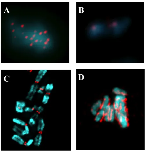
Figure 1: Presence of CENP-A is a conserved feature of centromeres in different organisms.
A. Point centromeres in S. cerevisiae. B. Chromosomes and centromeres of S. pombe. C. Regional centromeres in H. sapiens. D. Holocentric chromosmes of C. elegans. Blue indicates
A
B
DAPI staining of DNA. Red indicates CENP-A staining pattern on individual chromosomes (Vagnarelli et al., 2008).
Figure 2: CENP-A-containing nucleosomes generate the chromosomal foundation upon which inner and outer kinetochore proteins are assembled.
A. The spatial relationship between CENP-A nucleosomes, pericentric heterochromatin and the mitotic kinetochore in a condensed chromosome. B. Close-up model for the 3D configuration of CENP-A nucleosomes relative to H3 nucleosomes and pericentric heterochromatin (Allshire and Karpen, 2008).
Within the histone fold domain, deuterium exchange measured by mass spectrometry identified a unique structural element, termed the CENP-A targeting domain (CATD) comprising of the L1 and α2 helices (Black et al., 2007a). Chimeric molecules where the CATD of CENP-A is exchanged with the corresponding region in H3 (and vice versa) revealed that the CATD is a major determinant for targeting of CENP-A to the centromeres (Black et al., 2007b). Interestingly, structural data indicate that CATD also serves as an interface between CENP-A and H4 in the sub-nucleosomal (CENP-A:H4)2 complex (Sekulic
et al., 2010) as well as the CENP-A:CENP-A dimer in the putative octameric CENP-A nucleosomes (Bassett et al., 2012; Sekulic et al., 2010). Importantly, the CATD has been shown to interact with the holliday junction recognition protein (HJURP) which is an essential chaperone for CENP-A centromeric deposition (Black et al., 2004; Foltz et al., 2009; Hu et al., 2011). Despite detailed structural data discussed above, questions remain as to the precise nature of CENP-A chromatin. Complications in this regard mainly arise from inconsistencies between data obtained from biochemical work using in vitro nucleosome reconstitution approaches, proposing a canonical nucleosome-like octameric entity for CENP-A nucleosomes, and some lines of evidence obtained from studying endogenously purified centromeric chromatin revealing that CENP-A may not be present in a nucleosome form (octamers) and might exist in cells as some sort of tetrameric half-nucleosomes (hemisomes) at the centromere. Here we review our current understanding of the structure of CENP-A containing complexes highlighting the biological outcomes of the octamer vs. tetramer debate.
Soluble CENP-A:
Upon production of CENP-A in G2 (Howman et al., 2000; Shelby et al., 2000), the newly synthesized protein is thought to form a dimer with histone H4 and further in the cell
cycle recognized by HJURP (known as suppressor of chromosome missegregation 3, Scm3 in yeast) resulting in an equimolar complex of CENP-A:H4:HJURP (figure 3) (Cho and Harrison, 2011; Hu et al., 2011). In yeast, Scm3 binds the CATD of Cse4 (CENP-A homolog), and α2 and α3 of H4 via its Cse4 binding domain (CBD), with key residues conserved in HJURP (Zhou et al., 2011).
Figure 3: Proposed mechanism for the formation of putative CENP-A octameric nucleosomes.
Proposed mechanism for the formation of putative CENP-A octameric nucleosomes. Newly synthesized CENP-A along with histone H4 is suggested to bind HJURP to form a pre-nucleosomal trimeric complex. Next, in order to interact with DNA, HJURP has to be released leaving a (CENP-A:H4)2 tetramer. Addition of H2A:H2B dimers then complete the octamer
This interaction of HJURP with CENP-A is required to stabilize CENP-A as depletion of HJURP in human cells results in dramatically decreased CENP-A protein levels (Dunleavy et al., 2009; Foltz et al., 2009; Shuaib et al., 2010). Structural data suggest that this complex (Cse4:H4:Scm3) is not capable of interacting with DNA due to the induction of major conformational alterations in Cse4 and H4 (e.g. displacement of DNA-binding Loop 2 of H4) (Zhou et al., 2011). Moreover, it has been suggested that the presence of Scm3 in the pre-nucleosomal complex prevents the sub-pre-nucleosomal (Cse4:H4)2 tetramer formation (figure 3),
a step required for the nucleosome assembly (Zhou et al., 2011). Therefore, it is intuitive to assume that Scm3 needs be recognized by another/other component(s) in order to bind the chromatin and that it has to be removed for stable incorporation of CENP-A on to centromeres.
CENP-A nucleosomal structure The octamer model:
The crystal structure of the human CENP-A-containing nucleosome reconstituted in
vitro from bacterially purified histones indicates homotypic octamers containing two copies of
each histone molecule (Tachiwana et al., 2011). This study also revealed key features of CENP-A nucleosomes distinguishing them from canonical H3 nucleosomes. For instance, CENP-A contains a shorter αN helix lacking a key Arginine in position 49, which is an essential amino acid for DNA interaction. These findings are consistent with the data obtained independently from stepwise assembly of CENP-A nucleosomes not only confirming the octameric structure of CENP-A nucleosomes but also the loosening of the interaction between DNA superhelical termini and CENP-A (Conde e Silva et al., 2007; Panchenko et al., 2011).
CENP-A octamers formed in vitro have also been reported to induce conventional left-handed negative supercoiling to DNA (Barnhart et al., 2011; Conde e Silva et al., 2007; Panchenko et al., 2011; Tachiwana et al., 2011; Yoda et al., 2000). It was recently demonstrated that the mutation of the putative CENP-A:CENP-A dimer interface can abrogate centromeric targeting of CENP-A in Drosophila and mammalian tissue culture cells (Bassett et al., 2012; Zhang et al., 2012). In agreement with an octamer, over-expression of Cse4 (the CENP-A homolog) in budding yeast was reported to result in misincorporation of octamer-sized nucleosomes in chromosome arms (Camahort et al., 2009). These observations, along with the crystal structures available, provide solid evidence supporting the existence of octameric CENP-A nucleosomes at the centromere (Figure 6).
The Tetramer (hemisome) model:
In an effort to determine the native in vivo form of CENP-A chromatin, various purification and analysis techniques have been employed. One of the most extensive efforts has focused on nucleosome cross-linking followed by imunoprecipitation and atomic force
microscopy (AFM) to investigate CID-containing nucleosomes (CID for centromere identifier,
a Drosophila homologue of CENP-A). Challenging the octameric nucleosome concept, AFM data revealed that the height of the CID-containing interphase chromatin is half the height of canonical H3 nucleosomes (approximately 1 nm vs 2 nm) (Dalal et al., 2007). Moreover, in the beads-on-a-string structure of CID chromatin, the linker DNA is reported to be 2-3 times longer than that of conventional nucleosomes (Dalal et al., 2007). Surprisingly, the electrophoretic behavior of the purified CID-nucleosomal core particles corresponds to the presence of only one copy of each histone. This composition (CID:H4:H2A:H2B) is referred to as a tetramer, half-nucleosome or hemisome (Figure 4). Work done in human cells resulted
in similar observations regarding the equimolar presence of core histones with particle heights and volumes fitting well with the half-nucleosome model as compared to H3 nucleosomes (Dimitriadis et al., 2010).
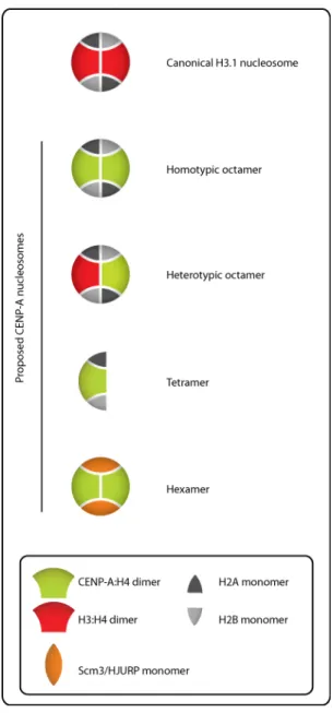
Figure 4: Different models for the composition of CENP-A nucleosomes.
These models differ in size, DNA wrapping and number as well as identity of their components. For a more detailed description of the differences between each model, please see the text.
Immuno-electron microscopy (Immuno-EM) data also suggest that the quantity of histones within each CENP-A nucleosomal particle matches with the half-nucleosome structure (Dimitriadis et al., 2010).
Recently, it was reported that the budding yeast centromeres are composed of a single Cse4-containing nucleosome wrapping about 80 bp of DNA, half the length of a canonical nucleosome, once in a right-handed manner (Henikoff and Henikoff, 2012; Krassovsky et al., 2012). The right-handedness of DNA wrapping in centromeric nucleosomes has been reported by other studies as well (Furuyama and Henikoff, 2009; Huang et al., 2011). ChIP data also demonstrated the occupancy of H2A at these sites all together consistent with the existence of a Cse4 hemisome at the centromere (Krassovsky et al., 2012). In support of this, the same study that found Cse4 nucleosomes (octamers) in chromosome arms reported tetramers in centromeres (Camahort et al., 2009). It should be noted that in vivo calibrated fluorescence intensity measurements of GFP:Cse4 are not consistent with a single copy of Cse4 at each centromere (Coffman et al., 2011; Lawrimore et al., 2011). Thus it is entirely possible that the conditions used for purification induce a hemisome like artifact and that does not exist or is not stable in vivo.
Intriguingly, the observations presented above about centromeric nucleosomes, are reminiscent of an early model suggesting that octameric nucleosomes are in fact constituted from symmetrical half-nucleosome pairs capable of independent existence (Weintraub et al., 1976). This was supported by work on SV40 minichromosome which primarily consists of about 20-25 nucleosomes as shown by electron microscopy (EM). Incubation of purified minichromosomes at low ionic strengths was however reported to induce the doubling of the number of beads-on-a-string, reduction of the dimensions of resulting particles and more interestingly longer inter-particle distances, all suggesting the splitting of octameric nucleosomes into half-nucleosomes (Lavelle and Prunell, 2007; Oudet et al., 1978). Similar
observations were made with cellular chromatin (Oudet et al., 1978). However, the occurrence of this conversion under cellular conditions remains to be shown to date.
On the other hand, the observation that nucleosomes can be found in various conformational states (Lavelle and Prunell, 2007), examples of which include Archaeal nucleosomes consisting solely of (H3:H4)2 tetramers (Reeve et al., 1997); eukaryotic
reversomes generated upon depletion of H2A:H2B dimers with right-handed DNA wrapping (Lavelle and Prunell, 2007) and the more recent proposed heterotetramer formation of CENPs-T:W:S:X (Nishino et al., 2012) capable of supercoiling DNA similar to nucleosomes, supports the possibility of tetrameric half-nucleosomes (hemisomes) residing in certain regions of the genome such as the centromere.
Other proposed forms of CENP-A nucleosomes:
Using a modified sequential immuno-precipitation technique in budding yeast, H3 was recently reported to co-occupy the centromeric DNA along with Cse4 and other core histones in a cell cycle independent manner (Lochmann and Ivanov, 2012) suggesting the potential existence of (Cse4:H4)(H3:H4)(H2A:H2B)2 heterotypic octamers (Figure 4). However, it is
not clear if stable association of H3 with Cse4 containing nucleosome is in the form of a heterotypic octamer or non-nucleosomal associations.
Additionally, a (Cse4:H4)2(Scm3)2 hexameric organization has also been proposed for
the centromeric chromatin in budding yeast (Figure 4) (Mizuguchi et al., 2007). However, a number of key observations soon detracted support for stable occurrence of this structure in centromeric chromatin. These include the previously mentioned structural barriers occluding Cse4 and H4 interaction with DNA (Zhou et al., 2011) and the fact that over-expression of
Cse4 in an Scm3Δ background, can rescue the Scm3 null phenotype (Camahort et al., 2009) suggesting that Scm3 is dispensable for centromere organization.
The tetramer to octamer transition model; towards a dispute settlement?
The controversial observations regarding the nature of CENP-A nucleosomes possibly stem from different chromatin preparation techniques, the stabilization of transient intermediates or the co-existence of more than one CENP-A nucleosome type under certain conditions. No matter the technical difference, a potential structural dynamics model for CENP-A containing nucleosomes through the cell cycle would be an important step forward in understanding centromere biology.
In this regard, an octamer to tetramer conversion model had been previously proposed based on which octameric CENP-A nucleosomes are split into tetrameric half-nucleosomes upon the passage of the replication fork in S phase allowing the equal inheritance of the epigenetic mark to the daughter strands (Figure 5A) (Allshire and Karpen, 2008; Probst et al., 2009). The resultant tetramers were proposed to be maintained throughout G2/M but converted into octamers in G1 following incorporation of new CENP-A by HJURP. This model, while providing a possible mechanism for the preservation of centromeric identity, had never been experimentally validated.
Interestingly, two recent studies co-published in Cell (Bui et al., 2012; Shivaraju et al., 2012) provide evidence for a novel cell cycle-coupled structural transition of CENP-A nucleosomes in human cells and budding yeast (Figure 5B). AFM-based analysis of immuno-precipitated CENP-A nucleosomes from cell cycle staged human cells revealed that centromeric nucleosomes changed in size depending on cell cycle timing.
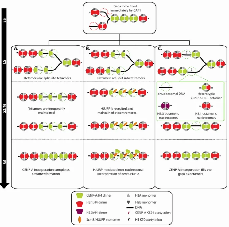
Figure 5: Proposed structural dynamics of centromeric nucleosomes throughout the cell cycle.
A. An octamer to tetramer transition model in which passage of the replication fork splits the pre-existing octameric CENP-A nucleosomes into tetramers allowing equal inheritance of the epigenetic mark to daughter strands. In this model, HJURP exclusively is found at the centromere in G1 and mediates the reconstruction of octamers by incorporation of new CENP-A. B. The tetramer to octamer transition model. CENP-A:H4:HJURP complex is recruited to the centromeric chromatin in G1 and is assumed to associate to the pre-exisiting CENP-A tetramers extra-nucleosomally. Post-translational modifications (PTMs) of tetrameric CENP-A nucleosomes along with the presence of HJURP impede the stable incorporation of new CENP-A:H4 dimers into nucleosomes. In late G1/early S, action of certain protein chaperones and remodeling complexes is proposed to facilitate the tetramer to octamer transition by affecting the PTMs of histones and chromatin structure resulting in tetramer to octamer transition. Likewise to the previous model, passage of the replication fork splits the octamers to tetramers. The tetramers are further stabilized by the reassociation of HJURP in G2 and presumed to be maintained in mitosis to form the kinetochore plate. C. In an alternative model, where CENP-A exists in octameric nuclesomes throughout the cell cycle, gaps generated as a result of the passage of the replication fork will be either maintained or could be transiently filled with multiple possible placeholder structures. In this model, HJURP is also found at the centromere exclusively in G1 to participate in new CENP-A assembly.
Tetrameric dimensions of CENP-A nucleosomes in G1 were reported to undergo a transition to octameric dimensions during S phase and revert back to tetrameric state in G2 and maintained through mitosis (Bui et al., 2012). Intriguingly, the authors also report cyclic association, dissociation and re-association of HJURP to the centromeric chromatin in G1, S and G2 phases respectively (Figure 5B). Purification of DNA-bound CENP-A:H4 from G1/S arrested cells followed by mass-spectroscopic analysis identified two previously unknown covalent modifications: acetylation of CENP-A K124 and H4 K79. These modifications and the presence of HJURP were proposed to prevent stable octamer formation in G1/S. However, as the cell enters the S phase, the authors speculated that opening of centromeric chromatin would be concomitant with the resolution of these modifications. This biochemical change could be coupled to the release of HJURP physically allowing the action of chromatin remodelers to trigger the generation octamers. The dynamics of HJURP at the centromere and the reformation of tetramers in G2 might indicate the role of HJURP in this reversal transition following DNA replication. However, HJURP in human cells has been previously shown to localize to centromeres exclusively during CENP-A loading in G1(Dunleavy et al., 2009; Foltz et al., 2009) and the G2 reappearance of HJURP at the centromeres has never been reported by other groups. Given the fact that nascent CENP-A is chaperoned by HJURP after synthesis in G2, cytoplasmic contamination of chromatin lysate could be a potential source for the detection of HJURP in G2/M CENP-A pull down. On the other hand, the authors report that HJURP is absent from S phase CENP-A pull down. If the possibility of cytoplasmic contamination is true, this would nicely explain the absence of HJURP in S phase CENP-A pull down since HJURP has already completed deposition of its cargo and thus even if the
chromatin prep does contain cytoplasmic contamination, HJURP will not copurify with CENP-A any longer.
Work in Saccharomyces cerevisiae and Candida albicans using fluorescence correlation spectroscopy (FCS) also suggest the presence of a single copy of Cse4 at each centromere (Shivaraju et al., 2012). This seems to be the case for G1, S, G2 and metaphase, however during anaphase B the authors note a “doubling” of Cse4 and speculate this is due to a tetramer to octamer transition. FCS can very accurately measure protein complexes in living cells by recording peak intensities of diffusing molecules and comparing these over varying time scales (Bulseco and Wolf, 2007) (usually up to 2 minutes), detecting auto-correlations. A major caveat with this technique is that while extremely accurate for freely diffusing complexes, it is less accurate for slow diffusing structures (Krichevsky and Bonnet, 2002) (in this case mitotic centromeres bound to microtubules). This is in part due to the fact that FCS is based on peak intensity of a diffraction limited spot. Thus, slowly diffusing or slightly dispersed (greater than the measurement spot) structures such as centromeres will not all be measured in the time scale required.
Interestingly, centromeres are much less dispersed in anaphase (compared to metaphase) (Pearson et al., 2001), which could allow for more accurate measurements and thus explain the difference reported. Nonetheless, using FRET and sequential ChIPs the authors show that Cse4:Cse4 interaction does indeed take place increasingly in anaphase B (Shivaraju et al., 2012). The tetramer to octamer transition is also concomitant with the transient disappearance of Scm3 from centromeres in a short time window corresponding to anaphase B. The mutually exclusive relationship between presence of Scm3/HJURP and the Cse4/CENP-A dimers could be attributed to the unique HFD of CENP-A harboring a shared interface for interaction with Scm3/HJURP or another molecule of CENP-A.
Even though the generation of differential CENP-A nucleosome types and the transition mechanisms remain largely unknown, it is presumable that such a dynamic behavior may require a tight regulation for timing and the concerted action of chromatin remodeling factors. It would be interesting to investigate the occurrence of K124- and K79-like modifications in yeast Cse4 and H4. In addition, the previous detection of CID-nucleosomes corresponding in dimensions to tetramers in interphase but octamers in mitotic Drosophila cells (Dalal et al., 2007) might reflect a similar cell-cycle regulated transition formerly unexplored.
Implications of the structure of CENP-A nucleosomes: why does it matter after all? Emerging evidence suggests that the assembly of CENP-A and thus propagation of the epigenetic mark occurs through three major steps: licensing by KNL-2/M18BP1 (Fujita et al., 2007; Maddox et al., 2007) and Mis18 (Hayashi et al., 2004), incorporation via HJURP (Bernad et al., 2011; Dunleavy et al., 2009; Foltz et al., 2009) and maintenance by
MgcRacGAP (Lagana et al., 2010). During S phase, as the replication fork forges ahead, the pre-existing population of CENP-A nucleosomes is halved and thereby inherited to daughter strands to preserve centromeric identity (Allshire and Karpen, 2008). The dilution of CENP-A nucleosomes and the replication-independent incorporation of CENP-A raise the possibility of the formation of various placeholder structures in S phase (Figure 5C). In the case of human cells, according to recent data, the CENP-A nucleosomal population is proposed to undergo the tetramer to octamer transition in front of the replication fork approaching the centromeric DNA (Bui et al., 2012). Figure 6 depicts possible steps in this transition.
It is not clear what exactly signals the speculated reversion of octamers into tetramers at the end of S phase. Passage of the fork may, via unknown mechanisms or interactions, trigger not only the splitting of the pre-existing CENP-A nucleosomes, but also the reversion of octamers into tetramers. This may in turn coincide with the reappearance of HJURP at the end of S phase in human cells. In contrast to canonical nucleosomes, incorporation of newly synthesized CENP-A nucleosomes does not accompany DNA replication (Jansen et al., 2007). CENP-A assembly in the mammalian system requires exit from mitosis (Jansen et al., 2007) and takes place during late M/G1 phase of the cell cycle in the mammalian and embryonic
Drosophila systems (Mellone et al., 2011; Schuh et al., 2007). However, the mechanism of
specific recognition of the centromere by CENP-A assembly proteins remains largely unknown. It is assumed that these proteins might recognize a specialized chromatin structure, certain contact sites on CENP-A-containing nucleosomes or a non-conventional nucleosome form exclusively found at the centromeric chromatin. Given the proposed atypical CENP-A nucleosomes, regardless of the model, an entertaining speculation would be that the heteroclite structure of CENP-A nucleosomes might provide the green light for CENP-A assembly
machinery to repopulate the centromere in preparation for the subsequent mitosis. In addition, these atypical structures may serve as recognition sites for kinetochore assembly during mitosis.
Figure 6 : Possible steps of the tetramer to octamer transition.
Upon transition of the cell to S phase, HJURP is released from the centromeric chromatin and certain posttranslational modifications and of CENP-A tetramers are resolved by yet-to-be-identified factors. In the first scenario, H2A:H2B dimers are also temporarily removed
allowing the tetrameric (CENP-A:H4)2 complex to form and interact with DNA. This reaction
is speculated to be mediated by NAP1, which is a chromatin remodeler. Addition of a pair of H2A:H2B dimers will complete the octamer formation. In the second scenario, however, incorporation of new CENP-A:H4 as well as H2B:H2B dimers does not require disassembly of the pre-existing CENP-A tetramers.
Conclusion:
This review summarizes major features of CENP-A-containing complexes on sub-nucleosomal, nucleosomal and chromatin levels. The debate over the true molecular nature of the CENP-A epigenetic mark remains to be resolved as many questions are still unanswered. For example, what could be the biological significance of the tetramer to octamer transition in S phase for mammalian cells or anaphase B in case of yeast? What are the factors and mechanisms involved? In the coming exciting years of research, high-resolution imaging and biochemical approaches hold promise to pave the way for answering these questions.
Acknowledgment:
We are grateful to all members of Paul and Amy Maddox labs. Special thanks to Joel Ryan for his valuable assistance with the figures. P.S.M. is the Canada Research Chair in Cell Division and Chromosomal Organization and supported by research grants from the CIHR (MOP-106548) and CCSRI (700824).
References:
Allshire, R.C., and Karpen, G.H. (2008). Epigenetic regulation of centromeric chromatin: old dogs, new tricks? Nature reviews Genetics 9, 923-937.
Barnhart, M.C., Kuich, P.H., Stellfox, M.E., Ward, J.A., Bassett, E.A., Black, B.E., and Foltz, D.R. (2011). HJURP is a CENP-A chromatin assembly factor sufficient to form a functional de novo kinetochore. The Journal of cell biology 194, 229-243.
Bassett, E.A., DeNizio, J., Barnhart-Dailey, M.C., Panchenko, T., Sekulic, N., Rogers, D.J., Foltz, D.R., and Black, B.E. (2012). HJURP uses distinct CENP-A surfaces to recognize and to stabilize CENP-A/histone H4 for centromere assembly. Developmental cell 22, 749-762.
Bernad, R., Sanchez, P., Rivera, T., Rodriguez-Corsino, M., Boyarchuk, E., Vassias, I., Ray-Gallet, D., Arnaoutov, A., Dasso, M., Almouzni, G., et al. (2011). Xenopus HJURP and condensin II are required for CENP-A assembly. The Journal of cell biology 192, 569-582.
Black, B.E., Brock, M.A., Bedard, S., Woods, V.L., Jr., and Cleveland, D.W. (2007a). An epigenetic mark generated by the incorporation of CENP-A into centromeric nucleosomes. Proceedings of the National Academy of Sciences of the United States of America 104, 5008-5013.
Black, B.E., Foltz, D.R., Chakravarthy, S., Luger, K., Woods, V.L., Jr., and Cleveland, D.W. (2004). Structural determinants for generating centromeric chromatin. Nature 430, 578-582.
Black, B.E., Jansen, L.E., Maddox, P.S., Foltz, D.R., Desai, A.B., Shah, J.V., and Cleveland, D.W. (2007b). Centromere identity maintained by nucleosomes assembled with histone H3 containing the CENP-A targeting domain. Molecular cell 25, 309-322.
Bui, M., Dimitriadis, E.K., Hoischen, C., An, E., Quenet, D., Giebe, S., Nita-Lazar, A., Diekmann, S., and Dalal, Y. (2012). Cell-Cycle-Dependent Structural Transitions in the Human CENP-A Nucleosome In Vivo. Cell 150, 317-326.
Bulseco, D.A., and Wolf, D.E. (2007). Fluorescence correlation spectroscopy: molecular complexing in solution and in living cells. Methods in cell biology 81, 525-559.
Camahort, R., Shivaraju, M., Mattingly, M., Li, B., Nakanishi, S., Zhu, D., Shilatifard, A., Workman, J.L., and Gerton, J.L. (2009). Cse4 is part of an octameric nucleosome in budding yeast. Molecular cell 35, 794-805.
Cho, U.S., and Harrison, S.C. (2011). Recognition of the centromere-specific histone Cse4 by the chaperone Scm3. Proceedings of the National Academy of Sciences of the United States of America 108, 9367-9371.
Cleveland, D.W., Mao, Y., and Sullivan, K.F. (2003). Centromeres and kinetochores: from epigenetics to mitotic checkpoint signaling. Cell 112, 407-421.
Coffman, V.C., Wu, P., Parthun, M.R., and Wu, J.Q. (2011). CENP-A exceeds microtubule attachment sites in centromere clusters of both budding and fission yeast. The Journal of cell biology 195, 563-572.
Conde e Silva, N., Black, B.E., Sivolob, A., Filipski, J., Cleveland, D.W., and Prunell, A. (2007). CENP-A-containing nucleosomes: easier disassembly versus exclusive centromeric localization. Journal of molecular biology 370, 555-573.
Dalal, Y., Wang, H., Lindsay, S., and Henikoff, S. (2007). Tetrameric structure of centromeric nucleosomes in interphase Drosophila cells. PLoS Biol 5, e218.
Dimitriadis, E.K., Weber, C., Gill, R.K., Diekmann, S., and Dalal, Y. (2010). Tetrameric organization of vertebrate centromeric nucleosomes. Proceedings of the National Academy of Sciences of the United States of America 107, 20317-20322.
Dunleavy, E.M., Roche, D., Tagami, H., Lacoste, N., Ray-Gallet, D., Nakamura, Y., Daigo, Y., Nakatani, Y., and Almouzni-Pettinotti, G. (2009). HJURP is a cell-cycle-dependent maintenance and deposition factor of CENP-A at centromeres. Cell 137, 485-497.
Foltz, D.R., Jansen, L.E., Bailey, A.O., Yates, J.R., 3rd, Bassett, E.A., Wood, S., Black, B.E., and Cleveland, D.W. (2009). Centromere-specific assembly of CENP-a nucleosomes is mediated by HJURP. Cell 137, 472-484.
Fujita, Y., Hayashi, T., Kiyomitsu, T., Toyoda, Y., Kokubu, A., Obuse, C., and Yanagida, M. (2007). Priming of centromere for CENP-A recruitment by human hMis18alpha, hMis18beta, and M18BP1. Developmental cell 12, 17-30.
Fukagawa, T. (2004). Centromere DNA, proteins and kinetochore assembly in vertebrate cells. Chromosome research : an international journal on the molecular, supramolecular and evolutionary aspects of chromosome biology 12, 557-567.
Furuyama, T., and Henikoff, S. (2009). Centromeric nucleosomes induce positive DNA supercoils. Cell 138, 104-113.
Guse, A., Carroll, C.W., Moree, B., Fuller, C.J., and Straight, A.F. (2011). In vitro centromere and kinetochore assembly on defined chromatin templates. Nature 477, 354-358.
Hayashi, T., Fujita, Y., Iwasaki, O., Adachi, Y., Takahashi, K., and Yanagida, M. (2004). Mis16 and Mis18 are required for CENP-A loading and histone deacetylation at centromeres. Cell 118, 715-729.
Henikoff, S., and Henikoff, J.G. (2012). "Point" centromeres of Saccharomyces harbor single centromere-specific nucleosomes. Genetics 190, 1575-1577.
Howman, E.V., Fowler, K.J., Newson, A.J., Redward, S., MacDonald, A.C., Kalitsis, P., and Choo, K.H. (2000). Early disruption of centromeric chromatin organization in centromere protein A (Cenpa) null mice. Proceedings of the National Academy of Sciences of the United States of America 97, 1148-1153.
Hu, H., Liu, Y., Wang, M., Fang, J., Huang, H., Yang, N., Li, Y., Wang, J., Yao, X., Shi, Y., et al. (2011). Structure of a CENP-A-histone H4 heterodimer in complex with chaperone HJURP. Genes & development 25, 901-906.
Huang, C.C., Chang, K.M., Cui, H., and Jayaram, M. (2011). Histone H3-variant Cse4-induced positive DNA supercoiling in the yeast plasmid has implications for a plasmid origin of a chromosome centromere. Proceedings of the National Academy of Sciences of the United States of America 108, 13671-13676.
Jansen, L.E., Black, B.E., Foltz, D.R., and Cleveland, D.W. (2007). Propagation of centromeric chromatin requires exit from mitosis. The Journal of cell biology 176, 795-805.
Krassovsky, K., Henikoff, J.G., and Henikoff, S. (2012). Tripartite organization of centromeric chromatin in budding yeast. Proceedings of the National Academy of Sciences of the United States of America 109, 243-248.
Krichevsky, O., and Bonnet, G. (2002). Fluorescence correlation spectroscopy: the technique and its applications. Rep Prog Phys 65, 251-297.
Lagana, A., Dorn, J.F., De Rop, V., Ladouceur, A.M., Maddox, A.S., and Maddox, P.S. (2010). A small GTPase molecular switch regulates epigenetic centromere maintenance by stabilizing newly incorporated CENP-A. Nature cell biology 12, 1186-1193.
Lavelle, C., and Prunell, A. (2007). Chromatin polymorphism and the nucleosome superfamily: a genealogy. Cell cycle 6, 2113-2119.
Lawrimore, J., Bloom, K.S., and Salmon, E.D. (2011). Point centromeres contain more than a single centromere-specific Cse4 (CENP-A) nucleosome. The Journal of cell biology
195, 573-582.
Lochmann, B., and Ivanov, D. (2012). Histone H3 localizes to the centromeric DNA in budding yeast. PLoS genetics 8, e1002739.
Maddox, P.S., Hyndman, F., Monen, J., Oegema, K., and Desai, A. (2007). Functional genomics identifies a Myb domain-containing protein family required for assembly of CENP-A chromatin. The Journal of cell biology 176, 757-763.
Mellone, B.G., Grive, K.J., Shteyn, V., Bowers, S.R., Oderberg, I., and Karpen, G.H. (2011). Assembly of Drosophila centromeric chromatin proteins during mitosis. PLoS genetics
7, e1002068.
Nishino, T., Takeuchi, K., Gascoigne, K.E., Suzuki, A., Hori, T., Oyama, T., Morikawa, K., Cheeseman, I.M., and Fukagawa, T. (2012). CENP-T-W-S-X forms a unique centromeric chromatin structure with a histone-like fold. Cell 148, 487-501.
Oudet, P., Germond, J.E., Bellard, M., Spadafora, C., and Chambon, P. (1978). Nucleosome structure. Philosophical transactions of the Royal Society of London Series B, Biological sciences 283, 241-258.
Palmer, D.K., O'Day, K., Trong, H.L., Charbonneau, H., and Margolis, R.L. (1991). Purification of the centromere-specific protein CENP-A and demonstration that it is a distinctive histone. Proceedings of the National Academy of Sciences of the United States of America 88, 3734-3738.
Panchenko, T., Sorensen, T.C., Woodcock, C.L., Kan, Z.Y., Wood, S., Resch, M.G., Luger, K., Englander, S.W., Hansen, J.C., and Black, B.E. (2011). Replacement of histone H3 with CENP-A directs global nucleosome array condensation and loosening of nucleosome superhelical termini. Proceedings of the National Academy of Sciences of the United States of America 108, 16588-16593.
Pearson, C.G., Maddox, P.S., Salmon, E.D., and Bloom, K. (2001). Budding yeast chromosome structure and dynamics during mitosis. The Journal of cell biology 152, 1255-1266.
Probst, A.V., Dunleavy, E., and Almouzni, G. (2009). Epigenetic inheritance during the cell cycle. Nature reviews Molecular cell biology 10, 192-206.
Reeve, J.N., Sandman, K., and Daniels, C.J. (1997). Archaeal histones, nucleosomes, and transcription initiation. Cell 89, 999-1002.
Schuh, M., Lehner, C.F., and Heidmann, S. (2007). Incorporation of Drosophila CID/CENP-A and CENP-C into centromeres during early embryonic anaphase. Current biology : CB 17, 237-243.
Sekulic, N., Bassett, E.A., Rogers, D.J., and Black, B.E. (2010). The structure of (CENP-A-H4)(2) reveals physical features that mark centromeres. Nature 467, 347-351.
Shelby, R.D., Monier, K., and Sullivan, K.F. (2000). Chromatin assembly at kinetochores is uncoupled from DNA replication. The Journal of cell biology 151, 1113-1118.
Shivaraju, M., Unruh, J.R., Slaughter, B.D., Mattingly, M., Berman, J., and Gerton, J.L. (2012). Cell-cycle-coupled structural oscillation of centromeric nucleosomes in yeast. Cell
Shuaib, M., Ouararhni, K., Dimitrov, S., and Hamiche, A. (2010). HJURP binds CENP-A via a highly conserved N-terminal domain and mediates its deposition at centromeres. Proceedings of the National Academy of Sciences of the United States of America 107, 1349-1354.
Sullivan, K.F., Hechenberger, M., and Masri, K. (1994). Human CENP-A contains a histone H3 related histone fold domain that is required for targeting to the centromere. J Cell Biol 127, 581-592.
Tachiwana, H., Kagawa, W., Shiga, T., Osakabe, A., Miya, Y., Saito, K., Hayashi-Takanaka, Y., Oda, T., Sato, M., Park, S.Y., et al. (2011). Crystal structure of the human centromeric nucleosome containing CENP-A. Nature 476, 232-235.
Tomonaga, T., Matsushita, K., Yamaguchi, S., Oohashi, T., Shimada, H., Ochiai, T., Yoda, K., and Nomura, F. (2003). Overexpression and mistargeting of centromere protein-A in human primary colorectal cancer. Cancer Res 63, 3511-3516.
Vagnarelli, P., Ribeiro, S.A., and Earnshaw, W.C. (2008). Centromeres: old tales and new tools. FEBS letters 582, 1950-1959.
Weintraub, H., Worcel, A., and Alberts, B. (1976). A model for chromatin based upon two symmetrically paired half-nucleosomes. Cell 9, 409-417.
Yoda, K., Ando, S., Morishita, S., Houmura, K., Hashimoto, K., Takeyasu, K., and Okazaki, T. (2000). Human centromere protein A (CENP-A) can replace histone H3 in nucleosome reconstitution in vitro. Proceedings of the National Academy of Sciences of the United States of America 97, 7266-7271.
Zhang, W., Colmenares, S.U., and Karpen, G.H. (2012). Assembly of Drosophila centromeric nucleosomes requires CID dimerization. Molecular cell 45, 263-269.
Zhou, Z., Feng, H., Zhou, B.R., Ghirlando, R., Hu, K., Zwolak, A., Miller Jenkins, L.M., Xiao, H., Tjandra, N., Wu, C., et al. (2011). Structural basis for recognition of centromere histone variant CenH3 by the chaperone Scm3. Nature 472, 234-237.
Octameric CENP-A nucleosomes are present at human centromeres throughout the cell cycle
Abbas Padeganeh1, 3, Joël Ryan1ψ, Jacques Boisvert1,4ψ, Anne-Marie Ladouceur1, 3, Jonas F. Dorn1 and Paul S. Maddox1,2 *
1Institute for Research in Immunology and Cancer (IRIC) 2Department of Pathology and Cell Biology,
3Graduate programs in Molecular Biology-Systems Biology Option, 4Graduate programs in Bioinformatics,
Faculty of Medicine, University of Montreal, H3T 1J4, Quebec, Canada
ψ These authors contributed equally to this work *To whom correspondence should be addressed
Institute for Research in Immunology and Cancer (IRIC), Dept of Pathology and Cell Biology, Université de Montréal
P.O. Box 6128, Station Centre-Ville Montréal QC, H3C 3J7
In this chapter, I present the development of a high-resolution single-molecule imaging assay to address the molecular entity of the centromeric chromatin epigenetic mark and its compositional dynamics during the cell cycle.
The material presented herein was published in Current Biology. 2013 May 6;23(9):764-9 and featured in a News & Views article in Nat Struct Mol Biol. Jun 2013;20(6):648-50)
Author contribution:
PSM and AP designed the project. AP performed the wet lab experiments, wrote the manuscript and prepared the images. JR and AML assisted with the experiments. JB and JFD developed the automated data analysis software. PSM and AP and JR analyzed the data and PSM edited the manuscript.
Summary:
The presence of a single centromere on each chromosome that signals formation of a mitotic kinetochore is central to accurate chromosome segregation (Cleveland et al., 2003). The histone H3 variant CENP-A is critical for centromere identity and function; CENP-A chromatin acts as an epigenetic mark to direct both centromere and kinetochore assembly (Allshire and Karpen, 2008; Black et al., 2007a; De Rop et al., 2012). Interpreting the centromere epigenetic mark ensures propagation of a single centromere per chromosome to maintain ploidy. Thus, understanding the nature of CENP-A chromatin is crucial for all cell divisions. However there are ongoing debates over the fundamental composition of centromeric chromatin. Here we show that natively assembled human CENP-A nucleosomes are octameric throughout the cell cycle. Using TIRF-coupled photobleaching-assisted copy number counting of single nucleosomes obtained from cultured cells, we find that the majority of CENP-A nucleosomes contain CENP-A dimers. In addition, we detect the presence of H2B and H4 in these nucleosomes. Surprisingly, CENP-A associated with the chaperone HJURP can exist as either monomer or dimer, indicating possible assembly intermediates. Thus, our findings indicate that octameric CENP-A nucleosomes mark the centromeric region to ensure proper epigenetic inheritance and kinetochore assembly.
Highlights
1) CENP-A octameric nucleosomes epigenetically mark centromeres 2) CENP-A associated with the chaperone HJURP can exist as either monomer or dimer
Results and Discussion:
Conservation of ploidy requires inheritance of an equal number of chromosomes during each cell division. Central to this is the presence of a single kinetochore on each chromatid. Kinetochores are protein super-structures assembled during mitosis at centromere regions of chromosomes to link sister chromatids to microtubules emanating from opposite spindle poles. Consequently, centromere singularity generates accurate cell division (Cleveland et al., 2003). The defining feature of centromeres is understood to be the presence of a histone H3 variant referred to as Centromere Protein-A (CENP-A, also called CenH3) in centromeric nucleosomes (Black et al., 2007a; De Rop et al., 2012). CENP-A is proposed to mark centromeres epigenetically (Allshire and Karpen, 2008; Black et al., 2007a) as evidenced by rarely documented cases of neocentromeres, where CENP-A chromatin forms a functional centromere on an ectopic chromosomal region distinct from the original genomic locus. In sum, CENP-A-containing chromatin encodes the epigenetic information recognized, read and interpreted by proteins required for centromere (Foltz et al., 2009; Fujita et al., 2007; Lagana et al., 2010; Shuaib et al., 2010) and kinetochore assembly (Cleveland et al., 2003). Thus, understanding the biochemical nature of CENP-A chromatin is critical for understanding both epigenetic centromere propagation and mitotic chromosome segregation.
The majority of CENP-A containing complexes in cells contain two CENP-A molecules: Unlike other epigenetic codes (e.g. histone methylation patterns regulating promoter activity), the centromere epigenetic code is as yet largely unknown. Extensive efforts focused to determine the structure of CENP-A-containing nucleosomes have led to contradicting observations. The culmination is a series of hypotheses each differing from one another in the
species and number of centromere chromatin core components (Padeganeh et al., 2013). These include 1) the octamer model (Panchenko et al., 2011; Tachiwana et al., 2011; Tachiwana and Kurumizaka, 2011; Zhang et al., 2012) supported mainly by in vitro nucleosome reconstitution experiments proposing a (CENP-A:H4)2 (H2A:H2B)2 composition,
2) the tetramer model (Dalal et al., 2007; Dimitriadis et al., 2010; Krassovsky et al., 2012) stemming from Atomic Force Microscopy data on immunoprecipitated CENP-A chromatin proposing a (CENP-A:H4)(H2A:H2B) composition and 3) the hexamer model (Mizuguchi et al., 2007) proposing a hybrid of CENP-A:H4 and the chaperone protein HJURP (also called Scm3, hereafter HJURP).
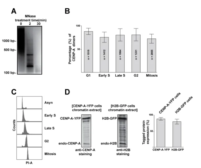
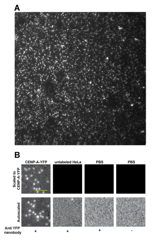
To determine the molecular composition of native CENP-A nucleosomes, we extracted single nucleosome core particles from clonal HeLa cells stably expressing a YFP fusion to CENP-A (see below and methods). For our analysis, we assumed that CENP-A-YFP and unlabeled CENP-A have equal probability of chromatin incorporation, and corrected for the incomplete labeling accordingly (see below and methods). In order to assess the stoichiometry of CENP-A in centromeric nucleosomes, we employed photo-bleaching-assisted copy number counting (PA-CNC, a modified version of SiM-Pull (Jain et al., 2011)). We assembled a homemade flow chamber and functionalized it with YFP-nanobodies (Rothbauer et al., 2008). This surface would act as a nanotrap for CENP-A-YFP containing complexes, effectively immunoprecipitating YFP containing particles from the whole chromatin extract (Figure 1A). We evaluated the specificity of our assay using control (no-YFP) HeLa chromatin lysates with and without YFP nanobodies and found minimal contamination, indicating that the analyzed signals were YFP derived (Figure 1B, and Supplement 1A). Importantly, nanobodies are monoclonal and single chained, therefore each nanobody isolates one and only one YFP
containing complex. The final chamber, when visualized by Total Internal Reflection Fluorescence (TIRF) microscopy, results in single isolated diffraction limited spots each representing a single CENP-A-YFP containing chromatin particle (Figure 1B). Thus, this system allows direct visual analysis of CENP-A-YFP stoichiometry in native assembled complexes. Our method has the additional benefit that there are very few manipulations of the sample as opposed to conventional fractionation techniques, reducing potential artifacts. A caveat of our method is that the TIRF illumination field is uneven, a common issue with objective-based TIRF systems. Uneven illumination precludes comparison of absolute intensities between individual complexes or experiments, however does not affect relative measurements of individual complexes.
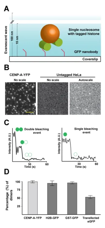
Figure 1: Asynchronous CENP-A-YFP expressing cells contain dimers of CENP-A at centromeric nucleosomes.
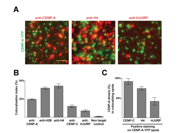
A. Schematic representation of our PA-CNC set up. Using anti-YFP single-chained nanobodies allows the isolation of single CENP-A-YFP nucleosomes on the surface of a coverslip. Samples are allowed to photobleach completely by TIRF illumination. By plotting the fluorescence intensity fluctuations, detected for each diffraction limited spot, over time, the number of fluorophores in each spot is quantified by counting the number of discrete bleaching events. B. Representative TIRF images are shown for samples with and without addition of CENP-A-YFP nucleosomes. C. Example traces of pixel intensity fluctuation over time in double and single bleaching events. Camera integration time for all movies is 900 ms. (A.U. = arbitrary unit). D.The percentage of double bleaching events detected using our PA-CNC assay with purified nucleosomes from CENP-A-YFP and H2B-GFP stable cell lines, purified GST-GFP and cellular lysate from eGFP-transfected HeLa cells. The data presented herein are corrected for the expression levels of the labelled proteins and pre-bleaching where applicable. (scale bar = 20 µm; error bars indicate standard deviation).
A key feature differing in models for the structure of centromeric chromatin is the presence of one or two CENP-A molecules per nucleosome. This difference has obvious implications towards the mechanisms interpreting the centromere epigenetic mark and therefore chromosome segregation. To quantify the number of CENP-A-YFP molecules per individual complex detected in discrete diffraction limited spots, consecutive images were acquired while illuminating the sample until all fluorophores photobleached (Supplemental movie S1). Plotting the intensity of each diffraction limited spot as a function of time revealed minor fluctuations (random noise) overlaid on large drops in intensity (Figure 1C, and Supplement 2). Large drops were generated by permanent bleaching of individual YFP molecules, visible in graphs of intensity versus time. Photobleaching is stochastic and a direct measure of the number of YFP molecules per isolated complex. Because measuring intensity drops by hand is very labor intensive and subject to human error, we developed a custom software package (QUBE for QUantitative Bleaching Estimation, written in MATLAB) to automatically detect, segment, and measure spot intensities over time. This greatly increased the number of spots analyzed (thousands per condition) and yielded similar, however far more statistically significant, results to those obtained by manual analysis (data not shown). Periodically, large intensity increases following large drops were observed (Supplement 2, sample blinking event). These were likely due to the reported blinking behavior of YFP, or to the polarized nature of the TIRF illumination light. Regardless of the origin, blinking events are indicative of single molecules and therefore enhance our confidence in the singularity of analyzed particles. In fact, fluorescence blinking is the basis for single molecule super-resolution techniques such as STochastic Optical Reconstruction Microscopy (STORM) (Rust et al., 2006). Analysis of CENP-A-YFP nucleosomes isolated from asynchronous cells