Déterminants moléculaires de la scoliose idiopathique de
l'adolescent
par Mohamed Elbakry Département de biochimie Faculté de médecineThèse présentée à la Faculté des études supérieures en vue de l’obtention du grade de Ph.D.
en Biochimie
Mai 2013
© Mohamed Elbakry, 2013
Faculté des études supérieures et postdoctorales
Cette thèse intitulée :
Déterminants moléculaires de la scoliose idiopathique de l'adolescent
Présentée par : Mohamed Elbakry
A été évaluée par un jury composé des personnes suivantes :
Dr Muriel Aubry, président-rapporteur Dr Alain Moreau, directeur de recherche
Dr Zoha Kibar, membre du jury Dr Fackson Mwale, examinateur externe
Résumé
La scoliose est la déformation de la colonne vertébrale la plus répendue. Elle atteint 3 à 4% de la population pédiatrique et dans 85% des cas, aucune cause n’a été identifiée. Ces cas sont appelés idiopathiques et les symptômes apparaissent durant la puberté; d’où le terme de ‘scoliose idiopathique de l’adolescent (SIA). Cette pathologie atteint le plus souvent les jeunes filles, en nombre et en sévérité. Ces dernières années, plusieurs hypothèses ont été proposées afin d’élucider l’étiologie de cette pathologie. Celles-ci ont mis de l’avant différents facteurs génétiques, biochimiques, méchaniques, neurologiques, musculaires ou hormonaux. Plusieurs études ont rapporté des formes familiales de scoliose, soutenant la thèse d’une prédisposition génétique. Nous avons démontré que les patients souffrant de SIA présentent un défaut de signalisation cellulaire médiée par les protéines Gi et un taux élevé d’ostéopontine (OPN) circulante. En utilisant une approche de type ‘gène candidat’, nous avons montré que la protéine tyrosine phosphatase µ (PTPµ) régule l’activité du complexe d’intégrines α5/β1 (récepteur de l’OPN) via la protéine kinase PIPKIγ. Dans ce but, nous avons utilisé des cultures primaires d’ostéoblastes issues de biopsies de patients et de cas traumatiques comme sujets contrôles. Les biopsies ossscuses de patients ont été obtenues lors de l’intervention chirurgicale à partir des vertébres T3 à L4, selon les différentes procédures. Les biopsies issues de cas traumatiques proviennent d’autres types d’os (tibia, crête iliaque, fémur). Les profils d’expression du gène PTPRM (codant pour la protéine PTPµ) ont été étudiés par PCR quantitative (qPCR). Les taux de protéines PTPµ ont été analysés par immunoprécipitation suivi d’un western blot. Pour évaluer le rôle de cette protéine, nous avons bénéficié d’un modèle murin. Machida et al. ont démontré qu’il existe un taux plus élevé de scoliose parmi les souris C57Bl/6 bipèdes obtenues suite à l’amputation des membres supérieurs, sous anesthésie, cinq semaines après la naissance. Nous avons utilisé des cultures primaires d’ostéoblastes issues de la colonne
vertébrale de souris C57Bl/6 bipèdes, délétées du gène PTPRM (souris dites ‘KO’), afin d’évaluer le niveau de signalisation cellulaire spécifique des protéines Gi par un test fonctionnel: la technique de spectroscopie cellulaire di-électrique (SCD). Selon nos données, 85% des souris bipédales ‘KO’ pour le géne PTPRM développent une scoliose (modérée à sévère) contre 55% des souris contrôles C57Bl6 bipèdes. De plus, les niveaux de PTPµ exprimée par les ostéoblastes de 34 patients SIA se trouvent diminués par comparaison à 17 sujets contrôles. Nos études de souris bipèdes ont montré que l’inactivation du gène PTPRM augmente l’incidence et la sévérité de la scoliose, sans pour autant affecter les taux circulant d’OPN ou l’expression de ses récepteurs. Par ailleur, dans ce même contexte, nous avons remarqué une augmentation de l’intéraction entre l’OPN et l’intégrine β1 en l’abscence du gène PTPRM. Les cellules issues de ces souris bipèdes KO montrent une réduction dans leurs niveaux de signalisation cellulaire médiée par les protéines Gi après stimulation par l’OPN. Cette diminution est en grande partie récupérée après traitement des cellules par un siRNA spécifique de la protéine PIPK1γ, substrat de PTPµ qui favorise la fixation de ligands aux intégrines. Ces études apportent les premières indications que la perte d’expression de PTPµ est impliquée dans le développement de la SIA, en amplifiant probablement l’effet inhibiteur de l’OPN sur la signalisation cellulaire médiée par les protéines Gi.
Ces études permettent une meilleure compréhension de l’étiologie de la SIA. Elles pourraient avoir une contribution importante dans le développement futur de méthodes diagnostique et thérapeuthique dans le but d'arrete l’apparition et l’évolution de la maladie chez les enfants atteints.
Mots-clés : Scoliose idiopathique adolescente (SIA), protéines G, signalisation, osteopontin, protéine tyrosine phosphatase µ (PTP µ)
Abstract
Adolescent idiopathic scoliosis (AIS) is the most common form of scoliosis that affects 3-4% of the global pediatric population. In more than 85% of cases, no specific cause can be identified. Such cases are called idiopathic and occur mostly during adolescence. AIS affects mainly females in number and severity. Over the past years, many hypotheses were proposed to explain the etiology of AIS, including genetic, biochemical, mechanics, neurological, muscular and hormonal factors. Several studies have reported a high incidence of scoliosis in some families, which argues for a genetic cause of this disease. We demonstrated that AIS patients have a Gi protein signaling defect and exhibit high levels of circulating Osteopontin (OPN). The goal of this thesis is to identify the mechanisms regulating OPN signaling activity in AIS patients. We have used a candidate gene driven approach and discovered that protein tyrosine phosphatase µ (PTP µ) regulates α5β1 integrin (a known OPN receptor) through phosphate kinase type 1 gamma (PIPK1γ). To achieve our goal, we have used primary osteoblast cell cultures derived from AIS patients and biopsies from control subjects. Bone specimens were obtained intraoperatively from vertebras (varying from T3 to L4 according to the surgical procedure performed) while with trauma cases used as non-scoliotic controls, bone specimens were obtained from other anatomical sites (tibia, femur or iliac crest). Expression profiles of the RPTPM gene (encoding for PTPµ) were studied by qPCR. On the other hand, PTPµ protein levels were determined by immunoprecipitation followed by western blot. To evaluate the role of this protein in AISetiopathogenesis, we took advantage of an animal model exhibiting a higher scoliosis incidence when maintained in a bipedal state. [1], [2] Bipedal surgeries were performed on C57Bl/6 mice after weaning (5-weeks after birth) by amputation of the forelimbs and tail under anesthesia as reported by Oyama et al. (2006). [1] We used the same approach with PTPµ knockout mice and primary osteoblast culture were derived from
the spine of these mice to assess Gi protein signaling through a functional assay termed Cellular Dielectric Spectroscopy (CDS).
Bipedal PTPµ knockout mice develop scoliosis more often (85%) in number and severity, than control C57Bl/6 bipedal mice (55%). Interestingly, functional analysis of osteoblasts derived from PTPµ KO mice by CDS method showed a flaw in the transmission of Gi protein coupled receptor signaling similar to a specific AIS patient subgroup. Furthermore, the clinical relevance of PTPµ was strengthened by the fact that a decrease in the gene expression level of PTPµ was observed in 34 AIS patients when compared to 17 control subjects. Such a decrease was also confirmed at the protein level. We demonstrated that genetic deletion of PTPµ enhances the incidence and severity of scoliosis without affecting plasma levels of OPN or the expression of its receptors. In contrast, increased interaction of OPN with β1 integrin was noticed in cells depleted of PTPµ. Furthermore, reduction of Gi- protein coupled receptor GiPCR signaling by OPN was also enhanced in these cells, while their response to GiPCR stimulation was improved with siRNA of phosphatidylinositol-phosphate kinase type 1 gamma (PIPK1γ), a PTPµ substrate that favours ligand binding to integrins. These studies provide the first indication that the loss of PTPµ influences the nature of idiopathic scoliosis, possibly by amplifying the inhibitory effect of OPN on GiPCR signaling.
This study allows a better understanding of AIS etiology and could have an impact for the future development of innovative diagnostic methods and eventual pharmacological approaches in order to prevent AIS and stop its progression in affected children.
Keywords : Adolescent idiopathic scoliosis (AIS), G proteins, signaling, osteopontin and protein tyrosine phosphatase µ (PTP µ)
Table of contents
Résumé ... i Abstract ... iii Table of contents ... v List of tables ... x List of figures ... xi Abbreviation: ... xii Acknowledgements ... xvi1 RATIONALE OF THE PROJECT ... 2
2 REVIEW OF LITERATURE ... 3
2.1 Scoliosis: Definition and types of scoliosis ... 3
2.2 Adolescent idiopathic scoliosis (AIS) ... 4
2.2.1 Diagnosis of AIS ... 5
2.2.2 Treatment of AIS ... 8
2.2.3 Etiology of AIS ... 10
2.2.4 Genetic Factors of AIS ... 13
2.2.5 Animal models of AIS ... 15
2.2.6 Clinical implication of melatonin deficiency ... 19
2.2.7 Melatonin signaling in AIS ... 22
2.2.8 G proteins ... 25
2.2.9 Osteopontin (OPN) and AIS ... 27
2.2.10 Protein tyrosine phosphatases (PTPases) ... 33
3 GENERAL OBJECTIVE ... 38
3.1 Key issues ... 38
3.2 Hypothesis 1 ... 39
3.4 Hypothesis 3 ... 41
4 The first manuscript: Disrupted Gi-coupled receptor signaling occurs in adolescent idiopathic scoliosis ... 42 4.1 Abstract ... 44 4.2 INTRODUCTION ... 45 4.3 Results ... 47 4.4 DISCUSSION ... 58 4.5 ACKNOWLEDGEMENTS ... 65 4.6 METHODS ... 67
4.6.1 French-Canadian patients (Montreal’s cohort) ... 67
4.6.2 Italian patients (Milano’s cohort) ... 67
4.6.3 Cell preparation and culture ... 67
4.6.4 Functional classification... 68
4.6.5 Functional evaluation of G proteins ... 69
4.6.6 RNA interference ... 69
4.6.7 Quantitative real-time PCR ... 69
4.6.8 G protein expression in osteoblasts ... 70
4.6.9 Assessment of phosphorylation of Gi protein isoforms in osteoblasts ... 70
4.6.10 Statistical analysis ... 71
4.7 References ... 72
4.8 Figure Legends ... 76
5 The second manuscript: Osteopontin contributes to the development of idiopathic scoliosis ... 103
5.1 ABSTRACT ... 105
5.2 INTODUCTION ... 106
5.3 MATERIALS AND METHODS ... 108
5.3.1 Derivation of primary osteoblast cultures ... 108
5.3.2 Functional evaluation of G proteins ... 108
5.3.4 Generation of bipedal C57Bl/6j OPN-null mice and bipedal C57Bl/6j
CD44-null mice ... 109
5.3.5 Cell line and transfections ... 110
5.3.6 Quantitative reverse transcription-polymerase chain reaction (qPCR) ... 110
5.3.7 The evaluation of the effect of OPN on GPCR and Gi proteins expression .. 111
5.3.8 Effect of OPN on interaction of Gi proteins with GPCR and β1 integrin ... 112
5.3.9 Statistical analysis ... 112
5.4 RESULTS ... 112
5.4.1 Genetic deletion of OPN protects bipedal C57Bl/6 from scoliosis... 112
5.4.2 OPN deficiency improves Gi protein-mediated receptor signal transduction 113 5.4.3 Extracellular OPN disrupts Gi protein-mediated receptor signal transduction 115 5.4.4 CD44 is not involved in the inhibition of GiPCR signaling by extracellular OPN 116 5.4.5 RGD-dependent integrins mediate the inhibitory effect of OPN on GiPCR signaling ... 117
5.4.6 Identification of integrins involved in the inhibition of GiPCR signaling by OPN 118 5.4.7 OPN is without effect on expression of Gi proteins and their cognate receptors 120 5.4.8 OPN reduces the availability of Gi proteins for their cognate receptors ... 120
5.4.9 OPN enhances the phosphorylation of Gi proteins ... 121
5.4.10 Phosphorylation of Gi proteins induced by OPN involves various kinases122 5.4.11 The effects of OPN on Gi and Gs proteins favour GsPCR signaling ... 123
5.5 Discussion ... 125
5.6 References ... 130
5.7 Figures Legends ... 136
6 The third manuscript: PTPµ deficiency aggravates the harmful role of osteopontin in Idiopathic Scoliosis ... 149
6.1 ABSTRACT ... 151
6.2 INTRODUCTION ... 152
6.3 Materials and Methods ... 154
6.3.1 Patient recruitment ... 154
6.3.2 Experimental animal models ... 154
6.3.3 Derivation of primary osteoblast cultures ... 155
6.3.4 Quantitative reverse transcription-polymerase chain reaction (qPCR) ... 156
6.3.5 Isolation of plasma membrane (PM) protein from cell culture ... 157
6.3.6 Immunoprecipitation and western blot ... 158
6.3.7 Analysis of G protein signaling ... 160
6.3.8 siRNA transfection ... 160
6.3.9 Osteopontin immunosorbent assays ... 161
6.3.10 Statistical analysis ... 161
6.4 RESULTS ... 162
6.4.1 Lack of PTPµ influences the nature of scoliosis associated with high plasma OPN in bipedal mice ... 162 6.4.2 Lack of PTPµ amplifies the defective GiPCR signaling in bipedal mice ... 163
6.4.3 Lack of PTPµ influences the interaction of OPN with integrin in osteoblasts 164 6.4.4 Silencing of PIPK1γ selectively enhances GiPCR signaling in PTPµ -/-osteoblasts ... 166
6.4.5 PTPµ is downregulated in osteoblasts from patients with idiopathic scoliosis 167 6.5 DISCUSSION ... 168 6.6 CONCLUSION ... 171 6.7 References ... 172 6.8 Figures Legends ... 175 7 GENERAL DISCUSSION ... 186
7.1 Methodological considerations ... 187
7.1.1 Choice of analytical methods ... 187
7.1.2 Choice of animal models ... 190
7.2 Physiopathological considerations ... 192
7.2.1 Nature of defective Gi protein-mediated signaling in adolescent idiopathic scoliosis (AIS) ... 193
7.3 Contribution of OPN in the development of scoliosis in bipedal mice ... 197
7.4 Impact of PTPµ deficiency on spinal deformity progression ... 199
7.5 Pathomechanism leading to the development of scoliosis in bipedal mice ... 200
8 General conclusion ... 204
9 Perspective ... 205
List of tables
Table 1: Summary of genetic studies for AIS ... 14
Table 2: Summary of the surgical animal models for AIS ... 16
Table 3: Genetic models for AIS... 19
Table 4: Clinical data representing the role of melatonin in scoliosis ... 20
Table 5: OPN is associated with many diseases ... 30
List of figures
Figure 1: Adam's forward bend test...6
Figure 2: Scoliometer measures the spinal deformity...7
Figure 3: Cobb angle measurement...8
Figure 4: Scoliosis treatment by bracing and surgery...9
Figure 5: AIS etiology...12
Figure 6: Melatonin signaling through Gi, Gs and Gq proteins……...23
Figure 7: Melatonin signaling dysfunction in AIS...24
Figure 8: PTPµ structure...36
Abbreviation:
ACs: Adenylate cyclases
AIS: Adolescent Idiopathic Scoliosis AJs: Adherens junctions
ATP: Adenosine triphosphate
BL: Black
BMPs: Bone morphogenetic proteins BSP-1: Bone sialoprotein I
cAMP: Cyclic adenosine monophosphate CD44: Cluster of differentiation 44 cdc25: cell division cycle 25
cDNA: Complementary deoxyribonucleic acid CDS: Cellular dielectric spectroscopy cGMP: Cyclic Guanosine triphosphate DAG: Diacylglycerol
DNA: Deoxyribonucleic acid
ECM: Extracellular matrix EGF: Epidermal growth factor
ERK: Extracellular signal-regulated kinases ETA-1: Early T-lymphocyte activation
FN: Fibronectin
GBM: Glioblastoma multiform GDP: Guanosine diphosphate GPCR: G protein coupled receptor
GiPCR: G inhibitory protein coupled receptor GTP: Guanosine triphosphate
Gαi: G alpha inhibitory Gαs: G alpha stimulatory
IL: Interleukin
IP3: Inositol trisphosphate
JM: Justamembrane
JNK: c-Jun N-terminal kinases
MAM: Meprin-A5 antigen-PTP domain
MKP-1: Mitogen-activated protein kinase phosphatase 1 MT1: Melatonin receptor 1
MT2: Melatonin receptor 2 NF-kB: Nuclear factor kappa B
OMIM: Online Mendelian Inheritance in Man OPG: Osteoprotegerin
OPN: Osteopontin
PDGF: Platelet derived growth factor PI3K: Phosphatidylinositide 3-kinases PIP2: Phosphatidylinositol 4, 5-bisphosphate
PIPK1γ: Phosphatidylinositol-phosphate kinase type 1 γ PKCδ: Protein kinase C delta
PLC β: Phospholipase C beta
PLCγ1: Phospholipase C gamma 1 PNX: Pinealectomized
PTH: Parathyroid hormone
PTPµ: Protein tyrosine phosphatase mu PTPases: Protein tyrosine phosphatases
PTPRM: Receptor-type tyrosine-protein phosphatase mu PTX: Pertussis toxin
RGD: Arginine- Glycine- Aspartic acid
SHP-1: Src-homology 2 domain–containing PTPase 1 SPP1: Secreted phosphoprotein 1
TGF: Transforming growth factor TNF: Tumor necrosis factor
TRAIL: Tumor necrosis factor -related apoptosis-inducing ligand α: Alpha
β: Beta γ: Gamma
I dedicate this work to my family: To my father’s soul To my mother To my wife, my son and my daughter
Acknowledgements
I would like to express my deep gratitude and appreciation to my supervisor Dr. Alain Moreau for accepting me in his lab, for his constant encouragement and support throughout the period of study. I am especially grateful for the constructive and clear guidance on how to approach certain research problems, and for teaching me to be persistent in order to achieve my goals. Many thanks to him for showing me the joy of research, for creating an engaging working environment, for always lending and ear, for his endless encouragement and support, and finally for not only being a supervisor, but being a friend too.
Special thanks go to Dr. Marie-Yvonne Akoume, for all the fundamental counsel, useful comments, help, support and for the intense yet interesting time while writing papers.
Special thanks go to my examiner committee, Dr. Muriel Aubry, Dr. Zoha Kibar and Dr. Fackson Mwale for enthusiastically accepting to be my examiners and for reviewing my thesis.
Special thanks go to Dr. Michel Bouvier, Dr. Mounib Elchebly and Dr. Zoha Kibar who have enthusiastically accepted to chair my doctoral committee.
I would like to thank my colleague and dear friend Dr. Bouziane Azeddine, the first person I met in Montreal, for his help, suggestions and support.
Many thanks go to Ms. Anita Franco and Mr. Saadallah Bouhanik, for the help and support through the years.
I would also like to thank my lab teammates Dr. Kristen Gorman, Dr. Cédric Julien, Dr. Jean-François Lavoie, Ms. Dina Nada, Ms. Nancy Karam, Dr. Da Shen Wang and Ms. Ginette Lacroix for all the interesting technical discussions, for their honest criticisms and
suggestions. I would also like to thank Dr. Maryam Tahiri and Ms. Isabelle Turgeon for their support.
I would like to thank Ms. Sylvie Beauchemin, the secretary of the biochemistry Department at University of Montreal for her help and support.
I would like to thank Dr. Aziz Kafafy, Faculty of science, Tanta University, Egypt for his help and support.
I am grateful to my beloved mother, for her love, help and support.
I would like to thank the administration team of the research center of Sainte Justine University Hospital especially Ms. Pauline Barrette, Ms. Sandy Lalonde, Ms. Dominika Kozubska, Ms. Cristina Pulciani, Ms. Chantal Dagenais and Ms. Marise Daigle for their support.
I would like to thank the orthopedic surgeons for their valuable collaboration and many thanks to the patients who participated in this study. You allow us to perform this study for the better understanding of this disease and improve the diagnosis, prognosis and treatment.
Many thanks go to my beloved wife, Wesam Elremaly, and my kids (Abdallah Elbakry and Sarah Elbakry), for their love and understanding during all these years. I would like also to thank them for their patience during my periods of absence for research purposes. I love you.
This research was funded by Higher Ministry of Education of Egypt, FESP, Sainte Justine research center and Star Foundation, I am grateful to them for supporting my Ph.D. and giving me the opportunity to come and study at University of Montreal, Canada.
Finally I will not achieve this thesis without the constant encouragement, support and motivation of the people around me. Thank you very much.
1 RATIONALE OF THE PROJECT
Adolescent idiopathic scoliosis (AIS) is a complex spinal deformity of high prevalence and of extreme variability that occurs most commonly in girls rather than in boys, around the time of puberty. [3], [4], [5] Approximately 4 % of teenagers are affected worldwide and the disease demonstrates a rapid onset with a clinical course which is often unpredictable. In its extreme form, AIS is manifested by a pronounced lateral curvature accompanied by various co-morbidities and dangerous complications, such as cardiopulmonary failure which may cause death. [6], [7] Although this disease has been clinically recognized for centuries, the standard of care for patients has not changed during the last three decades in any significant manner. As a first approach, patients are treated with braces to limit the progression of the spinal deformity, and then as a last resort patients are faced with invasive spinal fusion surgery. However, constraints associated with bracing and complications from surgery, as well as changes in life style caused by these conservative treatments highlight the need for new therapeutic options. Efforts toward developing therapeutic options that could target the cause of AIS and offer a better outcome for patients are largely limited by the disease’s multifactorial origin. [8], [9], [10] Indeed, despite decades of intensive research, no unified cause has emerged, to explain the exact pathomechanism of AIS. Nevertheless, there is an emerging concept that Gi protein coupled receptor signaling Dysfunction via Gi, proteins, combined with high circulating osteopontin (OPN) levels, is a decisive event in the development and progression of AIS. Therefore, the identification of molecular determinants regulating such pathological process and a full understanding of the mechanistic aspect of Gi-mediated signaling under high plasma OPN conditions could provide a molecular platform for the development of innovative pharmacological options and personalized strategies for the treatment of AIS.
REVIEW OF LITERATURE
Scoliosis: Definition and types of scoliosis
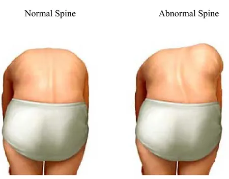
Scoliosis can be defined as an abnormal lateral curvature of the spine (as defined in Webster’s College Dictionary), and is also known as a three-dimensional deformity of the spine with lateral and vertebral rotation, where the Cobb angle is greater than 10 degrees as measured using the Cobb method on a standing radiography. Scoliosis can be classified as non-structural or structural.
The non-structural scoliosis occurs in a structurally normal spine with a lateral curve, developed as a secondary response to a problem occurring elsewhere in the body. This type of scoliosis can arise due to differing leg lengths or a pelvic tilt. These flexion deformities of the hip or knee are often referred to as postural or compensatory scoliosis. The spinal curvature in non-structural scoliosis can be resolved while the patient is seated or lying down. [11]
The structural scoliosis is the type where the spine has a lateral curve with a vertebral rotation. This form of scoliosis does not resolve when the patient lies down or sits upright and is the most known and familiar form. [12] Structural scoliosis is sub-classified to congenital, neurological, myopathic, traumatic and idiopathic. The congenital scoliosis refers to congenital vertebral anomalies occurring at birth and leads to a lateral curvature of the spine. The neurological and myopathic scoliosis are usually classified in a category called neuromuscular scoliosis. In this type of scoliosis, the spinal curvature is secondary to
a neurological or muscular disease, [13] like spina bifida and Duchenne muscular dystrophy. [12], [14] Idiopathic scoliosis is the most common form of spinal deformity for which the cause remains unknown. Idiopathic scoliosis is further classified according to the age of onset. The three subgroups of idiopathic scoliosis are infantile (1 day - 3 years), juvenile (4- 9 years) and adolescent (10-18 years). [15]
Adolescent idiopathic scoliosis (AIS)
AIS occurs during the time of puberty, when a child experiences a rapid growth. [15] AIS affects mainly females, both in number and severity, suggesting a possible contribution of estrogens.
According to the Terminology Committee of the Scoliosis Research Society (SRS) (1976) and Online Mendelian Inheritance in Man (OMIM 181800), AIS represents 80% of all observed spinal deformities, affecting 3-4% of the global pediatric population. [16], [17], [18], [19] The annual estimation report of the National Scoliosis Foundation demonstrated that AIS incurs more than 600,000 visits to physicians in North America; with 30,000 children requiring a brace and 38,000 who undergo a spinal fusion surgery. In the United States, the estimated cost of treating patients hospitalized with a diagnosis of idiopathic scoliosis in 2004 was over $3 billion dollars. [20], [21] According to the National Scoliosis Foundation statistics, there are six millions persons in the United States afflicted with scoliosis, and there is still no cure.
Diagnosis of AIS
In order to diagnose AIS correctly, it is important for doctors to determine the nature and symptoms of the deformity. An initial evaluation is performed, which includes physical examination, risk assessment based on familial history of the disease, as well as imaging techniques if deemed necessary. This evaluation enables the physicians to determine if the spinal deformity is primary or secondary to other diseases or disorders.
Physical examination
For the screening of scoliosis in schools, physicians, still today, use the Adam's forward bend test (figure 1). This physical examination is also used in the offices of pediatricians and primary care physicians. This test, which does not require any equipment whatsoever, has been used regularly since 1865 for the screening of spine curvature. Nevertheless, in 2005 the US Preventive Services Task Force determined that the test was ineffective for the detection of spinal curvatures and recommended against routine IS screening. [22]
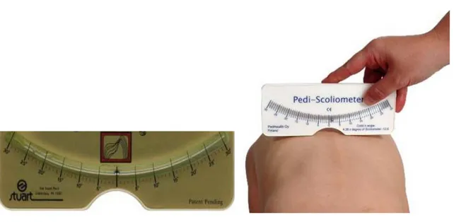
Another widely used method for the physical examination of scoliosis is the use of a scoliometer (figure 2). Using a scoliometer, a measurment of seven degrees equates to twenty degrees in Cobb angle [23], [24].
These routine tests are not sufficient for the proper diagnosis of idiopathic scoliosis and often give rise to false positives, which can lead to incorrect treatment, such as
unecessary bracing or referral to specialty care. For accurate diagnosis, an imaging test is required.
Figure 1: Adam's forward bend test, used for the evaluation of the spinal deformity (adapted from Reamy, et al). [25]
Figure 2: Scoliometer measures the spinal deformity. It is a simple instrument for the detection the spinal curvature (adapted from Franko, et al). [26]
Imaging test
The imaging technique is a more accurate and sensitive method for the detection of the disease, and allows the physicians to follow the progression of the spinal deformity. Additional tests are ordered, in the event of abnormal neurologic examinations, history of severe pain, or presence of rare left thoracic curves, to screen for other factors such as tumors. Additional tests may include X-rays, Computed Tomography (CT) scan or Magnetic Resonance Imaging (MRI). [27] The x- rays are an effective method to establish the severity of the disease and follow its progression by measuring the Cobb angle. However, special precautions need to be taken to limit radiological exposure to avoid the development of tumors.
F T c p v t d Figure 3: C The Cobb an curvature an Scoliscore Scoliscore is patients and spinal curve value of this to Caucasian
Treatmen
Two date, no pha and 25° are obb angle m ngle as show nd the lower s a recent ge then perform e progression s test remain n girls aged bnt of AIS
main treatm armacologica simply foll measurement wn above is t curvature (a enetic test de ming genetic n in AIS pati ns to be prove between 9 an ment options al treatment lowed by th . the intersecti adapted from eveloped for c analysis. B ients, curve p en, as well a nd 13 years exist for AI exists. Spin he orthopaed ion between m Greiner et the prognos By using 53 g progression as its clinical old. [29], [3 IS today: bra ne curvatures dic surgeons n the right an al). [28] sis of AIS us genetic mark can be predi l utility. Thi 30] acing and su s with Cobb s, referring t Upper Lower ngle of the up sing saliva fr kers associat icted. The po s test is also urgery (figur angles betw to this perio Cobb angle curvature r curvature pper rom the ted with ositive limited re 4). To ween 10° od as the estage of observation. Treatment of IS usually begins when the deformity has developed to 25° to 35°, using different types of braces. In cases where the spine continues to deform (≥ 45°), spinal surgery is the only remaining solution. [31], [11], [32], [33], [34] Alternatively, other treatments such as biofeedback, electric stimulation, physical therapy or chiropractic care have not been shown to alter the natural history of scoliosis. [35], [36] Despite many studies on its etiology, the cause of AIS remains unclear. Therefore, a better understanding of the etiopathogenesis of the disease will enable us to better diagnose, prognose and facilitate treatment.
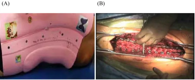
Figure 4: Scoliosis treatment by bracing and surgery.
The right panel (A) shows a brace which treats children with spinal deformities (25° to 35°) while the left panel (B) shows the surgery intervention to treat children with spinal deformities (≥ 45°) (adapted from Moe et al). [37]
Etiology of AIS
Even though widespread research hypotheses have been proposed, and many studies undertaken to examine the roles of the nervous system, environmental factors, spinal growth abnormalities, endocrine system, connective tissue abnormalities, hormonal factors and genetic factors in the etiology of the AIS, none of these studies really explain the cause of IS. [38], [39] [40], [41]
Many studies were undertaken to investigate the effects of the connective tissues on AIS. Researchers studied the collagen and the elastic fiber, the main structural elements of the spinal column. Others showed that proteoglycan and collagen contents are abnormal in the intervertebral disc of AIS patients, [42], [43] which raised controversy. [44] Another study suggested that the abnormalities of these elements are secondary to AIS and not the main cause. [45] Furthermore, elastic fiber abnormalities were observed in the skin and in the spinal ligaments of AIS. [46], [47] As a whole, most researchers postulated that these abnormalities are secondary to the spinal deformity. [48], [49]
Over the past years, the neurological abnormalities associated with AIS were investigated. It was thought that an abnormal sway pattern caused scoliosis but [50] the augmentation of the sway pattern now appears to be secondary to the scoliosis. [51] Machida and coworkers postulated that the development of the scoliosis in pinealectomized (PNX) chicken is due to somatosensory evoked potentials abnormalities. [52] On the other
hand, another group postulated that PNX leads to melatonin reduction, in turn affecting the nervous system maturation. [53]
Many studies were undertaken to assess the role of growth factors in AIS etiopathogenesis. These studies demonstrated that there is no difference between AIS and controls when growth hormone levels were increased. [54] Trontelj et al. demonstrated that the height of the vertebral bodies in girls is greater than in boys. [55] Further investigations are required to study the effects of these growth factors, hormones and their modulators.
Biomechanical factors were also studied in AIS. The mechanical properties of the spinal tissues, the way that the spine is supported and the forces which lead to abnormal loading may also contribute to scoliosis. However, researchers postulated that these factors too, are rather secondary to AIS. [56]
The similarity between the contractile systems of the platelets and the skeletal muscle led some researchers to postulate that the abnormalities of the contractile system could be involved in AIS etiology. The elevation of the platelet intracellular calcium and phosphorous in AIS patients was reported. [57] The elevation of calcium was observed in the dense bodies of platelets and they suggested that the defect of the calcium transport in the cell membrane or contractile complex could lead to spinal deformity. [57] Calmodulin, a well known calcium modulated protein, is a key player, which mediates calcium function and regulation in many processes in the eukaryotic organism. The calcium flux via the sarcoplasmic reticulum is mediated by the interaction of the calmodulin with (actin and
m p c t m m F T A l myosin) the et al. demon associated w platelet calm confirmed b the increase melatonin le melatonin co Figure 5: A This figure AIS. The f laboratory. contractile nstrated that with curve se modulin tha by Lowe in 2 e of the pla evels, which ould lead to AIS etiology summarizes factors in or system prot the augment everity. The an those exh 2002. [59] M atelet calmod lead to the b AIS. [60] the differen range repres eins of the p tation of the e patients wi hibiting a s Machida et a dulin levels belief that an nt axes of re sent topics platelets and platelet calm ith a progres stable curve al. also postu
in AIS pat ny change o esearch focu of research d the skeleta modulin leve ssive curve . [58] This ulated that th tients and t of the metabo using on the h investigate l muscle. Ki els in AIS pa had higher l demonstrat here is link the reduction olism or syn etiopathoge ed in Dr. M indsfater atients is levels of tion was between n of the nthesis of enesis of Moreau’s
Genetic Factors of AIS
Extensive research is being done on the role of the genetic factors in the development of AIS. Indeed some studies indicate a higher prevalence of AIS among the relatives of AIS patients when compared to the normal unaffected population. [61], [62],
] 63
[ , [64]
The main conclusion of most genetic studies (table 1) regarding AIS is its multi-factorial nature and possible crosstalk with environmental factors, which remain to be identified. However, for most, if not all of these studies, the number of patients and families needs to be increased and results replicated in distinct cohorts to confirm their validity and clinical utility.
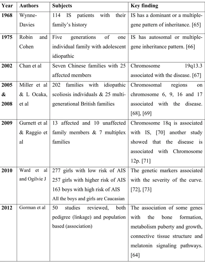
Table 1: Summary of genetic studies for AIS
Year Authors Subjects Key finding
1968 Wynne-Davies
114 IS patients with their family’s history
IS has a dominant or a multiple-gene pattern of inheritance. [65] 1975 Robin and
Cohen
Five generations of one individual family with adolescent idiopathic
IS has autosomal or multiple-gene inheritance pattern. [66]
2002 Chan et al Seven Chinese families with 25 affected members
Chromosome 19q13.3 associated with the disease. [67]
2005 & 2008 Miller et al & L Ocaka, et al
202 families with idiopathic scoliosis individuals & 25 multi-generational British families
Chromosomal regions on chromosome 6, 9, 16 and 17 associated with the disease. [68], [69]
2009 Gurnett et al & Raggio et al
13 affected and 10 unaffected family members & 7 multiplex families
Chromosome 18q is associated with IS, [70] another study showed that the disease is associated with Chromosome 12p. [71]
2010 Ward et al and Ogilvie J
277 girls with low risk of AIS 257 girls with higher risk of AIS 163 boys with high risk of AIS
All the boys and girls are Caucasian
The genetic markers associated with the severity of the curve. [72], [73]
2012 Gorman et al 50 studies reviewed, both pedigree (linkage) and population based (association)
The association of some genes with the bone formation, metabolism puberty and growth, connective tissue structure and melatonin signaling pathways. [64]
Animal models of AIS
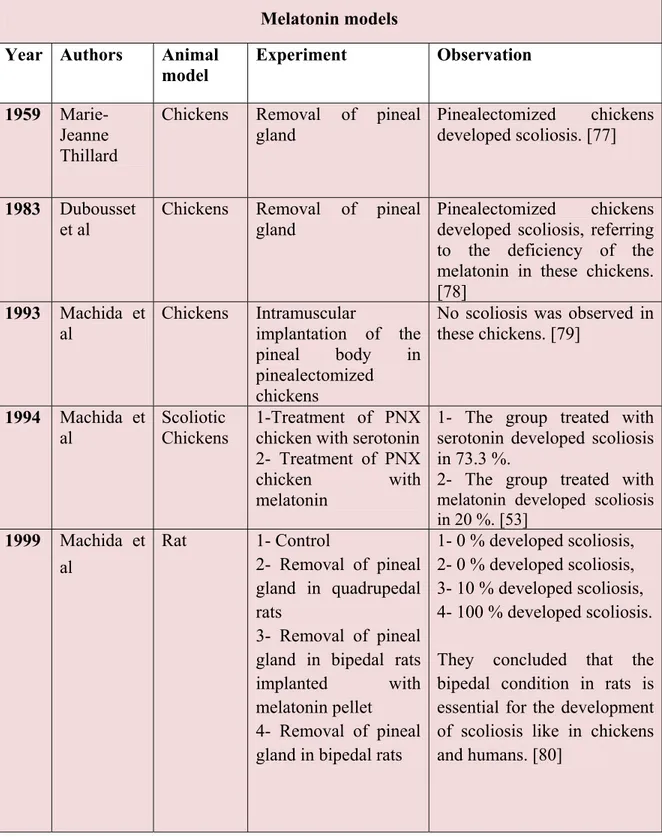
Animal models are important to enable us to understand many human diseases. These models help in the development of suitable treatments. The challenges are to find the suitable animal model representing the disease. In AIS, many animal models were investigated as shown in tables 2 and 3. These animal models were used to investigate many hypotheses like the role of melatonin, genetic, neurological and mechanical factors in AIS etiology. Most of the animal data collected supports the role of a melatonin deficiency as a cause of AIS. However the work of Cheung et al. [74] on monkeys raised controversy, suggesting that melatonin deficiency causes AIS only in lower animals. Gorman et al. demonstrated that guppies could be a relevant animal model for the study of scoliosis. In fact, this model is the only one that naturally develops scoliosis [75] while the curvature in all other scoliosis animal models is induced through surgical interventions. [2], [76]
Table 2: Summary of the surgical animal models for AIS Melatonin models Year Authors Animal
model
Experiment Observation
1959 Marie-Jeanne Thillard
Chickens Removal of pineal gland
Pinealectomized chickens developed scoliosis. [77]
1983 Dubousset et al
Chickens Removal of pineal gland
Pinealectomized chickens developed scoliosis, referring to the deficiency of the melatonin in these chickens. [78]
1993 Machida et
al Chickens Intramuscular implantation of the pineal body in pinealectomized
chickens
No scoliosis was observed in these chickens. [79] 1994 Machida et al Scoliotic Chickens 1-Treatment of PNX chicken with serotonin 2- Treatment of PNX
chicken with melatonin
1- The group treated with serotonin developed scoliosis in 73.3 %.
2- The group treated with melatonin developed scoliosis in 20 %. [53] 1999 Machida et al Rat 1- Control 2- Removal of pineal gland in quadrupedal rats 3- Removal of pineal gland in bipedal rats implanted with melatonin pellet
4- Removal of pineal gland in bipedal rats
1- 0 % developed scoliosis, 2- 0 % developed scoliosis, 3- 10 % developed scoliosis, 4- 100 % developed scoliosis. They concluded that the bipedal condition in rats is essential for the development of scoliosis like in chickens and humans. [80]
Year Authors Animal model
Experiment Observation 2001 Bagnall et
al
Chickens Study of the
relationship between melatonin levels and scoliosis in the chicken
The decrease in melatonin levels in young chickens led to scoliosis development.[81]
2005 Cheung et
al Monkeys Removal of pineal gland None of the pinealectomized (PNX) monkeys developed scoliosis. They concluded that the melatonin deficiency leads to scoliosis in the lower animals only and not in humans. [36] 2006 Machida et al and Oyama et al C57Bl/6j mice (naturally deficient in melatonin) Removal of the forelimbs of these mice (bipedal model)
Bipedal C57Bl/6j mice developed scoliosis; is considered a good model for the researchers who are working on scoliosis. [2], [82], [1]
2009 Fagan et al Chickens Removal of the pineal gland from one chicken group and used another group as a control without removal of pineal gland
75% of pinealectomized chickens developed scoliosis. Chickens developed scoliosis naturally in 19%. [83]
2011 Kono et al Chickens Removal of the pineal gland from two groups of chicken, one treated with melatonin
Deficiency of melatonin is suggested to lead to the development of scoliosis and osteoporosis. [84]
Year Authors Animal model
Experiment Observation
1989 Suk et al Rabbits Removal of the anterior and posterior rhizotomies
separately and together in different cohorts
They postulated that anterior root paralysis and posterior root paralysis may induce scoliosis. [85]
2009 Lambert et al Xenopus laevis (frog)
At the larva stage, removal of the
labyrinthine end organ
The young frog after the metamorphosis developed spinal curvature similar to AIS. [86]
Year Authors Animal
model Experiment Observation 1980 Beguiristain et al Pigs Epiphysiodesis in 4 to 5 consecutive vertebrae of neurocentral cartilage using cancellus screws
Pigs developed structural scoliosis. [87]
1991 Poussa et al Rabbits Use of an external splint to stimulate lordosis
- Developed lordosis in the thoracolumbar junction - 53 % developed scoliosis - Lordosis is a precondition for development of scoliosis. [88]
1997 Mente et al Rats Application of
external ring fixators to check the mechanical effect (asymmetrical load) on the spine -Vertebral wedging is formed
- Rats developed scoliosis. [89]
2009 Zhang et al Goat Tethering of the goats unilaterally by pedicle screws and contralateral rib resections
The goats developed scoliosis and the curvature increased over time and is similar to the idiopathic deformity. [90]
Mechanical models Neurological models Table 2 cont’d
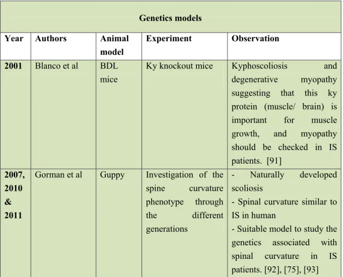
Table 3: Genetic models for AIS
Year Authors Animal
model
Experiment Observation 2001 Blanco et al BDL
mice
Ky knockout mice Kyphoscoliosis and
degenerative myopathy suggesting that this ky protein (muscle/ brain) is important for muscle growth, and myopathy should be checked in IS patients. [91] 2007, 2010 & 2011
Gorman et al Guppy Investigation of the spine curvature phenotype through the different generations - Naturally developed scoliosis
- Spinal curvature similar to IS in human
- Suitable model to study the genetics associated with spinal curvature in IS patients. [92], [75], [93]
Clinical implication of melatonin deficiency
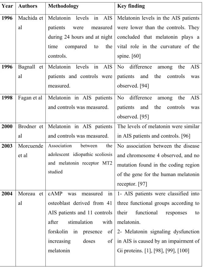
Despite the fact that most of the animal data pointed to a crucial role of melatonin deficiency in spinal deformity formation, the majority of the clinical results (table 4) clearly demonstrated that melatonin levels were normal in AIS patients.
Table 4: Clinical data representing the role of melatonin in scoliosis Year Authors Methodology Key finding 1996 Machida et
al
Melatonin levels in AIS patients were measured during 24 hours and at night time compared to the controls.
Melatonin levels in the AIS patients were lower than the controls. They concluded that melatonin plays a vital role in the curvature of the spine. [60]
1996 Bagnall et al
Melatonin levels in AIS patients and controls were measured.
No difference among the AIS patients and the controls was observed. [94]
1998 Fagan et al Melatonin in AIS patients and controls was measured.
No difference among the AIS patients and the controls was observed. [95]
2000 Brodner et al
Melatonin in AIS patients and controls was measured.
The levels of melatonin were similar in AIS patients and controls. [96] 2003 Morcuende
et al
Association between the adolescent idiopathic scoliosis and melatonin receptor MT2 studied
No association between the disease and chromosome 4 observed, and no mutation found in the coding region of the gene for the human melatonin receptor. [97]
2004 Moreau et al
cAMP was measured in osteoblast derived from 41 AIS patients and 11 controls after stimulation with forskolin in presence of increasing doses of melatonin
1- AIS patients were classified into three functional groups according to their functional responses to melatonin.
2- Melatonin signaling dysfunction in AIS is caused by an impairment of Gi proteins. [1], [98], [99], [100]
Table 4 cont’d
Year Authors Methodology Key finding 2007 Suh et al Metabolism of the pineal
gland in the AIS patients in comparison with the control subjects studied
The pineal gland metabolism was normal in AIS patients. [101]
2007 Qui et al Verification of the melatonin receptor in AIS patients
Melatonin receptor MT1 was not associated with AIS occurrence but MT2 was. Expression of the MT2 receptor expressed asymmetrically in the AIS patients. [102], [103], [104] 2007 Azeddine
et al
cAMP measured as a secondary messenger in 90 AIS patients and 10 controls
They confirmed what Moreau et al concluded in 2004. [105]
2010 Akoume et al
A new technique called cellular dielectric spectroscopy (CDS) was used to verify the melatonin signaling in the AIS patients
Moreau et al. results confirmed. AIS patients were classified into three different functional groups. Gi signaling defect found. [106]
2013 Yim et al Expression of melatonin receptors MT1& MT2 verified.
MT2 expression shown to be low in AIS girls. This abnormal expression shown to be associated to an abnormal systemic skeletal growth (abnormal arm span). [107]
Melatonin signaling in AIS
Melatonin is a neuro-hormone synthesized mainly by the pineal gland in the brain. [108], [109] Its secretion is stimulated by the dark and inhibited by the light, and is an important regulator of the circadian rhythm. Melatonin is a highly important antioxidant molecule [110], [111] and its biological effects are through its receptors. In humans and other mammals, melatonin classically binds to two G protein-coupled receptors (GPCR) called melatonin receptor 1 (MT1 or MTNR1A or Mel1A) and melatonin receptor 2 (MT2
or MTNR2B or Mel2B), whereas it binds to Xenopus melatonin receptors in
non-mammalian species. Melatonin acts through the activation of Gi protein coupled receptors
which inhibits the adenylate cyclase activity and stops the conversion of adenosine triphosphate (ATP) to cyclic adenosine monophosphate (cAMP). [112], [113], [114] Chan et al. showed that in MCF-7 cells melatonin stimulates the Gs and increases the cAMP accumulation, suggesting a coupling between MT1 or MT2 and Gs, activating adenylate cyclase. [115] Melatonin receptor 1 (MT1) also binds to Gq protein, which activates
phospholipase C (PLC), which in turn converts the phosphatidylinositol 4, 5-bisphosphate PIP2 into secondary messengers, IP3 and diacylglycerol (DAG) (figure 6). [116], [117]
F P c r m c Figure 6: M al) [113] H PLC=phosph cyclase, DA Our and involves responded d melatonin, t cellular diel Extracellula Plasma mem Intracellula Melatonin sig HMTR = holipase c, AG= Diacylg team demon s a different differently w the level of lectric spectr ar mbrane ar gnaling throu human mel PKC= prote lycerol, IP3= nstrated that tial impairme when their os cAMP was roscopy (CD ugh Gi, Gs a latonin rece ein kinase C = Inositol tri melatonin s ent of Gi pro steoblasts w higher when DS) techniqu and Gq prot eptor (MT1 C, PKA= pro isphosphate signaling dys oteins (figur were treated w n compared ue led our t eins (adapte 1 & MT2) otein kinase & PLC= Ph sfunction oc re 7). Additi with differe to normal c team to the ed from Slom ), Mel= me A, AC= A hospholipase ccurs in AIS ionally, AIS nt concentra cells. The us same result minski et elatonin, Adenylate e C patients patients ations of se of the ts. [106]
T F M H A L i F I This finding according to Figure 7: M Melatonin b However in ATP to cAM In os Letellier et a and in the p improvemen FG3 subgro estradiol (10 Extracellula Plasma mem Intracellular g led to th o the variabil Melatonin sig binds to eith AIS patient MP, culminat steoblasts de al. showed t presence of nt in Gi-sign oups exhibite 0-10 M). [118 ar mbrane r e classificat lity of their c gnaling dysfu her melaton s Gαi protein ting in the in erived from that a prefer 17-β-estradi naling. Howe ed a more p 8] tion of AIS cellular resp unction in A nin receptor nsdo not inh ncrease of cA AIS patients rential coupl iol (10-10M) ever, osteobl profound Gi S patients in ponse. [16], [ AIS 1 (MT1) o hibit adenyla AMP in AIS s classified a ling of MT2 such coupli lasts from A i signaling d nto three fu [106] or melatonin ate cyclases S patients. as functiona 2 receptor w ing was red AIS patients defect in th unctional su n receptor 2 or the conve al subgroup with Gs alpha duced, sugge classified as e presence ubgroups (MT2). ersion of 1 (FG1), a protein esting an s FG2 or of
17-β-G proteins
G proteins represent small guanine nucleotide-binding proteins that play an important role in signal transduction of many membrane receptors termed G protein coupled receptor (GPCR). G proteins act as molecular switches, transferring the signal from the outside of the cell to the inside. The signal is transferred to the cell through several effectors such as enzymes, peptides, hormones or channel ions. G proteins are involved in many physiological processes and their malfunction has been found in many pathologies. [119] The G proteins are heterotrimeric proteins which consist of three subunits called alpha (α), beta (β) and gamma (γ). [120] The G proteins are classified according to their α subunit: Gαi/o, Gαq, Gα12 and Gαs. There are 16 genes which encode α subunits, 5 genes
encode β subunits and 12 genes encode γ subunits in humans. Each alpha subunit binds with one beta and one gamma subunit. [121], [122] This classification has gone under review following the discovery in recent years of the roles of the βγ subunits. [123]
The largest subunit is the α subunit (33-55 kDa), which forms a complex with the β and γ subunits when it is linked to a guanosine diphosphate (GDP). This is called the inactive form of G proteins. Once the GDP converts to guanosine triphosphate (GTP), the α subunit dissociates from β and γ subunits and the G protein becomes active. It is in the GDP form that it is recognized by the proper receptor, which is activated by specific effectors and this receptor is called G protein coupled receptor (GPCR). During the interaction of the GPCR and the G protein, GDP is dissociated from the α subunit and is replaced by GTP, resulting in a conformational change and dissociation of the α subunit
from the βγ subunits. It is at this moment that the α and βγ subunits are able to bind to the effector proteins and control their respective functions. The alpha subunits have GTPase activity which regress the G protein to the inactive form. [124], [125], [126], [127], [125]
G proteins play a key role in many biological processes. The impairment of G protein signaling is associated with several diseases. Albright’s hereditary osteodystrophy (Pseudohypoparathyroidism type 1) is an example and is associated with low serum calcium, high serum phosphate, high parathyroid hormone (PTH), bone abnormalities and mental retardation, due to the lack of response to the parathyroid hormone. This disease is caused by a mutation in Gαs protein leading to a loss of function. [128], [129] Conversely,
the McCune–Albright syndrome, which is characterized by premature puberty as well as bone and skin pigmentation abnormalities, is caused by an activating mutation of the α subunit of Gs proteins. [130] Gαi2 deficient mice were used as the animal model to study
inflammatory bowel disease because they developed similar symptoms, and thus led to the discovery of pharmaceutical treatments. [131] There are two bacterial toxins impeding the G-protein inactivation-activation cycle and these toxins are associated with severe diseases. These toxins play a crucial role in understanding of G-protein signaling. Pertussis toxin (PTX) affects the respiratory system and causes severe coughing spells ending with Whooping cough. PTX catalyzes the ribosylation of adenosine diphosphate (ADP) of Gi proteins not Gs or Gq, and hinders the coupling of the GiPCR to the Gi proteins. Conversely, cholera toxin blocks the GTPase activity of Gs α- subunit through its internal ADP-ribosylation for the arginine receptors and Gs protein becomes constitutively
activated causing severe dehydration and diarrhea. [119], [132], [133] These toxins help researchers to study the specificity of agonists to G protein coupled receptors and their cognate G proteins.
Osteopontin (OPN) and AIS
Osteopontin (OPN) is also known as secreted phosphoprotein 1 (SPP1), bone sialoprotein I (BSP-1 or BNSP) and early T-lymphocyte activation (ETA-1). [134], [135], [136], [137]
The presence of OPN in skeletal muscle, proprioceptive sensory organs, postural control centers such as the cerebellum, and inner ear structures are of particular interest, since AIS patients also exhibit defects in postural control, proprioception and equilibrium. [138], [139], [140]
OPN is an extracellular matrix protein with high negative charges. It is composed of approximately 300 amino acid residues. Among the mammalian species, OPN cDNA exhibits a high degree of homology. OPN contains different binding sites, which includes a polyaspartic acid motif to be able to bind to the hydroxyapatite and the calcium channel. [141], [142] It includes two heparin binding domains and contains an arginine-glycine-aspartic acid (RGD) motif. It also has sites for N – O glycosylation in addition to the serine – threonine phosphorylation sites. These sites generate functional forms of OPN in different tissues. Alternatively, OPN binds to different receptors like integrin beta 1 (β1),
integrin beta 3 (β3), integrin beta 5 (β5) and CD44 isoforms. This diversity of interactions gives OPN different functions in different tissues.
OPN is expressed in different tissues like cartilage, dentin, cementum, skeletal muscle, sensory organ, cerebellum, inner ear, sweat and salivary gland, the pancreatic and bile ducts, the activated lymphocytes and macrophages, the gut and the kidney. [143], [144] OPN is expressed in the bone through osteoblast, osteocyte and osteoclast cells. [145] OPN is expressed by fibroblastic cells in the wound healing sites and the connective tissues. The secreted protein was observed in the biological fluids like blood, breast milk, urine and the seminal fluid. [143], [146], [147], [148], [149] OPN expression is affected by many factors such as cytokines, hormones and growth factors, factors which affect OPN transcription, translation and its posttranslational modifications. The regulation of OPN expression is not completely understood. Vitamin D is known to stimulate OPN expression in bone cells. Conversely, Vitamin D deficient mice have low OPN mRNA expression. Platelet derived growth factor (PDGF), transforming growth factor βs (TGF-βs), inflammatory cytokines, bone morphogenetic proteins (BMPs), angiogensin II and epidermal growth factor (EGF) are factors that up-regulate OPN expression. [144] The tumor necrosis factor α (TNF α) and interleukin-1β (IL-1β) are strong stimulators of OPN expression. On the other hand, OPN is down-regulated by bisphosphonates in the bone and by cGMP dependent protein kinase in the vascular smooth muscle cells. [142], [141]
Many studies have been undertaken to better understand the biological functions of OPN. OPN plays an important role in calcification, development, bone remodeling, immunoprotection and skeletal tissue homeostasis. OPN protects the cells from apoptosis and stimulates the proliferation and the survival of cells, especially smooth muscle and epithelial cells. This was shown by inducing apoptosis, by serum deprivation, in endothelial cells incubated in a plate coated with OPN. [150], [151], [152], [153]
There are studies that show that OPN plays a crucial role in bone remodeling, a process in which osteoclasts remove old bone and osteoblasts form new bone. Both osteoblasts and osteoclasts express OPN and it is found in cement lines (bone formation sites) and at the bone surface. The migration of the osteoclasts is stimulated by OPN through its receptors, CD44 and integrin αvβ3, aside from the stimulation of CD44 expression, which is important for motility. [154], [155]
OPN protects endothelial cells through the activation of nuclear factor kappa B (NF-kB) when it binds with integrin alpha v beta 3 (integrin αvβ3) and this stimulates osteoprotegerin (OPG). OPG blocks apoptosis when it forms a complex with the TNF-related apoptosis-inducing ligand (TRAIL). [156], [157] Non-adherent cells on the other hand, are protected against apoptosis by OPN through the CD44 receptor and the activation of the PI3K-Akt signaling pathway. [158] Taken together, we deduce that OPN can protect different types of the cells from apoptosis through different receptors and different pathways.
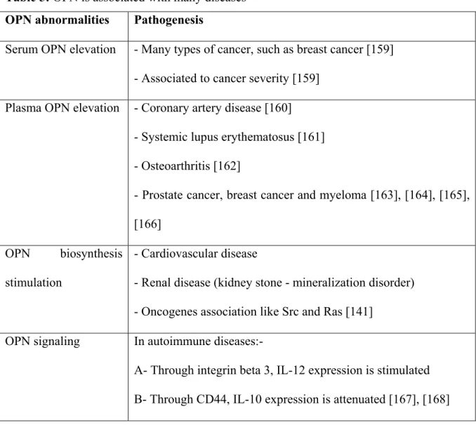
OPN is a multifunctional cytokine and is involved in many adult pathological conditions. [144], [141] Levels of OPN in the blood, OPN biosynthesis and OPN signaling are associated with different diseases. (Table 5)
Table 5: OPN is associated with many diseases OPN abnormalities Pathogenesis
Serum OPN elevation - Many types of cancer, such as breast cancer [159] - Associated to cancer severity [159]
Plasma OPN elevation - Coronary artery disease [160] - Systemic lupus erythematosus [161] - Osteoarthritis [162]
- Prostate cancer, breast cancer and myeloma [163], [164], [165], [166]
OPN biosynthesis stimulation
- Cardiovascular disease
- Renal disease (kidney stone - mineralization disorder) - Oncogenes association like Src and Ras [141]
OPN signaling In autoimmune diseases:-
A- Through integrin beta 3, IL-12 expression is stimulated B- Through CD44, IL-10 expression is attenuated [167], [168]
OPN receptors
OPN exhibits different functions through its interactions with distinct receptors. This can be explained by the fact that OPN binds to its receptors through different binding sites. These receptors are mainly CD44 and integrin receptors.
CD44 is the receptor for hyaluronic acid and also binds to OPN. CD44 is expressed in different types of the cells like osteocytes, fibroblasts, osteoblasts, epithelial and endothelial cells and smooth muscle cells. [142] There are many types of cancer like glioma, prostate, leukemia initiating cells and breast cancer that express the CD44 receptor. This receptor was shown to play a role in the stimulation of the invasion and metastasis in different types of cancer. [169], [170]
Integrins are other known receptors for OPN. They are transmembrane heterodimeric receptors which contain alpha (α) and beta (β) subunits. Integrins play a role in cell adhesion, allowing the cell to attach to the extracellular matrix (ECM), as well as a role in cell signaling. [171], [172]
The integrins have 18 α subunits and 8 β subunits, which associate together, to give no less than 24 heterodimers. [173] These heterodimers play a role in biological processes. One of the most widely expressed integrins is integrin β1 which has 12 complexes and invokes the binding of the cell with the matrix protein. [173] OPN and integrins can protect the cells from programmed cell death (apoptosis) and this was observed in solid cancers, for instance breast cancer. It was observed that integrin β1 inhibits apoptosis of cancer cells
normally induced by vincristine and paclitaxel, used as chemotherapy by stimulating apoptosis, leading to a drug resistance. [174] Other studies found that high expression of integrin β1 is associated with poor prognosis due to drug resistance in small cell lung cancer. [175], [176]
The integrin receptors play important roles in OPN-mediated cell adhesion through receptors like integrin beta 1, integrin beta 3 and integrin beta 5. [177], [178], [179] Some of these integrin receptors bind to OPN through the RGD motif like αvβ1, αvβ3,αvβ5 α5β1, and α8β1 but there are other integrin receptors interacting with OPN through a different amino acid sequence, like α4β1 and α9β1. CD44 binds to OPN in a RGD independent manner. [180], [181], [182], [183], [184], [185], [186], [187]
The activation of the integrins requires an interaction with talin. Talin has the ability to bind to the cytoplasmic tail of the integrins leading to a conformational change of the latter by the separation of the heterodimer subunits of the cytoplasmic tails. This conformational change of the integrins allows their binding to cognate agonists. [188] Talin needs to be phosphorylated to bind and activate the integrins. The phosphorylation of the talin needs serine/ threonine kinases to be phosphorylated, and this process is regulated by protein tyrosine phosphatases (PTPases). [189], [190]
Protein tyrosine phosphatases (PTPases)
Protein tyrosine phosphatases (PTPases) are enzymes removing the phosphate group from phosphorylated tyrosine residues. PTPases play a vital role in the regulation of the signal transduction, which controls many biological processes like differentiation, proliferation and apoptosis. [191] The human genome encodes approximately 112 PTPases. The specific function of these phosphatases is dependent on their substrate and their catalytic domain. [192]
PTPases are classified according to their function, which mainly removes the phosphate from the tyrosine residues. However, other PTPases target the serine and threonine residues, and thus these PTPases have a dual function. PTPases work on the phospholipids as well as phosphoproteins. [193]
The PTPases are further classified according to their localization: Non receptor PTPases, which are intracellular, and Receptor-like PTPases, which are transmembranous and contain PTPase domains, have an extracellular domain, a transmembrane region and catalytic cytoplasmic domain. In some cases, the extracellular domains of the receptor PTPases contain MAM domains, immunoglobulin-like domains and fibronectin type III (FN-III) repeats. While the cytoplasmic region contains two copies of the PTPase domain, only one has enzymatic activity while the other remains inactive. [194], [195]
PTPases and their roles in the pathogenesis
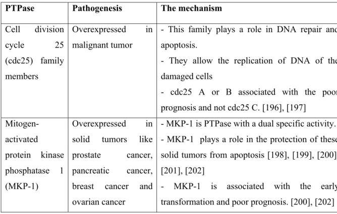
Once the identification and classification based on function and location is defined, it is necessary to discuss the association between PTPases and the pathogenesis (table 6).
Table 6: PTPases and their roles in diseases
PTPase Pathogenesis The mechanism Cell division cycle 25 (cdc25) family members Overexpressed in malignant tumor
- This family plays a role in DNA repair and apoptosis.
- They allow the replication of DNA of the damaged cells
- cdc25 A or B associated with the poor prognosis and not cdc25 C. [196], [197]
Mitogen-activated protein kinase phosphatase 1 (MKP-1) Overexpressed in solid tumors like prostate cancer, pancreatic cancer, breast cancer and ovarian cancer
- MKP-1 is PTPase with a dual specific activity. - MKP-1 plays a role in the protection of these solid tumors from apoptosis [198], [199], [200], [201], [202]
- MKP-1 is associated with the early transformation and poor prognosis. [200], [202]
The knockdown of the MKP-1 expression led to the reduction of the tumorigenicity in mice, the increase of programmed cell death and the decrease of cell growth in the cancer cells. [203], [204]
Src-homology 2 domain–containing PTPase 1 (SHP-1) is an example of a PTPase acting as a negative regulator. SHP-1 is a negative regulator for immune cell activation and cytokine signaling. [205], [206]
Hence, Protein tyrosine phosphatase mu (PTPµ) plays a crucial role in AIS; in the following section I will describe its structure and functions.
Protein tyrosine phosphatase mu (PTPµ): structure, function and role in diseases. In 1991 Gebbink et al, isolated 5.4 kb of mouse cDNA, which encodes for a protein called receptor protein tyrosine phosphatase mu (RPTPµ), which belong to the receptor-like protein tyrosine phosphatases family. RPTPµ has an extracellular region [contains a meprin-A5 antigen-PTP mu (MAM) domain, an Ig-like domain and four fibronectin type III-like repeats], a single transmembrane region, a long juxtamembrane domain and two tyrosine phosphatase domains, D1 and D2 (D1 is catalytically active, while D2 is not) (figure 8). Then in 1993, Suijkerbuijk RF et al. mapped the human receptor-like protein tyrosine phosphatase gene (PTPRM) to 18p11.2 using fluorescence in situ hybridization. [207], [208]
F R r p j c i c c j Figure 8: PT RPTPµ has an Ig-like d region, a lon PTPµ proteins are junctions. T catenins (al inhibits the cadherins-ca cadherin and junctions. [2 Extracellula Intracellula Plasma mem TPµ structur an extracell domain and ng juxtamem µ-cadherins-e important The stabiliza lpha, beta a growth of t atenins com d p120 cate 209], [210], [ ar ar mbrane re. lular region d four fibron mbrane doma -catenins are in the sta ation of the and p120) w
the actin, thu mplex which nin. PTPµ i [211], [212]
[contains a nectin type ain and two t
e proteins a abilization o cell-cell jun which then us keeping t h interacts is thus impo meprin-A5 III-like rep tyrosine pho associated w of cell-cell nctions occu binds to ac the cells fix and/or dep ortant in the
antigen-PTP peats], a sin osphatase dom
with cell adh junctions a urs through ctin cytoske xed. PTPµ is phosphoryla cell-cell ad Pµ (MAM) ngle transme mains (D1 a hesion. The c and called a the interacti eleton. This s a regulator ates E-cadhe dhesion and domain, embrane and D2). cadherin adherens ion with process r for the erin, N-cell-cell
It was demonstrated that PTPµ regulates integrins through the dephosphorylation of the phosphatidylinositol-phosphate kinase type 1 γ (PIPK1γ). The integrin needs a phosphorylated talin to be activated, and PIPK1γ plays this role. PIPK1γ needs to be phosphorylated to be activated and this occurs in order to stimulate the formation of adherens junctions (AJs). Once integrin completes its role, PTPµ dephosphorylates PIPK1γ, inactivating it and indirectly inactivating β integrins. [189] [213], [190] Another group demonstrated that β integrins regulate the expression of PTPµ and cell-cell adhesion. [214]
PTPµ also plays a role in the regulation of the neurite outgrowth. It was shown that the presence of PTPµ as a substrate in the dish led to neurite outgrowth stimulation. Also, the blocking of PTPµ in the developing retina affects the development of the neural retina. The dephosphrylation of the tyrosine residues, which is required for both temporal neurite repulsion and mediation of nasal neurite outgrowth, needs PTPµ. This tyrosine residue dephosphrylation is achieved through the main elements of the cadherin-catenin complex and this regulates the axonal migration. [215], [216], [217], [218], [219] Several related proteins were discovered such as protein kinase C delta (PKCδ), phospholipase C gamma 1(PLCγ1) and others. The first two proteins were found to be important substrates for PTPµ and which participate in the regulation of neurite outgrowth and cell migration. [220], [221] PTPµ downregualtion was observed in the Glioblastoma multiform (GBM) and interestingly, the expression level of PTPµ diminished in breast cancer. It is also noteworthy, that PTPµ is important for cell invasion and migration through the control of ERK and JNK tyrosine phosphorylation. [222], [223]
GENERAL OBJECTIVE
A series of studies from our laboratory initially demonstrated the occurrence of a melatonin signaling dysfunction in osteoblasts from progressive AIS patients, due to a hyperphosphorylation of Gi proteins. The differential degree of this defect among AIS patients allowed their classification into three functional subgroups. In addition, our studies have demonstrated that this defect occurs before the development of the spinal deformity. In parallel, analysis of plasma of AIS patients has revealed high levels of circulating OPN in both non-progressive and progressive AIS patients, as well as in asymptomatic children with a family history of AIS. Our general objective is to examine whether there is a cause-and-effect relationship between these findings in order to gain more insights into the potential pathomechanisms leading to the development and progression of AIS.
Key issues
The pharmacological aspect of melatonin is not simple. Indeed, melatonin has been demonstrated to regulate and influence a variety of biological functions by initiating signal transduction through two high affinity receptors coupled to G proteins from the Gi and Gq families. [224], [225] Our demonstration that Gi protein-mediated melatonin signaling is reduced at different degrees among AIS patients is suggestive of a disparity in melatonin signaling pathways. This signaling disparity does not seem to be related to melatonin receptors, [97], [16] but rather lies at the Gi protein level. Indeed, we have attributed the reduced melatonin signaling to the phosphorylation of Gi proteins by demonstrating an increased phosphorylation of serine residues through the three Gi1, Gi2 and Gi3 isoforms in