EVALUATION OF MITOCHONDRIAL FUNCTION IN A MODEL OF DEVELOPMENTAL PROGRAMMING OF HYPERTENSION ASSOCIATED WITH TRANSIENT NEONATAL OXYGEN EXPOSURE
par Zachary Anstey
Physiologie Médecine
Mémoire présenté à la Faculté des études supérieures en vue de l’obtention du grade de Maîtrise (MSc)
en Physiologie
Août 2012
Faculté des études supérieures et postdoctorales
Ce mémoire intitulé :
EVALUATION OF MITOCHONDRIAL FUNCTION IN A MODEL OF DEVELOPMENTAL PROGRAMMING OF HYPERTENSION ASSOCIATED WITH TRANSIENT NEONATAL OXYGEN EXPOSURE
Présenté par : Zachary Anstey
a été évalué par un jury composé des personnes suivantes :
Dr. Yan Burelle, président-rapporteur Dr. Anne Monique Nuyt, directeur de recherche
RÉSUMÉ
UNE EXPOSITION NÉONATALE À L’OXYGÈNE MÈNE À DES MODIFICATIONS DE LA FONCTION MITOCHONDRIALE CHEZ LE RAT ADULTE
Introduction: L’exposition à l’oxygène (O2) des ratons nouveau-nés a des conséquences à l’âge adulte dont une hypertension artérielle (HTA), une dysfonction vasculaire, une néphropénie et des indices de stress oxydant. En considérant que les reins sont encore en développement actif lors des premiers jours après la naissance chez les rats, jouent un rôle clé dans le développement de l’hypertension et qu’une dysfonction mitochondriale est associé à une augmentation du stress oxydant, nous postulons que les conditions délétères néonatales peuvent avoir un impact significatif au niveau rénal sur la modulation de l’expression de protéines clés du fonctionnement mitochondrial et une production mitochondriale excessive d’espèces réactives de l’ O2.
Méthodes: Des ratons Sprague-Dawley sont exposés à 80% d’O2 (H) ou 21% O2 (Ctrl) du 3e au 10e jr de vie. En considérant que plusieurs organes des rats sont encore en développement actif à la naissance, ces rongeurs sont un modèle reconnu pour étudier les complications d’une hyperoxie néonatale, comme celles liées à une naissance prématurée chez l’homme. À 4 et à 16 semaines, les reins sont prélevés et les mitochondries sont extraites suivant une méthode d’extraction standard, avec un tampon contenant du sucrose 0.32 M et différentes centrifugations. L’expression des protéines mitochondriales a été mesurée par Western blot, tandis que la production d’ H202 et les activités des enzymes clés du cycle de Krebs ont été évaluées par spectrophotométrie. Les résultats sont exprimés par la moyenne ± SD.
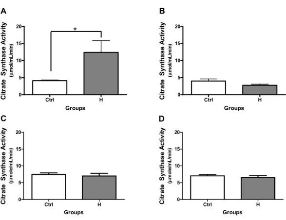
Résultats: Les rats mâles H de 16 semaines (n=6) présentent une activité de citrate synthase (considéré standard interne de l’expression protéique et de l’abondance mitochondriales) augmentée (12.4 ± 8.4 vs 4.1 ± 0.5 μmole/mL/min), une diminution de l’activité d’aconitase (enzyme sensible au redox mitochondrial) (0.11 ± 0.05 vs 0.20 ± 0.04 μmoles/min/mg mitochondrie), ainsi qu’une augmentation dans la production de H202 (7.0 ± 1.3 vs 5.4 ± 0.8 ρmoles/mg protéines mitochondriales) comparativement au groupe Ctrl (n=6 mâles et 4 femelles). Le groupe H (vs Ctrl) présente également une diminution dans l’expression de peroxiredoxin-3
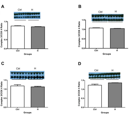
(Prx3) (H 0.61±0.06 vs. Ctrl 0.78±0.02 unité relative, -23%; p<0.05), une protéine impliquée dans l’élimination d’ H202, de l’expression du cytochrome C oxidase (Complexe IV) (H 1.02±0.04 vs. Ctrl 1.20±0.02 unité relative, -15%; p<0.05), une protéine de la chaine de respiration mitochondriale, tandis que l’expression de la protéine de découplage (uncoupling protein)-2 (UCP2), impliquée dans la dispersion du gradient proton, est significativement augmentée (H 1.05±0.02 vs. Ctrl 0.90±0.03 unité relative, +17%; p<0.05). Les femelles H (n=6) (vs Ctrl, n=6) de 16 semaines démontrent une augmentation significative de l’activité de l’aconitase (0.33±0.03 vs 0.17±0.02 μmoles/min/mg mitochondrie), de l’expression de l’ATP synthase sous unité β (H 0.73±0.02 vs. Ctrl 0.59±0.02 unité relative, +25%; p<0.05) et de l’expression de MnSOD (H 0.89±0.02 vs. Ctrl 0.74±0.03 unité relative, +20%; p<0.05) (superoxide dismutase mitochondriale, important antioxidant), tandis que l’expression de Prx3 est significativement réduite (H 1.1±0.07 vs. Ctrl 0.85±0.01 unité relative, -24%; p<0.05). À 4 semaines, les mâles H (vs Ctrl) présentent une augmentation significative de l’expression de Prx3 (H 0.72±0.03 vs. Ctrl 0.56±0.04 unité relative, +31%; p<0.05) et les femelles présentent une augmentation significative de l’expression d’UCP2 (H 1.22±0.05 vs. Ctrl 1.03±0.04 unité relative, +18%; p<0.05) et de l’expression de MnSOD (H 1.36±0.01 vs. 1.19±0.06 unité relative, +14%; p<0.05).
Conclusions: Une exposition néonatale à l’O2 chez le rat adulte mène à des indices de dysfonction mitochondriale dans les reins adultes, associée à une augmentation dans la production d’espèces réactives de l’oxygène, suggérant que ces modifications mitochondriales pourraient jouer un rôle dans l’hypertension artérielle et d’un stress oxydant, et par conséquent, être un facteur possible dans la progression vers des maladies cardiovasculaires.
ABSTRACT
EVALUATION OF MITOCHONDRIAL FUNCTION IN A MODEL OF DEVELOPMENTAL PROGRAMMING OF HYPERTENSION ASSOCIATED WITH TRANSIENT NEONATAL OXYGEN EXPOSURE
Introduction: Rats exposed to oxygen (O2) as newborns suffer complications in adulthood, including: arterial hypertension, vascular dysfunction, nephropenia and indices of oxidative stress. Although the rats are born at term, their organ development is equivalent to that of a preterm fetus, allowing organs of interest such as the kidney to be compared to premature infants. Given that impaired nephrogenesis or reduced nephron numbers has been shown to promote the development of hypertension and mitochondrial dysfunction is associated with increased oxidative stress, we hypothesised that exposure to high oxygen concentrations in the neonatal period would significantly impact the expression and activity of key proteins involved in renal mitochondrial function and lead to an excessive production of reactive oxygen species by the mitochondria.
Methods: Sprague-Dawley rat pups were exposed to 80% O2 (Hyperoxic (H) group; O2 exposed) or 21% O2 (Control (Ctrl) group) from day 3 to day 10 of life. At 4 and 16 weeks of age, kidneys were rapidly excised and the mitochondria isolated following a standard protocol; with a buffer containing 0.32 M sucrose and differential centrifugations. Expression of mitochondrial proteins was assessed by Western blot, whereas the release of hydrogen peroxide (H202), activities of key citric acid cycle enzymes and mitochondrial swelling were assessed by spectrophotometry. Results are expressed as the means ± SE. Both male and female offspring were studied.
Results: In male H rats at 16 weeks of age (n=6), citrate synthase activity (internal standard and measure of relative mitochondrial abundance) was significantly increased (12.4 ± 8.4 vs 4.1 ± 0.5 μmole/mL/min), whereas aconitase activity (sensitive to ROS) was significantly decreased (0.11 ± 0.05 vs 0.20 ± 0.04 μmoles/min/mg mitochondria) and H202 release was significantly increased (7.0 ± 1.3 vs 5.4 ± 0.8 ρmoles/mg mitochondrial protein) compared to the controls (Ctrl, n=6 males and 4 females). The H group (vs Ctrl) also demonstrated a reduction in the expression of
peroxiredoxin-3 (Prx3) (H 0.61±0.06 vs. Ctrl 0.78±0.02 relative units, -23%; p<0.05), a protein involved in the elimination of H202 and in the expression of cytochrome C oxidase (Complex IV) (H 1.02±0.04 vs. Ctrl 1.20±0.02 relative units, -15%; p<0.05), a protein in the mitochondrial respiratory chain, whereas the expression of uncoupling protein-2 (UCP2), a protein involved in dissipating the proton gradient, was significantly increased (H 1.05±0.02 vs. Ctrl 0.90±0.03 relative units, +17%; p<0.05). Female H rats (n=6) (vs Ctrl, n=6) at 16 weeks of age demonstrated a significant increase in aconitase activity (0.33±0.03 vs 0.17±0.02 μmoles/min/mg mitochondria), in the expression of ATP synthase β subunit (H 0.73±0.02 vs. Ctrl 0.59±0.02 relative units, +25%; p<0.05) (involved in ATP production) and in the expression of MnSOD (H 0.89±0.02 vs. Ctrl 0.74±0.03 relative units, +20%; p<0.05) (mitochondrial antioxidant involved in scavenging superoxide), whereas Prx3 expression was significantly reduced (H 1.1±0.07 vs. Ctrl 0.85±0.01 relative units, -24%; p<0.05 ). In male H rats (vs Ctrl) at 4 weeks of age, the expression of Prx3 was significantly increased (H 0.72±0.03 vs. Ctrl 0.56±0.04 relative units, +31%; p<0.05). Female H rats (vs Ctrl) at 4 weeks of age demonstrated a significant increase in the expression of UCP2 (H 1.22±0.05 vs. Ctrl 1.03±0.04 relative units, +18%; p<0.05) and in the expression of MnSOD (H 1.36±0.01 vs. 1.19±0.06 relative units, +14%; p<0.05).
Conclusion: The findings of this study demonstrate that transient oxygen exposure in the neonatal rat modifies protein expression, enzymatic activity and leads to indices of mitochondrial dysfunction (increase in ROS) in the adult kidney; these adverse changes in the mitochondria were more pronounced in adult males than in females. Overall, these findings, suggest that impaired mitochondrial function is associated with and could play a role in the development of arterial hypertension, oxidative stress and cardiovascular disease associated with transient neonatal hyperoxic stress.
LIST OF ABBREVIATIONS
Δp Proton-motive force
ACON Aconitase
ADP Adesonine diphosphate
ANT Adenine nucleotide translocase
ATP Adesonine triphosphate
BDP Bronchopulmonary dysplasia
BH4 Tetrahydrobiopterin
CAT Catalase
CLD Chronic lung disease
CO2 Carbon dioxide CoQ Quinone CoQH Semi-quinone CoQH2 Quinol COX Cyclooxygenase CS Citrate synthase Ctrl Control
Cu/Zn SOD Copper/Zinc superoxide dismutase
Cyt c Cytochrome c
DNA Deoxyribonucleic acid
eNOS Endothelial nitric oxide synthase ESRD End-stage renal disease
ETC Electron transport chain
FAD+/FADH2 Flavin adenine dinucleotide oxidized/reduced FCCP Trifluorocarbonylcyanide phenylhydrazone
FT-NSVD Full-term normal spontaneous vaginal delivery
GPX Glutathione peroxidase
GS/GSH Glutathione oxidized/reduced
GTP Guanosine triphosphate
H Hyperoxic group
HIF Hypoxia-inducible factor
H2O2 Hydrogen peroxide
IMT Intima-media thickness
IUGR Intrauterine growth restriction LPT Larger preterm infants
MnSOD Manganese-dependent superoxide dismutase MOMP Mitochondrial outer membrane permeabilization MRC Mitochondrial respiratory chain
mtDNA Mitochondrial DNA
NAD+/NADH+H+ Nicotinamide adenine dinucleotide oxidized/reduced NEC Necrotizing enterocolitis
NO Nitric oxide
NOS Nitric oxide synthase
O2 Diatomic oxygen
O2- Superoxide anion
OH- Hydroxyl radical
OXPHOS Oxidative-phosphorylation system
PPROM Premature pre-labour rupture of membranes (PPROM)
Prx Peroxiredoxin
PTP Permeability transition pore RNS Reactive nitrogen species ROP Retinopathy of prematurity ROS Reactive oxygen species SBP Systolic blood pressure
SHR Spontaneously hypertensive rats SPT Smaller preterm infants
TCA cycle Citric acid cycle
UCPs Uncoupling proteins
VDAC Voltage Dependant Anion Channel
RÉSUMÉ ... i
ABSTRACT ... i
LIST OF ABBREVIATIONS ... v
1.0 INTRODUCTION
...
1
1.1 Prematurity ... 1
1.2 Long-term
consequences of preterm birth ... 2
1.3 Reactive
Oxygen Species ... 4
1.4 Oxidative Stress ... 10
1.5
Preterm- high oxygen after birth ... 11
1.6
Oxidative Stress and its role in programming ... 14
1.7 Mitochondria ... 16
1.8 Hyperoxia
exposure
and mitochondria ... 29
1.9
Hypothesis, Aims and Study Design ... 30
1.9.1
Hypothesis ... 30
1.9.2
Aims
... 31
1.9.3
Study
Design ... 31
REFERENCES ... 32
2.0 MANUSCRIPT ... 46
INTRODUCTION ... 49
METHODS ... 51
Animals ... 51
Mitochondrial isolation ... 51
Western Blot ... 52
Citrate Synthase Enzymatic Activity Assays ... 52
Aconitase Activity ... 52
H
2O
2production ... 53
Statistical Analysis ... 53
RESULTS ... 54
Enzymatic Activity of Citrate Synthase ... 54
Enzymatic Activity of Aconitase ... 55
Protein Expression of Complex 3 ... 56
Protein Expression of Complex 4 ... 57
Protein Expression of ATP Synthase β subunit ... 58
Protein Expression of Uncoupling Protein-2 ... 59
Protein Expression of Manganese Superoxide Dismutase ... 60
Protein Expression of Catalase ... 61
Protein Expression of Peroxiredoxin-3 ... 62
Levels of Hydrogen Peroxide (H
20
2) Release ... 63
Mitochondrial Permeability Transition Pore Opening ... 64
DISCUSSION ... 65
REFERENCES ... 70
3.0 DISCUSSION
...
75
3.1
Summary of Main Findings ... 76
3.1.1
Citric
Acid Cycle ... 76
3.1.2 Electron Transport Chain (ATP/Energy) ... 76
3.1.3
Antioxidants/Oxidants ... 77
3.1.4
NADPH/NADP+ ratio ... 78
3.1.5
Mitochondrial
swelling (PTP) ... 78
3.2
Male and female differences ... 79
3.3
Relation to programming of hypertension ... 79
3.4 Future
Directions ... 81
4.0 CONCLUSION ... 82
REMERCIEMENTS
I would like to thank the following people for all of their support, encouragement, advice and assistance throughout my studies.
To Dr. Anne Monique Nuyt, thank you so much for the wonderful opportunity, the experiences, your support, your encouragement, for believing in me and for all of your help along the way. To Anik Cloutier, thank you for all of your help with experiments, for staying late or coming in early to help me when I needed it, for helping me with new techniques, for conveying your passion for research, for being there in general when I needed it and for your advice.
To Megan Sutherland, thank you for all of your help with my thesis, for your input, your advice, for showing me how to use various computer programs, and for everything else in general.
To Fanny Huyard, thank you for helping me with experiments, for your suggestions concerning different techniques, and for always being willing to help or offer advice.
To Mariane Bertagnolli, thank you for your advice concerning experiments, your willingness to help whenever I had a question, and for all of your suggestions in general.
To everyone, thank you all very much for this very enjoyable experience. I learned so much being apart of this team and I greatly appreciate all of your help.
1.0 INTRODUCTION
It is now well recognized that low birth weight can significantly impact adult health and disease, particularly the cardiovascular system1-3; this concept is termed ‘developmental programming’. Low birth weight (classically defined as <2500g) can result from intrauterine growth restriction and/or premature birth. Human and experimental studies have shown that developmental programming of hypertension is associated with vascular dysfunction, vascular oxidative stress and impaired nephrogenesis. The exact mechanisms underlying programming of elevated blood pressure are only partly understood. Among the many factors implicated in adverse perinatal conditions and developmental programming of hypertension, oxidative stress seems an important common denominator. Prematurely born infants are at high risk of sustaining oxidative stress because of their immature antioxidant defenses and the exposure to prooxidant conditions (increase in arterial pO2 upon birth, exposure to infection, inflammation and supplemental oxygen). The current master’s studies will examine in a rodent model of developmental programming of hypertension associated with neonatal exposure to hyperoxic stress (as a model of prematurity associated prooxidant conditions), whether renal mitochondria function is impaired, and contribute to enhance oxidative stress later in life.
Clinical relevance of current studies:
1.1 Prematurity
Prematurity is defined as the birth of a child prior to 37 completed weeks of gestational age4. Infants born before 32 weeks are defined as ‘very preterm’ and those born before 28 weeks as ‘extremely preterm’. Prematurity affects about 8% of births in Canada5 and almost 13% in the United States6.
There are many conditions associated with preterm birth, such as spontaneous preterm labour, premature pre-labour rupture of membranes (PPROM), infection (both overt and subclinical such as bacterial vaginosis and periodontal disease), multiple gestations, pre-eclampsia, intrauterine growth restriction, antepartum haemorrhage, cervical insufficiency, and uterine malformations6.
The incidence of preterm birth has increased over recent decades8. Preterm birth is a global phenomenon with varying prevalence according to differences in health care throughout the world. For example, the prevalence of preterm births exceeds 12% in the United States6 with an even higher prevalence amongst certain ethnic backgrounds (highest risk in black and Indigenous women, lowest risk in Hispanic and east Asian women11). These high rates are comparable to those found in some third world countries and significantly exceed rates in Europe and the Scandinavian countries9. The preterm birth rate remained stable in Canada between 2004 and 2008, fluctuating between 7.7% and 8.2%, with an average of 7.9%5.
Preterm birth is the leading cause of perinatal morbidity and mortality9. The most prominent short-term complications of preterm birth include brain injury, bronchopulmonary dysplasia and retinopathy of prematurity9. Technological advances in prenatal care have led to an increase in survival rates into young adulthood of these prematurely born infants9, causing long term consequences of preterm birth to become more clinically evident. In a national cohort examined by Moster et al. which included children who were born at a wide range of gestational ages and who were followed until adulthood, the risk of severe medical disabilities increased sharply with decreasing gestational age at birth12. Impact of preterm birth on neurodevelopment has been extensively studied12, 13; however, long term impact on other organs/systems is much less known.
1.2
Long-term consequences of preterm birth
As mentioned, the concept that many adult conditions or diseases can have their origins traced back to fetal and early postnatal life has been termed ‘developmental programming’15. There are multiple insults or factors that can affect a developing fetus leading to the programming of adult diseases such as nutrition, oxidative stress and inflammation, glucocorticoids, fetal hypoxia, and epigenetic changes16. Revolutionary studies spanning several decades, with David Barker as the pioneering figure, demonstrated that low birth weight (assimilated to impaired fetal growth in most studies) is associated with raised blood pressure17, hypertension17 and cardiovascular mortality later in life in both males and females18. Not only is cardiovascular health affected in the
long-term but also other markers of metabolic syndrome, including: type 2 diabetes or insulin resistance19-21, dyslipidemia22, 23 and obesity21, 24, as well as chronic kidney diseases25. The ‘fetal or developmental origins/programming of disease’ concept is now well accepted but the mechanisms of programming remain poorly understood26, 27. Developmental studies taking into account gestational age, and therefore prematurity, into their analyses are fewer and examine younger populations of individuals.
Recently, a number of studies have shown an increased risk of developing cardiovascular disease in children and adults born preterm28-31. Teenagers and young adults born preterm showed a 5 to 6 mmHg greater mean systolic blood pressure (SBP) compared to those born at term, even after taking (restricted) fetal growth into account32. Similarly, Vohr et al.33 demonstrated higher systolic blood pressure and higher rates of systolic prehypertension and hypertension in former preterm 16-year-old adolescents compared to term controls. A study by Skilton et al., demonstrated that maximum aortic thickness, measured as maximum aortic intima-media thickness (IMT) was significantly higher in babies with intrauterine growth restriction (810 μm [SD 113]) than in those without (743 μm [76])34. An increased aortic thickness increases the probability of developing atherosclerosis and consequently cardiovascular risk. This follows the trend that birth weight is highly important with regards to the onset of diseases in adulthood. In addition, a study by Jiang et
al. found that 9 year-old children (both male and female) who had weighed less at birth (after
adjusting for current body size and maternal pre-pregnancy size) had significantly smaller total coronary artery diameter, aortic root diameter and left ventricular outflow diameter35. A smaller coronary artery diameter has been linked to a higher prevalence of atherosclerotic lesions36, and a poorer outcome after cardiac interventions or invasive procedures such as coronary bypass surgery or angioplasty37. An important element in the development of hypertension and cardiovascular disease is endothelial dysfunction which precedes the disease outcome and therefore may be an important target for prevention strategies38. In 17-28 years old prematurely born adults ( 1000g), endothelial function (evaluated by brachial artery flow mediated vasodilation) is attenuated and correlated with gestational age at birth39. These cardiovascular system dysfunctions may also correspond to the observed increased risk of cardiovascular events.
Renal development and function has also been shown to be impaired as a result of preterm birth25, 40, 41. In the human fetus, nephrogenesis reaches completion at approximately 34-36 weeks of gestation with more than 60% of nephrons being formed during the last trimester9, 42. In infants born prior to 36 weeks of gestation, nephrogenesis is still ongoing making them highly susceptible to a reduced nephron number, be it through intrauterine stress or prenatal or postnatal perturbations43. Lackland et al.44 reported an inverse relationship between birth weight and renal failure from all causes of ESRD in Caucasians and African Americans in both males and females in the south-eastern United States. In addition a Norwegian cohort comprised of 526 subjects examined with 38 years of follow-up by Vikse et al45 revealed that low birth weight conferred a 70% increased risk for end-stage renal disease (ESRD) in those born with a birth weight 10th percentile compared within the 10th to 90th percentiles. Similarly, the outcome was observed in both men and women and persisted after adjustments for other birth-related variables. Brenner et
al.46 proposed a mechanism for this observed phenomenon, suggesting that impaired or retarded fetal growth leads to a reduced number of nephrons, which in turn leads to increased hydrostatic pressure in the glomerular capillaries, glomerular hyperfiltration, and the development of glomerular sclerosis. This sclerosis can lead to further loss of nephrons as the glomerular hypertension and hyperfiltration worsen46, ultimately contributing to elevation and maintenance of high blood pressure.
1.3
Reactive Oxygen Species
Reactive oxygen species (ROS) is a term used to describe a variety of molecules and free radicals (chemical species with one unpaired electron) derived from molecular oxygen. The superoxide anion (O2-); the product of a one-electron reduction of oxygen is central to ROS chemistry as it is the precursor of most ROS and a mediator in oxidative chain ractions47, 48. Dismutation of O2 (either spontaneously or through a reaction catalyzed by superoxide dismutases) produces hydrogen peroxide (H202), which in turn may be fully reduced to water or partially reduced to hydroxyl radical (OH-), one of the strongest oxidants in nature49. The formation of OH- is catalyzed
by reduced transition metals, which can be reduced again by O2-, driving this process. ‘Oxidative stress’ is an expression used to describe an imbalance between pro-oxidants (excessive formation of ROS and/or reactive nitrogen species) and limited antioxidant defences as a result of various deleterious processes50 or immature in development.
Reactive oxygen species are one of the major contributors of oxidative stress and are also essential signalling molecules required by the cell for normal cellular function. However, when there is an imbalance between ROS production and antioxidant levels, ROS act as oxidizing agents that can potentially trigger cell death via the induction of apoptosis or necrosis51. Excessive ROS production is thought to underpin many pathologies associated with neurological degenerative diseases (e.g. Alzheimer & Parkinson diseases), obesity, diabetes (types 1 and 2), metabolic syndrome, cardiovascular disease and ageing52.
ROS include free radicals such as superoxide anion (O2-), hydroxyl radical (OH), lipid radicals (ROO-) and nitric oxide (NO(ROO-). Other reactive oxygen species, hydrogen peroxide (H202), peroxynitrite (ONOO-) and hypochlorous acid (HOCl), although they are not free radicals, have oxidizing effects that contribute to oxidative stress53. ROS has been implicated in cell damage, necrosis and cell apoptosis due to its direct oxidizing effects on macromolecules such as lipids, proteins and DNA48.
1.3.1 INTRACELLULAR SOURCES OF REACTIVE OXYGEN SPECIES
1.3.1.1 NADH/NADPH oxidase system
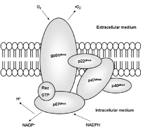
NADH/NADPH oxidases are membrane-associated enzymes that catalyse the 1-electron reduction of oxygen using NADH or NADPH as the electron donor (Figure 1). NADH/NADPH oxidases, initially described in neutrophils, are the most important oxidases in vascular tissue and in cardiac cells54, are important sources of endovascular ROS and are also integral to the generation of ROS in phagocytic cells48, 55.
NADH/NADPH activity is regulated by a number of factors known to be involved in the pathogenesis of cardiovascular disease including cytokines, hormones, local metabolic changes and haemodynamic forces56. Shear stress (of vascular wall) can have variable effects on NADPH oxidase, be it a transient elevation or a sustained increase48. NADH/NADPH-dependent oxidase activity and consequently NADH/NADPH driven O2- production are also increased in vascular smooth muscle cells by stimulation with angiotensin II48.
Figure 1: Structure of NAD(P)H oxidase (Reprinted from Rabelo et al.57). NADPH oxidase consists of the membrane-bound gp91phox and p22phox, the cytosolic proteins p47 and p67, and a low molecular-weight G protein (rac).
1.3.1.2 Xanthine oxido-reductase system
Xanthine oxido-reductase exists in two interconvertible forms, either as xanthine dehydrogenase or xanthine oxidase58. The first form reduces NAD+ whereas the latter produces superoxide anion and hydrogen peroxide via a reaction with O248, 59. The dual role means that it is an important regulator of cellular redox state. Xanthine oxidase generates ROS via purine metabolism pathway and is involved in causing endothelial dysfunction in patients with coronary disease and contractile dysfunction in heart failure48. Studies have shown that xanthine oxidase expression and activity are increased in cardiomyocytes isolated from failing hearts60-63.
1.3.1.3 Nitric oxide synthase uncoupling
Recent studies have demonstrated the crucial role of endothelial NOS (eNOS) as a ROS producing enzyme; resulting in vascular endothelial dysfunction48, 64. When tetrahydrobiopterin (BH4) is limited, electron transfer becomes uncoupled to L-arginine oxidation, leading to the dissociation of the ferrous dioxygen complex and superoxide is produced64. Endothelial NOS can produce both NO via its oxygenase function and superoxide through its reductase function (dependent on NADPH)48. The product of the reaction between NO and O2- can oxidize BH4 which may lead to further eNOS uncoupling. Imbalance between endothelial NO and ROS production is one of the major contributors of endothelial dysfunction which plays an important part in the development of atherosclerosis and cardiovascular disease48.
1.3.1.4 Mitochondrial electron transport chain
The major source of ROS is considered to be the mitochondrial electron transport chain in the inner mitochondrial membrane, with respiratory complexes I and III being the major sites of superoxide formation in the electron transport chain65 (Figure 2).
F a s o s f p a Figure 2: Site al.66) Various superoxide a or be convert superoxide co fenton reacti peroxynitrite and cause da es of superoxi s electron tr nion (O2-). Su ted to hydrog oncentration ions producin . Both hydrox mage to nucl ide formation ransport chai uperoxide can gen peroxide s increase to ng hydroxyl xyl radicals a leic acids, lipi
n in the elect in complexes n then reduc (H2O2) (in bo a high enou radicals (OH) nd peroxynit ds and protei tron transpor s leak electr e cytochrom th the matrix gh level, they ) or may rea trite are very
ins66.
rt chain (Repr rons to oxyg e c (in the in x and the inte y may reduce act with nitric strong oxida rinted from T gen producin termembran ermembrane e transition m c oxide (NO) ants which re Turrens et g mainly e space), space). If metals via to form eact with,
In the vasculature and the kidneys, ROS derive mainly from the mitochondrial electron transport chain, NAD(P)H oxidases and uncoupled nitric oxide synthase (NOS)67. For the current research, we will focus on the impact of mitochondria (dys)function. The roles of NAD(P)H oxidase and of (uncoupled) NOS-derived ROS in vascular dysfunction after neonatal hyperoxic stress were examined in a series of recent studies from the laboratory.
1.4 Oxidative
Stress
Oxidative stress is mainly caused by an imbalance between the activity of endogenous pro-oxidative enzymes (such as NADPH oxidase, xanthine oxidase, uncoupled NO synthase or the mitochondrial respiratory chain) and antioxidant enzymes (such as superoxide dismutase, glutathione peroxidase, heme oxygenase, thioredoxin peroxidase/peroxiredoxin, catalase and paraoxonase)68.
Under oxidative stress, excessive superoxide also releases free iron from iron-containing molecules, which further generate highly reactive hydroxyl radicals (OH) by reacting with hydrogen peroxide via the Fenton reaction69. ROS can also induce the opening of the mitochondria membrane permeability transition pore (PTP) and cause a release in cytochrome c and other factors that can lead to apoptosis-mediated cell death48.
Under physiological conditions, cells increase activities of antioxidant enzymes and other antioxidant defences in order to counter-act the occurrence of oxidative stress. These antioxidants include Copper/Zinc superoxide dismutase (Cu/Zn SOD), manganese-dependent superoxide dismutase (MnSOD), glutathione peroxidase, glutathione reductase and catalase (CAT). MnSOD and Cu/Zn SOD convert O2- to hydrogen peroxide, which is then transformed to water by glutathione peroxidase, catalase or peroxiredoxin48, 70. The thioredoxin system is composed of several proteins including peroxiredoxin, that are important in redox homeostasis due to their ROS scavenging ability71. Other antioxidant defences include radical scavengers such as vitamin E, beta carotene and vitamin C48. (Figure 3)
F s b g
1
T l m r r O t i A o p Figure 3: Red scavengers. T by the variou glutathione p1.5 Prete
The human e life72. This is may protect radicals72. A reproductive O2 (atmosphe that must be incomplete lu Among the m oxygen, and promoted, w dox homeost The steady-sta us scavenger m peroxidase, caerm- high ox
embryo devel thought to b the developi study by Fi tract of rhes eric O2 is appr overcome, in ung developm most commo finding the while minimizi tasis (Reprint ate levels of R mechanisms. atalase and thxygen after
lops in an oxy be responsibl ing embryo f ischer et al. sus monkeys, roximately 21 ncluding: diffi ment, inadeq on treatment optimum ox ng the risks oted from Dro ROS are dete
Antioxidative hioredoxin.
birth
ygen-poor en le for the sw from the delerevealed th hamsters an 1%)73. Preterm iculties in gas quate respira ts in neonat xygen concen of hyperoxia oge70). Balan rmined by th e enzymes inc nvironment c witch from em eterious effec hat the mea nd rabbits wa m infants the s exchange re tory drive, a tal intensive ntration whe is of utmost nce between e rate of prod clude superox ompared wit mbryonic to f cts of high o n oxygen te as only 40% o refore face a esulting from nd poor clea care is the reby growth importance75 ROS produc duction and c xide dismuta th later stage fetal hemoglo xygen levels ension in the or less of atm number of d surfactant de arance of lun use of supp and develo 5. A study by ction and clearance se (SOD), s of fetal obin, and and free e female mospheric ifficulties eficiency, ng fluid74. plemental pment is Vento et
al. found that initiating resuscitation with 30% versus 90% oxygen for infants 28 weeks’ gestation
or less significantly reduced the risk of developing bronchopulmonary dysplasia74, 76. Even brief exposure to high levels of oxygen induces oxidative stress, and the subsequent production of oxygen free radicals can be extremely dangerous, particularly for preterm infants who lack adequate protection from indigenous antioxidants75. Since fetal life takes place in a relatively hypoxic environment, preterm birth results in the sudden and premature exposure to comparatively high levels of oxygen and oxidative stress results from the lack of protection (underdeveloped antioxidant system) coupled with the necessary supplemental oxygen therapy to treat this population. This increased risk of oxygen toxicity poses a significant threat of tissue damage in response to reactive oxygen species and excess free radicals75.
The vast spectrum of disease evident in the neonatal period includes those that stem from the interruption of normal organogenesis in the vascular tree, pancreas, lung and kidney9. Of particular relevance for the current Master thesis, kidney development (nephrogenesis) begins at approximately day 30 of gestation, with the majority of nephrons being formed between week 20 until 34 to 36 weeks of gestation14. Given that nephrogenesis is still ongoing prior to 34 weeks of gestation, it is very likely that neonates born at less than 34 weeks of gestation will experience alterations in the structural development of the kidney14. In addition, the extrauterine environment is suboptimal for organogenesis due to the exposure to a number of insults, including high oxygen concentrations14. Oxidative stress has emerged as a likely promoter of several pregnancy-related disorders, such as spontaneous abortions, embryopathies, preeclampsia, fetal growth restriction, preterm labor and low birth weight77.
Saugstad et al.78 described the implications of oxidative stress in a number of conditions affecting premature infants, including: bronchopulmonary dysplasia (BPD) or chronic lung disease (CLD), retinopathy of prematurity (ROP), necrotizing enterocolitis (NEC) and patent ductus arteriosus. A number of studies indicate that the development of CLD is related to oxidative stress detected by the augmentation of peroxidation products in the very first days of life79, 80. Retinopathy of prematurity (ROP) is thought to occur through the frequent hyperoxygenation of the retina and subsequent oxidative damage due to the limited antioxidant defense of the retina in the preterm
infant81. A study by Papp et al. discovered that the ratio of oxidized to reduced glutathione more than doubled in patients with ROP compared with controls, and may be used as a method of identifying infants at risk of developing ROP82.
Newborn infants possess an underdeveloped antioxidative capacity, and when exposed to a greater percent oxygen concentration relative to the intrauterine milieu83, they are at a significantly higher risk of oxidative stress; a fact supported by many studies in both human and animal models84-86. A study by Lee et al.87 showed that smaller preterm infants (SPT) had significantly lower levels of GSH and NADPH ratio compared to larger preterm infants (LPT). GSH and the NADPH ratio are believed to be important indices of the cellular redox state88 and when impaired can lead to ROS generation48. It was further demonstrated that preterms had increased glucose-6-phosphate dehydrogenase (G6PD) activity compared to full term appropriate-for-age infants, suggesting a compensatory mechanism to combat an adverse environment87, as patients with G6PD deficiency often have lower antioxidant capacities89. Georgeson et al.90 reported significantly higher catalase (CAT), glutathion peroxidase (GPX), and Cu/Zn-SOD activity in healthy neonates, in the full-term normal spontaneous vaginal delivery (FT-NSVD) category compared to preterm normal spontaneous vaginal delivery (PT-NSVD)90. Of further importance for the current work is the role of oxidative stress and free radicals in renal injury. Studies by Vento et al.91 and Perrone et al.92, showed elevated concentrations of N-acetyl-glucosaminidase in the days following birth in preterm babies (a reliable marker for detecting renal tubular damage after neonatal anoxia). This increase in N-acetyl-glucosaminidase concentration demonstrates the correlation between indices of oxidative stress and kidney damage92. These results clearly demonstrate that preterm neonates possess an impaired antioxidant capacity relative to their term counterparts which may compromise their ability to deal with oxidative stress, predisposing them to the diseases discussed above.
1.6
Oxidative Stress and its role in programming
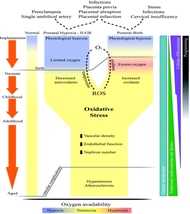
Oxidative stress both during fetal and neonatal life is considered a key element in programming of long-term cardiovascular consequences93-95 (see Figure 4). Regarding the kidney, Yzydorczyk et
al.83 explored the relationship between neonatal O2 exposure and long-term renal damage. A significant reduction in nephron number was observed in both male and female rats exposed to 80% oxygen (hyperoxic group) from day 3 to day 10 of life compared to the control (room air) and examined at 25-35 weeks of age (which corresponds to an adult in humans). Nephrogenesis in rats proceeds until 5 to 8 days postnatally83 compared to 32-36 weeks in humans96, allowing the kidney to be compared to that of a preterm infant. Since nephron number is decreased in adult patients with primary hypertension97, and reduced nephron endowment is a function of impaired renal growth, the kidney is an important organ to explore in the development (programming) of hypertension. Furthermore, oxygen tension has been shown to possess a regulatory role in nephrogenesis through hypoxia-inducible factor (HIF)98 which regulates expression of genes involved in angiogenesis and rat organogenesis appears to be enhanced in low (1-3%) O2 concentration compared to the standard (21%) O2 concentration83. All of these factors clearly indicate that the kidney is adversely affected under high oxygen concentrations and since mitochondria are known to be the primary producers of ROS65, it is important to investigate possible effects of oxidative stress and programming of adult disease on mitochondria function in the kidney.
Figure 4: Representation of potential common pathways in the ‘fetal origins of adult disease’ theory (Reprinted from Davidge et al.99).
1.7 Mitochondria
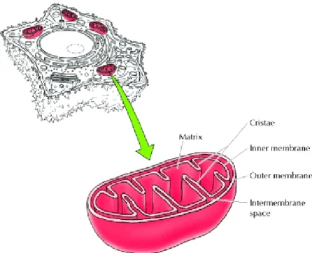
Mitochondria (shown in Figure 5) are surrounded by a double-membrane system, consisting of inner and outer mitochondrial membranes separated by an intermembrane space. The inner membrane forms numerous folds (cristae), which extend into the interior (or matrix) of the organelle and provide a greater surface area for the production of ATP. Each component is responsible for distinct functional roles, with the matrix and inner membrane accounting for the vast majority of mitochondrial work and ATP production100.
Mitochondria also contain their own genome, which is separate and distinct from the nuclear genome of the cell. Mitochondrial DNA (mtDNA) follows maternal inheritance and is termed ‘naked’ DNA since it lacks histone protection and has a weak repair capacity, making it more vulnerable to damage than nuclear DNA101. The level of oxidized bases in mtDNA is 10- to 20-fold higher than in nuclear DNA102.
1.7.1 Mitochondrial Function
Mitochondria are the primary energy-generating system in most eukaryotic cells and play a crucial role in energy metabolism through their involvement in ATP production by oxidative phosphorylation103. Apart from energy conversion, mitochondria take part in a number of other processes including calcium homeostasis, nucleotide precursor biosynthesis, cofactor biosynthesis, and apoptosis104. Given that mitochondria are involved in so many important and well-established functions, mitochondrial dysfunction has a multitude of effects in multicellular organisms, some of which remain poorly understood103. Importantly, dysfunction of the mitochondria has been linked to a wide range of human disorders, including, cancer, neurodegenerative diseases, Type 2 Diabetes and hypertension 105, 106 and at a cellular level can contribute to inflammation, ageing, apoptosis and alterations in cellular calcium handling104.
1.7.2 The Citric Acid Cycle
The citric acid cycle is located in the mitochondrial matrix and plays a crucial role in regulating cell metabolism through the production of reducing equivalents in the form of nicotinamide adenine dinucleotide (NADH) and flavin adenine dinucleotide (FADH2)107, 108. It is through the rate of delivery of these reducing equivalents to the electron transport chain, along with adenosine diphosphate that the citric acid cycle is able to regulate oxidative phosphorylation. This flux through the citric acid cycle is dependent upon the supply of acetyl units, activation of the three non-equilibrium reactions within the citric acid cycle and the total concentration of the citric acid cycle intermediates107. The cycle (dependent on oxygen) is also an important source of precursors, not only for the storage forms of fuels, but also for the building blocks of many other molecules such as amino acids, nucleotide bases, cholesterol, and porphyrin (the organic component of heme)109.
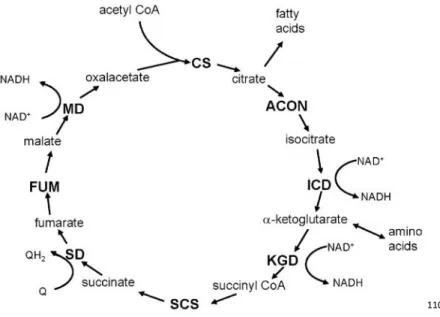
The citric acid cycle includes a series of oxidation-reduction reactions that result in the oxidation of an acetyl group to two molecules of carbon dioxide; transforming fuel molecules into ATP via the electron transport chain109. The overall pathway of the citric acid cycle is shown in Figure 6.
110
Figure 6: Overview of the Citric Acid Cycle (Reprinted from Berg et al.109) The citric acid cycle oxidizes two-carbon units, producing two molecules of CO2, one molecule of GTP, and high-energy electrons in the form of NADH and FADH2. A four-carbon compound (oxaloacetate) condenses with a two-carbon acetyl unit to yield a six-carbon tricarboxylic acid (citrate), which undergoes a series of decarboxylations producing succinate. Oxaloacetate is then regenerated from succinate. Two carbon atoms enter the cycle as an acetyl unit and two carbon atoms leave the cycle in the form of two molecules of carbon dioxide. Three hydride ions (six electrons) are transferred to three molecules of nicotinamide adenine dinucleotide (NAD+), whereas one pair of hydrogen atoms (two electrons) is transferred to one molecule of flavin adenine dinucleotide (FAD). The function of the citric acid cycle is the harvesting of high-energy electrons from carbon fuels109. CS, citrate synthase; ACON, aconitase; ICD, isocitrate dehydrogenase; KGD, α-ketoglutarate dehydrogenase; SCS, succinyl-CoA synthetase; SD, succinate dehydrogenase; FUM, fumarase; MD, malate dehydrogrenase.
Aconitase (mitochondrial, m-aconitase) is localized in the mitochondrial matrix. It catalyzes conversion of citrate to isocitrate as part of the citric acid cycle. The enzyme is inactivated upon oxidation of its iron-sulfur cluster by superoxide. Upon inactivation, isolated aconitase induces production of hydroxyl radical, most likely mediated by the release of Fe2+. 111
Experimental studies have shown that specific enzymes of the citric acid cycle are susceptible to oxidation112-114. Aconitase exhibits the most significant age-associated decline in activity and is known to be highly susceptible to oxidative damage110. Citrate is a key intermediate in the citric acid cycle and in fatty acid synthesis, and increased citrate synthase activity may contribute to progressive renal injury115.
1.7.3 Electron Transport Chain
The respiratory chain, located in the inner mitochondrial membrane, consists of five multimeric protein complexes116 (Figure 8). Mitochondrial ATP production relies on the electron transport chain, composed of respiratory chain complexes I-IV, which transfer electrons in a stepwise fashion until they finally reduce oxygen to form water117. Mitochondria contain their own genome, 13 polypeptides, all of them structural subunits of the oxidative phosphorylation complexes I, III, IV and V are encoded118. As explained by Mailloux et al.119, NADH: ubiquinone oxidoreductase (complex I) is composed of approximately 45 subunits and is the site of NADH oxidation. The flavin mononucleotide of complex I accepts the electrons from NADH and passes them through a series of eight iron-sulfur clusters to ubiquinone. Ubiquinol: cytochrome c oxidoreductase (complex III) has 11 subunits and contains three hemes and a Fe-S cluster center. Complex III plays a crucial role in funneling electrons from the ubiquinol generated by complexes I and II to cytochrome c. Upon binding to the Qo site of complex III, one electron from ubiquinol is ferried through the Rieske Fe-S cluster protein to the electron acceptor, cytochrome c. The resulting unstable semiquinone then donates the remaining electron to the cytochrome b hemes (cytochrome bL and bH). The electron in cytochrome b is then used to re-reduce ubiquinone in the Qi site to produce ubiquinol. Two electrons from semiquinones in the Qo are required for the reduction of ubiquinone to ubiquinol in the Qi site. This is referred to as the Q-cycle (Figure 7) because lone electrons remaining in semiquinone are reused to reduce ubiquinone back to ubiquinol. This electron cycling mechanism
i C a t F a c t c o is crucial for Complex III is ability to gen the build-up o Figure 7: Me al.66). Ubiqu cytochrome b transfers one cytochrome C oxygen produ preventing t s very well re nerate ROS on of semiquino echanism of e uinone (Q) is b on the inne e electron to C. The termin ucing water (H the use of th egarded as th n either side ne radicals in electron flow s reduced eit er side of the o oxygen via nal oxidase is H20)66.
hese lone ele e major sour of the mitoc n the Qo site1 w in the Q-cy ther by com e membrane a the Rieske s reduced by ectrons for u rce of ROS fro chondrial inne 19. ycle in Comp mplex I or II (i), producin iron-sulfur p four electron univalent red om the respir er membrane
plex III (Repri or by electr ng ubiquinol ( protein (ISP), ns which in tu duction of O2 ratory chain27 e can be attri nted from Tu ons transferr (QH2). Ubiqu , cytochrome urn are trans
2 to ROS. 7, 120. The ibuted to urrens et red from inol then e C1 and ferred to
1.7.4 Mitochondrial ATP production
Mitochondria play a central role in energy metabolism through their significant contribution to adenosine triphosphate (ATP) production; this process depends on oxygen. When oxygen is limited, the less efficient process of anaerobic respiration metabolizes glycolytic products directly in the cytosol121, 122. NADH and FADH2 are energy rich molecules formed in glycolysis, fatty-acid oxidation and the citric acid cycle that donate electrons to the ETC, establishing an electrochemical gradient122. Electrons move toward compounds with more positive oxidative potentials and the incremental release of energy during the electron transfer is used to pump protons (H+) into the intramembrane space. As described in a recent review by Bratic and Trifunovic (2010)121 complexes I, III and IV function as H+ pumps that are driven by the free energy of coupled oxidation reactions. During the electron transfer, protons pumped from the mitochondrial matrix to the intermembrane space produce a potential of around 150-180 mV121, 123. The proton gradient generates a chemiosmotic potential, also known as the proton motive force, which drives the ADP phosphorylation via the ATP synthase (F0F1-ATPase-Complex V). The rate of mitochondrial respiration depends on the phosphorylation potential expressed as [ATP]/[ADP][Pi] ratio across the inner mitochondrial membrane that is regulated by the adenine nucleotide translocase (ANT). In the case of increased cellular energy demand, when the phosphorylation potential is decreased and more ADP is available, a respiration rate is increased leading to an increased ATP synthesis. Tight coupling between the electron transport and the ATP synthesis and an inhibition of ATP synthase will therefore also inhibit the electron transport and cellular respiration. Under certain conditions, protons can re-enter into mitochondrial matrix without contributing to the ATP synthesis and the energy of proton electrochemical gradient will be released as heat. This process, known as proton leak or mitochondrial uncoupling could be mediated by protonophores (such as FCCP) and uncoupling proteins (UCPs)121.
Mitochondrial ATP production is fuelled by the chemical energy stored within the carbon-to-carbon bonds of various energy substrates and relies on the great oxidative power of diatomic oxygen (O2). After the oxidation of substrates in the citric acid cycle, the resulting electron carriers, NADH and FADH2, are oxidized by complexes I and II respectively. The liberated electrons/reducing equivalents are then transferred through the respiratory complexes III and IV via prosthetic groups arranged in increasing redox potential to the terminal electron acceptor, O2. This generates a
proton-motive force (Δp) across the mitochondrial inner membrane. This stored potential energy is then tapped by the F0F1-ATP synthase as protons move through it back to the matrix and drive ATP formation from ADP and Pi. This process is referred to as coupled respiration because the energy derived from the transfer of electrons to the terminal electron acceptor O2 is completely harnessed to drive ATP production. However, the reduction of dioxygen to water during aerobic respiration is accompanied by variable extents of reactive intermediate formation, because the two outermost electrons in molecular oxygen occupy separate orbitals. Consequently, the univalent reduction of O2 results in the formation of various singlet-electron-containing intermediates that can damage the cell119 (see Figure 8).
Figure 8: Mitochondrial energy metabolism (Reprinted from DiMauro et al.116). Overview of the numerous tasks performed by mitochondria, including : pyruvate oxidation, the citric acid (TCA) cycle, metabolism of amino acids, fatty acids, and steroids, but the most important is the generation of adenosine triphosphate (ATP) via the electron-transport chain and the oxidative-phosphorylation system (the ‘respiratory chain’)116. ADP, adenosine diphosphate; ANT, adenine nucleotide translocator; CACT, carnitine-acylcarnitine translocase; CoQ, coenzyme Q; CPT, carnitine palmitoyltransferase; DIC, dicarboxylate carrier; ETF, electron-transfer flavoprotein; ETF-DH, electron-transfer dehydrogenase; FAD, flavin adenine dinucleotide; FADH2, reduced FAD; NADH, reduced nicotinamide adenine dinucleotide; PDHC, pyruvate dehydrogenase complex; TCA, tricarboxylic acid; I, complex I; II, complex II; III, complex III; IV, complex IV; V, complex V.
1.7.5 ROS production and handling in the mitochondria
Approximately 90% of cellular oxygen is consumed within the mitochondria, accounting for nearly 90% of the total ROS produced in the cell121, 124. ROS have dual roles within aerobic cells, whereby they may act as signalling molecules in a wide range of normal cellular processes or as oxidizing agents that can potentially trigger cell death119. The mitochondrial electron transport chain is the major ROS production site but the mitochondria are also a prime target for oxidative damage. With accumulating oxidative damage, such as with aging or in disease states, mitochondria become larger and less numerous with age, accumulating vacuoles, cristae abnormalities and intra-mitochondrial paracrystalline inclusions125. Mitochondrial respiratory chain enzymes activities decrease, as well as mitochondrial membrane potential (on which the production of ATP is dependent); the amount of oxidative damage to proteins and mtDNA increases, with an associated accumulation in the quantity of mtDNA mutations124, 126. Age-associated decline in the mitochondrial respiratory chain capacity has also been reported in various tissues121, including the kidney127.
Approximately 1-2% of the O2 consumed in the mitochondria is reduced (univalently) to superoxide (O2-), the main molecule of ROS signalling and damage102, 119. Superoxide is then usually converted into H202 by the manganese-superoxide dismutase or copper/zinc-superoxide dismutase,found in the mitochondrial matrix and in the intermembrane space, respectively128. The high amount of O2 and the low concentration of superoxide, makes superoxide formation a very energetically favorable process102, 129. Superoxide can also react with nitric oxide radical (NO) to generate peroxynitrite (ONOO), a lipid soluble and highly toxic reactive nitrogen species. Of greater importance, superoxide production by the respiratory chain can increase significantly when the mitochondrial membrane potential is high (e.g. when ATP production is low)130, 131. Unlike superoxide, H202 is more stable and is able to oxidize low-molecular weight thiolating
agents such as glutathione (GSH). This stability allows H202 to play a key role as a mitochondrial signalling molecule. It is when H202 is produced in an uncontrolled fashion that it can damage the cell by generating hydroxyl radical through Fenton or Haber-Weiss reactions132.
Figure 9: Overview of mitochondrial ROS production (Reprinted from Murphy133). ROS production by mitochondria can impair their ability to synthesize ATP and to carry out normal cellular functions, including the citric acid cycle, fatty acid oxidation, the urea cycle, amino acid metabolism, heme synthesis and FeS centre assembly. Mitochondrial oxidative damage can also increase the tendency of mitochondria to release proteins such as cytochrome c (cyt c) to the cytosol via the mitochondrial outer membrane permeabilization, thereby activating the cell’s apoptotic machinery. In addition, mitochondrial ROS can affect redox signalling and lead to the induction of the mitochondrial permeability transition pore (PTP), rendering the inner membrane permeable to small solutes in situations such as ischemia/reperfusion133.
1.7.6 Antioxidant Defense Systems
The balance between oxidants and antioxidants is commonly referred to as the ‘redox state’48. Antioxidant defense is comprised of a large set of enzymes and low-molecular weight compounds that sequester the excess ROS molecules generated by aerobic respiration. Cells engage in a complex array of short and long term preventative measures such as the mild uncoupling of the oxidative phosphorylation system in the short term and possible upregulation of antioxidant enzyme expression in the long term. Some antioxidative systems can be energetically costly because they are reliant on both ATP and NADPH119.
The peroxiredoxin (Prx) and GSH/glutaredoxin (Grx)/glutathione peroxidase (GPx) systems are the most efficient at eliminating H202 in the mitochondria and cytosol because of their low Km values and abundance134-136. The presence of catalase in the mitochondria is also strongly debated because it seems to be localized primarily in peroxisomes and to a lesser extent the cytosol. For example, Kirkinezos et al.137, describe mitochondria as being catalase free, whereas Radi et al.138, demonstrate the presence of catalase in rat hearts. The antioxidative defense system is rejuvenated by the reductive power of NADPH (requires energy)119. They require ATP to synthesize small-molecule antioxidants and molecules for the sequestration of ROS and ROS by-products. The reactivation of H202-scavenging system requires the reductive power of NADPH (requires energy). The second layer of ROS defenses is formed by enzymes dealing with the primary ROS generated in mitochondria, superoxide radical and hydrogen peroxide. Superoxide is a substrate for mitochondrial manganese-containing superoxide dismutase (MnSOD, also known as SOD2). This enzyme is localized exclusively to the mitochondrial matrix, and its only function is to facilitate dismutation of superoxide radical to H202, thereby protecting mitochondrial iron-sulfur cluster containing enzymes from superoxide attack111. MnSOD does not require any cofactors so the efficiency of this system is determined solely by the amount of enzyme present.
Catalase is an antioxidant enzyme (ubiquitously expressed by aerobic cells containing the cytochrome system and is localized to the peroxisome, the nucleus, or mitochondria) that converts H202 into O2 and H20137. Two isoforms of peroxiredoxins (Prx3 and Prx5) are found in mammalian mitochondria. Prx3 (or SP-22) is ubiquitously present in various rat tissues, including the kidney139,
whereas Prx5 is mainly expressed in bovine tissues, with the highest level found in testis111. Importantly, Prx3 gene expression is known to be induced by oxidative stress111.
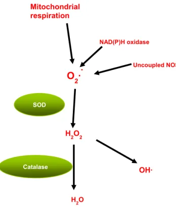
Figure 10: Selected pathway of ROS formation and neutralization
Mitochondrial respiration Uncoupled NOS NAD(P)H oxidase
O
2·
-SOD H2O2 H2O Catalase OH·1.7.7 Mitochondria and Uncoupling Proteins
Mild uncoupling of mitochondrial oxidative phosphorylation is effectively short circuiting the OXPHOS system due to leak of protons from the intermembrane space to the matrix, which means that the protons bypass ATP synthase and cause a decrease in the mitochondrial inner membrane potential119. A strong correlation exists between mitochondrial membrane potential and ROS production; small increases in the membrane potential induce ROS formation whereas slight decreases can substantially diminish the production of these reactive intermediates. Although the mechanisms responsible for the mitochondrial proton leak are still debated140-144, the proteins thought to be involved include adenine nucleotide translocase (ANT) and the uncoupling proteins, UCPs 1-3. ROS and ROS derivatives (specifically superoxide and 4-hydroxy-2-nonenal) have been described as activators of UCPs 1-3. Negative feedback mechanism has been suggested in which increased ROS induce uncoupling to decrease the formation of these toxic species. It is generally thought that the two major sites of mitochondrial ROS production are complexes I and III of the respiratory chain119.
UCPs are mitochondrial transporters present in the inner mitochondrial membrane and are found in all mammals145. Uncoupling proteins function as proton channels to allow proton leakage back across the inner mitochondrial membrane without creating ATP. It has been shown that O2- can activate UCP2146, possibly as a protective mechanism against excessive mitochondrial ROS formation52.
High proton motive force comes with an additional cost, the production of ROS. Because ROS production is highly dependent on the proton motive force, proton leak might help to limit the oxidative damage. There are presently 3 identified isoforms of UCPs (UCP1, UCP2, UCP3). UCP2 and UCP3 are recently identified UCP1 homologues but their function in normal cellular physiology is still unclear145, 147. UCP2 and UCP3 proteins might be involved in the proton conductance only upon activation with certain activators (such as ubiquinone, superoxide, reactive alkenals and other alkenal analogues)121.
ANT and UCPs 1-3 contribute to inducible proton leak. However, only UCPs 2 and 3 have been found to catalyze a proton leak that decreases ROS emission from mitochondria. UCP2 is widely expressed in a number of cells and tissues including the spleen, thymus, macrophages,
hypothalamus, pancreatic β-cells, stomach and the kidney148. UCP3 is expressed almost exclusively in skeletal muscle and brown adipose tissue and to a lesser extent in the heart148. In several studies Brand and colleagues149, 150 provided empirical evidence that superoxide and derivatives of ROS, namely 4-HNE, activated proton leak through UCPs 1-3. In this mechanism, the overproduction of ROS by the respiratory chain activates proton leak through the UCPs, thereby creating a negative feedback loop that regulates ROS formation by the mitochondria119.
1.7.8 Mitochondrial Permeability Transition Pore
Apart from energy conversion, mitochondria take part in a number of other processes including calcium homeostasis and apoptosis151. It is well documented that an increased permeability of the mitochondrial membranes is a key event in controlling apoptotic pathways, with the permeability transition pore (PTP) thought to play a major role152-156. Prolonged opening of the PTP, a nonspecific high-conductance proteinaceous channel of the inner membrane, leads to collapse of the membrane potential, mitochondrial matrix swelling, rupture of the outer mitochondrial membrane and release of cytochrome C 157, 158, which can ultimately lead to energy failure of the cell.
1.8
Hyperoxia exposure and mitochondria
A study by Clerch et al.159, found that neonatal rats exposed to 95% O2 (hyperoxia) for 48h exhibited a fall in lung activity of MnSOD. This suggests that the increased production of superoxide during hyperoxia results in the observed decrease in MnSOD activity which worsens the effect of the enhanced formation of superoxide and thereby contributes in a major way to the loss of mitochondrial function during hyperoxia159. Further studies have shown that oxidative damage to mitochondrial enzymes plays a significant role in oxygen toxicity. Schoonen et al.160, found that exposure of Chinese hamster ovary cells to hyperoxia resulted in the inactivation of three key mitochondrial enzymes (NADH dehydrogenase, succinate dehydrogenase, and
α-ketoglutarate), a 2.5-fold decrease in cellular ATP, and cell death159, 160. Fischer et al.161 found reduced oxidative metabolism and a fall in ATP concentration in pulmonary macrophages following a 72-hour oxygen exposure ( 95% O2). Campian et al.27 reported that hyperoxia tolerant HeLa-80 cells, as compared to wild-type HeLa-20 cells generate substantially less ROS through modification of cytochrome C oxidase activity and a tighter coupling of the electron transport chain (ETC). This was assessed by the oxidation of three fluorescent probes, the hyperoxia-mediated inactivation of aconitase, and the accumulation of mitochondrial protein carbonyls under hyperoxic conditions. Li et al.162 demonstrated that the absence of MnSOD rapidly leads to impairment of mitochondrial function in several organs, with the heart suffering the most pronounced consequences, followed by the liver and the brain. Impaired fatty acid metabolism was also discovered through accumulation of lipid in the liver and skeletal muscle, metabolic acidosis and ketosis. Interestingly, no evidence of mtDNA deletions or of structural damage to the mitochondria were found162. Schriner et al.163 demonstrate that catalase over expression increases lifespan in the mouse. Furthermore, several studies164-166 have shown decreased levels of SOD, GPx and catalase in newborns and small for gestational age infants exposed to various toxins (alcohol, tobacco, etc.) in utero. In addition, Yarian et al.110 report that among the citric acid cycle enzymes, aconitase exhibits the most significant age-associated decline in activity, while NADPH :NADP+ ratio also declined with age (important for redox balance) in mitochondria isolated from rat kidneys. Maintaining redox homeostasis by regulation of antioxidant defense systems or ROS production is therefore one of the key mechanisms for tissues to adapt to hyperoxia-induced oxidative stress167. An altered neonatal environment could have lasting effects on antioxidant enzymes and result in the observed disease phenotype. Currently not much is known about the precise mechanisms involved, making it especially important to determine the long-term effects.
1.9
Hypothesis, Aims and Study Design
It was hypothesised that a transient neonatal hyperoxic stress would lead to chronic alterations in cellular oxidant/antioxidant coupling through the modulation of the expression of key proteins involved in mitochondrial function, enzymatic activity of key citric acid cycle enzymes and an excessive production of ROS.
1.9.2
Aims
To evaluate the expression and function of key proteins and enzymes participating in mitochondrial function and antioxidant capacity in the kidney of adult rats exposed to high oxygen as newborns; at 4 and 16 weeks of age.
1.9.3
Study
Design
To address the aims of this study, Sprague-Dawley rat pups were exposed to 80% O2 from day 3 to day 10 of life and studied at 4 and 16 weeks. Although the rats are born at term, their organ development is equivalent to that of a human preterm fetus, allowing organs of interest such as the kidney to be compared to premature infants99. Therefore the rat is an ideal model to examine the effects of neonatal hyperoxia exposure on mitochondrial function in the developing kidney, especially since they also contain a large number of mitochondria due to their high aerobic capacity and their dysfunction is important in cardiovascular disease and hypertension52.
REFERENCES
(1) Barker DJ, Osmond C, Golding J, Kuh D, Wadsworth ME. Growth in utero, blood pressure in childhood and adult life, and mortality from cardiovascular disease. BMJ 1989 March 4;298(6673):564-7.
(2) Osmond C, Barker DJ, Winter PD, Fall CH, Simmonds SJ. Early growth and death from cardiovascular disease in women. BMJ 1993 December 11;307(6918):1519-24.
(3) Ingelfinger JR, Woods LL. Perinatal programming, renal development, and adult renal function. Am J Hypertens 2002 February;15(2 Pt 2):46S-9S.
(4) World Health Organisation. Complications of labour and delivery (O60-O75).
International classification of diseases and related health problems, 10th revision. WHO: Geneva;
1992.
(5) Liu S, Allen A, Fraser W. Canadian Perinatal Health Report 2008 Edition. Ottawa, Ontario, Canada; 2008.
(6) Poplawska K, Dudek K, Koziarz M et al. Prematurity-related hypertension in children and adolescents. Int J Pediatr 2012;2012:537936.
(7) Goldenberg RL, Culhane JF, Iams JD, Romero R. Epidemiology and causes of preterm birth. Lancet 2008 January 5;371(9606):75-84.
(8) Shennan AH, Bewley S. Why should preterm births be rising? BMJ 2006 April 22;332(7547):924-5.
(9) Abitbol CL, Rodriguez MM. The long-term renal and cardiovascular consequences of prematurity. Nat Rev Nephrol 2012 February 28.
(10) Slattery MM, Morrison JJ. Preterm delivery. Lancet 2002 November 9;360(9344):1489-97.
(11) Goldenberg RL, Hauth JC, Andrews WW. Intrauterine infection and preterm delivery. N Engl J Med 2000 May 18;342(20):1500-7.
(12) Moster D, Lie RT, Markestad T. Long-term medical and social consequences of preterm birth. N Engl J Med 2008 July 17;359(3):262-73.
(13) Fanaroff AA, Stoll BJ, Wright LL et al. Trends in neonatal morbidity and mortality for very low birthweight infants. Am J Obstet Gynecol 2007 February;196(2):147-8.
(14) Gubhaju L, Sutherland MR, Black MJ. Preterm birth and the kidney: implications for long-term renal health. Reprod Sci 2011 April;18(4):322-33.
(15) Nijland MJ, Ford SP, Nathanielsz PW. Prenatal origins of adult disease. Curr Opin
Obstet Gynecol 2008 April;20(2):132-8.
(16) Nuyt AM, Alexander BT. Developmental programming and hypertension. Curr Opin
Nephrol Hypertens 2009 March;18(2):144-52.
(17) Barker DJ, Bull AR, Osmond C, Simmonds SJ. Fetal and placental size and risk of hypertension in adult life. BMJ 1990 August 4;301(6746):259-62.
(18) Barker DJ, Forsen T, Eriksson JG, Osmond C. Growth and living conditions in childhood and hypertension in adult life: a longitudinal study. J Hypertens 2002
October;20(10):1951-6.
(19) Hales CN, Barker DJ, Clark PM et al. Fetal and infant growth and impaired glucose tolerance at age 64. BMJ 1991 October 26;303(6809):1019-22.
(20) McCance DR, Pettitt DJ, Hanson RL, Jacobsson LT, Knowler WC, Bennett PH. Birth weight and non-insulin dependent diabetes: thrifty genotype, thrifty phenotype, or surviving small baby genotype? BMJ 1994 April 9;308(6934):942-5.
(21) Valdez R, Athens MA, Thompson GH, Bradshaw BS, Stern MP. Birthweight and adult health outcomes in a biethnic population in the USA. Diabetologia 1994 June;37(6):624-31.
(22) Fall CH, Barker DJ, Osmond C, Winter PD, Clark PM, Hales CN. Relation of infant feeding to adult serum cholesterol concentration and death from ischaemic heart disease. BMJ 1992 March 28;304(6830):801-5.
(23) Barker DJ, Martyn CN, Osmond C, Hales CN, Fall CH. Growth in utero and serum cholesterol concentrations in adult life. BMJ 1993 December 11;307(6918):1524-7.
(24) Forsen T, Eriksson JG, Tuomilehto J, Osmond C, Barker DJ. Growth in utero and during childhood among women who develop coronary heart disease: longitudinal study. BMJ 1999 November 27;319(7222):1403-7.
(25) Brenner BM, Garcia DL, Anderson S. Glomeruli and blood pressure. Less of one, more the other? Am J Hypertens 1988 October;1(4 Pt 1):335-47.