Skin te nsile stre ngth m odulation by comp re ssive
garm e nts in burn patie nts. A pilot stud y
R. Fournier and G. E. Pie´rard*
Belgian SSTC Research Center 5596, Department of Dermatopathology, University Medical Center Sart Tilman, Lie`ge, Belgium
Compression therapy is frequently used to prevent hypertrophy of post-burn scars. This pilot study was performed in 6 patients to assess non-invasive changes induced in the tensile strength of the skin before any clinical improvement can be perceived. Assessments were performed using a computerized suction device delivering three 5 s cycles of 500 mbar depression. Measurements were made at one-month intervals for three months after initiating the garment compression therapy. Comparisons were made between the intact skin, the ungrafted and grafted post-burn scars and the graft donor sites. Data show that garment compression therapy alters the tensile strength in the skin of all test sites. The most reliable variations consist of an increase in both the extensibility and elasticity of the tissues submitted to traction.
Introduction
Hypertrophic scars are a common complication of second degree or deeper burn injuries. Compressive garments are used to limit such tissue reactions. Clinical experience has shown the ef cacy of such a treatment [1]. Objective measurements of the kinetics of improvement would be welcome in order to bring further improvements in the management of these patients.
The expected bene t of garment compression therapy on a post-burn hypertrophic scar consists of reducing and softening the connective tissue growth. For the functional part of the problem, measuring the tensile strength of the skin might appear to be a good means to objectivate the improvement. Such a non-invasive monitoring is made possible using computerized devices [2, 3]. Only a few studies have evaluated the in vivo tensile strength of scars using such objective assessments [4 – 11].
The present pilot study was undertaken to assess the effect of compressive garments on the skin tensile strength on intact areas, post-burn hypertrophic scars at grafted and ungrafted sites, and on the healing donor site of the grafts. A longitudinal survey over 3 months was chosen without enrolling untreated control patients which would not have been ethical.
Patients and methods
Six patients, victims of second degree burns, treated at the Burn Unit of the Lie`ge University Medical Center, were enrolled in the study (table 1). Autologous grafts had been used on some burned areas in order to improve wound healing. Compression therapy using
specially designed garments (TricolastÒ , Deinze) was
initiated a few weeks after the burns at a time when the scarring process appeared to enter a hypertrophic phase.
Objective non-invasive assessments of the skin tensile strength were made before wearing the garments and at one-month intervals for three consecutive months
of compression therapy. The Cutometer SM474Ò
(C+K electronic, Cologne, Germany) was used with a hollow probe centred by a suction aperture of 4 mm in diameter [12]. The time – strain recording was used with 3 cycles of 5 s traction under negative pressure of 500 mbar separated by 5 s relaxation phases. The biomechanical parameters were similar to those previously described [2, 12, 13]. They are summarized in gure 1 and table 2. In each patient, measurements were taken on different sites, which were kept identical at each evaluation time. Data were tabulated in four distinct groups, namely, ungrafted post-burn scars, grafted post-burn scar, healing donor site of graft and control normal skin symmetrical to the burns. The latter data were pooled with measure-ments performed on healthy skin at similar reference sites in all volunteers. These were located on the volar forearm at 17 cm from the wrist and on the lateral aspect of the arm at 5 cm beneath the acromion.
For each patient and each of the four types of lesional and healthy skin, measurements of each biomechanical parameter were averaged at each evaluation time. Data from the 6 patients were pooled and the median was calculated for each parameter. Differences and percen-tage variations were calculated between sites at a given evaluation time, and between the successive session assessments for each test site. Statistical comparisons were made using the non-parametric paired Wilcoxon test. The effect of garment compressive therapy over time was assessed using the Friedman test followed by the Dunn test. In the case of signi cant changes, regression analysis models were applied to evaluate the best tted relationship, either linear, logarithmic, exponential or power. The coef cient of correlation r was calculated. For all statistical tests, a p value lower than 0.05 was considered signi cant.
277
Journal of Medical Engineering & Technology
ISSN 0309-1902 print/ ISSN 1464-522X onlineÓ 2000 Taylor & Francis Ltd http:/ / www.tandf.co.uk / journals
DOI: 10.1080/ 03091900010013733
* Author for correspondence: Email: [email protected]
Journal of Medical Engineering & Technology, Volume 24, Number 6, (November/December 2000) , pages 277 – 280
J Med Eng Technol Downloaded from informahealthcare.com by Univ De Liege on 05/09/14
278 Results
Between site comparisons at entry in the study
When the patients were asked to enter the garment compression therapy, the tensile strength of the skin was markedly different on the scars compared to the normal control sites ( table 3). MD and DD were markedly reduced at the lesional sites, particularly the post-burn scars. EF, RER and VER were also decreased on the same sites although to a lesser degree. BE was little affected by the scarring process.
Evolution of the tensile strength of the skin during garment compression therapy
The salient data are presented in table 4. Garment compression therapy resulted in MD increase at all test sites, showing almost a linear trend in time. Signi cance was reached on normal skin and the graft donor site. The Dunn test indicated that the effect started after the rst month of therapy.
Variations in DD and VER were erratic at all the test sites and did not reach signi cance.
BE increased on all test sites with an exponential trend over time. Signi cance was reached except for on the
grafted lesions. The Dunn test showed a signi cant change after the second month of therapy. EF also increased on all test sites with an exponential trend on normal skin.
RER showed a linear trend increasing over time, reaching signi cance on normal and ungrafted post-burn scars.
In the overall evaluation, the combination of MD and BE variations appeared to be the best representative aspect of the skin tensile changes occurring during garment compression therapy ( gure 2) .
Discussion
The tensile strength of the skin can be assessed in health and disease using various methods including stretching, elevation, indentation, vibration, torsion and suction devices. The latter approach has been exten-sively used to study the functional tensile properties of the dermis. However, only a few studies have addressed the problem of abnormal scarring [4 – 11]. The present pilot study intended to disclose some physical char-acteristics, if any, which could objectively show the effect of garment compression therapy on evolving hypertrophic post-burn scars.
As expected, the tensile strength was different on normal and scarring skin at entry in the study before applying compressive therapy. Our data are in line with previous reports indicating a decreased skin extensibility and altered viscoelastic properties at the site of the scars [8]. Autologous grafts used to improve wound healing [14] did not appear to in uence the
R. Fournier and G. E. Pie´rard Skin tensile strength and modulation in burn patients
Table 1. Patients under garment compressive therapy (GCT).
Burn Post-burn
duration before Patient Gender Age Type Extent (% ) GCT (month)
1 M 26 ame 13.5 5 2 M 27 chemical 7 6 3 M 41 water 9 2 4 M 47 ame 15 1.5 5 M 56 ame 14 3.5 6 F 51 contact 3 3.5
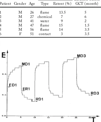
Figure 1. Example of skin deformations induced by three 5 s cycles of 500 mbar suction applied to an area of 4 mm in diameter. Elevation (E) of the skin is recorded in time (T). See text for the depicted parameters.
Table 2. Tensile parameters. Elevation
MD, maximum distension
VER, viscoelastic ratio = 102(MD1 Ð ED1) ED1Ð 1
DD, differential distension (lm) = MD3 Ð MD1 Retraction
RER, relative elastic recovery = 102 (MD1Ð ER1) MD1Ð 1
BE, biological elasticity = 102(MD1 Ð RD1) MD1Ð 1
EF, elastic function 102(MD1 Ð ER1) ED1Ð 1
Table 3. Tensile strength of the skin according to the test site before entering the garment compression therapy. Values represent the median percentages of variation compared to normal control skin (100% ).
Tensile Ungrafted Grafted Graft
property post-burn scar post-burn scar donor site
MD 41 39 64 DD 49 37 43 BE 86 82 90 VER 67 53 66 EF 50 51 61 RER 58 65 71
J Med Eng Technol Downloaded from informahealthcare.com by Univ De Liege on 05/09/14
279 tensile strength of scars compared to ungrafted
lesions.
It is acknowledged that the tensile strength of the skin is in uenced by the previous mechanical solicitations applied at the test site [2, 3]. The present study explores such a feature after applying sustained compression. Our data suggest a similar effect although with different intensities on healthy and damaged skin. Globally skin extensibility (MD) progressively increased while on compression treatment. Skin elasticity ( BE) also showed the same trend of evolution although to a lesser degree. Such overall ndings were already reported during compression therapy of oedematous legs in the gravita-tional syndrome [15].
The increased dynamic distensibility and elasticity after a relatively short period of compression therapy can hardly be explained by changes in the density and conforma-tion of the connective tissue bre networks. The mobilization of glycosaminoglycans may in uence the data. A resulting reduced amount should theoretically put less static tension on the bre networks at rest. As a result, any extrinsic superimposed force should allow apparent larger extensibility. Yet another explanation involves the natural intrinsic stress imposed by bro-blastic cells inside the dermis [16]. Any additional force
such as sustained compression applied to the tissues might alter the cell biology and mechanical function. The relevance of our ndings with regard to the control of hypertrophic scarring is undecided. During wound healing, intrinsic forces imposed by myo broblasts and broblasts are responsible for a retraction process. They are also likely to be a stimulus for dermal cell proliferation and accumulation. Hence, the distensi-bility of the skin is further decreased. Reducing the pathological limitation in static distensibility of the skin would be welcome to reverse the process of connective tissue hyperplasia. This seems to be effectively obtained using compression therapy.
Acknowledgments
We thank Professor M. Foidart-Desalle, Mrs A. Roesgen and Dr D. Soors-Jacquemin for their advice provided during the study.
References
1 BERMAN, B., and FLORES, F., 1998, The treatment of
hypertrophic scars and keloids. European Journal of
Dermatology, 8, 591 – 596.
2 PIE´RARD, G. E., and HENRY, F., 1995, Essai de classement
cate´goriel des proprie´te´s biome´caniques de la peau. Evaluation par la me´thode de succion. Nouvelles
Dermato-logiques, 14, 630 – 636.
3 PIE´RARD, G. E., and THE EEMCO GROUP, 1999, EEMCO
guidance to the in vivo assessment of tensile functional properties of the skin – Part 1: relevance to the structures and ageing of the skin and subcutaneous tissues. Skin
Pharmacology and Applied Skin Physiology, 12, 352 – 362.
4 LEUNG, K. S., CHENG, J. C. Y., LEUNG, Y. K., CARK, J. A., MA, G. F. Y., and LEUNG, P. C., 1984, In vivo study of the mechanical property of postburn hypertrophic scar tissues.
Journal of Burn Care and Rehabilitation, 5, 458 – 462.
5 KATZS. M., STEVEND. H., LEOPOLDG. R., and WACHTERT. L., 1985, Objective measurement of hypertrophic burn scar: a preliminary study of tonometry and ultrasonogra-phy. Journal of Plastic Surgery, 14, 121 – 127.
6 KRUSCHE, T., and WORRET, W. I., 1995, Mechanical
properties of keloids in vivo during treatment with intralesional triamcinolone acetonide. Archives for
Dermato-logical Research, 287, 289 – 293.
7 CLARK, J. A., CHENG, J. C., and LEUNG, K. S., 1996, Mechanical properties of normal skin and hypertrophic scars. Burns, 22, 443 – 446.
R. Fournier and G. E. Pie´rard Skin tensile strength and modulation in burn patients
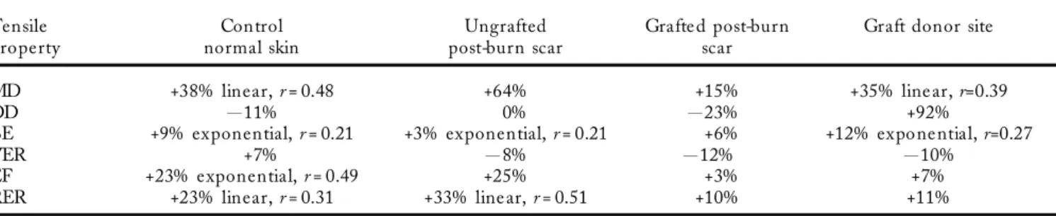
Table 4. Evaluation of the tensile strength of the skin during a garment compression therapy. Data are presented in % of variation yielded after three months. When the change is statistically signi cant, the correlation is indicated.
Tensile Control Ungrafted Grafted post-burn Graft donor site
property normal skin post-burn scar scar
MD +38% linear, r = 0.48 +64% +15% +35% linear, r=0.39
DD Ð 11% 0% Ð 23% +92%
BE +9% exponential, r = 0.21 +3% exponential, r = 0.21 +6% +12% exponential, r=0.27
VER +7% Ð 8% Ð 12% Ð 10%
EF +23% exponential, r = 0.49 +25% +3% +7%
RER +23% linear, r = 0.31 +33% linear, r = 0.51 +10% +11%
Figure 2. Scatterplot showing the mean percentage of change in maximum distensibility (MD) and biologic elasticity (BE) after a 3-month compression therapy on healthy skin (h ), grafted (e ), ungrafted postburn (s ), and donor graft sites (n ).
J Med Eng Technol Downloaded from informahealthcare.com by Univ De Liege on 05/09/14
280
8 FONG, S. S. L., HUNG, L. K., and CHENG, J. C. Y., 1997, The Cutometer and ultrasonography in the assessment of postburn hypertrophic scar – a preliminary study. Burns, 23(suppl 1), 512 – 518.
9 MCHUGH, A. A., FOWLKES, G. J., MAEVSKY, E. L., SMITH, D. J.,
RODRIGUEZ, J. L., and GARNER, W. L., 1997, Biomechanical
alterations in normal skin and hypertrophic scar after thermal injury. Journal of Burn Care and Rehabilitation, 18, 104 – 108.
10 DROBEV, H., 1999, Noninvasive monitoring of the
mechan-ical properties of keloids during cryosurgery. Acta Dermato
Venereologica, 79, 487 – 488.
11 HO, D. Q., BELLO, Y. M., GROVE, G. L., MANZOOR, J., LOPEZ, A. P., ZERWEK, C. R., PIERCE, E. A., WERKHEISER, J. L., and
PHILLIPS, T. J., 2000, A pilot study of noninvasive methods
to assess healed acute and chronic wounds. Dermatologic
Surgery, 26, 42 – 49.
12 PIE´RARD, G. E., NIKKELS-TASSOUDJI, N., and PIE´RARD-F
RAN-CHIMONT, C., 1995, In uence of test area on the
mechan-ical properties of skin. Dermatology, 191, 9 – 15.
13 PIE´RARD, G. E., NIZET, J. L., ADANT, J. P., AVILACAMACHO, M.,
PANS, A., and FISSETTE, J., 1999, Tensile properties of relaxed excised skin exhibiting striae distensae. Journal of
Medical Engineering and Technology, 23, 69 – 72.
14 RUSZCZAK, Z., and SCHWARTZ, A. R., 2000, Modern aspects of
wound healing: an update. Dermatologic Surgery, 26, 219 – 229.
15 PIE´RARD-FRANCHIMONT, C., LETAWE, C., FUMAL, I., VAN
CROMPHAUT, I., and PIE´RARD, G. E., 1998, Gravitational
syndrome and tensile properties of skin in the elderly.
Dermatology, 197, 317 – 320.
16 DELVOYE, P., MAUCH, C., KRIEG, TH., and LAPIE`RE, CH. M.,
1986, Contraction of collagen lattices by broblasts obtained from patients and animals with heritable disorders of connective tissue. British Journal of Dermatology, 115, 139 – 146.
R. Fournier and G. E. Pie´rard Skin tensile strength and modulation in burn patients
J Med Eng Technol Downloaded from informahealthcare.com by Univ De Liege on 05/09/14