Université de Montréal
Genetic Landscape of Joubert syndrome in
French Canadians
par
Myriam Srour
Biologie Moléculaire, Université de Montréal
Faculté de Médecine
Thèse présentée à la Faculté de Médecine
En vue de l’obtention du grade de doctorat
en Biologie Moléculaire
juin, 2015
i
Résumé
Le syndrome de Joubert est une maladie récessive caractérisée par une malformation congénitale distincte du tronc cérébral et du cervelet, associée à une anomalie des mouvements oculaires (apraxie oculomotrice), une respiration irrégulière, un retard de développement, et une ataxie à la démarche. Au cours de la dernière décennie, plus de 20 gènes responsables ont été identifiés, tous ayant un rôle important dans la structure et la fonction des cils primaires. Ainsi, le syndrome de Joubert est considéré une ciliopathie. Bien que le Syndrome de Joubert ait été décrit pour la première fois dans une famille canadienne-française en 1969, le(s) gène(s) causal demeurait inconnu dans presque tous les cas de syndrome de Joubert recensés en 2010 dans la population canadienne-française, soit début de mon projet doctoral.
Nous avons identifié un total de 43 individus canadiens-français (35 familles) atteints du syndrome de Joubert. Il y avait un regroupement de familles dans la région du Bas-Saint-Laurent de la province de Québec, suggérant la présence d'un effet fondateur. L’objectif de ce projet était de caractériser la génétique du syndrome de Joubert dans la population canadienne-française. Notre hypothèse était qu’il existait un effet fondateur impliquant au moins un nouveau gène JBTS.
Ainsi, dans un premier temps, nous avons utilisé une approche de cartographie par homozygotie. Cependant, nous n’avons pas identifié de région d’homozygotie partagée parmi les individus atteints, suggérant la présence d’une hétérogénéité génétique ou allélique. Nous avons donc utilisé le séquençage exomique chez nos patients, ce qui représente une approche plus puissante pour l’étude de conditions génétiquement hétérogènes.
Nos travaux ont permis l’identification de deux nouveaux gènes responsables du syndrome de Joubert: C5orf42 et TMEM231. Bien que la localisation cellulaire et la fonction de C5orf42 soient inconnus au moment de cette découverte, nos résultats génétiques combinés avec des études ultérieures ont établi un rôle important de C5orf42 dans la structure et la fonction ciliaire, en particulier dans la zone de transition, qui est une zone de transition entre le cil et le reste de la cellule. TMEM231 avait déjà un rôle établi dans la zone de transition
ii
ciliaire et son interaction avec d’autres protéines impliquées dans le syndrome de Joubert était connu. Nos études ont également identifié des variants rares délétères chez un patient JBTS dans le gène ciliaire CEP104. Nous proposons donc CEP104 comme un gène candidat JBTS. Nous avons identifié des mutations causales dans 10 gènes, y compris des mutations dans CC2D2A dans 9 familles et NPHP1 dans 3 familles. Au total, nous avons identifié les mutations causales définitives chez 32 des 35 familles étudiées (91% des cas). Nous avons documenté un effet fondateur complexe dans la population canadienne-française avec de multiples mutations récurrentes dans quatre gènes différents (C5orf42, CC2D2A, TMEM231, NPHP1).
Au début de ce projet de recherche, l’étiologie génétique était inconnue chez les 35 familles touchées du syndrome de Joubert. Maintenant, un diagnostique moléculaire définitif est identifié chez 32 familles, et probable chez les 3 autres. Nos travaux ont abouti à la caractérisation génétique du syndrome de Joubert dans la population canadienne-française grâce au séquençage exomique, et révèlent la présence d'un effet fondateur complexe avec une l'hétérogénéité allélique et intralocus importante. Ces découvertes ont éclairé la physiologie de cette maladie. Finalement, l’identification des gènes responsables ouvre de nouvelles perspectives diagnostiques ante-natales, et de conseils génétique, très précieuses pour les familles.
Mots-clés: Syndrome de Joubert, Cil, Ciliopathie, Zone de transition ciliaire, Séquençage exomique, Effet fondateur, Signe de la dent molaire, Canadiens-français, C5orf42, TMEM231,
iii
Abstract
Joubert syndrome (JBTS) is a primarily autosomal recessive disorder characterized by a distinctive mid-hindbrain/cerebellum malformation, eye movement abnormalities (oculomotor apraxia), irregular breathing, developmental delay, and ataxia. Over the past decade, over 20 causal genes have been identified, all of which have an important role in the structure and function of the primary cilia. Thus, JBTS joins an expanding category of diseases termed “ciliopathies”. Though JBTS was first described in affected siblings of a French Canadian (FC) family in 1969, the underlying genesis basis of the disorder was unknown in the overwhelming majority of FC cases at the onset of this doctoral project in 2010.
We identified a total of 43 FC individuals with JBTS from 35 families. We observed a clustering of the affected families in the Lower Saint-Lawrence region of the province of Quebec, suggesting the presence of a founder effect. The aim of this doctoral project was to characterize the genetic landscape of JBTS in the FC population, and we hypothesized the presence of a founder effect in novel JBTS gene(s). Therefore, we initially used a homozygosity mapping approach. However, we did not identify any shared regions of homozygosity amongst affected individuals, suggesting the presence of genetic and/or allelic heterogeneity. We therefore primarily used a whole exome sequencing approach in our JBTS patients, a strategy that is better suited for the study of genetically heterogeneous conditions.
Our work has resulted in the identification of two novel JBTS genes: C5orf42 and
TMEM231. In total, we have identified causal mutations in C5orf42 in 14 families (including
the original JBTS family described in 1969), and TMEM231 in 2 families. Though the function and cellular localization of C5orf42 was not known at the time of the publication of our manuscript, our genetic findings combined with subsequent animal and cellular work establish the important role of C5orf42 in ciliary structure and function, particularly at the ciliary transition zone. TMEM231 had been previously shown to localize to the ciliary transition zone and interact with several JBTS gene products. We also identified deleterious rare variants in one JBTS patient in the ciliary gene CEP104, implicating CEP104 as a strong
iv
candidate JBTS gene. We identified causal mutations in 10 JBTS genes, including CC2D2A in 9 families and NPHP1 in 3 families. Definite causal mutations were identified in 32 of 35 families (91% of cases). We documented a complex founder effect in the FC population with multiple recurrent mutations in 4 different genes (C5orf42, CC2D2A, TMEM231, NPHP1).
Prior to the start of this research endeavor, the underlying genetic etiology of Joubert syndrome was unknown in all 35 families. Now, a definite molecular diagnosis has been identified in 32 families, and a probable molecular diagnosis in the remaining 3. Therefore, our work has resulted in the unraveling of the genetic basis of JBTS in the French-Canadian population using WES, and reveals the presence of a complex founder effect with substantial locus and allelic heterogeneity.
Keywords: Joubert syndrome, Cilia, Ciliopathy, Ciliary transition zone, Whole exome sequencing, Founder effect, Molar tooth sign, French Canadians, C5orf42, TMEM231,
v
Table of Contents
Résumé………...………..i Summary...iiiList of Figures ...vii
List of Tables...viii
List of Abbreviations ... ix
Acknowledgements ... x
Chapter 1: Introduction………1
Clinical features associated with JBTS.……….………..4
Pathogenesis: JBTS is a prototypical ciliopathy………..………6
The primary cilium- basic biology………….………..7
Key role of the cilia in neurodevelopment……….………..9
Cilia and Sonic HedgeHog Signaling……….…..……….……...……….11
Cilia and the WNT pathway………..……….14
Founder effect underlies many autosomal recessive disorders in French Canadians………..……….………...….16
Landscape of Joubert syndrome in Quebec……….……..18
Hypothesis, aims and objectives………...…….21
Chapter 2: Mutations in C5orf 42 are responsible for Joubert syndrome in French Canadians (Manuscript 1)………23
Abstract………..25
Body of manuscript………26
Supplemental Data and figures………..39
Chapter 3: Mutations in TMEM231 cause Joubert syndrome in French Canadians (Manuscript 2)………44
Abstract………..47
Body of manuscript………48
Supplemental Figures and Tables………..58
Chapter 4: Genetics of Joubert Syndrome in French Canadians (Manuscript 3)………..62
Abstract………..64
Introduction………65
Methods………..66
Resullts………...67
vi
Chapter 5: General Discussion…….……….……….83
Mutations in C5orf42 are the most common cause of JBTS in French Canadians and worldwide……….…..84
Mutations in TMEM231 cause JBTS……….86
JBTS- a ciliary transition zone-opathy……….….87
Variability of JBTS phenotype………..88
The landscape of JBTS is French Canadians is largely unraveled………..…..89
Role of WES in JBTS gene discovery………...90
References……….92
ANNEX 1. Recessive and dominant mutations in retinoic acid receptor beta in cases with microphthalmia and diaphragmatic hernia……….100
vii
List of Figures
Figure 1: Molar Tooth Sign on MRI (p. 2)
Figure 2. Schematic representation of the primary cilia (p. 10) Figure 3: SHH signaling pathway schematic (p. 13)
Figure 4: WNT signaling pathway schematic (p. 15)
Figures 5 and 6. Distribution of individuals with Joubert syndrome in the Lower St-Lawrence region. (p. 20 and 27)
Figure 7: Segregation of C5orf42 mutations in families affected with JBTS (p. 32) Figure 8. C5orf42 mutations identified in individuals with Joubert Syndrome (p. 36) Figure 9: TMEM231 mutations identified in individuals with JBTS (p. 51)
Figure 10: Segregation of mutations in candidate JBTS genes. (p. 69)
Figure 11: Locus and allelic heterogeneity of JBTS in French Canadians. (p. 74)
Figure 12. Map of Quebec showing the geographic distribution and the genetic heterogeneity of FC families with JBTS. (p. 76)
viii
List of Tables
Table 1: Classification of JBTS based on clinical features (p. 3) Table 2: Genes associated with JBTS (p. 8)
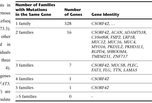
Table 3: Variant prioritization Steps in the analysis of combined exome sequences from 13 individuals with JBTS (p. 29)
Table 4: Genes with rare homozygous or multiple heterozygous variants from the combined exome sequences from 13 individuals with JBTS (p. 30)
Table 5: Clinical description of JBTS individuals with C5orf42 mutations (p. 34) Table 6: Genotypes and phenotypes of French Canadian individuals with JBTS (p. 53) Table 7: Clinical characteristics of new additional JBTS patients to this study (p. 72) Table 8: Summary of the clinical features of 43 FC patients with JBTS (p. 79)
ix
List of abbreviations
ARSACS: autosomal recessive spastic ataxia of Charlevoix-Saguenay CGNP: cerebellar granular neuronal precursors
COACH: Coloboma, Oligophrenia/developmental delay, Ataxia, Cerebellar vermis hypoplasia, Hepatic fibrosis syndrome
IFT: intraflagellar transport JBTS: Joubert syndrome JS: Joubert syndrome MKS: Meckel syndrome
MRI: magnetic resonance imaging MTS: molar tooth sign
OMA: oculomotor apraxia OFD: orofacio-digital syndrome PCP: planar cell polarity
SHH: Sonic Hedgehog
SNP: single nucleotide polymorphism TZ: transition zone
x
Acknowledgments
When I started my clinical Neurogenetics Fellowship in 2007, I could not have anticipated that I would embark in this exciting journey in the world of genetic research, such that I would eventually pursue a PhD in molecular biology.
I wish to thank Jacques Michaud, my Phd supervisor, for introducing me to these fascinating projects. In particular, I am grateful that he taught me how to always have a critical eye, and encouraged me to continually push the project further. He is the model of the clinician-scientist, a role that I wish to follow for the remainder of my career.
I would like to thank Bernard Brais, who encouraged me to start my Masters degree at the start of my fellowship, and started the entire process. He showed me how to engage science with enthusiasm and curiosity, and how to think ‘outside the box’ with patients.
I would like to thank Guy Rouleau, whose Neurogenetics clinics at the Montreal General Hospital during my residency first made me interested in this field, and for pointing me towards many compelling projects.
Thank you to Fadi Hamdan for all that he has taught me, for his advice, patience, support and friendship. I am eternally grateful.
Thank you to all the students in Jacques and Bernard’s Lab, who have made this journey so enjoyable and stimulating.
And of course thank you to my wonderful husband Ron who is always my pillar of support and encouragement, and to my parents, my unconditional cheerleaders.
1
Chapter 1:
Introduction
2
Joubert syndrome [JBTS, MIM 213300] is one of the most frequent forms of inherited congenital cerebellar ataxias. It was first described in 1969 by Dr. Marie Joubert. Dr Joubert described 4 French-Canadian siblings with agenesis of the cerebellar vermis, episodic breathing abnormalities (hypopnea/hyperpnea), abnormal eye movements (particularly oculomotor apraxia), gait ataxia and variable severities of intellectual disability (1). Following this seminal description, similarly affected individuals were reported throughout the world, and this constellation of clinical symptoms became characterized as “Joubert syndrome”.
Neuropathologic examination and advances in brain imaging have revealed that all JBTS patients have a distinctive midbrain and hindbrain malformation characterized by hypoplasia of the cerebellar vermis, thickened abnormally orientated superior cerebellar peduncles, and a deep inter-peduncular fossa. These features result in the hallmark “molar tooth sign” on axial brain magnetic resonance imaging (see Figure 1) first described by Maria
Figure 1. Molar Tooth Sign on MRI. Axial T2 brain MRI of a control individual (a) and patient with JBTS (b). Note the molar tooth sign (red circle), with deepened interpeduncular fossa (black arrow), and thickened and abnormally oriented superior cerebellar peduncles (yellow arrows).
3
et al. in 1997(2).
JBTS is a clinically heterogeneous condition. It combines hallmark neurologic signs with variable multi-organ involvement, mainly of the eyes, kidneys, skeleton and liver(3). The wide and variable clinical spectrum associated with JBTS is reflected by the large and confusing list of previously used syndromic designations associated with the various forms of JBTS. For example, the cerebello-oculo-renal syndrome designates individuals with ocular and renal involvement; Senior-Löken syndrome designates individuals with kidney and ocular disease; the acronym COACH refers to Coloboma, Oligophrenia/developmental delay, Ataxia, Cerebellar vermis hypoplasia, Hepatic fibrosis. These designations are no longer commonly used. The term “Joubert syndrome and related disorders” was coined to group all conditions sharing the molar tooth sign. Currently, a diagnosis of JBTS is made based on the mandatory presence of the molar tooth sign on imaging associated with the variable clinical features of hypotonia, ataxia, developmental delay, and oculomotor apraxia. Subtypes of JBTS are classified based on the extra-neural organ system involvement (see Table 1).
Table 1: Classification of JBTS based on clinical features
Clinical Subtype Mandatory features Preferentially
associated features
Previously used nosology
Pure Joubert syndrome (JS) MTS JS
JS with ocular defects (JS-O) MTS
Retinal dystrophy
JS
JS with renal defects (JS-R) MTS
Nephronophthesis JS with oculorenal defacts
(JS-OR) MTS Retinal dystrophy Nephronophthesis Cerebellooculorenal syndrome Senior-Löken syndrome JS with hepatic defects (JS-H) MTS
Congenital hepatic fibrosis
Colobomas Nephronophthesis
COACH syndrome Gentile syndrome JS with orofaciodigital defects
(JS-OFD)
MTS
Lobulated/bifid tongue (incl. hamartomas) Polydactyly
Cleft lip/palate Orofaciodigital VI syndrome
4 Clinical features associated with JBTS
Neurologic features
The cardinal features of JBTS are hypotonia, evolving into ataxia and developmental delay, often associated with intellectual disability, abnormal eye movements and altered respiratory pattern in the neonatal period. Hypotonia is observed in nearly all affected patients, and is generally the first sign observed on examination. Motor and language development is usually delayed in children with JBTS. In one series, the average age of sitting was 19 months, and of walking was 4 years in those who were able to develop these skills(4). Patients have ataxia, oromotor dyspraxia, and speech dyspraxia. Some children require assistive devices or need to use sign language for communication (5). Cognitive impairment is highly variable in JBTS(6). Intellectual disability, ranging widely from mild to very severe is common, with many patients retaining the capacity to attend special schools, learn specific job skills and work in protected conditions. It is important to note however that intellectual deficit is not inevitable, and that several individuals with borderline or even normal intelligence have been reported. Seizures have been reported, although they are not common. Behavioral problems, such as impulsivity, autism, perseveration and temper tantrums commonly occur (6).
Patients with JBTS can have other central nervous system abnormalities. These include occipital meningomyelocele, heterotopias, polymicrogyria, hydrocephalus, hypothalamic hamartoma and corpus callosal abnormalities (7).
Characteristic abnormal eye movements are almost always present(4, 6). Oculomotor apraxia is present in the large majority, and manifests as an inability to visually track objects, initiate saccades (high velocity large amplitude eye movements). This results in the compensatory head-thrust movement that is the hallmark of oculomotor apraxia. Oculomotor apraxia likely reflects abnormalities of axonal decussation. These abnormalities have been documented with diffusion tensor magnetic resonance imaging. As part of the axonal decussation abnormality, mirror movements (voluntary movements in a limb are coupled with
5
involuntary ‘mirror’ movements of the contralateral limb) are occasionally observable. Nystagmus is also common. Both oculomotor apraxia and nystagmus improve with age.
The respiratory abnormalities consist of short alternating episodes of apnea and hyperpnea, or isolated episodic hyperpnea. These tend to start shortly after birth, intensify with emotional stress, and improve with age. They generally disappear around 6 months of age.
Neuropathological examination of the midbrain-hindbrain in patients with JBTS reveals a characteristic association of abnormalities. The cerebellar vermis is invariably hypoplastic or absent, the cerebellar nuclei are often hypoplastic or fragmented, and there is heterotopia of Purkinje-like neurons. Pontine and medullary structures, such as the basis pontis, reticular formation, inferior olivary nucleus and dorsal column, are dysplastic (8, 9). There is typically failure of the decussation of the superior cerebellar peduncles and corticospinal tracts, as seen both on histological examination and imaging of fiber tracts on MRI(10).
Ocular features
The retina is among the organs the most frequently involved in JBTS. Overall, clinically-significant retinal involvement occurs in approximately one third of subjects. Retinal involvement can range from extremely severe Leber Congenital amaurosis to pigmentary retinopathies evident on fundus exam. In some cases milder forms of retinal dystrophy can be detected only on electroretinogram. There are two main forms of retinal disease,: severe congenital blindness with a flat electroretinogram (known as congenital amaurosis), and a later-onset pigmentary retinopathy that manifests with night-blindness and has a variable course. Colobomas can be unilateral or bilateral, and mostly affect the posterior segment of the eye. Colobamas are frequent in patients with liver abnormalities, occurring in up to 30%, as part of the COACH syndrome(11).
6
Renal involvement
Renal involvement is present in approximately 25% of JBTS patients (12). It is usually in the form of nephronophthesis, a progressive condition characterized by tubulointerstitial nephritis and cysts concentrated at the corticomedullary junction. Most patients present around the age of 9 years, with defects in urinary concentration, and progress to end stage renal disease late in the second decade(13).
Skeletal defects
Polydactyly is reported in approximately 8 to 16% of cases(12). The most common form is post axial affecting hands and feet. Mesoaxial polydactyly is associated with orofacial digital syndrome type 6, a subtype of JBTS characterized by the presence of oral frenulae, lingual tumors or hamartomas, and craniofacial abnormalities that include wide-spaced eyes and a midline lip groove.
Pathogenesis: JBTS is a prototypical ciliopathy
Over the past decade, advances in genetic research have revealed that JBTS is not only a clinically heterogeneous, but also a genetically heterogeneous disorder. Indeed, 24 causal genes have been identified to date. The first known causal gene, AHI1 (MIM# 608894), was identified in 2004 (14), and the second gene NPHP1 (MIM# 6071000) identified in 2005 (15). An updated list of JBTS genes is provided in Table 2. All these genes encode proteins that localize to the cilia, and all have an important role in ciliary structure or function. JBTS is thus considered a ciliopathy, and joins an expanding groups of disorders characterized by dysfunction of the cilia. To date, more than 30 human diseases are classified as ciliopathies, Examples of other ciliopathies include Meckel syndrome (MIM# 249000), Bardet-Biedl syndrome (MIM# 209900), Nephronophthisis (MIM# 256100), Orofacial digital syndrome (MIM# 311200), Alstrom syndrome (MIM# 203800) and Jeune Asphyxiating Thoracic Dysplasia (or Short-rib thoracic dysplasia, MIM# 208500). Overall, at least 80 ciliopathy
7
genes have been implicated so far. Ciliopathy disorders in general share overlapping features. All typically have multi-organ involvement, because cilia are present on almost all cell types. Defects in the primary cilium can affect the kidneys, retina, liver, brain and bones. The phenotype is a varying combination of cystic kidneys, polydactyly, retinopathy, liver fibrosis, developmental delay, intellectual disability and central nervous system abnormalities. Though ciliopathies may involve any of these organs and the significant clinical overlap between them, each disorder has a classical clinical presentation, such that most patients can be diagnosed in the clinical setting. For examples, Bardet-Biedl syndrome is characterized by intellectual disability, anosmia, obesity, polydactyly, hypogonadism, and retinal degeneration; Meckel syndrome is an early lethal disorder characterized by occipital encephalocele, cystic kidney and polydactyly; Alstrom syndrome is associated with obesity and retinopathy, but unlike JBTS or Bardet-Biedl syndrome, intellectual disability, polydactyly and hypogonadism are not a feature. Although individually rare, ciliopathy disorders may collectively have a prevalence as high as 1 in 2000 (based on the prevalence of the three common disease traits: renal cysts 1 in 500, retinal degeneration 1 in 3000 and polydactyly 1 in 500)(16).
8
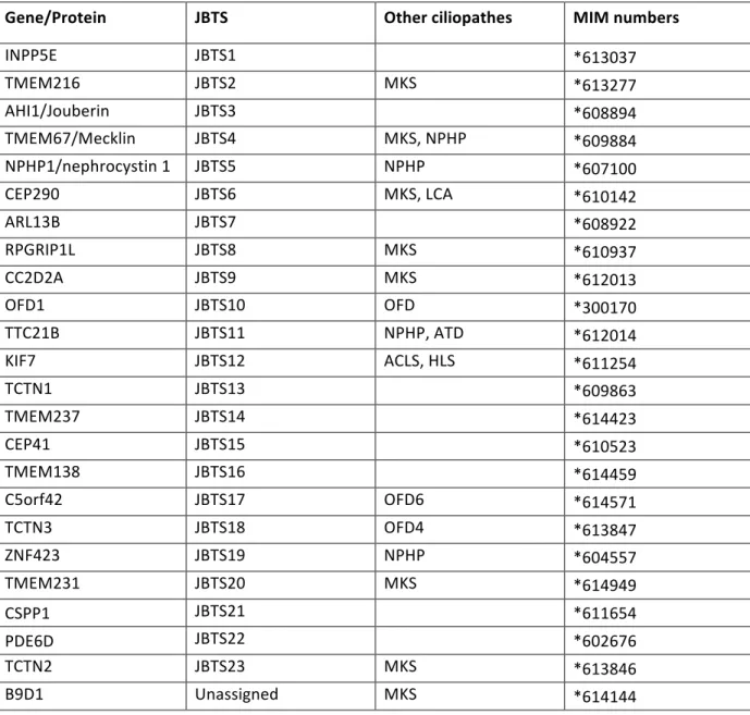
Table 2: Genes associated with JBTS. (MKS- Meckel syndrome, LCA- leber congenital amaurosis , ATD- asphyxiating thoracic dystrophy, ACLS- acrocallosal syndrome, HLS-hydrolethalus syndrome)
Gene/Protein JBTS Other ciliopathes MIM numbers
INPP5E JBTS1 *613037 TMEM216 JBTS2 MKS *613277 AHI1/Jouberin JBTS3 *608894 TMEM67/Mecklin JBTS4 MKS, NPHP *609884 NPHP1/nephrocystin 1 JBTS5 NPHP *607100 CEP290 JBTS6 MKS, LCA *610142 ARL13B JBTS7 *608922 RPGRIP1L JBTS8 MKS *610937 CC2D2A JBTS9 MKS *612013 OFD1 JBTS10 OFD *300170 TTC21B JBTS11 NPHP, ATD *612014 KIF7 JBTS12 ACLS, HLS *611254 TCTN1 JBTS13 *609863 TMEM237 JBTS14 *614423 CEP41 JBTS15 *610523 TMEM138 JBTS16 *614459 C5orf42 JBTS17 OFD6 *614571 TCTN3 JBTS18 OFD4 *613847 ZNF423 JBTS19 NPHP *604557 TMEM231 JBTS20 MKS *614949 CSPP1 JBTS21 *611654 PDE6D JBTS22 *602676 TCTN2 JBTS23 MKS *613846 B9D1 Unassigned MKS *614144
9 The primary cilium – basic biology
The primary cilium is a hair-like organelle protruding from the cell membrane, and is present in almost all cell-types. Each cell contains only one non-motile cilium, which is remarkably conserved throughout evolution. In the brain, motile cilia are restricted to the ependymal cells lining the ventricle and some of the choroid plexus cells, whereas primary cilia are present on all brain cells, including neural progenitors and mature neurons, glial cells and astrocytes(17).
The axoneme is the microtubule-based core of the cilia (Figure 2). In the non-motile cilia, it is formed by nine doublet microtubules (9+0), whereas in the motile cilia, it contains nine doublet microtubules and an extra pair of tubules (9+2) that are attached to a dynein motor to generate movement. The proximal end of the axoneme is anchored to the cell by the basal body, a modified centriole. The region between the basal body and the base of the axoneme is the transition zone. The main body of the transition zone is characterized by multiple rows of Y-shaped linkers projecting from the outer doublets of the axoneme and attaching to the ciliary membrane, as well as transitional fibers attaching the basal body to the peri-ciliary membrane. The transition zone isolates the cytoplasm of the cilia from the rest of the cell by acting as a selective barrier to protein diffusion. It also functions as a loading-unloading zone for transport into and out of the cilium. Cilia lack the necessary machinery for protein synthesis. Thus both soluble and membrane-bound ciliary proteins need to be trafficked to the basal body and taken up into the cilium. Proteins that enter the cilia from its base are carried along the axoneme via a microtubule-based transport system called intraflagellar transport (IFT). Anterograde transport toward the ciliary tip is regulated by complex B, consisting of at least 14 proteins (IFT172, IFT88, IFT81, IFT80, IFT74, IFT70, IFT57, IFT54, IFT52, IFT46, IFT27, IFT25, IFT22 and IFT20), in association with kinesin motors (KIF3A, KIF3B, and KAP3). The retrograde transport (i.e. from the tip of the cilia towards the base) is provided by complex A, which is composed of six IFT proteins (IFT144, IFT140, IFT139, IFT122, IFT121, and IFT43), in association with a dynein motor (DYNC2H1 and DYNC2L1)(18).
10
The antennae-like shape of the cilia (typically 5-10um in length) extending into the surrounding extracellular environment makes its ideally positioned to detect changes in chemical factors, morphogens or growth factors present in the extracellular medium. Additionally, the fact that the ciliary compartment is distinct from the remainder of the cellular compartment (both at the level of the intracellular compartment and the membrane) allows the partitioning of the sensory and signaling proteins away from the main body of the cell, and fine-tuning of biological responses to various stimuli. For example, the passive bending of the cilia present on renal tubular epithelial cells by fluid flow mediate the mechanosensation of extracellular urine flow; bending of the cilia within the kidney tubules leads to calcium influx through the polycystin-1 (PC-1)/polycystin-2 (PC-2) calcium channel. Mutations in both PC-1 and PC-2 are responsible for polycystic kidney disease. Retinal photoreceptors have specialized primary cilium that connects the inner segments which contains the cellular nucleus, to the outer nucleus which contains the photo-transducing pigments such as rhodopsin (19).
Axoneme' Transi-on'Zone' Basal'Body' Ciliary'necklace' Y7shaped'linkers' Transi-onal'fibers' External'cellular'' membrane' IFTA'(dynein'motor)' IFTB'(kinesin'motor)'
Figure 2. Schematic representation of the primary cilia. IFT: intraflagellar transport
11 Key role of the cilia in neurodevelopment
Primary cilia sense and transduce extracellular signals that influence a wide range of processes such as cell proliferation and polarity, developmental processes and neuronal growth. As exemplified with JBTS, perturbation in cilium formation or function can lead to profound defects in embryogenesis, with involvement of various organs including brain, retina, kidneys, liver, skeleton and heart. The primary cilium’s role in brain and organ patterning is mediated by morphogen signaling and its central role in the regulation of the sonic hedgehog (Shh), canonical and non-canonical Wnt pathways. The primary cilium also plays a pivotal role in a number of major growth factor-regulated signaling pathways, including platelet-derived growth factor (PDGF), fibroblast growth factor (FGF), and Notch pathways, with receptors for these ligands concentrated on the ciliary membranes(17).
Cilia and Sonic HedgeHog Signaling (Shh)
Shh signaling is essential for embryonic development, acting as a morphogen that is involved in patterning of multiple tissues, notably the central nervous system and limbs(20). In the central nervous sytem, Shh and the cilia have critical roles in neural tube/spinal cord patterning, telencephalic patterning, migration and placement of interneurons in the developing cortex, hippocampal neurogenesis, and formation of adult neural stem cells.
The cilium is required for amplification of the Shh signal when the Shh ligand is present. In the absence of Shh, its transmembrane receptor Patched (Ptc) localizes to the base of the cilium. There, Ptc represses the activity of Smo, a transmembrane protein with a structure reminiscent of G-protein-coupled receptors, by preventing its localization to the cilium. Upon binding of Shh to Ptc, Ptc is translocated outside the compartment allowing Smo to enter it, in a process that depends on the anterograde IFT machinery. The ciliary accumulation of Smo in turn facilitates the activation of Smo, which ultimately results in the accumulation of the active forms of three Gli (Gli-A) transcription molecules (Gli1, Gli2 and Gli3) (21). In the absence of Shh, Gli1, Gli2 and Gli3 are proteolytically processed into
12
repressor forms (Gli-R) by the removal of their carboxy tails. The activation of Smo inhibits Gli protein processing and results in the accumulation of the full Gli proteins containing the transactivator domain, which can enter the nucleus and activate target genes (Figure 3). Shh-related tissue patterning stems from the regulation of the balance between Gli transcriptional activators and repressors in the developing tissues. In the absence of Shh, Gli2 and Gli3 are cleaved to repressor forms (Gli2R, Gli3R), whereas in its presence, proteolysis is inhibited and Gli2 and Gli3 function as activators (Gli2A, Gli3A). Gli2A is the primary activator of Shh target genes, Gli3R the main repressor.
Shh signaling and activation of the Gli pathway are particularly important in cerebellar development (which is highly abnormal in patients with JBTS(22)). Most cell types of the cerebellum arise from the ventricular zone progenitors that are migrating through the forming cortex during embryogenesis. The cerebellar granule cells originate from a secondary germinal zone in the rostral rhombic lip. In the mouse, progenitors called cerebellar granular neuronal precursors (CGNP) undergo a first wave of proliferation at Embyonic day13.5. Then, from E17.5 on, they migrate over the nascent cerebellum to form the external granule layer where their population massively expands. This expansion is mediated by Shh, which is secreted by the Purkinje cells and diffuses up to the outer External Granule Layer where the CGNPs are located. The CGNPs highly express Gli1, the readout of the Shh pathway(23). Among the identified mitogens in the cerebellum (e.g. FGF-2, IGF-1 and EGF), Shh is the most potent mitogenic factor, able to trigger up to a 100-fold increase of CGNP proliferation
in vitro (24). The proliferation phase of the CGNPs peaks at P5-8 and declines thereafter to
stop at around P15. From P8, CGNPs start extending processes and initiate their onwards migration. The duration and intensity of the proliferation phase generating the pool of CGNPs is critical for the final shape and function of the cerebellum. Conditional removal of ciliary genes in CGNPs gives rise to striking dysgenesis and abnormal foliation of the cerebellum, as well as decreased target genes of and proliferation of CGNPS during cerebellar development(23).
13
Shh signaling is also essential for spinal cord and neural tube patterning. Disruption of Shh signaling leads to dorso-ventral patterning defects of the spinal cord, and neural tube defects such as failed closure of the neural tube (occipital encephalocele or exencephaly) and midline defects (colobomas, corpus callosum defects, defects of decussation of the
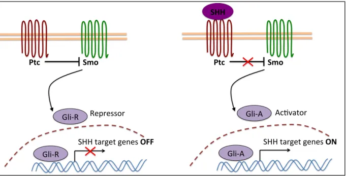
corticospinal tracts)(17). Shh protein is produced from the ventral pole of the neural tube and forms a ventral-to-dorsal concentration gradient. This gradient establishes the differential spatial patterns of gene expression in neural progenitor cells. High levels of Shh signaling induce the expression of the homeodomain protein Nkx2.2; consequently, Nkx2.2 is most strongly expressed ventrally. Low levels of Shh are sufficient to induce the expression of the transcription factor Olig2, which is expressed more dorsally than Nkx2.2.(25-27). Regulation of the balance between activated and repressor forms of Gli is critical in Shh-mediated patterning, and is regulated in part by the cilia. This explains why disruption of ciliary function can lead to different patterning abnormalities that reflect overactivity of either Gli-R or Gli-A. Ptc$ Smo$ Gli$R& Repressor& Gli$R& SHH&target&genes&OFF$ Ptc$ Smo$ Gli$A& Ac4vator& Gli$A& SHH&target&genes&ON$ SHH$
Figure 3: SHH signaling pathway schematic. In the absence of SHH, the receptor Ptc is located in the cilium and suppresses Smoothened (Smo). Full-length Gli transcription factors are truncated into their repressor forms (Gli-R). The binding of SHH to Ptc facilitates the activation of Smo, which ultimately results in the accumulation of the active forms of Gli (Gli-A) which can enter the nucleus and activate target genes. (Modified from Goetz and Anderson, Nature Reviews Genetics, 2010)
14
Disruption of Shh signaling also accounts for the limb patterning defects associated with ciliopathies, as Shh plays an important role in anteroposterior patterning of the limbs. Shh initially acts as morphogen and originates from posterior margin of the limb bud in a region called the zone of polarizing activity. Subsequently, it has mitogenic activity, which ensures the production of a sufficient number of cells to promote the normal complement of digits. Together, these two activities of Shh are responsible for specifying the identity of each digit and, as the limb bud expands, the position within the limb bud in which each digit forms. Loss-of-function mutations to Shh results in a loss of digits, whereas mutations in Gli3 cause polydactyly (28). Though the exact mechanism underlying polydactyly in ciliopathies has not been elucidated, disruption of Shh transduction signalling and abnormal Gli3 processing are key central processes (29) .
Cilia and the WNT pathway
The canonical and non-canonical (planar cell polarity, PCP) Wnt pathways are other important signaling pathways also regulated in part by the cilia. The Wnts are a family of secreted factors that bind Frizzled (Fzl) receptors to activate distinct signaling cascades, depending on the specific Wnt ligand, Wnt receptor, and the activity of Disheveled (Dvl), which acts as a molecular switch between signaling cascades (Figure 4). Canonical β-catenin-dependent Wnt signaling regulates proliferation, cell cycle progression and differentiation in the nervous system(30). The PCP Wnt signaling pathway provides cells with the positional clues required for concerted multicellular actions, such as the convergent extension cell movements that lead to neural tube closure (31). The PCP pathway is increasingly implicated in neuronal migration and axon guidance, in particular in the orderly development of large axon tracts. In absence of Wnt ligand, cytoplasmic β-catenin is constitutively phosphorylated by several kinases and targeted for proteosomal degradation(32). In canonical Wnt signaling, the binding of Wnt ligands to Fzl and LRP5 or LRP6 coreceptors transduces a signal across the plasma membrane that results in the activation of the Dvl. Activated Dvl inhibits the degradation and leads to the stabilization and accumulation of β-catenin in the cytoplasm. β-catenin then migrates into the nucleus where it
15
can act as a co-activator for TCF/LEF- mediated transcription of neurodevelopmental genes. Several JBTS genes, such as AHI, which encodes jouberin, regulate canonical Wnt signaling by facilitating β-catenin translocation to the nucleus. It has been shown that the primary cilium serves to modulate Wnt pathway responsiveness by sequestering β-catenin and jouberin within the cilium, thus limiting their nuclear entry. Similarly, other studies investigating the roles of the ciliary and basal body genes Kif3a, Ift88, and Ofd1 have revealed an inhibitory role of the primary cilium on canonical Wnt signaling, suggesting that loss of ciliary or basal body components leads to hyper-responsiveness to external canonical Wnt stimuli (33, 34). Loss of Kif3a in mice leads to enhanced β-catenin-dependent transcriptional activation. Embryonic fibroblasts from both Kif3a and Ofd1 mutant mice, as well as Ofd1-deficient embryonic stem cells have a hyper-responsive canonical Wnt response.
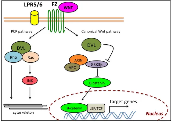
Figure 4: WNT signaling pathway schematic. The extracellular signalling molecule WNT activates the canonical pathway (right) and the planar cell polarity (PCP) pathway (left). In the canonical pathway, interaction of WNT with the transmembrane receptor frizzled (FZ) activates dishevelled (DVL), which induces the disassembly of a complex (axin, APC and GSK3β), and results in the accumulation of β-catenin in the cyctoplasm. β-catenin then translocates where it can activate the transcription of target genes. In the PCP pathway, FZ functions through G-proteins to activate DVL, which thereupon signals to Rho GTPases (Rho or Rac or both). Activated Ras signals through the c-Jun amino (N)-terminal kinase (JNK). Activation of Rho-GTPases induces changes in the cytoskeleton. (Modified from Moon et al, Nature Reviews
Genetics, 2004) target&genes! WNT!
FZ!
LPR5/6!
DVL& Rho& Ras& JNK& DVL& AXIN& GSK3β& APC& Β<catenin& Β<catenin& LEF/TCF& Nucleus' cytoskeleton& PCP&pathway& Canonical&Wnt&pathway&
16
The non-canonical PCP pathway is also activated via the binding of Wnt to Fzl and its co-receptor, however, it does not result in the downstream accumulation of β-catenin. Fzl recruits Dvl to form a complex with the Dvl-associated activator of morphogenesis 1 (DAAM) which then activates the small G-protein Rho. In turn, Rho activates Rho-associated kinase (ROCK), which is one of the major regulators of the cytoskeleton. Mutations in several ciliopathy-related genes, such as TMEM237, TMEM216 or TMEM67, result in abnormal PCP cascade activity, and conversely, mutations in key PCP pathway proteins such as inturned (intu), fuzzy (fuz) or dvl, were found to result in abnormal ciliogenesis.
In conditions of normal ciliary signaling, the PCP pathway is favored over the canonical pathway by inhibition of Dvl and activation of B-catenin destruction complex. With disruption of normal the basal body or ciliary function, there is decreased PCP signaling and upregulation of canonical Wnt signaling. Dysregulation of the Wnt pathway plays a critical role in the formation of renal cysts associated with ciliopathies. The Wnt pathway is required for the induction of the metanephric mesenchyme to develop the proximal portions of the nephron and for regulation of cell proliferation. Both PCP defects and hyperactivity of the canonical wnt signaling in transgenic mice overexpressiving B-catenin can result in the formation of cysts. The underlying mechanism is thought to be related to the fact that normal diving cells in the renal tubules orient their mitotic spindles parallel to the lumen, resulting in tubular elongation, whereas mis-orientated mitotic spindles, as observed in animal models of cystic kidney disease, result in tubular dilation(35).
Founder effect underlies many autosomal recessive disorders in French Canadians
There are a large number of genetic disorders that have a markedly increased incidence in the French-Canadian population. These include Autosomal recessive spastic ataxia of Charlevois-Saguenay (ARSACS, MIM# 270550), Agenesis of corpus callosum and peripheral neuropathy (ACCPN, Andermann syndrome (MIM# 218000), Leigh syndrome French-Canadian type (MIM# 220111), Oculopharyngeal muscular dystrophy (OPMD, MIM# 164300), Hereditary sensory and autonomic neuropathy type II (HSAN II, MIM#
17
201300), Myotonic dystrophy (MIM# 160900) and many others. The increased incidence of these hereditary disorders in the FC population reflects the presence of founder effects. A founder effect refers to the reduction in genetic variation that results when a small population establishes a new colony. The gene pool of the first generation of settlers (which may not represent the gene pool of the entire population it is derived from) contributes disproportionately to the ensuing population structure. Thus, if the founding settlers carry rare gene mutations, these will be passed on to future generations. Alleles that may have been rare in the original population will occur with a higher frequency in the settlers and their descendants.
The French-Canadian population arose following successive recent immigration waves. Of Quebec’s current >7 million inhabitants, 6 million are descendants from French Settlers. The French Colony of Nouvelle-France was first settled in 1608, with the foundation of the City of Quebec. From 1608 till the time it passed into British rule, approximately 8,500 permanent settlers arrived in Quebec, including only 1,600 women. The 2600 settlers who arrived before 1680 have contributed two thirds of the gene pool of the current French-Canadian population. Though settlers came from regions throughout France, the major source of immigrants to Nouvelle-France were the Atlantic seaports and the regions around Paris. French settlement in North America occurred in two main regions along the Saint Lawrence River and Acadia (now New Brunswick and Nova Scotia). In the 17th century, the population
groups had limited social and geographical mobility; therefore there were limited marriages between families from different regions or social groups. By the English conquest in 1759, most of the French-Canadian population lived in farming settlements along the Saint Lawrence River and, because of language and religious barriers, intermixed minimally with the Protestant English-speaking newcomers. The French-Canadian population has had a high fertility rate, attributed to the encouragement by the clergy, and frequently referred to as “la revanche des berceaux” (the revenge of the cradles). The high fecundity rate, early marriage and overall low infant mortality rate resulted in a rapid expansion of the French-Canadian population. Thus, the founding of the French-Canadian population by a limited number of settlers, combined with the relative isolation of the population and the higher fecundity rate,
18
explains the presence of a number of founder effects. This has resulted in the increased incidence of these rare genetic disorders in the French-Canadian population (36, 37).
For the recessive disorders with a higher incidence in the French-Canadian population, a single common founder mutation is usually observed, present at a higher carrier frequency amongst French-Canadian individuals. This explains the increased incidence of disease despite very low rates of consanguineous marriages. For example, ARSACS affects 1 in 1519 individuals in Charlevoix and 1 in 1952 individuals in the Saguenay, where the estimated carrier frequency is 1/22. One mutation (c.6594delT) in the causal gene SACS represents 94% of mutated alleles. Similarly, Leigh syndrome affects 1/2916 live births in the Saguenay-Lac-Saint-Jean region. Again, in Leigh syndrome only one allele, A354V, represents 98% of mutant alleles in the causal gene LRPPRC (36).
Landscape of Joubert syndrome in Quebec
Despite the fact that JBTS was first described in Quebec, the overwhelming majority of French-Canadian patients with JBTS did not have causal mutations identified at the onset of my doctoral project in 2010. In fact, in 2010, only 8 JBTS genes were known. Determining the precise underlying genetic etiology of JBTS is of great importance for several reasons. For the patients and their families, it facilitates genetic counseling and prenatal testing. It also concludes the diagnostic process (providing closure for families), improves the accuracy of prognostication and allows tailored medical management. Critically, determining causal genes expands our knowledge of the pathophysiology of JBTS and allows improved understanding of mechanisms underlying normal brain development. Ultimately, the understanding of the pathophysiology of these disorders will enable the development of targeted treatments in the future.
The precise incidence of JBTS is not known, but is estimated to be between 1/80,000 and 1/100,000 live births(38). Our preliminary observation suggested that there is a high prevalence of JBTS in the French-Canadian population living in the Lower St-Lawrence
19
(“Bas-du-Fleuve” in French) region of the province of Quebec (Figure 5). In particular, there is a striking cluster of families from the east end of the region (Matapedia region), including one family from Mont-Joli (population of 6,568), 3 families from Amqui (population 6,261) and 3 other families from Sayabec (population 1,877). This distribution strongly suggested the presence of a founder effect for JBTS.
The population of the Lower St-Lawrence region was initially established as the result of the immigration of a limited number of settlers (6,000 individuals) from Quebec City and its surroundings in the late 17th and early 18th century followed by a rapid increase of the population due to a high fertility rate(39). The establishment of settlers in the region followed a west-to-east pattern, with later settlers migrating to regions further east. A small number of Acadians also contributed to the early population of the Matapedia region(40). Thus, the demographic growth of this population appears to be characterized by a series of bottlenecks that may have resulted in regional founder effects. We hypothesized that a founder effect could underlie the clustering of individuals with JBTS in the Lower St-Lawrence region. If this were true, a single common homozygous mutation might explain a large proportion of them.
20
It was initially established that the population of the Lower St. Lawrence region was a result of both the immi-gration of a limited number of settlers (6,000 individuals) from Quebec City and its surrounding areas in the late
17thcentury and beginning of the 18thcentury and a rapid
increase in settlers resulting from a high fertility rate.22
The establishment of settlers in the region followed a west-to-east pattern, and settlers later migrated to regions farther east. A small number of Acadians also contributed
to the early population of the Matapedia region.23 The
demographic growth of this population thus appears to be characterized by a series of bottlenecks that might have resulted in regional founder effects. We hypothesized that a founder effect could underlie the clustering of individuals with JBTS in the Lower St. Lawrence region, raising the possibility that a common homozygous muta-tion explains a large pormuta-tion of them. We performed whole-genome SNP genotyping in all 15 individuals with JBTS by using the Illumina Human 610 Genotyping BeadChip panel, which interrogates 620,901 SNPs, and
we used PLINK24 to search for homozygosity regions
con-taining >30 consecutive SNPs and extending over >1Mb. We identified several overlapping regions of shared homozygosity, but these regions were not found in more than five families, were small (1 megabase or less), and contained genes that are unlikely to play a role in cilia
development and/or function (Table S1, available online).
Altogether, the genotyping data suggest the presence of allelic and/or genetic heterogeneity within our cohort.
Given the lack of hints from genotype-based mapping, we decided to sequence the protein-coding exomes of all our JBTS-affected subjects in the hopes of identifying a unique candidate gene harboring private pathogenic vari-ants in a large fraction of the samples. Genomic DNA from each sample was captured with the Agilent SureSelect
Figure 1. Distribution of Individuals with JBTS in the Lower St. Lawrence Region
Numbers refer to families (pedigrees inFigure 2). Note the cluster of families along Route 132, which follows the Matapedia River.
50 Mb oligonucleotide library, and the captured DNA was sequenced with paired-end 100 bp reads on Illumina HiSeq2000. The result was an average of 14.7 Gb of raw sequence for each sample. Data were
analyzed as previously described.25 After
we used Picard (v. 1.48) to remove putative PCR-generated duplicate reads, we aligned the reads to human genome assembly hg19 by using a Burroughs-Wheeler algo-rithm (BWA v. 0.5.9). The median read depth of the bases in CCDS (consensus coding sequence) exons was 115
(deter-mined with Broad Institute Genome
Analysis Toolkit v. 1.0.4418).26On average,
88% (52.9%) of the bases in CCDS exons were covered by at least 20 reads. We called sequence variants by using custom scripts for Samtools (v. 0.1.17), Pileup, and varFilter, and we required at least three variant reads as well as >20% variant reads for each called position. Single-nucleotide variants (SNVs) had Phred-like quality scores of at least 20, and small insertions or deletions (indels) had scores of at least 50. We used Annovar to annotate variants according to the type of mutation, occurrence in dbSNP, SIFT score, and 1,000 Genomes allele
frequency.27 To identify potentially pathogenic variants,
we filtered out (1) synonymous variants or intronic vari-ants other than those affecting the consensus splice sites, (2) variants seen in more than one of 261 exomes from individuals with rare, monogenic diseases unrelated to JBTS (these individuals were sequenced at the McGill University and Genome Quebec Innovation Centre), and (3) variants with a frequency greater than 0.5% in the
1,000 Genomes Browser (Tables 1 and 2).
We first examined the exome datasets to look for rare variants in the 15 genes already associated with JBTS (these genes are INPP5E [MIM 613037], TMEM216 [MIM 613277], AHI1 [MIM 608894], NPHP1 [MIM 607100], CEP290 [MIM 610142], TMEM67 [MIM 609884], RPGRIP1L [MIM 610937], ARL13B [MIM 608922], CC2D2A [MIM 612013], CXORF5 [MIM 300170], KIF7 [MIM 611254], TCTN1 [MIM 609863], TCTN2 [MIM 613885], TMEM237
[MIM 614424], and CEP41)2–19 as well as in the JBTS
candidate gene, TTC21B (MIM 612014).28Two individuals
(II-1 from family 484 and II-2 from family 473, Figure S2)
that are not known to be related were each found to be carrying two heterozygous missense variants (c.4667A>T
[p.Asp1556Val] and c.3376G>A [p.Glu1126Lys]) in
CC2D2A (RefSeq accession number NM_001080522.2).13,14
These amino acids are highly conserved, and both
694 The American Journal of Human Genetics 90, 693–700, April 6, 2012
Figure 5. Distribution of individuals with Joubert syndrome in the Lower St-Lawrence region. Note the cluster of families along Route 132, which follows the Matapedia river.
21 Hypotheses and objectives
JBTS remains largely unexplained in French Canadians. Given the documented presence of multiple founder effects in the French-Canadian population, and our preliminary observation that multiple affected families clustered in the Lower Saint Lawrence and Matapedia regions, we hypothesized that there existed a founder effect with a recurrent mutation in one gene that explains the majority of cases. In addition, given the fact that causal mutations in known JBTS genes were rarely identified in French-Canadian JBTS families on a clinical basis, we hypothesized the presence of a recurrent founder mutation in a novel JBTS gene. Therefore, our initial objective was to study these French-Canadian families using single nucleotide polymorphism (SNP) genotyping and homozygosity mapping, a strategy that is well suited for the study of families with an autosomal recessive disorder in which a founder effect is suspected.
Because JBTS is known to be a very genetically heterogeneous condition, we also hypothesized that, though a founder mutation likely existed and explained the majority of FC JBTS families, mutations in more than one gene probably accounted for the genetic etiology in Quebec. Thus, we anticipated using WES to study all JBTS families that remained
unexplained following our initial SNP genotyping and homozygosity approach. Indeed, WES
is an extremely powerful strategy to study disorders that are clinically and genetically heterogeneous, such as JBTS.
The specific objectives of this project were:
1) To recruit a cohort of French-Canadian families with JBTS and assess their geographic distribution throughout Quebec
2) To clinically characterize the affected JBTS individuals by extensive phenotypic evaluation of all JBTS patients when possible, supplemented by review of medical records and radiographic imaging.
22
3) To identify the causal genes underlying JBTS in the French-Canadian population using (a) a homozygosity mapping approach and/or (b) whole exome sequencing (WES).
4) To evaluate for the presence of a genetic founder effect in the French-Canadian population.
23
Chapter 2:
Mutations in C5ORF42 cause Joubert
syndrome in the French Canadian
Population
24
Manuscript 1:
Mutations in C5ORF42 cause Joubert syndrome in the French
Canadian Population.
Published in: Am J Hum Genet. 2012 Apr 6;90(4):693-700,
PMID:
22425360
Myriam Srour1,11, Jeremy Schwartzentruber2,11, Fadi F. Hamdan1, Luis H. Ospina3, Lysanne Patry1, Damian Labuda4, Christine Massicotte4, Sylvia Dobrzeniecka1, José Capo-Chichi1, Simon Papillon-Cavanagh4, Mark E. Samuels4, Kym M. Boycott5, Michael I. Shevell6, Rachel Laframboise7, Valérie Désilets4, FORGE Canada Consortium*, Bruno Maranda8, Guy A Rouleau9, Jacek Majewski10, Jacques L. Michaud1
1Centre of Excellence in Neurosciences of Université de Montréal and Sainte-Justine Hospital
Research Center, Montréal H3T 1C5, Canada; 2McGill University and Genome Quebec Innovation Centre, Montréal H3A 1A4, Canada; 3Department of Ophthalmology, Sainte-Justine Hospital Research Center, Montréal, H3T 1C5, Canada; 4Sainte-Justine Hospital Research Center, Montréal H3T 1C5, Canada; 5Children's Hospital of Eastern Ontario Research Institute, Ottawa, K1H 8L1 Canada; 6Division of Pediatric Neurology, Montreal Children's Hospital-McGill University Health Center, Montreal, H3H 1P3, Canada;
7Department of Medical Genetics, Centre Hospitalier Universitaire Laval, Québec, G1V 4G2,
Canada; 8Division of genetics, Centre Hospitalier Universitaire de Sherbrooke, Sherbrooke, J1H 5N4, Canada; 9Centre of Excellence in Neurosciences of Université de Montréal and Department of Medicine, Montréal H2L 2W5, Canada; 10Department of Human Genetics,
McGill University, H3A 1A4, Montréal, Canada.
11 Equal contribution
25 ABSTRACT
Joubert syndrome (JBTS) is an autosomal recessive disorder characterized by a distinctive mid-hindbrain malformation, developmental delay with hypotonia, ocular-motor apraxia and breathing abnormalities. Although JBTS was first described more than 40 years ago in French Canadian siblings, the causal mutations have not yet been identified in this family, nor in most French Canadian individuals subsequently described. We ascertained a cluster of 15 individuals with JBTS from 11 families living in the Lower St-Lawrence region. SNP genotyping excluded the presence of a common homozygous mutation that would explain the clustering of these individuals. Exome sequencing performed in all the 15 subjects showed that 9 affected individuals from 7 families (including the original JBTS family) carried rare compound heterozygous mutations in C5ORF42. Two missense variants (c.4006C>T [p.Arg1336Trp], c.4690G>A [p.Ala1564Thr]) and a splicing mutation (c.7400+1G>A), which causes exon skipping, were found in multiple subjects, not known to be related, whereas three other mutations, all truncating (c.6407delC [p.Pro2136Hisfs*31], c.4804C>T [p.Arg1602X], c.7477C>T [p.Arg2493X]), were identified in single individuals. None of the unaffected first-degree relatives were compound heterozygotes for these mutations. Moreover, none of the 6 putative mutations was detected among 477 French Canadian controls. Our data suggests that mutations in C5ORF42 explain a large fraction of French Canadian individuals with JBTS.
26
Joubert syndrome (JBTS; MIM 213300) is an autosomal recessive disorder characterized by the presence of hypotonia, apnea or hyperpnea in infancy, oculomotor apraxia, and variable developmental delay or intellectual impairment (reviewed in1). The diagnostic hallmark of JBTS is the presence of a complex malformation of the midbrain-hindbrain junction that comprises cerebellar vermis hypoplasia or aplasia, deepened interpeduncular fossa, and elongated superior cerebellar peduncles. This malformation takes the appearance of a molar tooth on axial brain MRI. In a subset of individuals, JBTS also involves other organs, resulting in cystic kidneys, retinopathy, or polydactyly. JBTS is a genetically heterogeneous condition with 15 genes described to date(41-58). All of these genes appear to play a role in the development and/or function of non-motile cilia. Although JBTS was first described in French Canadian siblings more than 40 years ago by Marie Joubert and colleagues, until now the causal mutations have not yet been identified in the original family nor in most French Canadians subjects(1).
There is a high prevalence of JBTS in the French Canadian population living in the Lower St-Lawrence (“Bas-du-Fleuve” in French) region of the province of Quebec (Fig. 6). In total, we identified 16 living affected individuals (from 11 unrelated families) with at least one grandparent originating from that region. Informed consent was obtained from all individuals or their legal guardians. This project was approved by our institutional ethics committee. We were initially able to collect blood-derived DNA from 15 of these individuals, including an affected individual (II-1 in family 394; individual BD in(1)) from the original JBTS family described by Marie Joubert and colleagues in 1969. There was a striking cluster of 7 families from the east end of the region (Matapedia region), including one family from Mont-Joli (population of 6,568), 3 families from Amqui (population 6,261) and 3 other families from Sayabec (population 1,877). Individual II-1 from family 394 did not undergo brain imaging studies but an MRI scan performed in her brother (II-2) showed the molar tooth sign (MTS)(59, 60). All the other affected individuals showed the MTS and variable expression of the classical JBTS features. The cohort included three families with two affected siblings, and in no case were parents affected, consistent with a recessive mode of transmission.
27
The population of the Lower St-Lawrence region was initially established as the result of the immigration of a limited number of settlers (6,000 individuals) from Quebec City and its surroundings in the late 17th century and beginning of the 18th century followed by a rapid increase due to a high fertility rate(39). The establishment of settlers in the region followed a west-to-east pattern, with later settlers migrating to regions further east. A small number of Acadians also contributed to the early population of the Matapedia region(40). The demographic growth of this population thus appears to be characterized by a series of bottlenecks that may have resulted in regional founder effects. We hypothesized that a founder effect could underlie the clustering of individuals with JBTS in the Lower
St-It was initially established that the population of the Lower St. Lawrence region was a result of both the immi-gration of a limited number of settlers (6,000 individuals) from Quebec City and its surrounding areas in the late
17thcentury and beginning of the 18thcentury and a rapid
increase in settlers resulting from a high fertility rate.22
The establishment of settlers in the region followed a west-to-east pattern, and settlers later migrated to regions farther east. A small number of Acadians also contributed
to the early population of the Matapedia region.23 The
demographic growth of this population thus appears to be characterized by a series of bottlenecks that might have resulted in regional founder effects. We hypothesized that a founder effect could underlie the clustering of individuals with JBTS in the Lower St. Lawrence region, raising the possibility that a common homozygous muta-tion explains a large pormuta-tion of them. We performed whole-genome SNP genotyping in all 15 individuals with JBTS by using the Illumina Human 610 Genotyping BeadChip panel, which interrogates 620,901 SNPs, and
we used PLINK24to search for homozygosity regions
con-taining >30 consecutive SNPs and extending over >1Mb. We identified several overlapping regions of shared homozygosity, but these regions were not found in more than five families, were small (1 megabase or less), and contained genes that are unlikely to play a role in cilia
development and/or function (Table S1, available online).
Altogether, the genotyping data suggest the presence of allelic and/or genetic heterogeneity within our cohort.
Given the lack of hints from genotype-based mapping, we decided to sequence the protein-coding exomes of all our JBTS-affected subjects in the hopes of identifying a
Figure 1. Distribution of Individuals with JBTS in the Lower St. Lawrence Region
Numbers refer to families (pedigrees inFigure 2). Note the cluster of families along Route 132, which follows the Matapedia River.
50 Mb oligonucleotide library, and the captured DNA was sequenced with paired-end 100 bp reads on Illumina HiSeq2000. The result was an average of 14.7 Gb of raw sequence for each sample. Data were
analyzed as previously described.25 After
we used Picard (v. 1.48) to remove putative PCR-generated duplicate reads, we aligned the reads to human genome assembly hg19 by using a Burroughs-Wheeler algo-rithm (BWA v. 0.5.9). The median read depth of the bases in CCDS (consensus coding sequence) exons was 115
(deter-mined with Broad Institute Genome
Analysis Toolkit v. 1.0.4418).26On average,
88% (52.9%) of the bases in CCDS exons were covered by at least 20 reads. We called sequence variants by using custom scripts for Samtools (v. 0.1.17), Pileup, and varFilter, and we required at least three variant reads as well as >20% variant reads for each called position. Single-nucleotide variants (SNVs) had Phred-like quality scores of at least 20, and small insertions or deletions (indels) had scores of at least 50. We used Annovar to annotate variants according to the type of mutation, occurrence in dbSNP, SIFT score, and 1,000 Genomes allele
frequency.27 To identify potentially pathogenic variants,
we filtered out (1) synonymous variants or intronic vari-ants other than those affecting the consensus splice sites, (2) variants seen in more than one of 261 exomes from individuals with rare, monogenic diseases unrelated to JBTS (these individuals were sequenced at the McGill University and Genome Quebec Innovation Centre), and (3) variants with a frequency greater than 0.5% in the
1,000 Genomes Browser (Tables 1 and 2).
We first examined the exome datasets to look for rare variants in the 15 genes already associated with JBTS (these genes are INPP5E [MIM 613037], TMEM216 [MIM 613277], AHI1 [MIM 608894], NPHP1 [MIM 607100], CEP290 [MIM 610142], TMEM67 [MIM 609884], RPGRIP1L [MIM 610937], ARL13B [MIM 608922], CC2D2A [MIM 612013], CXORF5 [MIM 300170], KIF7 [MIM 611254], TCTN1 [MIM 609863], TCTN2 [MIM 613885], TMEM237
[MIM 614424], and CEP41)2–19 as well as in the JBTS
candidate gene, TTC21B (MIM 612014).28Two individuals
(II-1 from family 484 and II-2 from family 473, Figure S2)
that are not known to be related were each found to be carrying two heterozygous missense variants (c.4667A>T Figure 6. Distribution of individuals with Joubert syndrome in the Lower
St-Lawrence region. Numbers refer to families (pedigrees in Figure 7). Note the cluster of families along Route 132, which follows the Matapedia river.