Metabolic Brain Disease
Brain edema: a valid endpoint for measuring hepatic encephalopathy?
--ManuscriptDraft--Manuscript Number: MEBR-D-15-00235R1
Full Title: Brain edema: a valid endpoint for measuring hepatic encephalopathy?
Article Type: SI: ISHEN
Keywords: brain edema; hepatic encephalopathy; astrocyte; Magnetic Resonance Imaging; neurobehavior
Corresponding Author: Christopher F Rose CANADA
Corresponding Author Secondary Information:
Corresponding Author's Institution: Corresponding Author's Secondary Institution:
First Author: Chantal Bémeur
First Author Secondary Information:
Order of Authors: Chantal Bémeur Cristina Cudulbu Gitte Dam
Alexander S Thrane Arthur JL Cooper Christopher F Rose Order of Authors Secondary Information:
Funding Information:
Abstract: Hepatic encephalopathy (HE) is a major complication of liver failure/disease which frequently develops during the progression of end-stage liver disease. This metabolic neuropsychiatric syndrome involves a spectrum of symptoms, including cognition impairment, attention deficits and motor dysfunction which eventually can progress to coma and death. Pathologically, HE is characterized by swelling of the astrocytes which consequently leads to brain edema, a common feature found in patients with acute liver failure (ALF) as well as in cirrhotic patients suffering from HE. The pathogenic factors involved in the onset of astrocyte swelling and brain edema in HE are unresolved. However, the role of astrocyte swelling/brain edema in the
development of HE remains ambiguous and therefore measuring brain edema as an endpoint to evaluate HE is questioned. The following review will determine the effect of astrocyte swelling and brain edema on neurological function, discuss the various possible techniques to measure brain edema and lastly to propose a number of neurobehavioral tests to evaluate HE.
Brain edema: a valid endpoint for measuring hepatic encephalopathy?
Chantal Bémeur1,7, Cristina Cudalbu2, Gitte Dam3, Alexander S. Thrane4,5, Arthur J. L.
Cooper6 and Christopher F. Rose7
1 Département de nutrition, Université de Montréal, Montréal, Québec, Canada 2Centre d'Imagerie Biomédicale (CIBM), Ecole Polytechnique Fédérale de Lausanne (EPFL), Lausanne, Switzerland
3 Department of Medicine V (Hepatology and Gastroenterology) , Aarhus , Denmark. 4 Department of Ophthalmology, Haukeland University Hospital, Bergen 5021, Norway. 5 Division of Glial Disease and Therapeutics, Center for Translational Neuromedicine, Department of Neurosurgery, University of Rochester Medical Center, Rochester, New York 14642.
6Department of Biochemistry and Molecular Biology, New York Medical College,
Valhalla, New York 10595
7 Hepato-Neuro Laboratory, CRCHUM, Université de Montréal, Montréal, Québec,
Canada
Corresponding author:
Christopher F. Rose Ph.D., Hepato-Neuro Laboratory, CRCHUM, Université de Montréal, Montréal, Québec, Canada
Phone: +1 514 890 8000, ext. 35739 email:[email protected]
List of abbreviations:
HE: hepatic encephalopathy; ALF : Acute Liver Failure; GFAP: glial fibrillary acid protein; CSF: cerebrospinal fluid; RVD: regulatory volume decrease; MRI: magnetic resonance imaging; DWI: diffusion weighted imaging; FLAIR: fast fluid-attenuated inversion recovery
Keywords:
Brain edema; Hepatic encephalopathy; astrocyte; magnetic resonance imaging; neurobehavior
Disclosures: The authors have no conflicts to disclose
1 2 3 4 5 6 7 8 9 10 11 12 13 14 15 16 17 18 19 20 21 22 23 24 25 26 27 28 29 30 31 32 33 34 35 36 37 38 39 40 41 42 43 44 45 46 47 48 49 50 51 52 53 54 55 56 57 58 59 60
Abstract
Hepatic encephalopathy (HE) is a major complication of liver failure/disease which frequently develops during the progression of end-stage liver disease. This metabolic neuropsychiatric syndrome involves a spectrum of symptoms, including cognition impairment, attention deficits and motor dysfunction which eventually can progress to coma and death. Pathologically, HE is characterized by swelling of the astrocytes which consequently leads to brain edema, a common feature found in patients with acute liver failure (ALF) as well as in cirrhotic patients suffering from HE. The pathogenic factors involved in the onset of astrocyte swelling and brain edema in HE are unresolved. However, the role of astrocyte swelling/brain edema in the development of HE remains ambiguous and therefore measuring brain edema as an endpoint to evaluate HE is questioned. The following review will determine the effect of astrocyte swelling and brain edema on neurological function, discuss the various possible techniques to measure brain edema and lastly to propose a number of neurobehavioral tests to evaluate HE.
Astrocyte swelling and brain dysfunction Astrocytes and disease
Astrocyte swelling has been implicated in a range of neurological disorders, involving stroke, migraine, epilepsy, and metabolic encephalopathies (including HE) (Felipo, 2013; Papadopoulos and Verkman, 2013; Thrane et al., 2014). Selective genetic (e.g. a mutation in glial fibrillary acid protein (GFAP) resulting in Alexander disease) or chemical impairment (fluorocitrate or fluoroacetate) of astrocyte function can also cause severe neurological impairment ranging from lethargy, stupor, ataxia, seizures to coma and death (Swanson and Graham, 1994; Messing et al., 2012). The cellular mechanisms causing these phenotypic manifestations can best be understood by first examining the physiological roles of astroglia.
Fundamental role of astrocytes in the central nervous system
Astrocytes are multifunctional glial cells responsible for key brain homeostatic functions such as regulating ion gradients, cerebral blood flow, blood-brain barrier (BBB) integrity, scar formation (reactive gliosis) and cellular metabolism (Ransom et al., 2003). They are electrically inactive, but signal with intracellular calcium and other secondary messengers (including adenosine triphosphate (ATP) and cyclic guanosine
monophosphate (cGMP)) (Cotrina et al., 2000; Sun et al., 2013). The importance of astrocytes for more complex nervous systems is highlighted by the fact that evolution has selectively increased the size, relative abundance and complexity of these cells, whilst leaving neurons relatively unchanged (Oberheim et al., 2006). Collectively, neuroglia make up approximately 41% of the volume fraction of human cortex, compared to 27% by neurons, and 12% by interstitial fluid, with the remainder comprising blood (10%) and cerebrospinal fluid (CSF); 10%)) (Syková and Nicholson, 2008). Although astrocytes were initially seen as simple star shaped structural cells on silver-chromate and
cytoskeletal (GFAP) staining, cytoplasmic labeling studies have revealed a more complex ‘bush-like’ morphology (Oberheim et al., 2008). Individual astrocytes are linked together into a syncytium by gap junctions, which have been suggested to facilitate faster ion movement, metabolic trafficking and neurovascular coupling (Rose and Ransom, 1997; 4 5 6 7 8 9 10 11 12 13 14 15 16 17 18 19 20 21 22 23 24 25 26 27 28 29 30 31 32 33 34 35 36 37 38 39 40 41 42 43 44 45 46 47 48 49 50 51 52 53 54 55 56 57 58 59 60
Rouach et al., 2008). Astrocytes have an intricate subcellular anatomy, with thousands of specialized processes that express a different subset of transporters depending on whether they abut blood vessels (peri-vascular end-feet), CSF (sub-ependymal end-feet) or
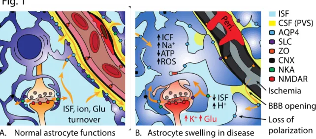
synapses (peri-synaptic processes). Peri-vascular processes, for instance, are selectively endowed with ion and water transport mechanisms (including Na+-K+-2Cl- cotransporter isoform 1(NKCC1), inwardly rectifying potassium channel Kir4.1, glutamate transporter-1 (GLTtransporter-1), glucose transporter (GLUT), Na+-K+-ATPase (NKA), system N transporter (SN1)), including the main brain water channel, aquaporin 4 (AQP4) (Figure 1). Metabolically, astrocytes are estimated to be responsible for about 30% of cerebral metabolism, and are amongst the most glycolytically active cells in the brain (Kasischke et al., 2004; Pellerin et al., 2007). Finally, a large body of evidence indicates that
astrocytes play an active role in synaptic transmission (tripartite synapse), for instance by regulating neurotransmitter turnover and perhaps gliotransmission, along with fine-tuning the structural and electrochemical synaptic environment (Araque et al., 1999; Nedergaard and Verkhratsky, 2012). Therefore, astrocytes are critical cells which play a pivotal role in health and disease (Rose et al., 2013).
Astrocytes: susceptible to swelling
Astrocyte responses to osmotic stress have been extensively studied in vitro and in situ. Astrocytes are thought to be able to cope with brief exposures (30-90 min) to mild or moderate amounts of swelling by offloading osmolytes, including potentially
excitotoxic neurotransmitters, a mechanism known as regulatory volume decrease (RVD) (Kimelberg, 1987; Ordaz et al., 2004; Thrane et al., 2011; Anderova et al., 2014).
Conversely, hyper-osmotic stress may be able to induce a regulatory volume increase (RVI) by uptake of osmolytes and water (Evanko et al., 2004; Risher et al., 2009). The molecular mechanisms underlying this response are incompletely understood, and thought to involve osmo- or stretch-sensitive intracellular signaling cascades involving [Ca2+]I transients, AQP4 and volume-regulated anion channels (VRACs) (Mulligan and MacVicar, 2006; Thrane et al., 2011; Qiu et al., 2014; Voss et al., 2014). The high expression of water transporting membrane proteins has also led many authors to suggest that astrocytes are more susceptible to swelling than neurons when the capacity for RVD is exhausted (Häussinger, 2000; Bosoi and Rose, 2013; Papadopoulos and Verkman, 2013; Thrane et al., 2015).
Astrocyte swelling and brain edema
On a larger scale, brain edema is believed to preferentially enter via astrocyte membranes by virtue of their strategic perivascular location and high water permeability (Papadopoulos and Verkman, 2013). A range of studies have shown that deleting or inhibiting AQP4 reduces the amount of brain edema following many types of insults, including hypoosmotic stress, stroke, traumatic brain injury, hepatic (but not
hyperammonemic) encephalopathy and meningitis (Manley et al., 2000; Amiry-Moghaddam et al., 2003; Papadopoulos and Verkman, 2005; Fukuda et al., 2013;
Rangroo Thrane et al., 2013; Rao et al., 2014). Conversely, AQP4 deletion can also slow edema resorption, and this mechanism likely explains why AQP4-/- animals display worse vasogenic edema compared to AQP4+/+ animals in the context of tumors, abscesses and following subarachnoid hemorrhage (Papadopoulos et al., 2004; Bloch et al., 2005; Tait 4 5 6 7 8 9 10 11 12 13 14 15 16 17 18 19 20 21 22 23 24 25 26 27 28 29 30 31 32 33 34 35 36 37 38 39 40 41 42 43 44 45 46 47 48 49 50 51 52 53 54 55 56 57 58 59 60
et al., 2010). Astrocyte swelling is accompanied by a shift of fluid from either interstitial or intravascular to the intracellular (astrocytic) compartment (Thrane et al., 2014). This fluid shift can have several detrimental effects. Net fluid entry to the brain from the vascular compartment (vasogenic or osmotic edema) increases the brain volume, raising intracranial pressure, and causing potentially fatal brainstem compression; complications which develop in 25% of patients with ALF (Lee, 2012). However, astrocyte swelling can hypothetically also occur if there is an isolated fluid shift from the interstitial to the intracellular (cytosol) compartment, with no net fluid entry into the brain. This is termed cytotoxic edema, and when seen in isolation it does by definition not lead to raised intracranial pressure (Simard et al., 2007). However, this definition of cytotoxic edema is perhaps controversial, as cytotoxic swelling would arguably always be accompanied by some degree of net brain edema through other mechanisms (e.g. osmotic gradient across the BBB). Moreover, cytotoxic edema has several direct detrimental effects, such as increasing the concentration of ions and neurotransmitters in the now shrunken interstitial space. This can potentially lower the seizure threshold (increased [K+]o) and cause
excitotoxicity (increased [Glutamate]o and consequent NMDA receptor activation). Additionally, cytotoxic edema can dilute intracellular ion and metabolite concentrations and thereby impair cellular metabolism. Generally, any form of tissue edema will also increase the distance for oxygen and metabolite diffusion, exposing micro-watershed areas to hypoxia (Takano et al., 2007; Thrane et al., 2013).
Brain edema and the ‘glymphatic’ system
Brain edema can also develop by net salt and water entry into the parenchyma in the presence of an intact BBB, termed ionic edema by some authors, to distinguish it from isolated (cytotoxic) extra-to-intra-cellular redistribution (Simard et al., 2007; Iliff et al., 2012; Thrane et al., 2014). One recent hypothesis that might explain this apparent paradoxical observation, proposes that physiological interstitial fluid turnover in the brain parenchyma is facilitated by continuous influx of peri-arterial CSF and efflux via the perivascular space of a subset of large veins (Iliff et al., 2012) or the recently discovered brain glymphatic vessels (Louveau et al., 2015). This pathway, termed the glymphatic system, might also explain the preponderance of astrocytes for swelling (Manley et al., 2000; Amiry-Moghaddam et al., 2003) and the extensive molecular machinery astrocytes express for volume regulation (Iliff et al., 2012; Nedergaard, 2013). This hypothesis is also particularly interesting in the context of astrocyte swelling, because the main gateway for net water entry into the brain parenchyma is thought to be via the AQP4-enriched perivascular membranes of astrocytes (Nielsen et al., 1997; Nagelhus and Ottersen, 2013; Papadopoulos and Verkman, 2013). Moreover, a derangement of the polarized perivascular expression of salt and water transporters along with decreased glymphatic interstitial fluid turnover appears to be a consistent feature of traumatized, infarcted, aged and even sleep-deprived brain tissue, which might make it more prone to astrocyte swelling and edema formation (Iliff et al., 2012; Wang et al., 2012; Ren et al., 2013; Xie et al., 2013). Taken together, recent studies therefore highlight how astrocyte water transport, and consequently volume change, is likely to be linked to vascular perfusion and highly compartmentalized, with most of it happening in the small
perivascular and perisynaptic processes, rather than astrocyte cell bodies. Future studies should therefore aim to use experimental models that best recapitulate the intimate 4 5 6 7 8 9 10 11 12 13 14 15 16 17 18 19 20 21 22 23 24 25 26 27 28 29 30 31 32 33 34 35 36 37 38 39 40 41 42 43 44 45 46 47 48 49 50 51 52 53 54 55 56 57 58 59 60
glio-vascular interplay seen in living brain tissue extrapolating findings from cell culture or even brain slice studies may sometimes lead to false conclusions.
Astrocyte swelling results in brain dysfunction
Astrocyte swelling can lead to neurological dysfunction in several ways.
Prolonged osmotic and/or metabolic stress has also been shown to cause the generation of reactive oxygen species (ROS), apoptotic pathways (such as mitochondrial permeability transition pore (MPTP)) and/or inflammatory signals (such as tumor necrosis factor-α (TNF-α), interferon-γ (INF-γ), transforming growth factor-β (TGF-β), matrix
metallopeptidase-9 (MMP-9) and interleukin-6 (IL-6)) (Schliess et al., 2004; Simard et al., 2007; Thrane et al., 2014). These mechanisms likely have physiological, as well as pathophysiological consequences. For example, perisynaptic astrocyte processes swell briefly and reversibly during normal synaptic transmission and this is thought to represent buffering of extracellular potassium and sodium released by active neurons (Binder et al., 2006; Haj-Yasein et al., 2012; Karus et al., 2015). However, prolonged osmotic stress and astrocyte swelling near the synapse could set up a vicious cycle where shrinkage of the interstitial space, volume-regulated excitotoxic neurotransmitter offloading,
extracellular K+ accumulation, lactic acidosis, neuronal Na+ and Cl- accumulation impairing inhibitory neurotransmission, ROS, and inflammatory signals further compound brain edema by promoting astrocytes to swell more readily (Mulligan and MacVicar, 2006; Cauli et al., 2007; Rodrigo et al., 2010) (Figure 1).
Techniques to measure brain edema
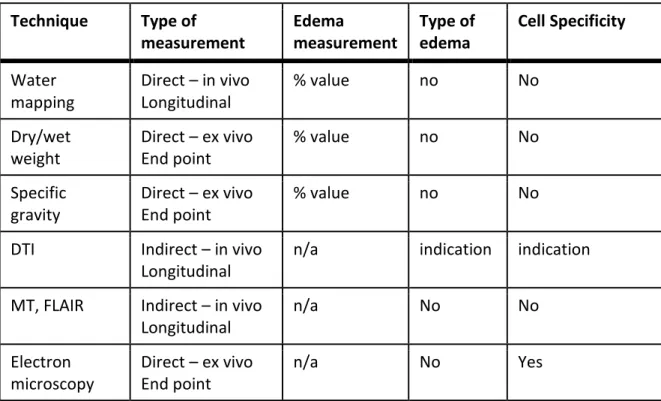
It is evident that the development of intracranial hypertension is associated with brain edema in ALF, however in chronic liver disease, where intracranial hypertension is rarely observed, brain edema is also present (O’Grady, 2008; Rovira et al., 2008; Shah et al., 2008; Sugimoto et al., 2008; Bosoi et al., 2012; Bosoi and Rose, 2013; Butterworth, 2015; Dam et al., 2015). The pressure-volume relationship between intracranial pressure and brain volume indicates that either low-grade edema or age-induced brain atrophy could explain the difference between ALF and chronic liver disease (Bosoi and Rose, 2013). To date several methods have been used to measure brain edema (i.e. brain water content) either directly or indirectly: 1) Direct/absolute value of the amount of water in the brain can be measured through; i) dry/wet weight technique (Marmarou et al., 1978), ii) specific gravity method (Marmarou et al., 1978; Hayazaki et al., 1995) and iii) brain water mapping using magnetic resonance imaging (MRI) (Shah et al., 2003, 2008; Neeb et al., 2006; Dam et al., 2015); 2) Indirect/relative information regarding the content of water in the brain can be calculated using several advanced MRI techniques; i) magnetization transfer (MT), ii) diffusion weighted or tensor imaging (DWI or DTI) and iii) fast fluid-attenuated inversion recovery (FLAIR) Table 1) (Häussinger et al., 1994; Córdoba et al., 2001; Spahr et al., 2002; McPhail et al., 2012; Braissant et al., 2013; Cudalbu, 2013).
The dry/wet weight and specific gravity methods are performed ex-vivo, using dissected tissue from sacrificed animals. The dry/wet weight method is the easiest way to measure the amount of water in extracted brain tissue. In this technique, brain samples are weighed before and after drying for 24 hours at 100°C (Marmarou et al., 1978) and the difference in weight reflects the amount of water (evaporated) in the tissue. Aside 4 5 6 7 8 9 10 11 12 13 14 15 16 17 18 19 20 21 22 23 24 25 26 27 28 29 30 31 32 33 34 35 36 37 38 39 40 41 42 43 44 45 46 47 48 49 50 51 52 53 54 55 56 57 58 59 60
from its simplicity, this technique is not sensitive enough for precisely measuring water evaporation in small regions and therefore is limited to whole or half brains (at least for rodents).
The specific gravity method involves placing small pieces of brain tissue into a graduated cylinder containing a layered mixture (gradient) of organic solvents of known density (kerosene and bromobenzene). The equilibration depth of the inserted tissue is recorded after 2 minutes (representing the specific gravity). This technique is excellent for measuring small changes in water of many different (including small) brain regions. The initial set-up for making the kerosene/bromobenzene gradient columns is cumbersome, but worth the investment. Following the complex mixing process, each column must be carefully calibrated but can be used to measure the specific gravity of about 20-25 samples. The percentage of water is calculated taking into consideration the specific gravity of the solid dry tissue (Marmarou et al., 1978). This method provides absolute water content in the brain and has been shown to detect changes in water content of 1-2% in a rat model of chronic HE (Bosoi et al., 2012).
Non-invasive in vivo measurements of water content in the human brain with MRI are often applied to explore brain swelling in human HE studies (Häussinger et al., 1994; Córdoba et al., 2001; Shah et al., 2003, 2008; Rovira et al., 2008). MRI is a non-invasive technique applicable in vivo and therefore be implicated in longitudinally studies on the same individual. MRI is primarily focused on imaging the single proton of the hydrogen nucleus (1H). Since hydrogen is by far the most common nucleus in the human body, it has become a valuable target for in vivo imaging. Several refined MRI techniques are presently available to detect subtle changes of approximately 1% in total brain water content. MT, FLAIR and DWI imaging are all sensitive to changes in brain tissue water of ~1 %. Although sufficiently sensitive, they all lack specificity in regards the etiology of the water accumulation. MT and FLAIR are both sensitive to exchange properties between bound and free protons, and therefore the output is determined by factors besides the total water content. The images, therefore, provide only indirect evidence of brain swelling. DWI allows the mapping of the diffusion process of primarily water molecules. However, molecular diffusion in tissues is not free, but reflects interactions with, for example, macromolecules, fibers, membranes. DWI can demonstrate changes in intra- or extracellular volume but no firm conclusions on the absolute water content can be drawn. Hence, the direct assessment of cerebral water changes in HE is tedious. A quantitative estimation of brain tissue water contents (water mapping) was validated on 1.5 and 3 Tesla platforms in Juelich in Germany from 2004-2008 (Neeb et al., 2006; Shah et al., 2008). This water mapping method enables an automated assessment of global water content changes in both grey and white matter as well as changes in the spatial distribution of water in the brain. It is sensitive and specific and can be performed within a clinically relevant measurement time (less 20 min). The water mapping method was applied to fifty-four patients with various grades of HE in 2008 (Shah et al., 2008).The average white matter water content was 2.1% higher in patients with overt HE compared to the healthy control group. There was no significant difference in grey matter water.
In conclusion, we believe that brain water mapping is the most precise and accurate method that can be applied to patients with HE for absolute quantification of their cerebral hydration status. However, increased water content remains an unspecific phenomenon that also occurs in trauma, tumors, focal inflammation and late stages of 4 5 6 7 8 9 10 11 12 13 14 15 16 17 18 19 20 21 22 23 24 25 26 27 28 29 30 31 32 33 34 35 36 37 38 39 40 41 42 43 44 45 46 47 48 49 50 51 52 53 54 55 56 57 58 59 60
cerebral ischemia. For measuring brain edema in animal models a multimodal approach would be the most suitable. For example, an approach combining in vivo and longitudinal measurements with an ex vivo technique measuring the absolute water value in the brain. This combination allows monitoring of the progression of the syndrome longitudinally and therefore provides additional information on the temporal resolution of the onset of brain edema. Since none of these techniques provides information on the type of the edema or which cell is involved, it would be also very useful to combine these techniques with electron microscopy (Kato et al., 1992).
Measuring hepatic encephalopathy in rodents; neurophenotyping
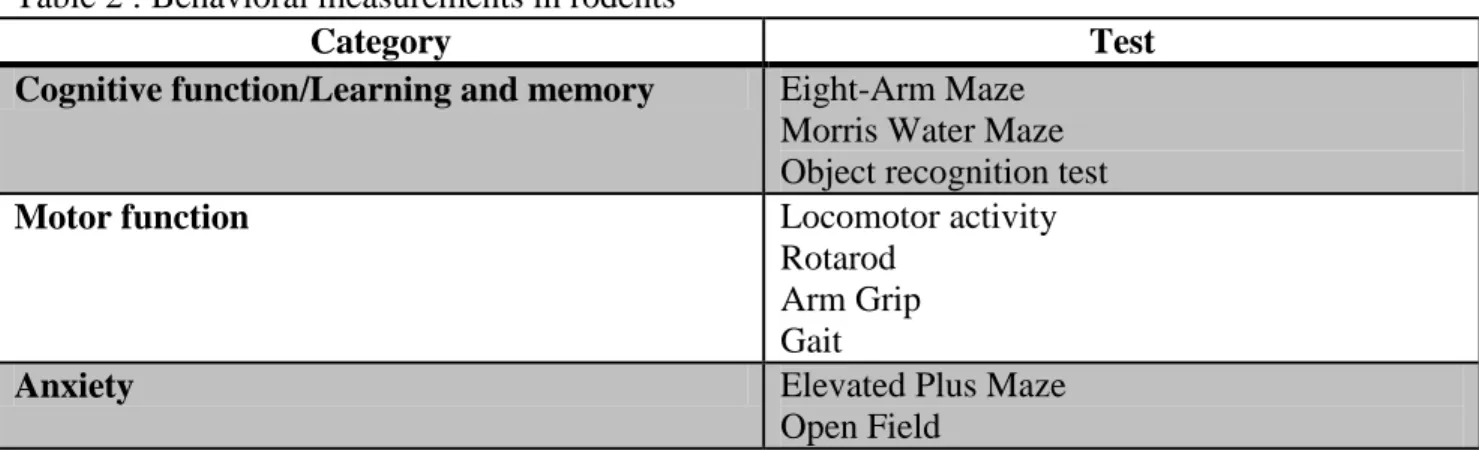
In humans, diagnosing HE, in particular minimal/covert HE, involves using sophisticated neuropsychometric and neurophysiological tests, such as the psychometric hepatic encephalopathy score (PHES) (Weissenborn et al., 2001; Ferenci et al., 2002; Amodio et al., 2008), the inhibitory control test (ICT) (Bajaj et al., 2007), the critical flicker frequency (CFF) test (Kircheis et al., 2002), the continuous reaction time (CRT) (Lauridsen et al., 2013) and the EncephalApp (Smart-phone based Stroop test) (Bajaj et al., 2013). Overall, these tests evaluate cognition, psychomotor processing speed, visuomotor coordination, memory, attention as well as motor function. As with HE patients, the best methods to evaluate HE in rodents are through the use of various behavioral tests (neurophenotyping). In general, behavioral measurements in rodents can be divided into three categories : 1) Cognitive function/learning and memory; 2) Motor function: and 3) Anxiety (Table 2). In the following sections, different tests used to evaluate the parameters in each category are discussed.
Cognitive function/learning and memory
Eight-Arm Maze
This test (Olton and Samuelson, 1976), which assesses spatial memory, consists of eight horizontal arms placed radially around a central platform above the floor. Food is placed at the end of all arms, and the animal must learn to enter each arm a single time. Errors are defined as repeat entries into already-visited arms. Although the simplest strategy to solve this task would be to enter adjacent arms, rodents do not typically adopt this tactic. As such, the analysis of arm entries can yield insight into such processes as planning and decision making and impulsivity in the rodent. Different variables are commonly used for the analysis of the performance, including: number of errors in each session (entering an arm that has been visited previously counted as an error) and the total number of errors during eight sessions; number of correct choices in the first eight arm entries; location of the first error in each session; time taken to visit each arm (total time to complete the session divided by the total number of arm entries); number of sessions to reach the criterion of one error or less, averaged over four consecutive days of training. Since the task is motivated by appetite, food restriction regimen are required, which represents a disadvantage of this behavioral test. Indeed, food restriction contrasts with cognitive tasks which are aversively motivated.
Morris Water Maze
This is a behavioral procedure widely used to study spatial learning and memory (Morris, 1984; D’Hooge and Deyn, 2001). Animals are placed into a pool of water in 4 5 6 7 8 9 10 11 12 13 14 15 16 17 18 19 20 21 22 23 24 25 26 27 28 29 30 31 32 33 34 35 36 37 38 39 40 41 42 43 44 45 46 47 48 49 50 51 52 53 54 55 56 57 58 59 60
which a platform is hidden beneath the surface. The animal must learn to use spatial cues located in the testing room to navigate to the platform. Longer latencies indicate poorer performance. Variations in the experimental protocol allow the experimenter to
determine whether the observed impairments are the result of working (more than once a day) or reference memory (once a day) systems. Cognitive flexibility can be assessed using a water maze paradigm in which the hidden platform is continually re-located. The earliest measure of learning is escape latency, which is the time it takes to find the platform. However, this measure is confounded by swimming speed, not necessarily a cognitive factor. Path length between point of origin and platform is a parameter more closely related to spatial learning. Stress of swimming may be a disadvantage of this test. Object Recognition test
This is a fast and efficient test to assess working memory (Ennaceur and Delacour, 1988). The animal is first placed into an arena containing two identical objects. After a
predetermined period of exploration, the animal is removed from the arena, and a delay is imposed. Following the delay, the animal is placed back into the arena, where one of the objects has been replaced by a novel object. Since rodents are curious, they typically avoid familiar objects and explore novel objects. The amount of time investigating the novel object is taken as the measure of working memory. Lower exploration of the novel object is thus interpreted as poorer working memory performance (for further details: (Antunes and Biala, 2011).
Motor function
Locomotor activity
This test is commonly used in rodents to qualitatively and quantitatively measure general locomotor activity and willingness to explore (Denenberg, 1969; Stanford, 2007). This test uses an arena with walls to prevent escape. Generally, the field is marked with a grid and square crossings. This test measures exploratory behavior in a novel, enclosed environment. Rearing and time spent moving are used to assess the activity of the rodent. The apparatus is equipped with infrared beams or video cameras with associated software that can be used to automate the assessment process.
Rotarod
The rotarod test is used to assess motor coordination and balance (Jones and Roberts, 1968). The test animal (usually a rodent) is placed on a cylinder that rotates at gradually increasing speed until the animal can no longer maintain itself on the cylinder. The speed of the rotarod may either be held constant or accelerated. The length of time that a given animal stays on the rotating rod is a measure of its balance and coordination. However, the physical condition of the animal may represent a bias to the analysis.
Arm Grip test
This test measures forepaw strength (Meyer et al., 1979). Animals are allowed to grasp the grip strength meter with their forepaws. They are then gently pulled from the base of the tail until the grip is released. A grip strength meter measures the maximum force applied to the meter.
4 5 6 7 8 9 10 11 12 13 14 15 16 17 18 19 20 21 22 23 24 25 26 27 28 29 30 31 32 33 34 35 36 37 38 39 40 41 42 43 44 45 46 47 48 49 50 51 52 53 54 55 56 57 58 59 60
Gait
This test consists of images of the underside of the animal that are taken as the animal ambulates on a clear treadmill. Measurements of stride length, base width, and fore and hind paw overlap give an indication of gait.
Anxiety
Elevated Plus Maze
This test has become the benchmark for assessing anxiety in rodents (Pellow et al., 1985). The test creates an approach-avoidance conflict between the natural tendency of the rodent to explore and its aversion for open spaces. The elevated plus maze itself consists of two enclosed arms and two open arms (arms without walls). Anxiety is typically measured by the amount of time the animal explores the open arms. The more anxious the animal, the less it will explore the open arms.
Open Field test
This test, which is used to assess motor function (see previous section), may also be used to assess anxiety (Prut and Belzung, 2003). The latter is assessed by including additional measures of defecation, time spent in the center of the field and the first few minutes of activity. Anxiety is measured by the amount of time the animal avoids the exposed center area of the field and remains in close proximity to the walls.
In conclusion, evaluating the behavioral phenotype in rodent models of liver disease is a valid way to define the presence of HE. However, due to costs, space and infrastructure, neurophenotyping is not routinely performed in laboratories. Furthermore, with
increasing use of these protocols, standardizing these tests will be an imperative.
Hepatic encephalopathy and brain edema
Undoubtedly, in the development of intracranial hypertension in ALF, brain edema plays a “physical” contributing role to the associated encephalopathy. However the pathophysiological role of astrocyte swelling/brain edema in neurological
deterioration remains elusive. Even in the absence of intracranial hypertension, it is suggested that astrocyte swelling can have important functional consequences and that HE represents a clinical manifestation of astrocyte swelling (Häussinger, 2000).
It is well documented that cirrhotic patients with brain edema present HE (Kumar et al., 2008; O’Grady, 2008; Rovira et al., 2008; Shah et al., 2008; Sugimoto et al., 2008; Bosoi and Rose, 2013; Dam et al., 2015, 2015)
. However, it is not known whether all patients with HE have an increase in brain water. In rats with bile-duct ligation (BDL)-induced CLD, a type-C model of HE, brain edema and HE are present (Bosoi et al., 2014). However, rats with portacaval anastomosis (PCA), a type-B model of HE, brain edema is not present (Bosoi et al., 2012). This raises the question regarding the role of brain edema in the neurological alterations related to HE. Other studies suggest that brain edema is not implicated in the pathogenesis of HE; in rats with ALF, it was shown that following attenuation of brain edema with the hypertonic solution mannitol, motor tract function did not improve (Oria et al., 2010). In addition, the same authors demonstrated that following an acute injection of ammonia to PCA rats, severe alterations of the motor tract function developed, without the
4 5 6 7 8 9 10 11 12 13 14 15 16 17 18 19 20 21 22 23 24 25 26 27 28 29 30 31 32 33 34 35 36 37 38 39 40 41 42 43 44 45 46 47 48 49 50 51 52 53 54 55 56 57 58 59 60
development of brain edema (Oria et al., 2010). It is however worth noting that in this study, attenuation in brain water was only measured in the cortex and brain stem whereas brain water content in the red nucleus, substantia nigra and basal ganglia (regions
implicated in the modulation of the motor tract function) were not evaluated. In another study by Wright and colleagues, BDL and sham-operated controls were challenged with lipopolysaccharides (LPS) and both groups developed brain edema. However, only the BDL rats presented with a neurological decline (Wright et al., 2007). In addition, acute hyperammonemia induced in mice resulted in severe encephalopathy without brain edema or astrocyte swelling (Rangroo Thrane et al., 2013). Furthermore, Aqp4-/- mice have 2-3% increased brain water content compared to wild-type animals, but no obvious neurological phenotype (Nagelhus and Ottersen, 2013). However, there are many studies implicating brain edema in the pathogenesis of HE. Rovira et al., elegantly demonstrated a decrease in brain volume and improvement in HE following liver transplantation (Rovira et al., 2007). Furthermore, BDL rats treated with ammonia-lowering agents or antioxidants result in an attenuation in brain edema as well as an improvement in neurological status (Bosoi et al., 2011, 2012). Whether these discrepancies are model-specific (i.e. HE type A vs B vs C) remains to be determined.
Conclusion
The role of brain edema, as a neuropathological feature or a cause of HE, remains a controversial topic. The correlation between brain edema and HE is strong, with ample supporting studies demonstrating that brain edema leads to neuronal dysfunction. We suggest that different degrees of astrocyte swelling/brain edema may inflict differential effects (physical stress as well as metabolic alterations) on cerebral function. Therefore, brain edema remains a valid endpoint in the evaluation of HE. However, in the setting of liver disease/failure, other factors in addition to brain edema may play a role in the severity of HE. In effect, brain edema may play a predisposing or precipitating role in the pathogenesis of severe/overt HE.
Understanding the obvious limitations of assessing similar neuropsychological tests to rodents as in humans, the worthiest assessment of HE in rodents remains evaluating behavioural changes. Measuring changes in cognitive function, learning, memory, anxiety and motor function are valid parameters in the assessment of HE. In fact, developing and standardizing a battery of tests to assess HE in small animals is highly warranted and worth considering in the future.
4 5 6 7 8 9 10 11 12 13 14 15 16 17 18 19 20 21 22 23 24 25 26 27 28 29 30 31 32 33 34 35 36 37 38 39 40 41 42 43 44 45 46 47 48 49 50 51 52 53 54 55 56 57 58 59 60
References
Amiry-Moghaddam M, Otsuka T, Hurn PD, Traystman RJ, Haug F-M, Froehner SC, Adams ME, Neely JD, Agre P, Ottersen OP, et al. (2003) An alpha-syntrophin-dependent pool of AQP4 in astroglial end-feet confers bidirectional water flow between blood and brain. Proc. Natl. Acad. Sci. U.S.A 100:2106–2111
Amodio P, Campagna F, Olianas S, Iannizzi P, Mapelli D, Penzo M, Angeli P, Gatta A (2008) Detection of minimal hepatic encephalopathy: normalization and optimization of the Psychometric Hepatic Encephalopathy Score. A neuropsychological and quantified EEG study. J Hepatol 49:346–353
Anderova M, Benesova J, Mikesova M, Dzamba D, Honsa P, Kriska J, Butenko O, Novosadova V, Valihrach L, Kubista M, et al. (2014) Altered Astrocytic Swelling in the Cortex of alpha-Syntrophin-Negative GFAP/EGFP Mice. PLoS ONE 9:e113444
Antunes M, Biala G (2011) The novel object recognition memory: neurobiology, test procedure, and its modifications. Cogn Process 13:93–110
Araque A, Parpura V, Sanzgiri RP, Haydon PG (1999) Tripartite synapses: glia, the unacknowledged partner. Trends Neurosci 22:208–215
Bajaj JS, Saeian K, Verber MD, Hischke D, Hoffmann RG, Franco J, Varma RR, Rao SM (2007) Inhibitory control test is a simple method to diagnose minimal hepatic encephalopathy and predict development of overt hepatic encephalopathy. Am J Gastroenterol 102:754–760
Bajaj JS, Thacker LR, Heuman DM, Fuchs M, Sterling RK, Sanyal AJ, Puri P, Siddiqui MS, Stravitz RT, Bouneva I, et al. (2013) The Stroop smartphone application is a short and valid method to screen for minimal hepatic encephalopathy. Hepatology 58:1122– 1132
Binder DK, Yao X, Zador Z, Sick TJ, Verkman AS, Manley GT (2006) Increased seizure duration and slowed potassium kinetics in mice lacking aquaporin-4 water channels. Glia 53:631–636
Bloch O, Papadopoulos MC, Manley GT, Verkman AS (2005) Aquaporin-4 gene deletion in mice increases focal edema associated with staphylococcal brain abscess. J Neurochem 95:254–262
Bosoi CR, Parent-Robitaille C, Anderson K, Tremblay M, Rose CF (2011) AST-120 (spherical carbon adsorbent) lowers ammonia levels and attenuates brain edema in bile duct-ligated rats. Hepatology 53:1995–2002
Bosoi CR, Rose CF (2013) Brain edema in acute liver failure and chronic liver disease: Similarities and differences. Neurochem. Int. 62:446–457
4 5 6 7 8 9 10 11 12 13 14 15 16 17 18 19 20 21 22 23 24 25 26 27 28 29 30 31 32 33 34 35 36 37 38 39 40 41 42 43 44 45 46 47 48 49 50 51 52 53 54 55 56 57 58 59 60
Bosoi CR, Yang X, Huynh J, Parent-Robitaille C, Jiang W, Tremblay M, Rose CF (2012) Systemic oxidative stress is implicated in the pathogenesis of brain edema in rats with chronic liver failure. Free Radic. Biol. Med. 52:1228–1235
Bosoi CR, Zwingmann C, Marin H, Parent-Robitaille C, Huynh J, Tremblay M, Rose CF (2014) Increased brain lactate is central to the development of brain edema in rats with chronic liver disease. J Hepatol 60:554–560
Braissant O, McLin VA, Cudalbu C (2013) Ammonia toxicity to the brain. J Inherit Metab Dis 36:595–612
Butterworth RF (2015) Pathogenesis of hepatic encephalopathy and brain edema in acute liver failure. J Clin Exp Hepatol 5:S96–S103
Cauli O, Rodrigo R, Piedrafita B, Boix J, Felipo V (2007) Inflammation and hepatic encephalopathy: ibuprofen restores learning ability in rats with portacaval shunts. Hepatology 46:514–519
Córdoba J, Alonso J, Rovira A, Jacas C, Sanpedro F, Castells L, Vargas V, Margarit C, Kulisewsky J, Esteban R, et al. (2001) The development of low-grade cerebral edema in cirrhosis is supported by the evolution of (1)H-magnetic resonance abnormalities after liver transplantation. J. Hepatol. 35:598–604
Cotrina ML, Lin JH, López-García JC, Naus CC, Nedergaard M (2000) ATP-mediated glia signaling. J. Neurosci. 20:2835–2844
Cudalbu C (2013) In vivo studies of brain metabolism in animal models of Hepatic Encephalopathy using 1H Magnetic Resonance Spectroscopy. Metab Brain Dis 28:167– 174
Dam G, Thomsen KL, Gras V, Vilstrup H, Ott P, Wintherdahl M (2015) P0113 :
Regional cerebral water content in hepatic encephalopathy measured by MRI. J Hepatol 62:S343
Denenberg VH (1969) Open-field behavior in the rat : what does it mean? Ann NY Acad Sci 159:852–859
D’Hooge R, Deyn PPD (2001) Applications of the Morris water maze in the study of learning and memory. Brain Res Rev 36:60–90
Ennaceur A, Delacour J (1988) A new one-trial test for neurobiological studies of memory in rats. 1: Behavioral data. Behav. Brain Res 31:47–59
Evanko DS, Zhang Q, Zorec R, Haydon PG (2004) Defining pathways of loss and secretion of chemical messengers from astrocytes. Glia 47:233–240
Felipo V (2013) Hepatic encephalopathy: effects of liver failure on brain function. Nat Rev Neurosci 14:851–858 4 5 6 7 8 9 10 11 12 13 14 15 16 17 18 19 20 21 22 23 24 25 26 27 28 29 30 31 32 33 34 35 36 37 38 39 40 41 42 43 44 45 46 47 48 49 50 51 52 53 54 55 56 57 58 59 60
Ferenci P, Lockwood A, Mullen K, Tarter R, Weissenborn K, Blei AT (2002) Hepatic encephalopathy–definition, nomenclature, diagnosis, and quantification: final report of the working party at the 11th World Congresses of Gastroenterology, Vienna, 1998. Hepatology 35:716–721
Fukuda AM, Adami A, Pop V, Bellone JA, Coats JS, Hartman RE, Ashwal S, Obenaus A, Badaut J (2013) Posttraumatic reduction of edema with aquaporin-4 RNA interference improves acute and chronic functional recovery. J Cereb Blood Flow Metab 33:1621– 1632
Haj-Yasein NN, Jensen V, Østby I, Omholt SW, Voipio J, Kaila K, Ottersen OP, Hvalby Ø, Nagelhus EA (2012) Aquaporin-4 regulates extracellular space volume dynamics during high-frequency synaptic stimulation: A gene deletion study in mouse
hippocampus. Glia 60:867–874
Häussinger D (2000) Hepatic encephalopathy in chronic liver disease: a clinical manifestation of astrocyte swelling and low-grade cerebral edema? J Hepatol 32:1035– 1038
Häussinger D, Laubenberger J, Dahl SV, Ernst T, Bayer S, Langer M, Gerok W, Hennig J (1994) Proton magnetic resonance spectroscopy studies on human brain Myo-inositol in hypo-osmolarity and hepatic encephalopathy. Gastroenterology 107:1475–1480
Hayazaki K, Matsuoka Y, Kan M, Hakuba A (1995) Variation in Equation Coefficients in the Gravimetric Method to Determine Brain Water Content. Neurologia Medico-Chirurgica 35:69–74
Iliff JJ, Wang M, Liao Y, Plogg BA, Peng W, Gundersen GA, Benveniste H, Vates GE, Deane R, Goldman SA, et al. (2012) A Paravascular Pathway Facilitates CSF Flow Through the Brain Parenchyma and the Clearance of Interstitial Solutes, Including Amyloid. Sci Transl Med 4:147ra111–ra147ra111
Jones BJ, Roberts DJ (1968) The quantitative measurement of motor inco-ordination in naive mice using an accelerating rotarod. J Pharm Pharmacol 20:302–304
Karus C, Mondragão MA, Ziemens D, Rose CR (2015) Astrocytes restrict discharge duration and neuronal sodium loads during recurrent network activity. Glia 63:936–957 Kasischke KA, Vishwasrao HD, Fisher PJ, Zipfel WR, Webb WW (2004) Neural activity triggers neuronal oxidative metabolism followed by astrocytic glycolysis. Science
305:99–103
Kato M, Hughes RD, Keays RT, Williams R (1992) Electron microscopic study of brain capillaries in cerebral edema from fulminant hepatic failure. Hepatology 15:1060–1066 Kimelberg HK (1987) Anisotonic media and glutamate-induced ion transport and volume responses in primary astrocyte cultures. J. Physiol. (Paris) 82:294–303
4 5 6 7 8 9 10 11 12 13 14 15 16 17 18 19 20 21 22 23 24 25 26 27 28 29 30 31 32 33 34 35 36 37 38 39 40 41 42 43 44 45 46 47 48 49 50 51 52 53 54 55 56 57 58 59 60
Kircheis G, Wettstein M, Timmermann L, Schnitzler A, Häussinger D (2002) Critical flicker frequency for quantification of low-grade hepatic encephalopathy. Hepatology 35:357–366
Kumar R, Gupta RK, Elderkin-Thompson V, Huda A, Sayre J, Kirsch C, Guze B, Han S, Thomas MA (2008) Voxel-based diffusion tensor magnetic resonance imaging evaluation of low-grade hepatic encephalopathy. J Magn Reson Imaging 27:1061–1068
Lauridsen MM, Thiele M, Kimer N, Vilstrup H (2013) The continuous reaction times method for diagnosing, grading, and monitoring minimal/covert hepatic encephalopathy. Metab Brain Dis 28:231–234
Lee WM (2012) Acute liver failure. Semin Respir Crit Care Med 33:36–45
Louveau A, Smirnov I, Keyes TJ, Eccles JD, Rouhani SJ, Peske JD, Derecki NC, Castle D, Mandell JW, Lee KS, et al. (2015) Structural and functional features of central nervous system lymphatic vessels. Nature 523:337–341
Manley GT, Fujimura M, Ma T, Noshita N, Filiz F, Bollen AW, Chan P, Verkman AS (2000) Aquaporin-4 deletion in mice reduces brain edema after acute water intoxication and ischemic stroke. Nat. Med. 6:159–163
Marmarou A, Poll W, Shulman K, Bhagavan H (1978) A simple gravimetric technique for measurement of cerebral edema. J Neurosurg 49:530–537
McPhail MJW, Patel NR, Taylor-Robinson SD (2012) Brain imaging and hepatic encephalopathy. Clin Liver Dis 16:57–72
Messing A, Brenner M, Feany MB, Nedergaard M, Goldman JE (2012) Alexander Disease. J Neurosci 32:5017–5023
Meyer OA, Tilson HA, Byrd WC, Riley MT (1979) A method for the routine assessment of fore- and hindlimb grip strength of rats and mice. Neurobehav Toxicol 1:233–236 Morris R (1984) Developments of a water-maze procedure for studying spatial learning in the rat. J Neurosci Methods 11:47–60
Mulligan SJ, MacVicar BA (2006) VRACs CARVe a Path for Novel Mechanisms of Communication in the CNS. Science STKE 2006:pe42–pe42
Nagelhus EA, Ottersen OP (2013) Physiological Roles of Aquaporin-4 in Brain. Physiol Rev 93:1543–1562
Nedergaard M (2013) Garbage Truck of the Brain. Science 340:1529–1530
Nedergaard M, Verkhratsky A (2012) Artifact versus reality-How astrocytes contribute to synaptic events. Glia 60:1013–1023
4 5 6 7 8 9 10 11 12 13 14 15 16 17 18 19 20 21 22 23 24 25 26 27 28 29 30 31 32 33 34 35 36 37 38 39 40 41 42 43 44 45 46 47 48 49 50 51 52 53 54 55 56 57 58 59 60
Neeb H, Zilles K, Shah NJ (2006) A new method for fast quantitative mapping of absolute water content in vivo. NeuroImage 31:1156–1168
Nielsen S, Nagelhus EA, Amiry-Moghaddam M, Bourque C, Agre P, Ottersen OP (1997) Specialized membrane domains for water transport in glial cells: high-resolution
immunogold cytochemistry of aquaporin-4 in rat brain. J. Neurosci. 17:171–180
Oberheim NA, Tian G-F, Han X, Peng W, Takano T, Ransom B, Nedergaard M (2008) Loss of Astrocytic Domain Organization in the Epileptic Brain. J Neurosci 28:3264–3276 Oberheim NA, Wang X, Goldman S, Nedergaard M (2006) Astrocytic complexity
distinguishes the human brain. Trends Neurosci 29:547–553
O’Grady JG (2008) Intracranial pressure and acute liver failure: a “finger on the pulse.” Liver Transpl. 14:915–916
Olton DS, Samuelson RJ (1976) Remembrance of places passed: Spatial memory in rats. J Exp Psychol Anim Behav Process. 2:97–116
Ordaz B, Tuz K, Ochoa LD, Lezama R, Peña-Segura C, Franco R (2004) Osmolytes and Mechanisms Involved in Regulatory Volume Decrease Under Conditions of Sudden or Gradual Osmolarity Decrease. Neurochem Res 29:65–72
Oria M, Chatauret N, Chavarria L, Romero-Giménez J, Palenzuela L, Pardo-Yules B, Arranz JA, Bodega G, Raguer N, Córdoba J (2010) Motor-evoked potentials in awake rats are a valid method of assessing hepatic encephalopathy and of studying its
pathogenesis. Hepatology 52:2077–2085
Papadopoulos MC, Manley GT, Krishna S, Verkman AS (2004) Aquaporin-4 facilitates reabsorption of excess fluid in vasogenic brain edema. FASEB J. 18:1291–1293
Papadopoulos MC, Verkman AS (2005) Aquaporin-4 Gene Disruption in Mice Reduces Brain Swelling and Mortality in Pneumococcal Meningitis. J Biol Chem 280:13906– 13912
Papadopoulos MC, Verkman AS (2013) Aquaporin water channels in the nervous system. Nat Rev Neurosci 14:265–277
Pellerin L, Bouzier-Sore A-K, Aubert A, Serres S, Merle M, Costalat R, Magistretti PJ (2007) Activity-dependent regulation of energy metabolism by astrocytes: an update. Glia 55:1251–1262
Pellow S, Chopin P, File SE, Briley M (1985) Validation of open : closed arm entries in an elevated plus-maze as a measure of anxiety in the rat. J Neurosci 14:149–167
Prut L, Belzung C (2003) The open field as a paradigm to measure the effects of drugs on anxiety-like behaviors: a review. Eur J Pharmacol 463:3–33
4 5 6 7 8 9 10 11 12 13 14 15 16 17 18 19 20 21 22 23 24 25 26 27 28 29 30 31 32 33 34 35 36 37 38 39 40 41 42 43 44 45 46 47 48 49 50 51 52 53 54 55 56 57 58 59 60
Qiu Z, Dubin AE, Mathur J, Tu B, Reddy K, Miraglia LJ, Reinhardt J, Orth AP,
Patapoutian A (2014) SWELL1, a Plasma Membrane Protein, Is an Essential Component of Volume-Regulated Anion Channel. Cell 157:447–458
Rangroo Thrane V, Thrane AS, Wang F, Cotrina ML, Smith NA, Chen M, Xu Q, Kang N, Fujita T, Nagelhus EA, et al. (2013) Ammonia triggers neuronal disinhibition and seizures by impairing astrocyte potassium buffering. Nat. Med. 19:1643–1648
Ransom B, Behar T, Nedergaard M (2003) New roles for astrocytes (stars at last). Trends Neurosci 26:520–522
Rao KVR, Verkman AS, Curtis KM, Norenberg MD (2014) Aquaporin-4 deletion in mice reduces encephalopathy and brain edema in experimental acute liver failure. Neurobiol Dis 63:222–228
Ren Z, Iliff JJ, Yang L, Yang J, Chen X, Chen MJ, Giese RN, Wang B, Shi X,
Nedergaard M (2013) `Hit & Run’ model of closed-skull traumatic brain injury (TBI) reveals complex patterns of post-traumatic AQP4 dysregulation. J Cereb Blood Flow Metab 33:834–845
Risher WC, Andrew RD, Kirov SA (2009) Real-time passive volume responses of astrocytes to acute osmotic and ischemic stress in cortical slices and in vivo revealed by two-photon microscopy. Glia 57:207–221
Rodrigo R, Cauli O, Gomez-Pinedo U, Agusti A, Hernandez-Rabaza V, Garcia-Verdugo J-M, Felipo V (2010) Hyperammonemia induces neuroinflammation that contributes to cognitive impairment in rats with hepatic encephalopathy. Gastroenterology 139:675–684 Rose CF, Verkhratsky A, Parpura V (2013) Astrocyte glutamine synthetase: pivotal in health and disease. Biochem. Soc. Trans. 41:1518–1524
Rose CR, Ransom BR (1997) Gap junctions equalize intracellular Na+ concentration in astrocytes. Glia 20:299–307
Rouach N, Koulakoff A, Abudara V, Willecke K, Giaume C (2008) Astroglial Metabolic Networks Sustain Hippocampal Synaptic Transmission. Science 322:1551–1555
Rovira A, Alonso J, Córdoba J (2008) MR imaging findings in hepatic encephalopathy. AJNR Am J Neuroradiol 29:1612–1621
Rovira A, Mínguez B, Aymerich FX, Jacas C, Huerga E, Córdoba J, Alonso J (2007) Decreased white matter lesion volume and improved cognitive function after liver transplantation. Hepatology 46:1485–1490
Schliess F, Foster N, Görg B, Reinehr R, Häussinger D (2004) Hypoosmotic swelling increases protein tyrosine nitration in cultured rat astrocytes. Glia 47:21–29
4 5 6 7 8 9 10 11 12 13 14 15 16 17 18 19 20 21 22 23 24 25 26 27 28 29 30 31 32 33 34 35 36 37 38 39 40 41 42 43 44 45 46 47 48 49 50 51 52 53 54 55 56 57 58 59 60
Shah NJ, Neeb H, Kircheis G, Engels P, Häussinger D, Zilles K (2008) Quantitative cerebral water content mapping in hepatic encephalopathy. Neuroimage 41:706–717 Shah NJ, Neeb H, Zaitsev M, Steinhoff S, Kircheis G, Amunts K, Häussinger D, Zilles K (2003) Quantitative T1 mapping of hepatic encephalopathy using magnetic resonance imaging. Hepatology 38:1219–1226
Simard JM, Kent TA, Chen M, Tarasov KV, Gerzanich V (2007) Brain oedema in focal ischaemia: molecular pathophysiology and theoretical implications. Lancet Neurol 6:258–268
Spahr L, Burkhard PR, Grötzsch H, Hadengue A (2002) Clinical significance of basal ganglia alterations at brain MRI and 1H MRS in cirrhosis and role in the pathogenesis of hepatic encephalopathy. Metab Brain Dis 17:399–413
Stanford SC (2007) The Open Field Test: reinventing the wheel. J Psychopharm 21:134– 135
Sugimoto R, Iwasa M, Maeda M, Urawa N, Tanaka H, Fujita N, Kobayashi Y, Takeda K, Kaito M, Takei Y (2008) Value of the apparent diffusion coefficient for quantification of low-grade hepatic encephalopathy. Am J Gastroenterol 103:1413–1420
Sun W, McConnell E, Pare J-F, Xu Q, Chen M, Peng W, Lovatt D, Han X, Smith Y, Nedergaard M (2013) Glutamate-dependent neuroglial calcium signaling differs between young and adult brain. Science 339:197–200
Swanson RA, Graham SH (1994) Fluorocitrate and fluoroacetate effects on astrocyte metabolism in vitro. Brain Res 664:94–100
Syková E, Nicholson C (2008) Diffusion in brain extracellular space. Physiol. Rev. 88:1277–1340
Tait MJ, Saadoun S, Bell BA, Verkman AS, Papadopoulos MC (2010) Increased brain edema in aqp4-null mice in an experimental model of subarachnoid hemorrhage. Neuroscience 167:60–67
Takano T, Tian G-F, Peng W, Lou N, Lovatt D, Hansen AJ, Kasischke KA, Nedergaard M (2007) Cortical spreading depression causes and coincides with tissue hypoxia. Nat Neurosci 10:754–762
Thrane AS, Rangroo Thrane V, Plog BA, Nedergaard M (2015) Filtering the muddied waters of brain edema. Trends Neurosci. 38:333–335
Thrane AS, Rappold PM, Fujita T, Torres A, Bekar LK, Takano T, Peng W, Wang F, Rangroo Thrane V, Enger R, et al. (2011) Critical role of aquaporin-4 (AQP4) in
astrocytic Ca2+ signaling events elicited by cerebral edema. Proc. Natl. Acad. Sci. U.S.A. 108:846–851 4 5 6 7 8 9 10 11 12 13 14 15 16 17 18 19 20 21 22 23 24 25 26 27 28 29 30 31 32 33 34 35 36 37 38 39 40 41 42 43 44 45 46 47 48 49 50 51 52 53 54 55 56 57 58 59 60
Thrane AS, Takano T, Thrane VR, Wang F, Peng W, Ottersen OP, Nedergaard M,
Nagelhus EA (2013) In vivo NADH fluorescence imaging indicates effect of aquaporin-4 deletion on oxygen microdistribution in cortical spreading depression. J Cereb Blood Flow Metab 33:996–999
Thrane AS, Thrane VR, Nedergaard M (2014) Drowning stars: reassessing the role of astrocytes in brain edema. Trends Neurosci 37:620–628
Voss FK, Ullrich F, Munch J, Lazarow K, Lutter D, Mah N, Andrade-Navarro MA, Kries JP von, Stauber T, Jentsch TJ (2014) Identification of LRRC8 Heteromers as an Essential Component of the Volume-Regulated Anion Channel VRAC. Science 344:634–638 Wang M, Iliff JJ, Liao Y, Chen MJ, Shinseki MS, Venkataraman A, Cheung J, Wang W, Nedergaard M (2012) Cognitive Deficits and Delayed Neuronal Loss in a Mouse Model of Multiple Microinfarcts. J Neurosci 32:17948–17960
Weissenborn K, Ennen JC, Schomerus H, Rückert N, Hecker H (2001)
Neuropsychological characterization of hepatic encephalopathy. J. Hepatol 34:768–773 Wright G, Davies NA, Shawcross DL, Hodges SJ, Zwingmann C, Brooks HF, Mani AR, Harry D, Stadlbauer V, Zou Z, et al. (2007) Endotoxemia produces coma and brain swelling in bile duct ligated rats. Hepatology 45:1517–1526
Xie L, Kang H, Xu Q, Chen MJ, Liao Y, Thiyagarajan M, O’Donnell J, Christensen DJ, Nicholson C, Iliff JJ, et al. (2013) Sleep Drives Metabolite Clearance from the Adult Brain. Science 342:373–377 4 5 6 7 8 9 10 11 12 13 14 15 16 17 18 19 20 21 22 23 24 25 26 27 28 29 30 31 32 33 34 35 36 37 38 39 40 41 42 43 44 45 46 47 48 49 50 51 52 53 54 55 56 57 58 59 60
Figure 1. Effects of astrocyte swelling on brain function. Left, physiological salt and water turnover in
the brain is functionally and anatomically compartmentalized to the peri-synaptic and peri-vascular regions.
Right, proposed effects of astrocyte swelling on brain function include both acute alterations in ion
concentrations, metabolism and blood flow, followed by more chronic or adaptive changes in transporter expression and polarization. Glutamate (Glu), interstitial fluid (ISF), intracellular fluid (ICF), adenosine triphosphate (ATP), reactive oxygen species (ROS), blood-brain barrier (BBB), cerebrospinal fluid (CSF),
perivascular space (PVS), aquaporin 4 (AQP4), solute carrier family protein (SLC, e.g. Na+-K+-Cl-
-cotransporter), zonula occludens (ZO, tight junctions), connexin (CNX, gap junctions), Na+-K+-ATPase,
Table 1: Short description of different techniques used to measure brain edema. Technique Type of measurement Edema measurement Type of edema Cell Specificity Water mapping Direct – in vivo Longitudinal % value no No Dry/wet weight Direct – ex vivo End point % value no No Specific gravity Direct – ex vivo End point % value no No
DTI Indirect – in vivo
Longitudinal
n/a indication indication
MT, FLAIR Indirect – in vivo Longitudinal n/a No No Electron microscopy Direct – ex vivo End point n/a No Yes
DTI: diffusion tensor imaging; MT : magnetization transfer; FLAIR : fast fluid-attenuated inversion recovery
Table 2 : Behavioral measurements in rodents
Category Test
Cognitive function/Learning and memory Eight-Arm Maze Morris Water Maze Object recognition test
Motor function Locomotor activity
Rotarod Arm Grip Gait
Anxiety Elevated Plus Maze