HAL Id: dumas-01628358
https://dumas.ccsd.cnrs.fr/dumas-01628358
Submitted on 3 Nov 2017HAL is a multi-disciplinary open access archive for the deposit and dissemination of sci-entific research documents, whether they are pub-lished or not. The documents may come from teaching and research institutions in France or abroad, or from public or private research centers.
L’archive ouverte pluridisciplinaire HAL, est destinée au dépôt et à la diffusion de documents scientifiques de niveau recherche, publiés ou non, émanant des établissements d’enseignement et de recherche français ou étrangers, des laboratoires publics ou privés.
Bilan du dépistage par télémédecine de la rétinopathie
du prématuré dans une maternité de niveau 3 en France
sur une période de 6 ans
Hélène Chan
To cite this version:
Hélène Chan. Bilan du dépistage par télémédecine de la rétinopathie du prématuré dans une maternité de niveau 3 en France sur une période de 6 ans. Médecine humaine et pathologie. 2017. �dumas-01628358�
Université de Bordeaux
U.F.R DES SCIENCES MEDICALES
ANNÉE 2017
THÈSE N°3193
Thèse pour l’obtention du
DIPLÔME D’ÉTAT de DOCTEUR EN MÉDECINE
DES OPHTALMOLOGIE
Présentée et soutenue publiquement par
CHAN Hélène, Sophie
Née le 20/05/1987 à Bordeaux (33)
Le 17/10/2017
BILAN DU DÉPISTAGE PAR TÉLÉMÉDECINE DE LA
RÉTINOPATHIE DU PRÉMATURÉ DANS UNE MATERNITÉ DE
NIVEAU 3 EN FRANCE SUR UNE PÉRIODE DE 6 ANS
Directeur de Thèse
Monsieur le Docteur PAYA Clément
Membres du jury
Président du Jury : Monsieur le Professeur KOROBELNIK Jean-François
Membres du Jury : Monsieur le Docteur PAYA Clément
Madame le Professeur DELYFER Marie-Noëlle
Monsieur le Professeur TOUBOUL David
Madame le Docteur COSTE Valentine
TABLE DES MATIÈRES
I. LISTE DES ABRÉVIATIONS………...……10
II. ARTICLE………...….11
1. ABSTRACT………..12
2. MANUSCRIPT……….…13
a. Introduction………....13
b. Materials and methods………...…13
c. Statistical analysis………..15 d. Results………15 e. Discussion………..17 f. References………..20 g. Figures ………...24 III. ANNEXE 1………..29 La rétinopathie du prématuré 1. Introduction………..29 2. Définition de la prématurité……….29 3. Epidémiologie………..29 4. Physiopathologie………..30 5. Facteurs de risques………...32 6. Classification de la ROP………..33 7. Dépistage……….39 8. Retcam……….41 9. Traitement………42
10. Evolution à long terme……….44
11. Conclusion………...44 12. Bibliographie………...45 IV. ANNEXE 2………..51 Le score d’Apgar V. ANNEXE 3……….52 Figures complémentaires
TABLE DES ILLUSTRATIONS : Tableaux et figures
a) MANUSCRIPT
a. Figure 1. Prevalence and severity of ROP by gestational age at birth……..…24 b. Figure 2. Prevalence and severity of ROP by birth weight………...…24 c. Table 1. Demographic and clinical characteristics of included infants (n=410) (2009-2015) ……….……….………25 d. Table 2. Multivariate analysis of risk factor for retinopathy of prematurity…26 e. Table 3. Univariate analysis of risk factor for treatment-requiring ROP versus
non-treatment requiring ROP (n=116) ……….………...….27 f. Table 4: Multivariate analysis of risk factor for treatment-requiring ROP versus non-treatment requiring ROP………...28 b) ANNEXE 1
a. Figure 1. Progression de la rétinopathie des prématurés……….…...…..31 b. Figure 2. Zones de la ROP……….……….……….………...34 c. Figure 3. Stades de sévérité de la ROP……….……….…...…35 d. Figure 4. Photographie montrant le bourrelet entre rétine vasculaire et avasculaire caractéristique du stade 2 de la ROP……….…………...…..36 e. Figure 5. ROP de stade 3, proliferation fibrovasculaire extra rétinienne…...36
f. Figure 6. Photographie du fond d’œil montrant un stade « plus »………..….37
g. Figure 7. Régression de la ROP après photocoagulation………..38
h. Figure 8. Âge post-conceptionnel, âge de vie, âge corrigé………..….40 i. Figure 9. Imagerie du fond d’oeil par la RetCam-120 Digital Retinal Camera………..42 j. Figure 10. Photographie d’un fond d’œil de ROP immédiatement après
photocoagulation de la rétine avasculaire……….………...……….43 k. Tableau 1. Délai du premier examen de dépistage de la rétinopathie du prématuré ……….……….……….………...…...…39
c) ANNEXE 2
a. Tableau 1. Le score d’Apgar……….……….………51
d) ANNEXE 3 a. Figure 1. Répartition du terme de naissance ……….…..…………52
b. Figure 2. Répartition du poids de naissance………...……….52
c. Figure 3. Nombre de fœtus par grossesse……….…………...……53
d. Figure 4. Causes de la prématurité……….………..53
e. Figure 5. Durée de séjour en réanimation……….…………...……54
f. Figure 6. Pathologies respiratoires durant la période post natale………..…..54
g. Figure 7. Durée d’oxygénothérapie totale……….………...…54
h. Figure 8. Durée de ventilation invasive……….………….……...55
i. Figure 9. Durée de ventilation non invasive……….………...……55
j. Figure 10. Durée d’oxygénothérapie nasale………..…….……….……55
k. Figure 11. Répartition du nombre d’examens du fond d’œil (FO) par nourrisson……….….……….….………...56
I.
LISTE DES ABRÉVIATIONS
BW : Birth Weight
CHU : Centre Hospitalier Universitaire EPO : Erythropoïétine
FiO2 : Fraction inspirée en Oxygène FO : Fond d’Oeil
HTA : Hypertension Artérielle GA : Gestational Age
IGF-1 : insuline-like growth factor 1 MAP : Menace d’Accouchement Prématuré RCF : Rythme Cardiaque Foetal
RCIU : Retard de Croissance Intra Utérin ROP : Retinopathy of Prematurity
RPM : Rupture Prématurée des Membranes SA : Semaines d’Aménorrhée
SD : Standard Deviation SpO2 : Saturation en Oxygène
II.
ARTICLE
Screening for Retinopathy of Prematurity by telemedecine in a tertiary level neonatal intensive care unit in France: review of a six-year period
Bilan du dépistage par télémédecine de la rétinopathie du prématuré dans une maternité de niveau 3 en France sur une période de 6 ans
Authors: H. Chan, A. Cougnard-Grégoire, JF. Korobelnik, MN. Delyfer, B. Mortemousque, D. Touboul, V. Coste, J. Sarlangue, C. Dutheil, C. Paya
Keywords: Retinopathy of prematurity, risk factors, retina, pediatric ophthalmology, prematurity, neonatology
ABSTRACT
Purpose: To analyze the prevalence and risk factors of retinopathy of prematurity (ROP) and of treatment-requiring ROP.
Methods: A retrospective study was conducted in a level III neonatal unit in Bordeaux, France, during 2009-2015. 419 preterm infants whose ROP was screened for ROP exclusively with RetCam were included.
Results: ROP of any degree was diagnosed in 27.68% of infants. Stage 1, 2, 3 and 4 ROP was found in 44%, 46%, 9% and 1% of subjects, respectively. No fifth stage of ROP was observed. 28/419 (6,6 %) infants were treated by exclusive laser photocoagulation. No intravitreal anti-VEGF injections or surgical treatments were performed. No infant born at>31 weeks or with BW>1110g needed ROP treatment. In multivariate analysis, risk factors for ROP development were low birth weight, low gestational age at birth, high total days of invasive mechanical ventilation and shock or vasoactive substances use. In multivariate analysis, risk factors for treatment-severe ROP were male gender, gestational age £ 27 weeks and Apgar score at 5 minutes £ 7.
Conclusion: In our 6-year series, ROP was successfully screened by exclusive telemedecine and no surgical treatment was required. This study identifies known ROP risk factors but Apgar score at 5 minutes as severe ROP risk factor requires furthers studies in order to be confirmed.
MANUSCRIPT
Introduction
Retinopathy of prematurity (ROP) is a vasoproliferative disorder of the eye affecting premature neonates. First described in the late 1940s, ROP was initially called retrolental fibroplasia and was characterized by a complete retinal detachment behind the lens. The cause of this first wave of ROP was the use of supplemental oxygen in closed incubators, which helped to improve the survival of preterm infants, but also contributed to blindness. (1) Although significant advances have been made in perinatal care, ROP remains a serious complication in prematurely born infants. Currently in developed countries it is one of the three most common causes of childhood blindness with cortical visual impairment and optic nerve hypoplasia. (2)
Low gestational age (GA) (3) (4), low birth weight (BW) (5) and prolonged exposure to supplementary oxygen are major proven risk factors for ROP. Oxygen administration is better controlled nowadays than in the past in developed countries, but ROP persists, partly because of the increased survival of infants with extremely low GA and BW. Identification of postnatal factors that affect the risk for and the course of ROP might allow neonatologists and ophtalmologists to attempt to prevent the disease and to limit comorbidities with which it shares modifiable risk factors.
The objective of this study was to evaluate the prevalence of ROP and to determine risk factors to develop ROP and severe ROP (requiring treatment) in our population after systematic screening of ROP by telemedecine.
Materials and Methods
Subjects
We conducted a retrospective descriptive study including all premature infants who were hospitalized and underwent ROP screening from July 15, 2009 to August 15, 2015, at the Centre Hospitalier Universitaire (CHU) de Bordeaux. This hospital is the largest tertiary level neonatal intensive care unit in the south west of France.
Inclusion criteria were: 1) Preterm infants hospitalized at the CHU de Bordeaux which have been screened for ROP exclusively with RetCam according to the recommendations of the American Academy of Pediatrics (6) 2) Consent of the parents or the legal representative for the screening of the ROP 3) Fundus images can be analyzed 4) Complete medical information.
Exclusion criteria were: 1) Non consent of parents or legal representative for ROP screening 2) Fundus images not available or not analyzable due to poor dilation or eye abnormality 3) Incomplete medical information 4) Funduscupic examination in indirect ophthalmoscopy performed in consultation when infants were no longer hospitalized 5) Early deaths.
Screening criteria
ROP screening followed the recommendations of the American Academy of Pediatrics: first exam between 4 and 9 weeks of postnatal life according to the age of birth in all the infants < 30 weeks’ GA or BW < 1500 g at birth or with an unstable clinical course.
Eye Examinations, monitoring and management of infants at risk of ROP
For screening, fundus images were obtained using RetCam 120 (Massie Research Laboratories, USA) after dilatation with tropicamide and neosynephrine 2.5% prior to the exam. Their realization was delegated to two pediatric nurses trained for this purpose.
The fundus images were interpreted within 72 hours. We did not consider these results but the images have been reinterpreted by two ophthalmologists specializing in pediatric ophthalmology.
ROP stages were classified according to the International Classification of Retinopathy of Prematurity Revisited and the most severe stage of one of the two eyes was considered. (7) Stage was defined by the severity of ROP (stage 1-5), the location (zone I, II, or III) and the presence or absence of plus disease.
The follow-up was adapted from the 1st examination results according to the recommendations of the American Academy of Pediatrics, using RetCam if they were still hospitalized or in consultation with indirect ophthalmoscopy, until complete retinal vascularization of zone III. The results of the examinations in consultation were not considered but none required ROP treatment.
Argon laser photocoagulation under general anesthesia was performed in cases of threshold ROP according to the recommendation by the American Academy of Pediatrics. (6) We have not used other therapeutic modalities such as intravitreal anti-VEGF injections or surgical treatments.
Collection of risk factors
For each infant, the medical records including maternal and neonatal factors were retrospectively reviewed from the network data of the hospital.
Statistical analysis
Univariate comparisons of risk factors between the two groups were evaluated using the χ2 test and the Fisher test as appropriate between subjects 1) With or without ROP and 2) ROP requiring treatment or ROP not requiring treatment. A value of P<0.05 was considered statistically significant.
Then multiple logistic regression analyses were performed with a step-up methodology using variables that were significant on univariate comparisons to determine adjusted odds ratios for the development of ROP and for the need of ROP treatment. All analyzes were performed with SAS software version 9.3 (SAS Institute, Cary, INC).
Results
A total of 419 premature infants, of which 203 girls (48%) and 216 boys (52%) were included. Mean GA was 28,4 +/- 1,8 weeks (range, 25-35 weeks) and mean BW was 1038 g +/- 263 g (range, 500-2070 g).
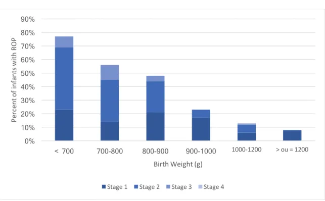
ROP of any degree was diagnosed in 116 (27.68%) infants. The prevalence was 25 % for girls (51/203) and 30 % for boys (65/216). Stage 1, 2, 3 and 4 ROP was found in 44% (51/116), 46% (54/116), 9% (10/116) and 1% (1/116) of subjects, respectively. No stage 5 ROP was observed.
The prevalence and severity of ROP decreased with GA at birth and BW, as shown in figure 1 and 2.
Characteristics of subjects with ROP and risk factors
The mean GA at birth in infants with ROP was 27.12 weeks (range, 25 to 31 weeks) versus 28.83 weeks (range, 25 to 35 weeks) in subjects without ROP. The mean BW in infants with ROP was 866 g (range, 560 to 1715 g) versus 1104 g (range, 500 to 2070 g) in subjects without ROP.
No ROP was noted in infants born at >31 weeks’ GA or with BW>1715 g. 13 infants had a term > 30 SA and a BW > 1500 g and none developed ROP.
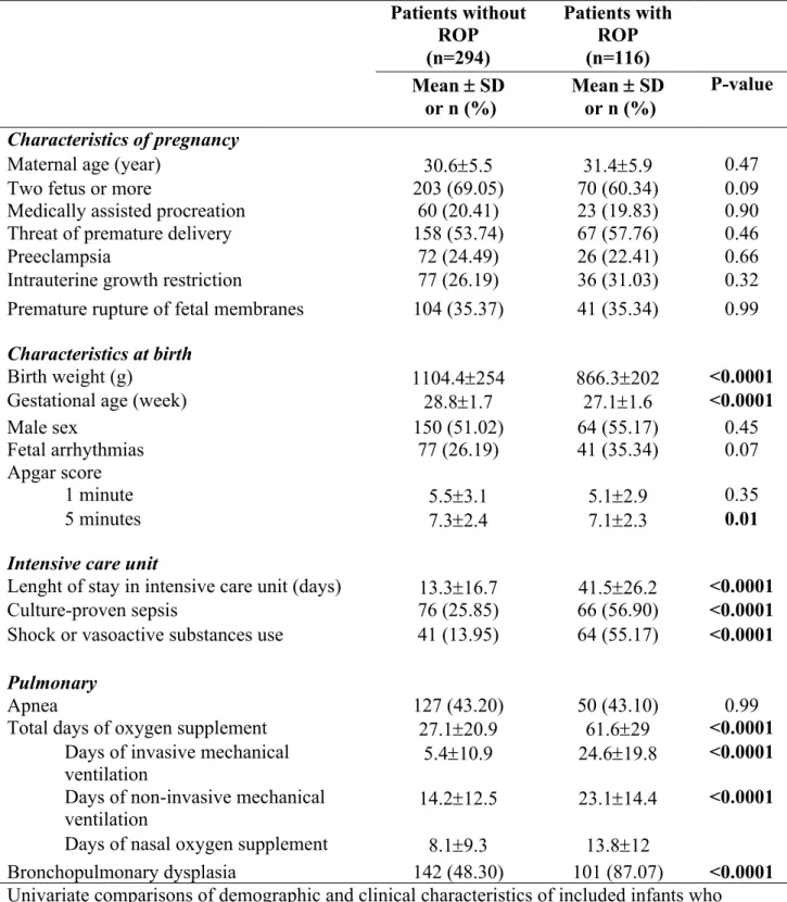
Demographic and clinical characteristics of infants are resumed in table 1.
Table 2 summarizes results from multivariate logistic regression analysis of risk factors for presence of ROP. In multivariate analysis, the independent risk factors for ROP development were low BW (OR=0.26 if BW ≥ 1120 g, 95 CI=0.12-0.59), p <0.0001), low GA at birth (OR=0.44 if GA ≥ 29 weeks, 95 CI=[0.21-0.91], p <0.008), high total days of invasive mechanical ventilation (OR=6.15 if ≥ 7 days, 95 CI=[2.36-15.98], p <0.0008) and shock or vasoactive substances use (OR=2.07, 95 CI=[1.02-4.18], p <0.004).
Characteristics of subjects with treatment-requiring ROP and risk factors
28/419 (6,6 %) infants (28/116 infants (24%) with ROP) were treated by exclusive laser photocoagulation (19 boys, 9 girls). 3 infants required laser retreatment. No intravitreal anti-VEGF injections or surgical treatments were performed. There was no recurrence of ROP after laser treatment.
The mean natal age at which ROP was treated was 12 weeks +/- 1.8, i.e. 36 weeks post-conception (range, 33 to 40 weeks). The mean post-post-conceptional age at birth in the treatment-requiring ROP group was 26.3 weeks 1.5 (range, 25 to 31 weeks) versus 27.4 weeks +/-1.5 (range, 25 to 30 weeks) in the non treatment-requiring ROP group and mean BW was 766.9 g +/- 104.9 (range, 590 to 1110 g) versus 897.9 g +/- 214.9 (range, 560 to 1715) in the non treatment-requiring ROP group.
Of the 28 subjects treated, there were 19 stage 2, 8 stage 3 and 1 stage 4 ; 22 had a "plus" disease. No infant born at>31 weeks or with BW>1110g needed ROP treatment.
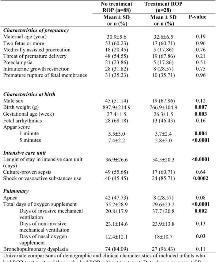
Table 3 summarizes demographic and clinical characteristics between the two groups.
Table 4 summarizes results from multivariate logistic regression analysis of risk factors for treatment-requiring ROP versus non-treatment-requiring ROP. In multivariate analysis, the independent risk factors for treatment-requiring ROP were male gender (OR=3.93, 95 CI=[1.26-12.32], p=0.02), gestational age £ 27 weeks (OR=0.21 if GA ≥ 27 , 95
CI=[0.06-0.67], p=0.009), Apgar score at 5 minutes £ 7 (OR=0.12 if ≥7, 95 CI=[0.04-0.38], p=0.0003] whereas days of non-invasive mechanical ventilation and days of nasal oxygen supplement ≥ 20 (OR=0.26, 95 CI=[0.08-0.80], p=0.02) were protective factor.
Discussion
ROP prevalence
Incidence of prematurity had decreased since the multicenter CRYO-ROP trial in 1986-1987, which reported an 81.6% incidence of ROP among infants with birth weight <1000 g. (8) Advances in neonatology, surfactant therapy, management of oxygen therapy, and better understanding of retinopathy of prematurity have decreased its prevalence. Hoogerwerf et al. found that the incidence of ROP decreased significantly over the last decade (40.9% in 1991-1995 vs 23.3 % in 2001-2005 in all preterm infants and 67.0% vs 41.8% in infants with a BW < 1000g). (9) In our study, 45.6 % of infants with BW < 1000 g had any stage of ROP. Previous studies found incidence of ROP in infants with BW < 1000 g between 41.8% and 59%. (9) (10) (11) (12). We found a prevalence of 53.9% in infants <28 weeks at birth and 69.1% in infants <27 weeks ; previous studies found incidence between 33% and 40% in infants <28 weeks (11) (13) (11) and 73% infants <27 weeks. (14) 6.6% of screened infants were treated, as described in studies between 5.9 and 19.6%. (15) (16) (14) (17) However, comparisons of the incidence of ROP from population-based studies is difficult because of variability in study designs, population studied, gestational ages of included infants, survival rates, and treatments used. Moreover, in most of the studies, ROP screening was performed by indirect ophthalmoscopy and not by exclusive telemedicine.
Finally in our study, ROP screening was performed in infants born until 35 weeks due to a long duration of oxygenotherapy in such cases.
Risk factors
Clinical ROP risk factors in this study are consistent with previously-published research. BW, GA and oxygen supplementation are the most important risk factors for ROP in published studies. (3) (18) (5) (19) Lower BW and GA reflects infants immaturity. The more profound the immaturity at birth and the persistence of developmental arrest due to exposure of the retinal to harmful factors, coupled with deficiencies of factors normally provided in utero, the more aggressive the later pathological response. Low oxygenation targets are associated with
increased mortality and the optimum timing and target concentration of oxygen treatment remain unanswered questions. Strict management of oxygen to minimize alternating hypoxia and hyperoxia and avoidance of undesired high oxygen saturation during the early phase of the pathology seem to be the most promising strategies to prevent ROP, although this outcome has to be balanced against the effect on other morbidities such as cerebral palsy and death. (20) (21) (22)
We also found shock or vasoactive substances use to be significant risk factors of any stage of ROP. We have not determined the etiology of the shock, but it is probable that some shock were secondary to a septic shock. Indeed, neonatal infections are also risk factors for ROP. (23) (24) (12) Furthermore, in our study culture-proven sepsis was a risk factor for ROP in univariate analysis but not in multivariate analysis. The increased risk associated with infection might be partly due to systemic inflammation, which could act synergistically with hyperoxia. (25) Furthermore, hemorrhagic shock and unstable hemodynamic state could be a risk factor for severe ROP. (26) (27)
There were no significant gender differences for ROP presence but male gender was associated with severe ROP (treatment required) in multivariate analysis (OR=3.93). Some studies found that male gender could contribute to severe ROP (3) (28) (29) while others found no correlation between gender and incidence of ROP. (11) (30) (8)
The other two protective factors for treatment-requiring ROP were gestational age ≥ 27 week and Apgar score at 5 minutes ≥ 7. Several studies found Apgar score at 5 minutes as risk factor for ROP (31) (32) (33) but only one found that an Apgar score at 5 minutes £ 6 was an independent risk factor for progression of ROP to stages requiring treatment. (34) Infants with a low 5-minute Apgar score have greater risk for mortality and morbidities associated with prematurity. (35) This association requires to be studied in a larger population before any conclusions can be drawn.
For ROP treatment, BW was not a significant risk factor because 71% of the subjects had a BW < 830 g.
There are several limitations to our study, and our findings should be interpreted with caution. This study was retrospective and it is possible that some data on clinical and demographic risk factors were incomplete. A second limitation is that ethnicity was not determined. Previous studies have demonstrated that ethnicity could be another important independent risk factor for the development of ROP. Hispanic (16) and Asian ethnicity (36) could be a risk factor
whereas black ethnicity could be less susceptible to ROP (37) (8) but this results are inconsistent. (36)
Finally, this study did not investigate the role of postnatal weight gain in the development or requiring treatment of ROP. These last years, studies have shown the importance of postnatal weight gain in the development of ROP and algorithms based upon postnatal weight gain such as the WINROP (weight, IGF, neonatal ROP) algorithms have been shown to have a high sensitivity for detecting severe ROP. (38) (39) (40) Missing weight gain data probably influenced the results of the multivariate analysis.
In our 6-year series, ROP was successfully screened by exclusive telemedecine and no surgical treatment was required. This study confirms several risk factors recognized in preview statistical analyses: low birth weight and gestational age, oxygen supplementation, shock or vasoactive substances use and male gender. Apgar score at 5 minutes as severe ROP risk factor requires furthers studies in order to be confirmed.
REFERENCES
1. Hart JT. Retrolental fibroplasia. A modern parable. J R Coll Gen Pract. 1982 Jun;32(239):371–2.
2. Steinkuller PG, Du L, Gilbert C, Foster A, Collins ML, Coats DK. Childhood blindness. J AAPOS Off Publ Am Assoc Pediatr Ophthalmol Strabismus Am Assoc Pediatr Ophthalmol Strabismus. 1999 Feb;3(1):26–32.
3. Darlow BA, Hutchinson JL, Henderson-Smart DJ, Donoghue DA, Simpson JM, Evans NJ, et al. Prenatal risk factors for severe retinopathy of prematurity among very preterm infants of the Australian and New Zealand Neonatal Network. Pediatrics. 2005 Apr;115(4):990–6.
4. Bardin C, Zelkowitz P, Papageorgiou A. Outcome of small-for-gestational age and appropriate-for-gestational age infants born before 27 weeks of gestation. Pediatrics. 1997 Aug;100(2):E4.
5. Allegaert K, Vanhole C, Casteels I, Naulaers G, Debeer A, Cossey V, et al. Perinatal growth characteristics and associated risk of developing threshold retinopathy of prematurity. J AAPOS Off Publ Am Assoc Pediatr Ophthalmol Strabismus Am Assoc Pediatr Ophthalmol Strabismus. 2003 Feb;7(1):34–7.
6. Fierson WM, Saunders RA, Good W, Palmer EA, Phelps D, Reynolds J, et al. Screening Examination of Premature Infants for Retinopathy of Prematurity. Pediatrics. 2013 Jan 1;131(1):189–95.
7. International Committee for the Classification of Retinopathy of Prematurity. The International Classification of Retinopathy of Prematurity revisited. Arch Ophthalmol Chic Ill 1960. 2005 Jul;123(7):991–9.
8. Palmer EA, Flynn JT, Hardy RJ, Phelps DL, Phillips CL, Schaffer DB, et al. Incidence and early course of retinopathy of prematurity. The Cryotherapy for Retinopathy of Prematurity Cooperative Group. Ophthalmology. 1991 Nov;98(11):1628–40.
9. Hoogerwerf A, Schalij-Delfos NE, van Schooneveld MJ, Termote JUM. Incidence of retinopathy of prematurity over the last decade in the Central Netherlands. Neonatology. 2010;98(2):137–42.
10. Shah VA, Yeo CL, Ling YLF, Ho LY. Incidence, risk factors of retinopathy of prematurity among very low birth weight infants in Singapore. Ann Acad Med Singapore. 2005 Mar;34(2):169–78.
1989-1997. Pediatrics. 1999 Sep;104(3):e26.
12. Liu P-M, Fang P-C, Huang C-B, Kou H-K, Chung M-Y, Yang Y-H, et al. Risk factors of retinopathy of prematurity in premature infants weighing less than 1600 g. Am J Perinatol. 2005 Feb;22(2):115–20.
13. Markestad T, Kaaresen PI, Rønnestad A, Reigstad H, Lossius K, Medbø S, et al. Early death, morbidity, and need of treatment among extremely premature infants. Pediatrics. 2005 May;115(5):1289–98.
14. Austeng D, Källen KBM, Ewald UW, Jakobsson PG, Holmström GE. Incidence of retinopathy of prematurity in infants born before 27 weeks’ gestation in Sweden. Arch Ophthalmol Chic Ill 1960. 2009 Oct;127(10):1315–9.
15. Chiang MF, Arons RR, Flynn JT, Starren JB. Incidence of retinopathy of prematurity from 1996 to 2000: analysis of a comprehensive New York state patient database. Ophthalmology. 2004 Jul;111(7):1317–25.
16. Lad EM, Nguyen TC, Morton JM, Moshfeghi DM. Retinopathy of prematurity in the United States. Br J Ophthalmol. 2008 Mar 1;92(3):320–5.
17. Port AD, Chan RVP, Ostmo S, Choi D, Chiang MF. Risk Factors for Retinopathy of Prematurity: Insights from Outlier Infants. Graefes Arch Clin Exp Ophthalmol Albrecht Von Graefes Arch Klin Exp Ophthalmol. 2014 Oct;252(10):1669–77.
18. Bardin C, Zelkowitz P, Papageorgiou A. Outcome of small-for-gestational age and appropriate-for-gestational age infants born before 27 weeks of gestation. Pediatrics. 1997 Aug;100(2):E4.
19. Patz A, Hoeck LE, De La Cruz E. Studies on the effect of high oxygen administration in retrolental fibroplasia. I. Nursery observations. Am J Ophthalmol. 1952 Sep;35(9):1248– 53.
20. Chow LC, Wright KW, Sola A, CSMC Oxygen Administration Study Group. Can changes in clinical practice decrease the incidence of severe retinopathy of prematurity in very low birth weight infants? Pediatrics. 2003 Feb;111(2):339–45.
21. Castillo A, Deulofeut R, Critz A, Sola A. Prevention of retinopathy of prematurity in preterm infants through changes in clinical practice and SpO₂technology. Acta Paediatr Oslo Nor 1992. 2011 Feb;100(2):188–92.
22. Lee J, Dammann O. Perinatal infection, inflammation, and retinopathy of prematurity. Semin Fetal Neonatal Med. 2012 Feb;17(1):26–9.
23. Tolsma KW, Allred EN, Chen ML, Duker J, Leviton A, Dammann O. Neonatal bacteremia and retinopathy of prematurity: the ELGAN study. Arch Ophthalmol Chic Ill
1960. 2011 Dec;129(12):1555–63.
24. Mittal M, Dhanireddy R, Higgins RD. Candida Sepsis and Association With Retinopathy of Prematurity. Pediatrics. 1998 Apr 1;101(4):654–7.
25. Chen M, Citil A, McCabe F, Leicht KM, Fiascone J, Dammann CEL, et al. Infection, oxygen, and immaturity: interacting risk factors for retinopathy of prematurity. Neonatology. 2011;99(2):125–32.
26. Jandeck C, Kellner U, Kössel H, Bartsch M, Versmold HT, Foerster MH. Retinopathy of prematurity in infants of birth weight > 2000 g after haemorrhagic shock at birth. Br J Ophthalmol. 1996 Aug;80(8):728–31.
27. Gaugler C, Beladdale J, Astruc D, Schaeffer D, Donato L, Speeg-Schatz C, et al. [Retinopathy of prematurity: 10-year retrospective study at the University Hospital of Strasbourg]. Arch Pediatr Organe Off Soc Francaise Pediatr. 2002 Apr;9(4):350–7.
28. Ying G-S, Quinn GE, Wade KC, Repka MX, Baumritter A, Daniel E, et al. Predictors for the development of referral-warranted retinopathy of prematurity in the telemedicine approaches to evaluating acute-phase retinopathy of prematurity (e-ROP) study. JAMA Ophthalmol. 2015 Mar;133(3):304–11.
29. Nødgaard H, Andreasen H, Hansen H, Sørensen HT. Risk factors associated with retinopathy of prematurity (ROP) in northern Jutland, Denmark 1990-1993. Acta Ophthalmol Scand. 1996 Jun;74(3):306–10.
30. Arrøe M, Peitersen B. Retinopathy of prematurity: review of a seven-year period in a Danish neonatal intensive care unit. Acta Paediatr Oslo Nor 1992. 1994 May;83(5):501–5. 31. Yang C-Y, Lien R, Yang P-H, Chu S-M, Hsu J-F, Fu R-H, et al. Analysis of incidence and risk factors of retinopathy of prematurity among very-low-birth-weight infants in North Taiwan. Pediatr Neonatol. 2011 Dec;52(6):321–6.
32. García-Serrano JL, Ramírez-García MC, Piñar-Molina R. [Retinopathy of prematurity in multiple births: risk analysis for plus disease]. Arch Soc Espanola Oftalmol. 2009 Apr;84(4):191–8.
33. Ke X, Ju R, Zhang J, Chen H, Wei E, Chen X. Risk factors for severe retinopathy of prematurity in premature infants: a single-center study. Nan Fang Yi Ke Da Xue Xue Bao. 2011 Dec;31(12):1963–7.
34. Marinov VG, Koleva-Georgieva DN, Sivkova NP, Krasteva MB. The 5-minute Apgar Score as a Prognostic Factor for Development and Progression of Retinopathy of Prematurity. Folia Med (Plovdiv). 2017 Mar 1;59(1):78–83.
outcomes in extremely low-birth-weight infants. J Perinat Neonatal Nurs. 2012 Jun;26(2):166–71.
36. Aralikatti AKV, Mitra A, Denniston AKO, Haque MS, Ewer AK, Butler L. Is ethnicity a risk factor for severe retinopathy of prematurity? Arch Dis Child Fetal Neonatal Ed. 2010 May;95(3):F174–6.
37. Saunders RA, Donahue ML, Christmann LM, Pakalnis AV, Tung B, Hardy RJ, et al. Racial variation in retinopathy of prematurity. The Cryotherapy for Retinopathy of Prematurity Cooperative Group. Arch Ophthalmol Chic Ill 1960. 1997 May;115(5):604–8. 38. Hellström A, Hård A-L, Engström E, Niklasson A, Andersson E, Smith L, et al. Early weight gain predicts retinopathy in preterm infants: new, simple, efficient approach to screening. Pediatrics. 2009 Apr;123(4):e638–45.
39. Löfqvist C, Andersson E, Sigurdsson J, Engström E, Hård A-L, Niklasson A, et al. Longitudinal postnatal weight and insulin-like growth factor I measurements in the prediction of retinopathy of prematurity. Arch Ophthalmol Chic Ill 1960. 2006 Dec;124(12):1711–8. 40. Wu C, Vanderveen DK, Hellström A, Löfqvist C, Smith LEH. Longitudinal postnatal weight measurements for the prediction of retinopathy of prematurity. Arch Ophthalmol Chic Ill 1960. 2010 Apr;128(4):443–7.
FIGURES
Figure 1. Prevalence and severity of ROP by gestational age at birth
Figure 2. Prevalence and severity of ROP by birth weight
0% 10% 20% 30% 40% 50% 60% 70% 80% 90% < 26 26-27 27-28 28-29 29-30 > ou = 30 Pe rc en t o f i nf an ts w ith R O P Gestational age (week)
Stage 1 Stage 2 Stage 3 Stage 4
0% 10% 20% 30% 40% 50% 60% 70% 80% 90% < 700 700-800 800-900 900-1000 1000-1200 > ou = 1200 Pe rc en t o f i nf an ts w ith R O P Birth Weight (g)
Table 1. Demographic and clinical characteristics of included infants (n=410) (2009-2015) Patients without ROP (n=294) Patients with ROP (n=116) Mean ± SD or n (%) Mean ± SD or n (%) P-value Characteristics of pregnancy
Maternal age (year) 30.6±5.5 31.4±5.9 0.47
Two fetus or more 203 (69.05) 70 (60.34) 0.09
Medically assisted procreation 60 (20.41) 23 (19.83) 0.90 Threat of premature delivery 158 (53.74) 67 (57.76) 0.46
Preeclampsia 72 (24.49) 26 (22.41) 0.66
Intrauterine growth restriction 77 (26.19) 36 (31.03) 0.32 Premature rupture of fetal membranes
Characteristics at birth
104 (35.37) 41 (35.34) 0.99
Birth weight (g) 1104.4±254 866.3±202 <0.0001
Gestational age (week) 28.8±1.7 27.1±1.6 <0.0001
Male sex 150 (51.02) 64 (55.17) 0.45
Fetal arrhythmias 77 (26.19) 41 (35.34) 0.07
Apgar score
1 minute 5.5±3.1 5.1±2.9 0.35
5 minutes 7.3±2.4 7.1±2.3 0.01
Intensive care unit
Lenght of stay in intensive care unit (days) 13.3±16.7 41.5±26.2 <0.0001
Culture-proven sepsis 76 (25.85) 66 (56.90) <0.0001
Shock or vasoactive substances use 41 (13.95) 64 (55.17) <0.0001
Pulmonary
Apnea 127 (43.20) 50 (43.10) 0.99
Total days of oxygen supplement 27.1±20.9 61.6±29 <0.0001 Days of invasive mechanical
ventilation 5.4±10.9 24.6±19.8 <0.0001
Days of non-invasive mechanical
ventilation 14.2±12.5 23.1±14.4 <0.0001
Days of nasal oxygen supplement 8.1±9.3 13.8±12
Bronchopulmonary dysplasia 142 (48.30) 101 (87.07) <0.0001 Univariate comparisons of demographic and clinical characteristics of included infants who developed any stage of ROP and those who did not. Data shown as mean ± SD or n(%).
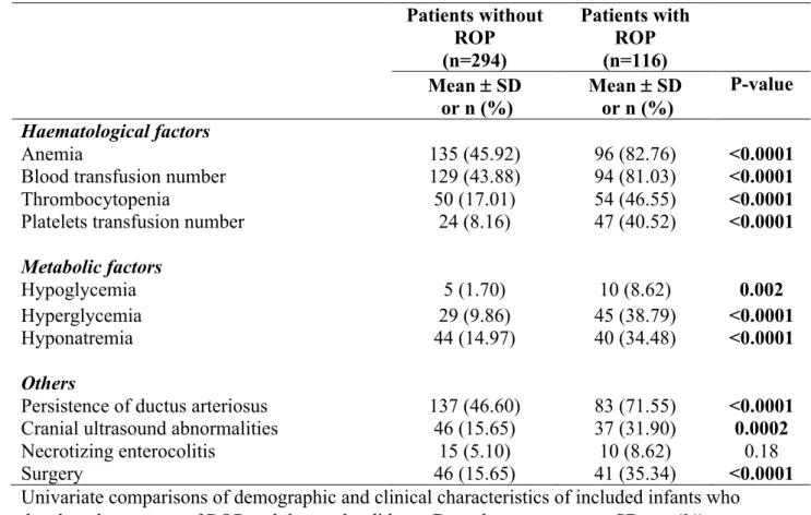
Table 1. Demographic and clinical characteristics of included infants (n=410) (2009-2015) Patients without ROP Patients with ROP (n=294) (n=116) Mean ± SD or n (%) Mean ± SD or n (%) P-value Haematological factors Anemia 135 (45.92) 96 (82.76) <0.0001
Blood transfusion number 129 (43.88) 94 (81.03) <0.0001
Thrombocytopenia 50 (17.01) 54 (46.55) <0.0001
Platelets transfusion number 24 (8.16) 47 (40.52) <0.0001
Metabolic factors
Hypoglycemia 5 (1.70) 10 (8.62) 0.002
Hyperglycemia 29 (9.86) 45 (38.79) <0.0001
Hyponatremia 44 (14.97) 40 (34.48) <0.0001
Others
Persistence of ductus arteriosus 137 (46.60) 83 (71.55) <0.0001 Cranial ultrasound abnormalities 46 (15.65) 37 (31.90) 0.0002
Necrotizing enterocolitis 15 (5.10) 10 (8.62) 0.18
Surgery 46 (15.65) 41 (35.34) <0.0001
Univariate comparisons of demographic and clinical characteristics of included infants who developed any stage of ROP and those who did not. Data shown as mean ± SD or n(%).
Table 2. Multivariate analysis of risk factor for retinopathy of prematurity
OR 95% CI P-value
Birth weight (g)
<890 <0.0001
890-1120 0.27 0.14 ; 0.52
≥1120 0.26 0.12 ; 0.59
Gestational age (week)
<28 0.008
28-29 0.32 0.15 ; 0.70
≥29 0.44 0.21 ; 0.91
Days of invasive mechanical ventilation
<1 0.0008
1-7 2.45 1.04 ; 5.80
≥7 6.15 2.36 ; 15.98
Shock or vasoactive substances use 2.07 1.02 ; 4.18 0.04 OR: Odds ratio
Table 3. Univariate analysis of risk factor for treatment-requiring ROP versus non-treatment requiring ROP (n=116)
No treatment ROP (n=88) Treatment ROP (n=28) Mean ± SD or n (%) Mean ± SD or n (%) P-value Characteristics of pregnancy
Maternal age (year) 30.9±5.6 32.6±6.5 0.19
Two fetus or more 53 (60.23) 17 (60.71) 0.96
Medically assisted procreation 18 (20.45) 5 (17.86) 0.76 Threat of premature delivery 48 (54.55) 19 (67.86) 0.21
Preeclampsia 21 (23.86) 5 (17.86) 0.51
Intrauterine growth restriction 28 (31.82) 8 (28.57) 0.75 Premature rupture of fetal membranes 31 (35.23) 10 (35.71) 0.96
Characteristics at birth
Male sex 45 (51.14) 19 (67.86) 0.12
Birth weight (g) 897.9±214.9 766.9±104.9 0.007
Gestational age (week) 27.4±1.5 26.3±1.5 0.003
Fetal arrhythmias 28 (68.18) 13 (46.43) 0.16
Apgar score
1 minute 5.5±3.0 3.7±2.4 0.004
5 minutes 7.4±2.2 5.8±2.0 <0.0001
Intensive care unit
Lenght of stay in intensive care unit
(days) 36.9±26.6 54.5±20.3 <0.0001
Culture-proven sepsis 49 (55.68) 17 (60.71) 0.64
Shock or vasoactive substances use 40 (45.45) 24 (85.71) 0.0002
Pulmonary
Apnea 42 (47.73) 8 (28.57) 0.08
Total days of oxygen supplement 55.2±28.9 79.6±23.2 <0.0001 Days of invasive mechanical
ventilation 20.8±17.9 37.7±20.8
0.002 Days of non-invasive
mechanical ventilation 23.1±14.6 23.9±13.8 0.13
Days of nasal oxygen
supplement 12.4±12.1 18±10.7
0.03
Bronchopulmonary dysplasia 74 (84.09) 27 (96.43) 0.11
Univariate comparisons of demographic and clinical characteristics of included infants who had ROP treatment and those who had ROP without treatment. Data shown as mean ± SD or n(%).
Tableau 3. Univariate analysis of risk factor for treatment-requiring ROP versus non-treatment requiring ROP (n=116)
No treatment ROP (n=88) Treatment ROP (n=28) Mean ± SD or n (%) Mean ± SD or n (%) P-value Haematological factors Anemia 71 (80.68) 25 (89.29) 0.40
Blood transfusion number 2.5±1.9 4.1±2.3 0.004
Thrombocytopenia 36 (40.91) 18 (64.29) 0.03
Platelets transfusion number 0.91±2.5 1.8±2.5 0.003
Metabolic factors
Hypoglycemia 7 (7.95) 3 (10.71) 0.70
Hyperglycemia 29 (32.95) 16 (57.14) 0.02
Hyponatremia 28 (31.82) 12 (42.86) 0.28
Others
Persistence of ductus arteriosus 57 (64.77) 26 (92.86) 0.004 Cranial ultrasound abnormalities 26 (29.55) 11 (39.29) 0.34
Surgery 26 (29.55) 15 (53.57) 0.02
First fundus stage
0 30 (34.09) 10 (35.71) 0.0006
1 44 (50.00) 6 (21.43)
2 10 (11.36) 3 (10.71)
NI 4 (4.55) 9 (32.14)
First interpretable fundus stage
0 31 (35.23) 11 (39.29) 0.05
1 47 (53.41) 9 (32.14)
2 10 (11.36) 8 (28.57)
Univariate comparisons of demographic and clinical characteristics of included infants who had ROP treatment and those who had ROP without treatment. Data shown as mean ± SD or n(%).
Table 4: Multivariate analysis of risk factor for treatment-requiring ROP versus non-treatment requiring ROP
OR 95% CI P-value
Male sex 3.93 1.26 ; 12.32 0.02
Gestational age (week) ≥27 0.21 0.06 ; 0.67 0.009
5 minutes Apgar score ≥7 0.12 0.04 ; 0.38 0.0003
Days of non-invasive mechanical ventilation and days of nasal oxygen supplement ≥20
0.26 0.08 ; 0.80 0.02
III. ANNEXE 1
La rétinopathie du prématuré
INTRODUCTION
La rétinopathie des prématurés (ROP) se caractérise par des anomalies du développement de la vascularisation rétinienne chez l’enfant prématuré.
Décrite pour la première fois en 1942, elle représente une cause importante et évitable de cécité de l’enfant. Dans les pays développés, elle fait partie des 3 causes les plus fréquentes de cécité de l’enfant avec la cécité d’origine corticale et l’hypoplasie du nerf optique. (1)
DEFINITION DE LA PREMATURITÉ
Toute naissance avant le terme de 37 semaines d’aménorrhée (SA) révolues est prématurée, le terme normal étant de 41 semaines.
On les classe en :
- Prématurité (de 33 à 37 SA)
- Grande prématurité (de 28 à 32 SA) - Très grande prématurité (de 25 à 27) - Extrême prématurité (moins de 24 SA)
Dans le monde, environ 10 % des naissances sont prématurées.
EPIDEMIOLOGIE
A la fin des années 1940, la rétinopathie des prématurés, appelée fibrodysplasie rétrolentale, apparaissait chez des enfants prématurés du fait de l’utilisation d’oxygénothérapie dans des incubateurs fermés. Depuis le contrôle de l’oxygénothérapie, une deuxième vague de ROP a eu lieu à la fin des années 1970 et 1980 du fait de la survie de prématurés de très faible poids de naissance (750-999 g).
Dans les pays industrialisés, la ROP est une cause importante de cécité de l’enfant puisqu’elle est responsable de 6 à 20 % des cécités de l’enfant.
La prévalence de la ROP varie de 33 à 73 % et de ROP sévère de 16 à 35 % pour un âge gestationnel inférieur à 27 à 29 SA selon les études. (2) (3)(4)(5)(6)(7)
PHYSIOPATHOLOGIE
Introduction
La rétinopathie des prématurés peut être vue comme un arrêt de la rétine neurosensorielle normale et du développement vasculaire chez un nourrisson prématuré, avec des mécanismes compensatoires pathologiques aboutissant à une vascularisation aberrante de la rétine. Plus l’immaturité à la naissance est profonde du fait de l’insuffisance de facteurs normalement présents in utero est importante, plus la réponse pathologique ultérieure sera importante.
Normalement, la rétine nasale devient complètement vascularisée aux alentours de 36 SA et les vaisseaux temporaux atteignent l’ora serrata à 40 SA environ.
La pathologie présente deux phases post natales (8) (9) (10), possiblement précédées par une phase anténatale de sensibilisation par l’inflammation. (11) La première phase commence par une croissance vasculaire rétinienne retardée après la naissance et la régression partielle des vaisseaux existants, suivie d’une seconde phase de croissance pathologique des vaisseaux induite par l’hypoxie.
Phase 1 ou phase vaso-oblitérative (de la 22e à 30e SA)
L’hyperoxie est un facteur important d’arrêt de la croissance vasculaire en phase 1. (10) (12) L’hyperoxie conduit à la suppression de facteurs de croissance angiogéniques régulés par l’oxygène, en particulier l’érythropoiétine (13) et le facteur de croissance vasculaire endothélial (VEGF) (14) qui provoque à la fois l’arrêt de la croissance des vaisseaux rétiniens et la perte de certains vaisseaux rétiniens existants. (15) D’autre part, chez les nourrissons nés avant la fin du troisième trimestre de grossesse, la diminution de facteurs de croissance tels que l’insuline-like growth factor 1 (IGF-1), normalement présent à des concentrations optimales in utero contribue à l’arrêt de la croissance vasculaire. (16) De plus,
la perte des acides gras polyinsaturés à longue chaîne oméga maternels semble avoir un rôle dans la pathogenèse de la ROP. (17)
Phase 2 ou phase vasoproliférative (de la 31e à 44e SA) :
Dans les cas sévères, la phase 2 débute lorsque la rétine peu vascularisée et métaboliquement active devient hypoxique. La phase 2 se caractérise par la prolifération de vaisseaux sanguins, de cellules gliales et par la formation de shunt artérioveineux en réponse à l’augmentation du VEGF et de l’EPO du fait de l’hypoxie. (18) Les néovaisseaux perfusent peu la rétine et sont peu perméables, ce qui conduit à la formation de fibrose et de décollement de rétine. (Figure 1)
Dans la plupart des cas, la rétinopathie régresse spontanément et la rétine est vascularisée normalement, bien que des déficits neuronaux (perte de la fonction des photorécepteurs) peuvent persister, même dans les cas bénins. (19)
Dans certains cas, les facteurs en cause dans la prématurité peuvent affecter la vascularisation rétinienne intra-utérine. Des facteurs tels qu’une inflammation ou une infection placentaire peuvent prédisposer la rétine fœtale à une rétinopathie du prématuré sévère et pourrait être un effet de sensibilisation constituant la préphase de la pathologie. (11)
Figure 1. Progression de la rétinopathie des prématurés (Retinopathy of Prematurity,
Hellström et al. (20))
A : la concentration en oxygène est faible in utero et la croissance vasculaire est normale. B : Phase 1 : après la naissance et jusqu’à l’âge de 30 semaines environ, la vascularisation de la rétine est inhibée en raison de l’hyperoxie et de la perte des nutriments et des facteurs de croissance maternels. La croissance vasculaire s’arrête alors que la rétine se développe, augmentant la demande métabolique, il en résulte une hypoxie.
C : Phase 2 : la rétine hypoxique stimule l’expression des facteurs régulés par l’oxygène tels que l’EPO et le VEGF qui stimulent la néovascularisation rétinienne. La concentration d’IGF-1 augmente lentement mais suffisamment pour activer la voie du VEGF.
D : La résolution de la ROP peut se faire par la prévention de la phase 1 en augmentant les concentrations d’IGF-A in utero et en limitant l’oxygénothérapie afin d’empêcher la suppression du VEGF. Ce dernier peut être diminué en phase 2 après néovascularisation avec le traitement par laser ou injection d’anticorps.
FACTEURS DE RISQUES
Bien que de nombreux facteurs ont été proposés pour la ROP, seuls le petit poids de naissance, l’âge gestationnel bas et l’oxygénothérapie postnatale ont été associés avec la ROP. (21) (22)(23)
Age gestationnel et poids de naissance
Le faible âge gestationnel et le faible poids de naissance sont des facteurs de risques majeur de ROP. (24) Ces deux facteurs sont liés au taux d’immaturité des neurones rétiniens et du développement vasculaire à la naissance et donc à l’agression de la rétine vulnérable. En outre, plus l’âge gestationnel et le poids de naissance sont faibles, plus la perte de facteurs intra-utérins pour lesquels le fœtus immature est incapable de produire est importante. De plus, le faible âge gestationnel augmente la durée d’exposition du nourrisson aux agressions post natales.
Oxygène
L’oxygénothérapie tient une place prépondérante dans le déterminisme de la ROP.
Les fluctuations de la concentration en oxygène durant les premières semaines de vies sont également associées à une augmentation du risque de ROP. (25) (26) (27) (28) (29) De plus, l’hypoxie intermittente durant les 8 premières semaines de vie est associée à une plus forte sévérité de la rétinopathie.
Le taux d’oxygénothérapie optimale pour diminuer les risques de ROP et optimiser la survie demeure encore inconnu. La saturation en oxygène (SpO2) cible devrait être différente en fonction des stades de développement et en fonction des stades de la ROP. La gestion rigoureuse de l’oxygène pour minimiser les hypoxies et les hyperoxies alternatives et la diminution de fortes SpO2 en phase 1 semble être la bonne stratégie pour prévenir la ROP bien que cela doit être équilibré contre les effets sur d’autres morbidités comme la paralysie cérébrale et le décès. (30)
Les autres facteurs de risques dont l’imputabilité est discutée, sont les suivants : - Facteurs génétiques : nourrissons caucasiens, sexe masculin
- Taille à la naissance et retard de croissance intra utérin (RCIU) (31) (32) - Faible prise de poids (33) (34)
- Hyperglycémie néonatale (35) (36) et administration d’insuline (37) - Infections néonatales (38) (39)
- Transfusions sanguines (40)(41)
- Neurologiques : hémorragies intraventriculaires - Diminution de l’IGF-1(42)
CLASSIFICATION DE LA ROP
Selon l’ICROP (The International Classification of Retinopathy or Prematurity), 4 paramètres permettent d’évaluer la ROP : (43)
- La sévérité, classée en stades 1 à 5
- La localisation classée en zones centrées par la papille
- Le stade « PLUS » qui correspond à une tortuosité vasculaire - L’étendue décrite en nombre de quadrants horaires (1 à 12)
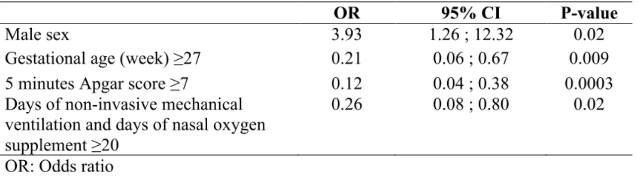
Localisation de la rétinopathie : séparation en zones
3 zones concentriques ont été définies. Chaque zone est centrée sur la papille plutôt que sur la car le processus de vascularisation normal de la rétine part du centre du nerf optique vers l’ora serrata de manière ordonnée. (Figure 2)
Les zones sont centrées sur la papille et sont définies de la manière suivante :
- Zone 1 (la plus interne) : il s’agit d’un cercle centré sur le nerf optique dont le rayon fait deux fois la distance papille-fovéa
- Zone 2 : elle s’étend du bord de la zone 1 jusqu’à l’ora serrata en nasal - Zone 3 : elle comprend le croissant de rétine résiduel antérieur à la zone 2. Par convention, les zones 2 et 3 sont considérées comme mutuellement exclusives.
Figure 2. Zones de la ROP. Schéma de la rétine de l’œil droit (RE) et de l’œil gauche (LE) montrant les limites des zones et les cadrans horaires utilisés pour décrire la topographie de la ROP (The International Classification of Retinopathy of Prematurity Revisited)
Stades de la rétinopathie
5 stades sont utilisés pour décrire la réponse vasculaire anormale au niveau de la jonction rétine vascularisée et avasculaire. Plusieurs stades peuvent être présents simultanément dans chaque œil, c’est donc le stade le plus sévère qui est retenu.
Les stades sont les suivants (Figure 3) :
• Stade 1 : ligne de démarcation. Cette ligne est une fine structure bien définie qui sépare antérieurement la rétine avasculaire de la rétine vasculaire postérieure
• Stade 2 : bourrelet. Le bourrelet témoigne d’une néovascularisation intrarétinienne qui atteint la ligne de démarcation et se prolonge au-dessus du plan de la rétine. De petites touffes de tissus néovasculaires isolées couchées à la surface de la rétine communément appelées « pop-corn » peuvent être vus de manière postérieure au bourrelet. (Figure 4)
• Stade 3 : prolifération fibrovasculaire extrarétinienne. La prolifération fibrovasculaire extrarétinienne s’étend depuis le bourrelet jusque dans le vitré. (Figure 5)
• Stade 4 : décollement de rétine partiel. Le stade 4 est divisé en décollement de
rétine partiel extrafovéal (stade 4A) et fovéal (stade 4B). Typiquement, le
décollement de rétine débute au niveau du point d’attache fibrovasculaire à la rétine vascularisée. En cas d’évolution, le tissu fibrovasculaire continue à se contracter et le décollement de rétine tractionnel augmente en hauteur, s’étendant de manière antérieure et postérieure
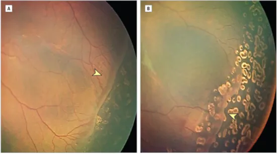
Figure 3. Stades de sévérité de la ROP. A) Stade 1 : ligne de démarcation entre la zone de rétine vasculaire et avasculaire. B) Stade 2 : Bourrelet. C) Prolifération fibrovasculaire extrarétinienne. D) Stade 4 : décollement de rétine partiel. (Stem Cell Therapy for Retinopathy of Prematurity. Ka Wai Lai et al.)
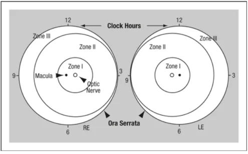
Figure 4. Photographie montrant le bourrelet entre rétine vasculaire et avasculaire caractéristique du stade 2 de la ROP (Image du CHU de Bordeaux)
Figure 5. ROP de stade 3, prolifération fibrovasculaire extra rétinienne (Image du CHU
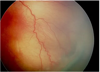
Formes « plus disease »
La ROP est qualifiée de « plus » s’il y a une tortuosité et une dilatation vasculaire veineuse
et artérielle des vaisseaux rétiniens postérieurs importante dans au moins deux cadrans.
Par exemple, le stade 2 combiné avec une dilatation et tortuosité vasculaire postérieure est notée « stade ROP 2 + ». Cela peut évoluer vers une dilatation des vaisseaux iriens, une mauvaise dilatation pupillaire et un haze vitréen.
Le stade plus est un signe inquiétant de progression de la rétinopathie. (Figure 6)
Figure 6. Photographie du fond d’œil montrant un stade « plus ». (Image du CHU de Bordeaux)
Stade pré-plus
Il s’agit de signes d’activité de la ROP avec une dilatation et une tortuosité anormale des vaisseaux du pôle postérieur mais dont l’intensité n’atteint pas le stade « plus ».
ROP postérieure agressive
La ROP postérieure agressive est une forme sévère, rare et rapidement progressive de
ROP. Si elle n’est pas traitée, elle évolue habituellement vers le stade 5. Elle est caractérisée
Régression de la ROP
La plupart des ROP régressent spontanément par un processus d’involution ou évoluent d’une forme vasoproliférative vers une phase fibreuse.
Les stades 1 et 2 ainsi que les rétinopathies en zone 3 sont susceptibles de régresser spontanément tandis que dans les stades 3, la néovascularisation extrarétinienne peut devenir assez sévère pour provoquer un décollement de rétine total (stade 5) et aboutir vers la cécité. Un des premiers signes de stabilisation est l’absence de progression vers le stade suivant.
Le processus de régression se produit en grande partie à la jonction entre la rétine vasculaire et avasculaire et la vascularisation progresse en périphérie. (Figure 7) Les séquelles involutionnelles comprennent un large éventail de modifications rétiniennes et vasculaires périphériques tels que des zones de rétine avasculaire, des ramifications vasculaires anormales et des télangiectasies. Des modifications pigmentaires peuvent être retrouvées ainsi que des modifications circonférentielles de l’interface vitréorétiniennes comme des lignes fines ou des bourrelets plus proéminents. Au niveau du pôle postérieur, on peut retrouver des phénomènes de traction qui peuvent varier de distorsions mineures de l’architecture fovéale jusqu’à l’hétérotopie ou ectopie maculaire (Figure 6). Enfin, les décollements de rétine tractionnels et rhegmatogènes et, plus rarement, exsudatifs peuvent compliquer des ROP ayant régressé.
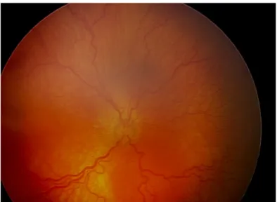
Figure 7. Régression de la ROP après photocoagulation. A) Retcam réalisée 10 jours après
le traitement par photocoagulation d’une ROP sévère. B) Un mois plus tard, progression de la vascularisation rétinienne et évolution de la différenciation rétinienne. (Images du CHU de
DEPISTAGE
Les dernières recommandations de l’American Academy of Pediatrics de 2013 (44) recommandent un dépistage chez :
• Les nourrissons dont le poids de naissance est inférieur ou égal à 1500 g ou l’âge gestationnel inférieur ou égal à 30 SA
• Ainsi que les nourrissons de 15000 à 2000 g ou d’âge supérieur à 30 SA mais ayant une évolution clinique instable
Il doit être réalisé après dilatation pupillaire en utilisant un ophtalmoscope indirect et un blépharostat en cas de nécessité et en réalisant une dépression sclérale avec anesthésie topique si nécessaire pour dépister une ROP.
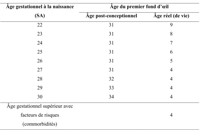
L’initiation du dépistage de la ROP est fondée sur l’âge post-conceptionnel (âge gestationnel + âge chronologique) plutôt que sur l’âge réel (de vie) (Figure 8).
Les délais du premier examen du fond d’œil sont résumés dans le tableau ci-dessous et permettrait un dépistage des stades « pré-seuil » dans 99 % des cas.
Âge gestationnel à la naissance (SA)
Âge du premier fond d’œil
Âge post-conceptionnel Âge réel (de vie)
22 31 9 23 31 8 24 31 7 25 31 6 26 31 5 27 31 4 28 32 4 29 33 4 30 34 4
Âge gestationnel supérieur avec facteurs de risques
(commorbidités)
4
Figure 8. Âge post-conceptionnel, âge de vie, âge corrigé
Les recommandations de suivi sont les suivantes :
Contrôle à 1 semaine ou moins :
- Vascularisation immature en zone 1 sans ROP
- Rétine immature en zone 2 postérieure, derrière la limite de la zone 1 - ROP stade 1 ou 2 : zone 1
- ROP stade 3 : zone 2
- Présence ou suspicion de ROP postérieure agressive Contrôle à 1 à 2 semaines :
- Vascularisation immature en zone 2 postérieure - ROP stade 2 : zone 2
- Régression de ROP : zone 1 Contrôle à 2 à 3 semaines :
- ROP stades 1 ou 2 : zone 3 - ROP zone 3 ayant régressé
- Vascularisation rétinienne en zone 3 sans ROP en zone 1 ou 2
- Vascularisation rétinienne complète proche de l’ora serrata sur 360° (ce critère devrait être utilisé pour tous les cas de ROP traités uniquement par bevacizumab)
- Age postmenstruel > 50 SA et ROP - Régression de ROP
Les examens du fond d’œil avec dilatation devraient être répétés jusqu’à ce que la rétine soit complètement vascularisée. Cependant les examens du fond d’œil peuvent être douloureux chez les nourrissons prématurés. (45) Dans un contexte de soins intensifs néonataux de qualité, seulement 5 à 10 % des enfants nécessiteront un traitement. Diminuer prudemment le nombre d’examens de dépistage stressant et coûteux serait bénéfique.
Pour cela, différents algorithmes ont été développés comme WINROP, afin d’identifier de manière précoce après la naissance les enfants à risque de développer une ROP sévère. (46) (47)
De plus, la télémédecine pourrait améliorer le dépistage de la ROP en réduisant la nécessité d’ophtalmologistes qualifiés dans les unités de soins intensifs néonataux grâce à la centralisation des lecteurs. (48)
RETCAM
Depuis les années 2000, le dépistage peut être réalisé par la RETCam 120 qui permet de capturer l’image du fond d’œil sur 120° numériquement. (49) (50) (51) L’acquisition des images, plus facile à réaliser qu’une ophtalmoscopie indirecte, peut être déléguée et l’interprétation est ensuite réalisée par l’ophtalmologiste. Le coût de cet appareil serait équivalent au coût des soins pour une cécité congénitale. (Figure 9)
L’imagerie par photographie grand champ permet d’obtenir une documentation objective des données de l’examen clinique, une amélioration du diagnostic et de la progression de la pathologie par la possibilité de réaliser une analyse comparative des rétinographies et de commenter ces résultats avec la famille et les néonatologistes. Une revue de la littérature récente a montré la supériorité de ce type d’imagerie par rapport à l’ophtalmoscopie dans le diagnostic de ROP, sans complications notables. (52)
Figure 9. Imagerie du fond d’oeil par la RetCam-120 Digital Retinal Camera (Massie Research Laboratories Inc., Dublin, CA)
TRAITEMENT
Le traitement par laser transpupillaire pour traiter la rétine avasculaire a remplacé la cryothérapie utilisée dans les années 1980 du fait de meilleurs résultats visuels et de moins d’effets secondaires locaux et systémiques. La photocoagulation est appliquée sur les zones de rétine ischémiques responsables de la réponse néovasculaire. Les impacts sont appliqués à partir du bourrelet néovasculaire jusqu’à l’orra serrata ; ils doivent être rapprochés mais non confluents. (Figure 10)
Le traitement devrait être réalisé, si possible, dans les 72 heures après que l’indication soit posée pour minimiser les risques de décollement de rétine. Un suivi est recommandé dans les 3 à 7 jours après le traitement afin de s’assurer qu’un traitement complémentaire ne soit pas nécessaire dans certaines zones non traitées.
Le traitement de la ROP proliférante (stade 2) avec l’injection intravitréenne d’anticorps anti-VEGF est une nouvelle piste thérapeutique, notamment pour les atteintes les plus postérieures. (53)(54) Le traitement serait plus efficace pour les ROP atteignant la zone 1, mais la surveillance doit être prolongée car les récidives sont plus tardives qu’après un
traitement par laser. De plus, l’incertitude sur les effets systémiques potentiels à long terme doit être prise en considération dans la décision thérapeutique.
Cependant, les résultats visuels et les effets systémiques indésirables n’ont pas été étudiés. Le Bevazizumab injecté en intravitréen diffuse dans la circulation systémique et réduit les concentrations systémiques de VEGF et pourrait diminuer la croissance vasculaire systémique ou avoir d’autres effets encore méconnus. Pour l’instant ce traitement n’est pas approuvé par la Food and Drug Administration des Etats-Unis pour le traitement de la ROP.
Le traitement des stades 4 et 5 (décollement de rétine) est chirurgical avec un pronostic sombre.
Les indications du traitement sont les suivantes (55) : - La présence d’un stade « plus » en zone 1 ou 2
- ROP en zone 1 : n’importe quel stade avec le stade « plus » - ROP en zone 1 : stade 3 sans le stade « plus »
- ROP en zone 2 : stade 2 ou 3 avec le stade « plus »
Figure 10. Photographie d’un fond d’œil de ROP immédiatement après photocoagulation de la rétine avasculaire (noter les impacts blanchâtres) (Image du CHU de Bordeaux)
EVOLUTION A LONG TERME
La ROP sévère conduit le plus souvent à la perte visuelle à long terme avec cécité dans les cas les plus graves. Sans traitement, la plupart des ROP non proliférantes régressent, mais même les pathologies non prolifératives sont associées à des déficits visuels puisque la prématurité en elle-même a des effets sur le développement visuel. La ROP est également associée à d’autres problèmes oculaires. (56) (57) Les nourrissons traités par laser transpupillaire pour ROP sévère ont un risque accru de myopie (jusqu’à 70 % des enfants). (58) La prématurité est un facteur de risque d’hypermétropie et d’astigmatisme. (59) 80 % des enfants ayant des antécédents graves de ROP vont développer un strabisme durant les 6 premières années de vie. (60) A l’âge de 8 ans, un tiers des enfants avec une ROP « threshold » dans la CRYO-ROP study nécessitaient une éducation spécialisée et plus de la moitié des performances scolaires plus faibles.
CONCLUSION
La rétinopathie des prématurés continue d’être un défi en néonatologie. Elle représente une pathologie inévitable dans les pays industrialisés où l’âge de réanimation des enfants prématurés est de plus en plus précoce. Elle bénéficie de traitements efficaces comme le laser à condition d’être dépistée à un stade précoce. La place des anti-VEGF est réelle mais encore mal codifiée. Lorsqu’apparaît un décollement de rétine, le pronostic visuel bascule dans le registre de la basse vision.
BIBLIOGRAPHIE
1. Steinkuller PG, Du L, Gilbert C, Foster A, Collins ML, Coats DK. Childhood blindness. J AAPOS Off Publ Am Assoc Pediatr Ophthalmol Strabismus Am Assoc Pediatr Ophthalmol Strabismus. 1999 Feb;3(1):26–32.
2. Austeng D, Källen KBM, Ewald UW, Jakobsson PG, Holmström GE. Incidence of retinopathy of prematurity in infants born before 27 weeks’ gestation in Sweden. Arch Ophthalmol Chic Ill 1960. 2009 Oct;127(10):1315–9.
3. Markestad T, Kaaresen PI, Rønnestad A, Reigstad H, Lossius K, Medbø S, et al. Early death, morbidity, and need of treatment among extremely premature infants. Pediatrics. 2005 May;115(5):1289–98.
4. Allegaert K, de Coen K, Devlieger H. Threshold retinopathy at threshold of viability: the EpiBel study. Br J Ophthalmol. 2004 Feb;88(2):239–42.
5. Darlow BA, Hutchinson JL, Henderson-Smart DJ, Donoghue DA, Simpson JM, Evans NJ, et al. Prenatal risk factors for severe retinopathy of prematurity among very preterm infants of the Australian and New Zealand Neonatal Network. Pediatrics. 2005 Apr;115(4):990–6.
6. Weber C, Weninger M, Klebermass K, Reiter G, Wiesinger-Eidenberger G, Brandauer M, et al. Mortality and morbidity in extremely preterm infants (22 to 26 weeks of gestation): Austria 1999-2001. Wien Klin Wochenschr. 2005 Nov;117(21-22):740–6.
7. Tommiska V, Heinonen K, Lehtonen L, Renlund M, Saarela T, Tammela O, et al. No improvement in outcome of nationwide extremely low birth weight infant populations between 1996-1997 and 1999-2000. Pediatrics. 2007 Jan;119(1):29–36.
8. Chen J, Smith LEH. Retinopathy of prematurity. Angiogenesis. 2007;10(2):133–40. 9. Heidary G, Vanderveen D, Smith LE. Retinopathy of Prematurity: Current Concepts in Molecular Pathogenesis. Semin Ophthalmol. 2009;24(2):77–81.
10. Hartnett ME, Penn JS. Mechanisms and Management of Retinopathy of Prematurity. N Engl J Med. 2012 Dec 27;367(26):2515–26.
11. Lee J, Dammann O. Perinatal infection, inflammation, and retinopathy of prematurity. Semin Fetal Neonatal Med. 2012 Feb;17(1):26–9.
12. Connor KM, Krah NM, Dennison RJ, Aderman CM, Chen J, Guerin KI, et al. Quantification of oxygen-induced retinopathy in the mouse: a model of vessel loss, vessel regrowth and pathological angiogenesis. Nat Protoc. 2009;4(11):1565–73.
vascular stability in mice. J Clin Invest. 2008 Feb;118(2):526–33.
14. Pierce EA, Avery RL, Foley ED, Aiello LP, Smith LE. Vascular endothelial growth factor/vascular permeability factor expression in a mouse model of retinal neovascularization. Proc Natl Acad Sci U S A. 1995 Jan 31;92(3):905–9.
15. Pierce EA, Foley ED, Smith LE. Regulation of vascular endothelial growth factor by oxygen in a model of retinopathy of prematurity. Arch Ophthalmol Chic Ill 1960. 1996 Oct;114(10):1219–28.
16. Hellström A, Engström E, Hård A-L, Albertsson-Wikland K, Carlsson B, Niklasson A, et al. Postnatal serum insulin-like growth factor I deficiency is associated with retinopathy of prematurity and other complications of premature birth. Pediatrics. 2003 Nov;112(5):1016– 20.
17. Connor KM, SanGiovanni JP, Lofqvist C, Aderman CM, Chen J, Higuchi A, et al. Increased dietary intake of omega-3-polyunsaturated fatty acids reduces pathological retinal angiogenesis. Nat Med. 2007 Jul;13(7):868–73.
18. Aiello LP, Pierce EA, Foley ED, Takagi H, Chen H, Riddle L, et al. Suppression of retinal neovascularization in vivo by inhibition of vascular endothelial growth factor (VEGF) using soluble VEGF-receptor chimeric proteins. Proc Natl Acad Sci U S A. 1995 Nov 7;92(23):10457–61.
19. Fulton AB, Hansen RM, Moskowitz A, Akula JD. The neurovascular retina in retinopathy of prematurity. Prog Retin Eye Res. 2009 Nov;28(6):452–82.
20. Hellström A, Smith LEH, Dammann O. Retinopathy of prematurity. Lancet. 2013 Oct 26;382(9902):1445–57.
21. Leo SW, Cheong PY. Incidence of retinopathy of prematurity in Singapore. Singapore Med J. 1997 Feb;38(2):54–7.
22. Arrøe M, Peitersen B. Retinopathy of prematurity: review of a seven-year period in a Danish neonatal intensive care unit. Acta Paediatr Oslo Nor 1992. 1994 May;83(5):501–5. 23. Seiberth V, Linderkamp O. Risk factors in retinopathy of prematurity. a multivariate statistical analysis. Ophthalmol J Int Ophtalmol Int J Ophthalmol Z Für Augenheilkd. 2000;214(2):131–5.
24. Darlow BA, Hutchinson JL, Henderson-Smart DJ, Donoghue DA, Simpson JM, Evans NJ, et al. Prenatal risk factors for severe retinopathy of prematurity among very preterm infants of the Australian and New Zealand Neonatal Network. Pediatrics. 2005 Apr;115(4):990–6.