HAL Id: tel-02984777
https://tel.archives-ouvertes.fr/tel-02984777
Submitted on 1 Nov 2020HAL is a multi-disciplinary open access
archive for the deposit and dissemination of sci-entific research documents, whether they are pub-lished or not. The documents may come from teaching and research institutions in France or abroad, or from public or private research centers.
L’archive ouverte pluridisciplinaire HAL, est destinée au dépôt et à la diffusion de documents scientifiques de niveau recherche, publiés ou non, émanant des établissements d’enseignement et de recherche français ou étrangers, des laboratoires publics ou privés.
Molecular mechanisms of Streptococcus pyogenes tissue
colonization and invasion
Antonin Weckel
To cite this version:
Antonin Weckel. Molecular mechanisms of Streptococcus pyogenes tissue colonization and invasion. Microbiology and Parasitology. Université Sorbonne Paris Cité, 2018. English. �NNT : 2018US-PCB097�. �tel-02984777�
Ecole doctorale BioSPC
Institut Cochin INSERM U1016 / Equipe Barrières et Pathogènes
Molecular mechanisms of
Streptococcus pyogenes tissue
colonization and invasion
By Antonin WECKEL
PhD Thesis in Microbiology
Directed by Agnès Fouet
Thesis defense: 30th of October 2018
Jury members: Pr Anna NORRBY-TEGLUND Dr Pascale SERROR Pr François VANDENESCH Dr Philippe BOUSSO Dr Agnès FOUET Reviewer Reviewer Examiner Examiner PhD supervisor
“We have to remember that what we observe is not nature herself,
but nature exposed to our method of questioning”
Werner Heisenberg
Physics and Philosophy: The Revolution in Modern Science (1958)
1
Acknowledgements
I would like to thank Pascale Serror, Anna Norrby-Teglund, François Vandenesch and Philippe Bousso for having kindly accepted to be part of my defense jury, in particular to Anna Norrby-Teglund who came from Sweden for my defense.
It has been 12 years now that I realized I want to work in life science research. Considering the beginning of my memories, this is more than half of my life having this idea stuck in my head. I want here to take the time to acknowledge all the people who contributed to this seed to grow into the doctor I’m about to become: the beauty of a flower resides less in the seed than in the quality of the environment it is exposed to during its growth. I consider that over the past years, I have been enlightened and watered by being surrounded by many wonderful people. While this dissertation and PhD are mine, this work belongs also to all of them.
Thanks to Agnès and Claire for accepting me in the team and project. Agnès was a wonderful supervisor, always here when I needed her. I learned so much with her; she was a very good GPS during my PhD, I never felt lost and her directions were always correct. A very kind, upright and mindful boss. Thanks to Claire for always being kind and frank with me.
Céline Méhats. Officially my “collaborator”. We had such a friendly and productive collaboration. You put so much energy into this project that started from nothing. You are a wonderful researcher and I am so glad I had the chance to work with you.
Thanks to Thomas Guilbert. He gave me the knowledge, the curiosity, the will to work in microscopy. I never ever did microscopy before I met him and I still remember his first “lesson” on the microscope. He replied to several calls while I was such in a state in the L2 microscope during my night experiments. 9pm, 10pm. He was in bars, restaurants. He shouldn’t have replied. But I am so glad he did!!
Thanks to Thierry Meylheuc for the SEM images. I will never forget the excitement of the first session with you, discovering the beautiful landscape of the infection at the nanometric level. Thanks to Magalie for all the help and support at the beginning of my PhD. Thanks to Samuel for being my biochemistry mentor and such a friendly colleague. Thanks to Constantin, my last year roommate, labmate, late evening-mate, weekend-mate and dissertation-mate. I am glad we had the opportunity to do our last year together, working next to you made everything easier to go through.
Thanks to all past and current members of my team. Abdel, Asmaa, Anne-Sophie, Céline, Clara, Gerald, Julie and Lionel. A very nice environment and people to work in and with. Thanks to all the interns I had the chance or not to supervise. You learn a lot from your own mistakes, but also from those of others. Special thanks to Dorian, Quentin and Sandra.
Thanks to the Donnadieu team. I owe you a lot, I had so much help and so many advice from all of you. I would not have done a 100th of what I did in microscopy without you. You always welcomed me and tried to help. Thanks to Elisa, Vincent, Emmanuel, Alain, Sarah, Chahrazade. Thanks to my former teacher and supervisors (Mr Poisson, Marie-Claude Serre, David Ermert and Anna Blom). You trusted me, and gave me all the knowledge and tools to start my PhD in the most comfortable way possible.
Thanks to the people from the facilities I used during this PhD: Cybio, Proteomic facility and the Imag’IC facility. Thanks to Maryline Bajolle for her kindness and help. Thanks to Méchain building, Florian, Gaëlle, Christophe, Marina, Mickaël, Roy, Maxence, Ghina, Fanny, and all the people at the Institute I’ve been working for a couple of years .
2
Thanks to JeCCo and associates, Armelle, Stéphane, Marion, Gabrielle, Gabriel, Simon, Robin, Cyril, Mariangela, Rozenn, Anaïs, Vincent, Milica. Thanks for all this time releasing pressure from our day work and from the beer tap.
I always think of Coralie, Béatrice, Nicolas and David as a special category of friends. We did secondary school together. I know them for more than 10 years. Thanks for all the time spent together and for being friends I wish to keep all my life. Who could have guessed while taking our daily lunch in the Agora we would ever be here for my PhD!
ENS was a great opportunity on many aspects. Meeting Pierre, Aurélien, Noémie, Anne, Justine, Thomas, Valentine, Milena, Julia, Ralitza and Xavier was one of the best. So many parties, so many holidays and memories altogether. So much fun. Thank you all.
Thanks to Mickael and Alex. Thanks for making me speak English and drinking beer on a regular base. You are true friends that always helped me through time of doubts.
I have two brothers and a sister, Adrien, Amélie and Louis. I am so glad we had the opportunity to spend monthly time together for several years in Paris when we were reunited. Though we are no longer on the same continent, I know I have a wonderful family I can count on at any time in my life and this is invaluable. Thanks to all the members of my family, for their care and love, especially my godmother Corinne. Thanks to my grandfather for saying since I knew I wanted to do research: “A researcher who searches, it is easy to find, but a researcher who finds, we are still searching for them”. To which I used to reply in my innocent and naive youth and with a sense of humor: “I will not become a researcher, but a finder”.
My parents definitely played a critical role in my calling to science. Though none is a scientist, I have been nurtured on science very early in life, and I will never ever be able to thank them sufficiently for that. They gave me all I needed to develop my curiosity and to have the best education. They kindly supported all my life decisions. My prepa was a difficult time not only for me but also for them, they bore and tolerated things many parents would not have. It was a time none of us regret, but I don’t know if they realize that I would not have done it without them. They have always trusted me, more than I ever did, or ever will trust myself. The further I get from childhood, the further I am from their life, the harder it is for me to retain tears thinking of my own mother not sleeping for me to grow up in the most suitable environment. I don’t know if anything can be worth what they did for me, but I want to thank them and dedicate this PhD to them.
So far I described many “categories” of people that helped me over the past years. Coralie enters all categories: she has always been here for me and for a long time, she cared for me, she was here at any time of the day and night for my tears and my laughs, she helped me a lot to release the pressure from the lab. You tolerated my bad mood and accepted that I come and leave our sweet home in the middle of the night, and the following days when I was tired and unpleasant. Thank you. We did it, together, hand in hand.
3
Remerciements
J’aimerais remercier Pascale Serror, Anna Norrby-Teglund, François Vandenesch et Philippe Bousso d’avoir gentiment accepté de faire partie de mon jury de thèse. Merci en particulier à Anna Norrby-Teglund qui a fait le déplacement depuis la Suède.
Cela fait maintenant 12 ans que j’ai réalisé que j’aimerais travailler dans la recherche en biologie. En prenant compte de l’âge de mes premiers souvenirs, j’ai eu cette idée coincée dans ma tête durant plus de la moitié de ma vie. J’aimerais ici prendre le temps de remercier toutes les personnes qui ont contribué à faire de cette graine le docteur que je suis sur le point de devenir : la beauté d’une fleur réside moins dans la graine que dans l’environnement auquel elle est exposée durant sa croissance. Je considère que durant ces dernières années, j’ai été nourri des lumières et « arrosé » par la présence autour de moi de nombreuses personnes fabuleuses que j’aimerais remercier. Car si ces travaux et ce manuscrit sont miens, ils appartiennent aussi à toutes ces personnes.
Merci à Agnès et Claire pour m’avoir donné l’opportunité de rejoindre l’équipe. Agnès a été une excellente encadrante, toujours disponible pour moi. J’ai beaucoup appris à ses côtés, elle a été un très bon GPS pour me guider durant cette thèse, je ne me suis jamais senti perdu et ses indications ont toujours porté leur fruit. Une cheffe gentille et sincère. Merci à Claire pour son honnêteté et sa gentillesse envers moi.
Céline Méhats. Ma « collaboratrice ». On a eu une collaboration si fructueuse et sympa. Tu as mis une telle énergie dans ce projet qui ne partait de rien. Tu es une chercheuse incroyable et c’est une chance que j’ai eu d’avoir pu travailler avec toi.
Merci Thomas, il m’a transmis la curiosité et l’envie de faire de la microscopie. J’ai débuté grâce à lui, et je me souviens encore de son premier « cours ». Il a été décisif à des moments où j’étais dans un état de désespoir avancé au microscope au milieu de mes nuits de manips. 21h, 22h. Au bar, restaurant. Il n’aurait pas dû répondre mais je le remercie mille fois de l’avoir fait. Merci à Thierry Meylheuc pour la microscopie électronique à balayage. Je ne pourrais jamais oublier l’excitation lors de notre premier voyage à l’échelle nanométrique dans le paysage des tissus infectés.
Merci à Magalie pour son aide et soutient en début de thèse. Merci à Samuel, mon mentor en biochimie et un collègue très sympa. Merci à Antonin Constantin, mon acolyte et compagnon de bureau, de labo, de fin de journée, de week-end et de thèse. Je suis ravi d’avoir partagé ma dernière année de thèse avec toi, travailler à tes côtés a permis de rendre tout plus facile à vivre. Merci aux membres actuels et passés de mon équipe. Abdel, Asmaa, Anne-Sophie, Céline, Clara, Gerald, Julie et Lionel. Un environnement agréable et des personnes sympas avec qui travailler. Merci aux stagiaires que j’ai eu la chance ou non d’encadrer. On apprend de ces erreurs, mais aussi de celles des autres. Un merci en particulier pour Sandra, Quentin et Dorian. Merci à l’équipe Donnadieu, je leur dois beaucoup. Je n’aurais guère fait qu’un centième du projet sans leurs conseils. Je vous ai surnommé ma « famille d’accueil » car je me suis toujours senti accueilli et bien reçu, avec de nombreux conseils et outils. Merci à Elisa, Vincent, Emmanuel, Alain, Sarah et Chahrazade.
Merci aux différents encadrants et professeurs (Mr Poisson, Marie-Claude-Serre, David Ermert et Anna Blom) qui m’ont donné envie de faire de la recherche, qui ont cru en moi et m’ont toujours poussé à me dépasser. Vous m’avez donné tous les outils et connaissances pour débuter ma thèse le mieux possible.
4
Merci aux membres des plateformes Cybio, Protéomique et Imag’IC. Merci à Maryline Bajolle, la clé de voûte de l’institut sans qui tout s’effondrerait. Merci au personnel de la laverie, Khemwantee et Romero. Merci à tous mes collègues de Méchain, Florian, Gaëlle, Christophe, Marina, Mickaël, Roy, Maxence, Ghina, Fanny. Merci aux autres membres de l’Institut avec qui j’ai travaillé pendant plusieurs années.
Merci aux Jecco et associés, Armelle, Stéphane, Marion, Gabrielle, Gabriel, Simon, Robin, Cyril, Mariangela, Rozenn, Anaïs, Vincent et Milica. Merci pour tous ces moments à relâcher la pression du travail et du fût de bière.
Coralie, Béatrice, Nicolas et David forment une catégorie d’amis à part. Nous avons été au lycée ensemble, je les connais depuis 10 ans. Merci pour tous ces moments passés ensemble. Qui aurait cru lors de nos déjeuners à l’Agora que nous nous retrouverions ici à ma thèse ?! L’ENS a été une opportunité sur de multiples aspects. Rencontrer Pierre, Noémie, Anne, Justine, Thomas, Valentine, Milena, Julia, Ralitza et Xavier fut la plus grande. Merci pour tous ces souvenirs de soirée et de vacances ensemble. On s’est bien marré.
Merci à Mickael et Alex. Merci de m’avoir permis de régulièrement parler anglais et boire des bières. Vous avez toujours été présents à des moments de doutes, merci.
J’ai deux frères et une sœur ; Adrien, Amélie et Louis. Durant plusieurs années nous avons eu la chance de partager de nombreux moments en région parisienne où nous habitions tous. Bien que nous ne soyons plus sur le même continent, je sais désormais à quel point j’ai une famille formidable sur laquelle je peux compter à tout instant, et c’est inestimable. Merci à tous les autres membres de ma famille pour leurs attentions et leur amour, en particulier à ma marraine Corinne. Merci aussi à mon grand-père, lui qui ne rate pas une occasion depuis qu’il a su que je voulais faire de la recherche de me sortir : « Des chercheurs qui cherchent, on en trouve, mais des chercheurs qui trouvent, on en cherche ». J’aimais dans ma jeunesse innocente et avec une pointe d’humour répondre que je ne serai pas chercheur, mais trouveur.
Mes parents ont indubitablement joué un rôle dans ma vocation scientifique, bien qu’aucun ne soit scientifique. J’ai été nourri de science très tôt dans ma vie et je ne saurais jamais comment leur en remercier. Ils m’ont donné tout ce dont j’avais besoin pour satisfaire ma curiosité et pour avoir le meilleur enseignement. Ils ont gentiment accompagné, appuyé et aidé chacune des décisions importantes que j’ai prise dans ma vie. La classe préparatoire n’a pas été qu’un moment pénible pour moi, mais aussi pour eux, un moment où ils ont fait preuve de patience et tolérance comme certains parents n’auraient jamais pu. Un temps que nul ne regrette, mais je ne sais s’il réalise que je n’aurais pas réussi sans eux. Ils ont toujours eu et ont confiance en ma réussite plus que je n’en aurai jamais en moi. Plus je m’éloigne de mon enfance et adolescence, plus je m’éloigne d’eux et de leur vie, plus il m’est difficile de retenir des larmes en pensant à ma mère qui ne dort pas afin de me permettre d’évoluer dans un environnement le plus favorable possible. Aucun mot ni rien ne pourra compenser pour ce qu’ils ont fait pour moi, mais je tiens à les remercier infiniment et leur dédier cette thèse.
Jusqu’ici j’ai décrit plusieurs « catégories » de personnes. Coralie entre dans de nombreuses : elle a toujours été là pour moi et depuis longtemps, elle a pris soin de moi et a toujours été présente jour et nuit pour mes rires et mes larmes, elle m’a beaucoup aidé notamment à relâcher la pression du travail. Tu as toléré ma mauvaise humeur et que je rentre et reparte de l’appartement au milieu de la nuit, et mes lendemains où j’étais épuisé et désagréable. Merci. On a réussi (survécu ?), ensemble, main dans la main.
5
Abstract
Streptococcus pyogenes (Group A Streptococcus, GAS), is a Gram-positive pathogen responsible for a wide range of diseases, from superficial infections such as pharyngitis to invasive infections such as necrotizing fasciitis and endometritis. Endometritis was a huge social burden in the 19th century, killing one woman out of ten after delivery, and still corresponds to 27% of woman invasive infections in France. GAS strains are genetically diverse and harbor specific repertoires. emm28 is the third most prevalent genotype in France and is associated with endometritis. By two complementary axes, we analyzed factors and mechanisms involved in GAS endometritis.
Using biochemical and cellular approaches, we characterized the interaction between emm28-specific surface protein, R28, and host cells. R28 N-terminal domain, R28Nt, and its two subdomains promote the binding to endometrial, cervical and decidual cells. R28Nt and its two subdomains directly interact with the integrins α3β1, α6β1 and α6β4. R28Nt also promotes adhesion to pulmonary and skin epithelial cells. Our results suggest that R28Nt-integrin interactions contribute not only to emm28-elicited endometritis, but also to the overall prevalence of the emm28 strains.
To further characterize the initial events involved in the establishment of GAS endometritis, we developed a novel approach in which we infected ex vivo the human decidua, the mucosal uterine linen during pregnancy. We analyzed the outcome using state-of-the-art imaging set-up, image processing and analysis. GAS adheres to the tissue and grows at its surface; secreted host factors triggering this growth. GAS readily forms biofilm at the tissue surface, thread-like and inter-chains filaments ultra-structures composing these biofilms. GAS invades the tissue and this depends on the expression of the cysteine protease SpeB. GAS induces the cell death of 50% of cells within 4 h and this cytotoxicity depends on secreted factors, including the Streptolysin O (SLO). Finally, GAS restrains the tissue immune response at the transcriptional and protein levels, the latter depending on the expression of SLO and SpeB.
7
Résumé
Streptococcus pyogenes, également appelé Streptocoque du Goupe A (SGA), est un pathogène à l’origine d’une grande diversité d’infections, allant d’infections superficielles comme l’angine aux infections invasives, comme la fasciite nécrosante et les endométrites. Au 19ème siècle, une femme sur dix mourait après l’accouchement de fièvre puerpérale, notamment d’endométrite. En France, les infections gynéco-obstétricales correspondent encore de nos jours à 27 % des infections invasives à SGA chez les femmes. Les souches de SGA présentent une forte diversité génétique et de répertoire de facteurs de virulence. Le génotype emm28 est le troisième génotype le plus prévalent en France et il est associé aux endométrites. Nous avons analysé par deux axes complémentaires les facteurs et mécanismes impliqués dans les endométrites à SGA. Par des approches de biochimie et de biologie cellulaire, nous avons caractérisé l’interaction entre les cellules de l’hôte et R28, une protéine de surface spécifique du génotype emm28. Le domaine N-terminal de R28 (R28Nt) et ses deux sous-domaines favorisent la fixation des bactéries à des cellules endométriales, cervicales et déciduales. Ils fixent de manière directe les intégrines α3β1, α6β1 et α6β4. Par ailleurs, R28Nt promeut aussi l’adhésion à des cellules épithéliales de la peau et des poumons. Ces résultats suggèrent que la fixation des intégrines par R28Nt concourt, non seulement, aux endométrites dues au génotype emm28, mais aussi, et de manière plus générale, à la prévalence de ce génotype.
Afin de mieux caractériser les étapes précoces essentielles au développement des endométrites à SGA, nous avons infecté ex vivo la décidue humaine, qui correspond à la membrane utérine durant la grossesse. Nous avons analysé les effets de l’infection de la décidue par des techniques de microscopie et d’analyse d’image de pointes. SGA adhère au tissu et se multiplie au contact de celui-ci grâce à des éléments sécrétés par le tissu. Sur ce tissu, SGA forme des biofilms composés d’ultrastructures ressemblant, pour certains, à des fils reliant deux coques d’une même chaine et, pour d’autres, à des filaments reliant plusieurs chaînettes ; certains s’organisent en réseau. GAS envahit en profondeur le tissu, ce qui dépend de l’expression de la cystéine protéase SpeB. SGA induit la mort de la moitié des cellules en moins de 4 h à travers la sécrétion de différents facteurs, dont la Streptolysine O (SLO). Enfin, GAS est capable de restreindre la réponse immunitaire du tissu à l’échelle transcriptomique et protéique, le contrôle protéique dépendant de l’expression de SLO et de SpeB.
9
Table of contents
Foreword ... 15
Introduction ... 17
1. Streptococcus pyogenes morphology, epidemiology and induced diseases ... 19
1.1. Streptococcus pyogenes history and morphology ... 19
1.1.1. The intimate relation between the history of S. pyogenes and puerperal fever ... 19
1.1.2. GAS microbiological characteristics ... 22
1.2. GAS emm-typing ... 23
1.3. GAS carriage and induced diseases... 24
1.3.1. Asymptomatic colonization ... 24
1.3.2. Superficial infections ... 24
1.3.3. Invasive infections ... 25
1.3.4. Post-infectious complications ... 27
1.4. Epidemiology of the emm-types strains... 27
1.4.1. Tissue tropism and emm patterns ... 29
1.4.2. The emm28 association with gyneco-obstetrical sphere infections ... 30
2. GAS virulence factors and their regulation ... 31
2.1. GAS virulence factors ... 31
2.1.1. Secreted factors ... 32
2.1.2. Surface-anchored virulence factors... 39
2.2. GAS regulation of virulence factors ... 50
2.2.1. Regulation of gene transcription ... 51
2.2.2. Small RNA regulation of virulence ... 56
2.2.3. GAS quorum sensing ... 56
2.2.4. Proteome regulation ... 58
3. GAS virulence mechanisms ... 59
3.1. Settling the infection and multiplication ... 59
3.1.1. Binding to the ECM ... 59
3.1.2. Biofilm formation ... 60
3.1.3. Adhesion to host cells and internalization ... 62
3.1.4. Bacterial multiplication ... 66
10
3.2.1. GAS crossing the epithelium ... 67
3.2.2. Tissue degradation ... 68
3.2.3. Interaction with the fibrinolytic system ... 69
3.2.4. Cytotoxicity ... 70
3.1. Control of the immune response ... 71
3.1.1. Resistance to anti-microbial peptides ... 71
3.1.2. Resistance to the complement system and phagocytosis ... 72
3.1.3. Modulation of the inflammation ... 76
4. Biopsies and experimental approaches to characterize GAS infections ... 80
4.1. Biopsies... 80
4.2. Animal models ... 81
4.2.1. Rodent models ... 81
4.2.1. Non-human primate models ... 84
4.3. 3D organotypic skin model and ex vivo infections ... 84
4.3.1. 3D organotypic skin model ... 84
4.3.2. Ex vivo models of infection ... 85
Research context ... 89
Results ... 93
1. R28 protein and puerperal fever ... 95
2. GAS interaction with the decidua ... 125
Discussion... 175
11
List of principal abbreviations
C-ter: C-terminal
ECM: extracellular matrix
EPS: extracellular polymeric substance Fg: fibrinogen
FH: factor H
FHL-1: factor-H-like protein 1 Fn: fibronectin
GAS: Group A Streptococcus GBS: Group B Streptococcus HVR: hypervariable region LTA: lipotechoic acid
mRNA: messenger ribonucleic acid NET: neutrophil extracellular traps NF: necrotizing fasciitis
N-ter: N-terminal Pg: plasminogen Pm: plasmin
QS : quorum sensing
SA regulator : stand-alone regulator SAgs: superantigens
SEM: scanning electron microscopy sRNA: small ribonucleic acid
STSS: streptococcal toxic shock syndrome TCS: two-component system
13
List of Figures of the introduction
Figure 1. Maternal death rate in the two clinics ... 20
Figure 2. Evolution of the maternal death rate between 1784 and 1848 ... 21
Figure 3. Monthly maternal rate between 1846 and 1847 ... 21
Figure 4. Morphology of GAS ... 23
Figure 5. Structure of the M protein ... 24
Figure 6. Number of invasive infections in adults in France ... 26
Figure 7. Distribution of GAS emm-types involved in invasive infections in France ... 29
Figure 8. emm pattern classification ... 29
Figure 9. RD2 region of emm28 strains. ... 30
Figure 10. Membrane repair after SLO damages. ... 32
Figure 11. Mechanism of action of superantigens ... 35
Figure 12. Mechanism of Ska transformation of plasminogen in plasmin. ... 39
Figure 13. Structure of the M protein from the different emm patterns ... 40
Figure 14. M and M-like proteins structure ... 43
Figure 15. R28 and the structure of the Alp family proteins ... 46
Figure 16. CovR/S system, genes regulated and pathways involved ... 52
Figure 17. Stand-alone regulators ... 53
Figure 18. Main genes regulated by Mga ... 54
Figure 19. General pathway of quorum sensing ... 57
Figure 20. Lantibiotics mechanisms of regulation. ... 58
Figure 21. The four stages of GAS biofilm formation ... 60
Figure 22. Different routes for GAS translocation across an epithelium ... 68
Figure 23 Extracellular matrix and its degradation by GAS... 69
Figure 24. The complement pathway ... 73
Figure 25. GAS resistance to the complement activation pathway... 74
Figure 26. GAS: a "two-faced" modulator of the inflammation ... 79
14
List of Tables of the introduction
Table 1. Semmelweis’ train of thoughts ... 20
Table 2. GAS gyneco-obstetrical invasive infections in France 2006-2015, risks factors and prevalence of the main emm-genotypes ... 27
Table 3. Main GAS emm-types involved in invasive infections in Europe from 1st January 2000 to 31st May 2017 ... 28
Table 4. SpeB interactions with host components ... 34
Table 5. Repartition of the SAgs gene ... 36
Table 6. M protein ligands ... 42
Table 7. Mrp and Enn host ligands... 44
Table 8. ECM components bound by GAS ... 59
Table 9. Factors involved in adhesion and invasion ... 64
Table 10 Virulence factors interacting with plasma components ... 70
15
Foreword
Streptococcus pyogenes is a strictly human pathogen responsible for a wide range of diseases, from superficial infections, such as pharyngitis, to life-threatening invasive infections, such as necrotizing fasciitis and endometritis. S. pyogenes is also involved in post-infectious complications such as rheumatic fever. Strains are genetically highly diverse and classified through the sequencing of the emm gene, with more than 200 emm-types identified. emm28 is the third most prevalent emm-type in France and is associated with gyneco-obstetrical infections. During the course of infections, numerous ubiquitous or strain-specific virulence factors are involved. However, there are few models that enable an integrative study of S. pyogenes early steps of infection which are involved in both superficial and invasive infections. The first part of this dissertation will be an introduction on S. pyogenes pathogenesis. The second part will present the main results of my thesis, composed of two complementary aspects addressing S. pyogenes and endometritis. The last part of my dissertation will discuss the results and conclude with some perspectives on this work. The introduction will review the most important features of S. pyogenes pathogenesis, focusing on the virulence factors, the mechanisms involved and the study models. In the result chapter, we will first present our results on the emm28-specific R28 protein involved in promoting adhesion to host cells and its receptors. In the second part, an original model of ex vivo infection of a human tissue will be presented, where bacterial colonization, biofilm formation, tissue invasion, induced cytotoxicity and modulation of the immune response are analyzed, with a focus on the role of the Streptolysin O (SLO) and the cysteine protease (SpeB) on several of these phenotypes.
17
19
Introduction
In this introduction, we will first describe the main bacteriological and clinical characteristics of S. pyogenes, then the virulence factors and their regulations, the mechanisms involved in the pathogenesis and finally, the main experimental approaches to understand S. pyogenes pathogenesis.
1. Streptococcus pyogenes morphology, epidemiology and induced
diseases
In this chapter, we briefly introduce the main morphological and microbiological characteristics of S. pyogenes, the strain classification, the different S. pyogenes induced diseases and their burden, genotypes prevalence and finally we focus on a specific association between emm28 strains and gyneco-obstetrical diseases.
1.1. Streptococcus pyogenes history and morphology
1.1.1. The intimate relation between the history of S. pyogenes and puerperal fever “Everything was in question; everything seemed inexplicable; everything was doubtful. Only the large number of deaths was an unquestionable reality.” Ignaz Semmelweis was a young Hungarian physician in 1847 who worked in a Vienna hospital when he made this statement. The Vienna hospital had two clinics, and as shown in Figure 1, the mortality rate was strikingly higher in one than in the other (1). In the 19th century, up to one woman out of 10 died after delivery and this was not the case before (Figure 2). This had a huge societal impact and was a great society burden. It is during his investigation on the origin of the differences between the mortality rates in the clinics that he discovered that hand washing with specific solution (chlorinated lime) could strongly reduce maternal death rate (Figure 3). Semmelweis’ train of thoughts that lead to his assumption that particles from cadavers were the cause of puerperal fever is summed up in Table 1.
Theodor Billroth is an Austrian surgeon thought to be the first to describe streptococcal infections, cases of erysipelas and wound infections. In 1874, he described “small organisms (Kettenkokken) as found in either isolated or arranged in pairs, sometimes in chains of four to twenty or more links” (2).Louis Pasteur became aware of these results as soon as 1875.
Introduction Streptococcus pyogenes history and morphology
20 Table 1. Semmelweis’ train of thoughts
Observation Interpretation
The maternal death rate in clinic 1 is higher than in clinic 2
Endemic specificities of clinic 1 induce puerperal fever
Student and professor only visit the
clinic 1 Students and professor could be the vector of the disease Professor Jakob Kolletschka, a forensic,
died after a student hit him accidentally with a knife that touched the dead body. Autopsy of the professor revealed similar clinical manifestations as puerperal fever patients
Something from the cadaver could explain both the professor and the maternal deaths
Rotten fruit transmit putrefaction to
living fruits “Disease” can be transmitted by contact
The death rate was lower in December to April 1847, where the assistant professor went less to the morgue
Physician, by coming from the morgue transfer the disease
Women with high dilatation were most
likely to die than others These women were “visited” several times, increasing the risk to get the disease
There are more death in the 19th century than before
Hospitals and the increase in knowledge in medicine correlates with the increase in maternal death rate
Figure 1. Maternal death rate in the two clinics
Clinic 1 was the normal clinic, where students and professors took care of pregnant women until delivery and after. In the Clinic 2, most women already had delivered and were taken care of by midwifes. Data from (1)
1841 1842 1843 1844 1845 1846 0 5 10 15 20 Clinic 1 Clinic 2 Year A n n u al m at er n al d ea th r at e
21
Figure 2. Evolution of the maternal death rate between 1784 and 1848
Before 1820, maternal death was low in the Vienna hospital (~1%), after 1820 and until handwashing was mandatory, the maternal death rate was around 6%. Data from (1).
Figure 3. Monthly maternal rate between 1846 and 1847
There was a change in assistant professor in December 1846 until March 1847. In April 1847, Semmelweis became assistant professor and went every morning to the morgue before taking care of pregnant women. In May 1847, chlorine lie handwashing became mandatory. Data from (1) 1790 1800 1810 1820 1830 1840 1850 0 2 4 6 8 10 12 14
Chlorine lime
handwashing
Year A n n u al m at er n al d ea th r at e in th e V ie n n a h o sp it al 01 /4 6 02 /4 6 03 /4 6 04 /4 6 05 /4 6 06 /4 6 07 /4 6 08 /4 6 09 /4 6 10 /4 6 11 /4 6 12 /4 6 01 /4 7 02 /4 7 03 /4 7 04 /4 7 05 /4 7 06 /4 7 07 /4 7 08 /4 7 09 /4 7 10 /4 7 11 /4 7 12 /4 7 0 5 10 15 20few morgue
visit
Chlorine lime
handwashing
Month/year M at er n al d ea th r at e in c lin ic 1 ( % )Introduction Streptococcus pyogenes history and morphology
22
On the 21st of January 1879, thirty years after Semmelweis’ discovery, Maurice Perrin, a French physician declared: “How can we explain that in the countryside there is not even a case out of a 1000 of puerperal fever, perhaps even one out of 10 000, whereas in hospitals postpartum women suffer dreadful epidemy”1 (3). In early 1879, Pasteur isolated microbes from a woman with puerperal fever, which looked exactly as the one described by Theodor Billroth. When Dr Hervieux, director of the Port-Royal maternity, claimed that a mysterious “miasma” was the explanation for puerperal fever and not a germ (called vibrion), he ended his speech by declaring: “I have an awful fear […]: it is to die before we ever find this vibrion”2 (4). Pasteur at this point had not validated the Koch postulate for the puerperal fever. However, Pasteur replied on the same day to Hervieux’s speech by stating “Let the Academy allow me to draw the dangerous microbe I believe is responsible for puerperal fever”3, and he draw a microbe that Pasteur describes himself as “an organism made of grains in couples or chains”4 (4). Later in 1879, Pasteur went to Hervieux’s clinic and isolated from multiple women suffering puerperal fever the same organism as that isolated by Theodor Billroth, confirming his initial statement (4). The name Streptococcus pyogenes (streptus: chain, pyo: pus, genes: forming) was coined 5 years later in 1884 by Friedrich Julius Rosenbach (5).
Streptococci are classified through one of the very first serotyping methods, developed by Rebecca Lancefield in 1919. Lancefield identified several immunogens to distinguish streptococci, and most of them were carbohydrates. S. pyogenes belongs to the Lancefield Group A family of Streptococcus (GAS) (6).
1.1.2. GAS microbiological characteristics
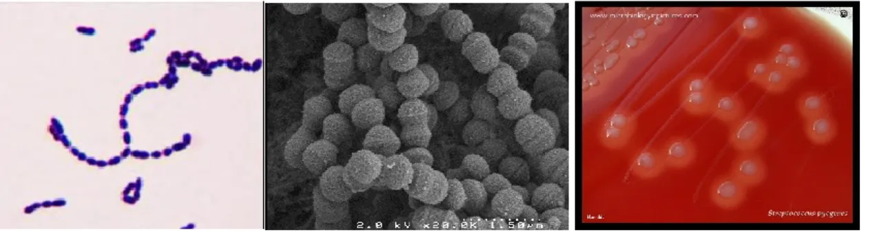
GAS is a Gram positive bacterium, β-hemolytic, with a low G+C % DNA content. Cocci are assembled in chains of minimum two cocci (Figure 4). GAS is grown on blood or Todd Hewitt broth supplemented with 0.2% yeast extract agar plates and its multiplication is favored by CO2 and anaerobia. GAS is auxotroph for 15 amino acids and its primary source of carbon is glucose [(7, 8), for review: (9)].
1 «Comment se fait-il qu'à la campagne on ne compte pas un cas de fièvre puerpérale sur mille, peut-être sur dix
mille accouchées, tandis que les hôpitaux de femmes en couches sont dévastés par d'effroyables épidémies?» p50
2 « Mais, faut-il l’avouer, j’ai une peur terrible, une peur dont je ne puis me défendre, et que l’Académie
comprendra : c’est celle de mourir avant qu’on n’ait découvert ce vibrion-là » p 256
3 « Eh bien, que l’Académie me permette de dessiner sous ses yeux le dangereux microbe auquel je suis porté en
ce moment à attribuer l’existence de cette fièvre » p 259
23
GAS is a non-motile, extracellular and human specific pathogen. The size of the GAS genome varies from 1.75 to 1.9 megabases, harboring roughly 2000 genes. Eighty-five % of the coding genome is the core genome conserved between all strains. Moreover, 10 % of the metagenome corresponds to prophages, largely contributing to strain diversity (10).
Figure 4. Morphology of GAS
Left: Gram-positive staining of GAS. Middle: Scanning electron microscopy of GAS. Right: GAS β-hemolysis on sheep blood agar plates.
1.2. GAS emm-typing
While identifying the carbohydrates involved in the Lancefield Group typing, Lancefield also isolated another major immunogen of GAS which is a protein, and she used this protein as a typing method for GAS strains. After discovering that a non-typable strain was less mucoid, she called this protein the M protein (11). She then set up the first collection and classification of GAS strains depending on the M protein expressed.
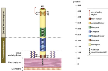
The main variations of the M protein are in the N-terminal domain, called the hypervariable region (HVR) (Figure 5) and the serotyping is based on antibodies directed against this region (12). This is an important functional domain, and antibodies against it highly decrease the strain virulence (13). The diversity of M proteins is explained by the immunological pressure and the critical importance of the M protein in virulence: people immunized for one M protein will no longer be affected by the strains presenting this M protein making it a major selective pressure. In addition to this, the more people are infected by a given strain, the smaller the pool of potentially neo-infected individuals is, reducing the strain prevalence. This immunological pressure implies a strong immunization against the M protein encountered, which occur to a lesser extent with antibiotic treatments which stop the infections before a strong immune response is established.
Introduction GAS carriage and induced diseases
24
Today the classification no longer stands on serotyping but on genotyping, by sequencing the 5’ of the emm gene, coding for the 50 first residues of the N-terminal domain of the M protein (14). The sequencing of an array of 1064 invasive strains identified 225 emm-types (15)
.
Figure 5. Structure of the M protein
Structure of the M5 protein to highlight the region used for serotyping (hypervariable region), and the 50 residues region now used for the emm-typing (in red). Adapted from (12).
1.3. GAS carriage and induced diseases
1.3.1. Asymptomatic colonizationGAS asymptomatic colonization or carriage is frequent in the throat: a meta-analysis showed that around 10% and 6% of children and 2.8% and 2% of adult are colonized in high-income and low-income countries, respectively (16). Some individuals are colonized for long periods of time (several years); the reasons for this persistent state are not well understood, but it appears that it has no role in post-infectious manifestations nor bacterial transmission (17, 18). Moreover, carrier children frequently switch emm-types throughout their lives (19).
1.3.2. Superficial infections
Pharyngitis and scarlet fever. Pharyngitis is a superficial infection of the oropharynx and GAS accounts for 4-10% pharyngitis cases in adults (20). GAS is annually involved in 616 million cases of pharyngitis in the world (21). Pharyngitis is more prevalent in OECD (National Organization for Economic Cooperation and Development) than non-OECD countries (16).
25
The complexity of GAS pharyngitis diagnostic is that most pharyngitis cases are not due to GAS and GAS is found asymptomatically in the throat. Consequently, a GAS positive swab does not systematically indicate a GAS-elicited pharyngitis. This could explain the dichotomy between GAS positive pharyngitis and serologically positive pharyngitis, with only around 50% of matches (16).
Impetigo and erysipelas without bacteremia. GAS is also involved in superficial skin infections, such as impetigo and erysipelas. In contrast to pharyngitis, impetigo is predominant in non-OECD countries, and there are an estimated 111 million cases of GAS-elicited impetigo per year (21).
1.3.3. Invasive infections
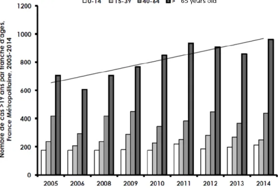
GAS invasive infections are infections during which GAS is found in normally sterile compartments or with Streptococcal Toxic Shock Syndrome (STSS). GAS invasive infections are responsible for 163 000 deaths per year (21) and, in France, their annual incidence is estimated to be 3.1/100 000, with a case-fatality ratio of 14% (22). Of note, the number of cases of invasive infections has been increasing in the world since 1990s (21), with an increase of 4% per year in France between 2007 and 2014, partly due to an increase in the number of invasive infections of people above 65 years of age (Figure 6).
Necrotizing fasciitis. Necrotizing fasciitis (NF) is the infection of the deeper layers of the skin, below the fascia, and is a rapidly progressing and life-threatening disease, with a mortality around 30% (23). In the United States of America, there are around 700 cases of NF per year and in France, in 2007, there has been 104 cases of GAS-elicited NF, of which 22 % were fatal (22, 24). NF risk factors are varicella, wound, burn and blunt trauma (25).
Bacteremia and septic shock. Bacteremia is the presence of bacteria in the blood and is associated with 60% of French invasive infections (https://cnr-strep.fr/). For some bacteremia, the origin of the bacterial translocation is unknown, and is called bacteremia without identified focus; this is the case for around 22% of French GAS invasive infections (22).
STSS. Bacterial toxins in the bloodstream or in a tissue can induce the hyperactivation of the immune system, further inducing a cytokine “storm” and shock followed by organ failures. This is called a septic shock and more specifically the STSS. STSS is the most life-threatening complication of GAS infections, with a mortality of up to 43% (22).
Introduction GAS carriage and induced diseases
26
Figure 6. Number of invasive infections in adults in France
Number of cases of invasive infections in France depending on the age. There is an overall increase of the number of invasive infections in France, driven by an increase in the number of invasive infections in people aged above 65 years. From (https://cnr-strep.fr/).
Endometritis and Puerperal fever. Puerperal fever, or childbed fever, is the increase of temperature in the 24 h following delivery and is due to an infection, referred to as puerperal infection or post-partum infection. The majority of these infections are in the gyneco-obstetrical tract, including the endometrium. In this thesis, endometritis was studied as a model of GAS invasive infections.
While less frequent than in the 19th century, puerperal fever is still responsible for 75 000 maternal deaths annually in the world, affecting 5-10% of pregnant women (26). A recent analysis in the UK showed an incidence of post-partum endometritis of 109 cases per 100 000 persons/year (27), slightly higher with a US rate of 59 per 100 000 persons/year (28). A population based study in Sweden showed that 2% of women suffered endometritis following delivery (29). The post-partum is a favorable condition for infections with several pathogens: Escherichia coli, Group B Streptococcus (GBS), Staphylococcus aureus, anaerobic bacteria and Listeria monocytogenes (30), but GAS is involved in 50 % of maternal sepsis, making it the pathogen most frequently responsible for maternal infections; furthermore, it is the most aggressive one. In France, infections of the gyneco-obstetrical sphere still correspond to 26.8%
27
of women, all age groups considered, GAS invasive infections (https://cnr-strep.fr/) (Table 2), of which 15.9% are endometritis. Women have a 20-fold increased incidence of GAS invasive diseases compared with non-pregnant women (31). The postpartum, abortion, in vitro fertilization and intrauterine devices all are risk factors for endometritis and are involved in more than half of GAS-elicited endometritis France (Table 2).
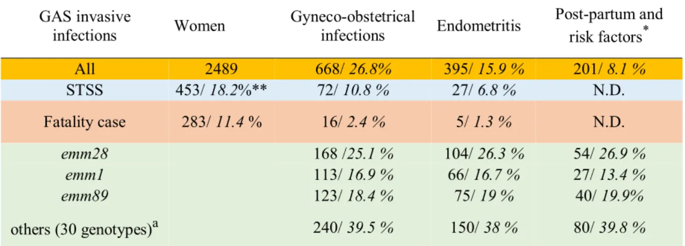
Table 2. GAS gyneco-obstetrical invasive infections in France 2006-2015, risks factors and prevalence of the main emm-genotypes
GAS invasive
infections Women Gyneco-obstetrical infections Endometritis
Post-partum and risk factors* All 2489 668/ 26.8% 395/ 15.9 % 201/ 8.1 % STSS 453/ 18.2%** 72/ 10.8 % 27/ 6.8 % N.D. Fatality case 283/ 11.4 % 16/ 2.4 % 5/ 1.3 % N.D. emm28 168 /25.1 % 104/ 26.3 % 54/ 26.9 % emm1 113/ 16.9 % 66/ 16.7 % 27/ 13.4 % emm89 123/ 18.4 % 75/ 19 % 40/ 19.9% others (30 genotypes)a 240/ 39.5 % 150/ 38 % 80/ 39.8 %
*risk factors: abortion, intra uterine device, in vitro fertilization. a 30 other genotypes.
**First numbers correspond to the number of cases and strains, the second ones, in italic, to the percentages: first line percentage of all women invasive infections; STSS and fatality case: percentage of the STSS/deaths for each column. The percentage for the strain genotypes corresponds to the percentage of strains for each column. N.D.: not determined. From (https://cnr-strep.fr/).
1.3.4. Post-infectious complications
GAS infections are the indirect cause of several pathologies, such as glomuronephritis and rhumatic arthritis. They all are auto-immune diseases due to the cross-reaction between GAS epitopes and host epitopes (32). Overall, these complications are responsible for 354 000 deaths per year and the incidence is very important in developing countries, with for example 15 to 16 million people suffering from GAS induced rheumatic heart diseases and 282 000 new cases per year (21).
1.4. Epidemiology of the emm-types strains
The diversity of strains responsible for superficial and invasive infections is higher in developing countries than in high income countries, which could be explained by climatic differences, genetic susceptibility and social economic reasons (33, 34). The distribution of
Introduction Epidemiology of the emm-types strains
28
strains responsible for invasive infections in France and in Europe is summarized in Figure 7 and Table 3, respectively.
Table 3. Main GAS emm-types involved in invasive infections in Europe from 1st January 2000 to 31st May 2017
Country 1st 2nd 3rd 4th
Czech
Republic emm1 emm81 emm28 Denmark emm1 emm28 emm89 England/Wales emm3 emm1
Finland emm28 emm89 emm1
France emm1 emm28 emm89 Germany emm1 emm28 emm3
Greece emm1 emm12
Hungary emm1
Iceland emm1 emm89 emm28
Ireland emm1 emm12 emm28
Italy emm1 emm12
Norway emm89 emm28 emm3 Poland emm1 emm12
Portugal emm1 emm89 emm3 Romania emm1
Scotland emm1
Spain emm1 emm3
Sweden emm89 emm81 emm28 emm1
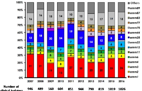
emm-types involved in more than 10% of the invasive infections are represented in the order of prevalence from left to right. The countries are in the alphabetical order. The emm28 genotype used in this thesis is underlined, the data do not include the most recent French epidemiological data (Figure 4), and emm28 are no longer the second most prevalent strain in France since 2012. Adapted from (35).
emm1 strains are the most prevalent strains in high income countries, with a clone emerging in the 1980s that is more efficient in colonizing and that increased the emm1 overall prevalence (36). Since 2008 in Europe, an emm89 clone has been rapidly emerging (37). While there are more than 200 described emm-types, in 2016, 60% of GAS invasive strains in France are from four genotypes: emm1, emm89, emm28 and emm12 (Figure 7) (https://cnr-strep.fr/). In Europe, the main genotypes responsible for invasive infections are emm1, emm28, emm89, emm3, emm12 and emm81 (35).
29
Figure 7. Distribution of GAS emm-types involved in invasive infections in France (National Center for Streptococcus activity report, https://cnr-strep.fr/images/CNR-STREP/rapport/rapport_CNR-strep_2016.pdf).
1.4.1. Tissue tropism and emm patterns
GAS induces a wide range of diseases and there are few associations between diseases and genotypes (38). GAS strains can be classified with a sequence typing in five gene patterns, A to E, which is based on the analysis of the patterns of the emm, emm-like genes and sof/sfbx genes of numerous clinical strains (Figure 8) (39, 40).
Figure 8. emm pattern classification
The organization of the Mga locus and the presence or not of sof/sfbx can discriminate GAS strains into 5 emm pattern, from A to E. From (41).
There is a clear association between specific emm patterns and skin/throat tropisms (40, 42). A-C strains represent ~47% of throat infections, but only 8% of impetigo isolates. In contrast, D
Introduction Epidemiology of the emm-types strains
30
strains represent ~50% of impetigo strains, but only 2% of pharyngitis strain, and E pattern strains are equally represented in both infections, thus called ubiquitous (42).
1.4.2. The emm28 association with gyneco-obstetrical sphere infections
The emm28 genotype is one of the three main genotypes encountered in Europe (Figure 7, Table 2). It belongs to the E pattern and it is associated with gyneco-obstetrical infections, representing around 25% of these infections while being responsible for only 15% of all invasive infections (43). More specifically, the association between puerperal infections and the emm28 was described in several countries (44–47). emm28 strains are, together with emm1, associated with severe cases of puerperal infection (46). The R28 protein expressed by emm28 strains has been implicated in the association of emm28 strains with puerperal infection (48). The first sequence of an emm28 genome identified two emm28 specific genomic regions (RD1 and RD2) that could explain this association. RD2 is a 37.4 kb region with genes encoding several cell-wall anchored proteins, including AspA, also known as AgI/II (M28_Spy1325) and R28 (M28_Spy1336) (Figure 9).
Figure 9. RD2 region of emm28 strains.
In green are shown inferred hypothetical proteins of unknown function, in red, extracellular proteins, in yellow, putative gene regulators and in blue, putative mobility/transfer ORFs. From (45)
The integrative conjugative element RD2 contains several genes related to GBS genes and was most likely horizontally transferred from GBS (45, 49). Since GBS colonizes 10-30% of healthy women uro-vaginal tract (50), it was suggested that RD2 harbors genes involved in the emm28 association with gyneco-obstetrical diseases.
31
2. GAS virulence factors and their regulation
2.1. GAS virulence factors
In this chapter, we will describe different factors whose implication in the GAS pathogenesis mechanisms will be discussed in chapter 3 and in the Results section. We will briefly discuss the concept of “virulence” factors in the light of GAS pathogenesis, and we will then describe the different virulence factors and then their regulation.
The virulence concept in the light of GAS pathogenesis
The concept of virulence and virulence factors, coined in the early 20th century aggressins and virulins by Rosenow, was based on the concept that some pathogenic elements could confer the pathogenic property to avirulent organisms (51). This was linked to the concept of intrinsic pathogenicity of a bacterium, in opposition to commensal and avirulent organisms, and to the intrinsic capacity of a factor to yield virulence. However, as described by Casadevall and Pirofski in 1999, virulence emerges from the intersection between the repertoires of factors of an organism and the host environment encountered (52). The pathogenicity of an organism is dependent on how much, where and when it expresses its factors. Moreover, many “virulence factors” expressed by highly pathogenic bacteria are also expressed by commensal bacterium, and the importance of factors in virulence depends on the genetic backgrounds (with the exception of certain toxins). Therefore, virulence properties and specificity of a factor are not intrinsic.
In the following paragraph, we will describe different factors which, in the host environment tested and the bacterial genetic background used, are implicated in the virulence of the tested strains. The virulence property of any factor cannot be extended to all GAS strains or diseases. The biochemical nature and main properties will be analyzed in this section, while the regulation is analyzed in section 2.2, and the role of these virulence factors in GAS infections is analyzed in section 3. Factors are discussed by their importance regarding my PhD work and in alphabetical order. A special focus is made on the R28 protein which is one of the main topics of this dissertation. The order of factors discussed and the focus on some factors/regulation pathways do not pretend to hierarchize them relatively to an importance in virulence.
Introduction GAS virulence factors
32 2.1.1. Secreted factors
2.1.1.1. SLO, NADase, SLS, SpeB and Superantigens
SLO and NADase. Streptolysin O (SLO) is a pore-forming cholesterol-dependent cytotoxin (53), secreted as a 69 kDa protein. SLO forms pores through oligomerization of the protein after cholesterol and galactose binding at the cell surface (54, 55). At low multiplicity of infection, SLO pores induce in keratinocytes an endoplasmic reticulum (ER) stress through a cytoplasmic calcium increase (56). This ER stress induces the unfolded protein response in HeLa cells (cervical cells), which further increases asparagine secretion, used by GAS to multiply (57). Ultimately, these pores induce cell death in different cell types (reviewed in section 3.2). In response to the pores, different pathways were shown to restrain their effect: endocytosis and ectocytosis of the damaged membrane (Figure 10).
Figure 10. Membrane repair after SLO damages.
Left. Ca2+ increase in the cytoplasm is detected by Synaptotagmin VII (sytVII, yellow), which activates lysosome fusion at the membrane. This releases acid sphingomyelinase (ASM, green) in the extracellular compartment, which transforms sphingomyelin in ceramide rich domain, further triggering endocytosis of damaged membrane (58–61). Right. Ca2+ influx is sensed by
annexin A1 which is recruited at the damaged membrane, which triggers ectocytosis of the pore, a mechanism described as “blebbing” (62, 63). From (64)
slo is cotranscribed with nga, coding for the streptococcal NAD-glycohydrase (NADase, Nga or SPN), and if coding for NADase inhibitor (IF). NADase is a SLO cofactor and is composed of two domains, one involved in the SLO mediated translocation and the second one is a domain wit β-NAD+ glycohydrolase activity (65, 66). In the bacterium, the NAD-glycohydrase activity of NADase is restrained through IF action (67–69). NADase is translocated into cells via a 70 amino acid domain of SLO but the pore formation is not required for NADase translocation
33
(66, 70). NADase binding to an unknown receptor forms a pore independently of the SLO mediated pores (54, 55). Intracellularly, NADase induces the depletion of NAD, which induces cell-death (67, 71). However, a NADase variant without the β-NAD+ glycohydrolase activity still triggers programmed cell death (72). Extracellular NADase reduces the release of IL-1β by macrophages, and this inhibition is independent from its translocation by SLO (73).
SLS. Streptolysin S, SLS, is a member of the thiazole/oxazole-modified microcins and the pro-SLS peptide is coded by the gene sagA. sagA is co-transcribed in an operon composed of nine genes (sagA to sagI) which encodes proteins that profoundly modify the initial 2.7 kDa peptide into the mature SLS (74). It is an oxygen stable cytotoxin that forms hydrophilic pores in multiple cell types, including immune cells, and it is, for example, responsible for GAS β-hemolysis. SLS cytotoxic activity is sevenfold higher when bacteria are in contact with cells than when they are not (75, 76), and its activity is stabilized by host high-density lipoprotein (HDL) or albumin (74, 77, 78). Recently, the mechanism of SLS red blood cells lysis was unraveled: SLS interaction with band 3 protein, also known as Anion exchanger 1 (AE1), induces Cl- influx and subsequent osmotic shock (79).
SpeB. Streptococcal pyrogenic erythrogenic toxin B (SpeB) or streptopain or SPC (80), is commonly known as the streptococcal cysteine protease. It is produced as a zymogen of 40 kDa that is converted into a 28 kDa mature active protein after multiple intramolecular autocatalytic cleavages and reduction of the cysteine residue (81–84).All clinical strains express SpeB (85) and in vitro, SpeB is predominantly produced during the stationary phase of growth. Structurally SpeB presents a papain fold and is active form is the dimer (86, 87). SpeB protease activity requires a three amino acid motif which makes it a broad-spectrum protease. Consequently, SpeB can degrade in vitro a very large number of host proteins (Table 4). SpeB also degrades several bacterial surface proteins (protein M and M-like, protein F1, Fba, C5a peptidase, protein H). Of note, SpeB capacity to degrade IgG was recently questioned (88), as SpeB only cleaves reduced IgGs, and reductive conditions might not be present in vivo (89). In addition to its soluble form, SpeB can be retained at the bacterial surface by the alpha2-M-binding protein bound to the bacterial surface G-related alpha2-M-alpha2-M-binding protein (GRAB) (90) and it remains active against several ligands. Noteworthy, surface bound SpeB degrades the anti-microbial peptide LL-37 (91). SpeB can also act as an adhesin; it binds laminin (92) and a variant of SpeB with a RGD motif present in 20 % of the tested strains directly binds to the integrins αVβ3 and αIIbβ3 (93).
Introduction GAS virulence factors
34 Table 4. SpeB interactions with host components
Protein Activity Reference
IgG C (94–97)
IgA,IgM, IgD, IgE D (97)
C3b C/D (98, 99)
Properdin D (100)
Chemokines D (101)
IL-1β A (102)
H-kininogen Bradykinin release (103)
Fibrinogen D (104, 105) Plasminogen D (106) Vitronectin D (107) Fibronectin D (107) Urokinase receptor C (108) MMPs A (109, 110) Decorin D (111) Integrins B (93) Laminin B (92) A1AT B (112) A2M C (91)
C= cleavage, A= activation, D= degradation, B= binding. Adapted from (89)
Superantigens. Superantigens (SAgs) are highly potent mitogens of the immune system: they induce hyper-activation and subsequent proliferation of human and some other mammalian T-lymphocytes. In conventional activation, peptides bind to the major histocompatibility class II (MHC) molecules expressed by antigen presenting cells (macrophage, B-lymphocytes and dendritic cells), and the recognition of the peptide/MHC complex by an antigen specific T-cell receptor (TCR) activates the specific T cells. The range of lymphocytes activated is limited by the epitope specificity of the TCR. In contrast, SAgs are able to induce activation of cells by binding non-specifically both the MHC and the TCR molecules (Figure 11).
SAgs are produced by a minority of organisms: some bacteria, among the most common bacterial geniuses, Group A, C and G Streptococcus and Staphylococcus, and viruses (113). Eleven SAgs have been described in GAS (114) and the overall SAg repertoires are conserved within emm-types. However, some strains within an emm-type express a specific repertoire of SAgs. The first GAS SAg, later termed SpeA, was identified in 1924 and called the “scarlet fever toxin”. SpeC was discovered in 1960 (115). Because SpeA, SpeB and SpeC are able to induce fever when injected into rabbits (pyrogenicity), Kim and collaborators renamed these toxins streptococcal pyrogenic exotoxin (Spe) A, B and C (116). The SpeB was later shown not to be a SAg but the cysteine protease SpeB (80).
35 Figure 11. Mechanism of action of superantigens
APC: antigen presenting cell. MHC II: major histocompatibility complex class II. T cell: T lymphocyte. Ag = antigen. SAg = superantigen. TcR = T-cell receptor.
The amino acid sequence of the streptococcal superantigen SSA is very similar to staphylococcal SAgs (117). The streptococcal mitogenic exotoxin Z (SMEZ) is expressed by 94% of invasive strains in France (https://cnr-strep.fr/). Genetic analysis of a sequenced strain identified seven other gene coding for SAgs: speG, speH, speI and speJ, speK, speL and speM (118–122). The genes are associated with bacteriophages, except for speG, speJ and smeZ which are chromosomally encoded. Genes encoding GAS SAgs are not highly polymorph. Few alleles exist with minor diversity, except for the smeZ gene with so far 50 alleles identified, some of them containing nonsense mutations (123, 124). The distribution of SAgs genes in GAS isolates are summed up in Table 5 (125). The strain used in this PhD was sequenced and its genome contains 5 genes coding superantigens: smeZ, speG, speJ, speC and speK (126).
SAgs bind the variable β chains (Vβ) of the TCR and the human T cell Vβ repertoire is composed of 50 different Vβ chains. Each SAg can bind more than one Vβ chain, and one SAg can activate up to 25% of the T cell repertoire. This is to be compared with the ability of a conventional peptide to activate only one subset of naïve T cells from the pool of 105 – 106 cells. In addition to Vβ chains and MHC class II molecules, SAgs bind CD28 (127), a co-stimulatory receptor expressed by T cells, and SAgs interact with B7 (CD80/CD86) molecules present on antigen presenting cells. The binding of TCR to MHC molecule is a prerequisite for activation of T cells, but the interaction between B7 molecules of APC and CD28 of T cells is also necessary for T cell activation and the induction of pro-inflammatory cytokine genes (127).
Introduction GAS virulence factors
36 Table 5. Repartition of the SAgs gene
smeZ speG speJ speC ssa speA speK speH speI speL/M % of GAS strains 96 86.9 32.7 51.5 35.4 32.1 24.6 17.1 15.2 9.2 480 nonduplicate GAS isolates from Portugal between 2000-2005 were tested by PCR. Adapted from (125). Binding of superantigens to T cells induces rapid release of TNF-α, TNF-β followed by IL-2, IL-6, IL-1 and IFN. In addition to Vβ specificities, each SAg induces responses of different intensities, as shown by the ability of SMEZ to induce ten times more cytokines than SpeA (128).
2.1.1.2. Other secreted virulence factors
DNases. DNases are enzymes that cleave DNA and play a potentially crucial role in defense against neutrophil extracellular traps (NETs) (129, 130). Eight types of DNases are expressed by GAS: sda1, sda2, spd1, spd3, spd4 and sdn; spn1 and sdaB are the only core genome encoded DNase genes in GAS, and they are present in all GAS strains (131, 132).
Sda1: the strain M1T1 acquired the DNase Sda1 via a bacteriophage and is the most important DNase for virulence of this clone (36, 130, 133). The importance of Sda1 in the emergence of the M1T1 strain and in its virulence is questioned (134–136).
Spd1 and Spd3 are bacteriophage encoded DNases (137, 138). Spd1 is reported to have an RNase activity but its role in virulence is unknown (137).
The EndoS family. Endo-beta-N-acetylglucosaminidase of S. pyogenes (EndoS) is a 108 kDa protein that removes carbohydrates from IgG in a specific manner, reducing Fc affinity for its receptors and diminishing the opsonophagocytosis of bacteria (95, 97).
EndoS2. A variant of EndoS was described in serotype M49, named EndoS2 (139). EndoS2 activity differs from that of EndoS in the hydrolysis of N-linked glycans on native IgG chains and biantennary and sialylated glycans of the alpha1-acid glycoprotein (AGP).
IdeS. The immunoglobulin-degradin enzyme of S. pyogenes (IdeS), also known as Mac-1or MspA is a secreted cysteine protease that specifically cleaves the hinge region of IgG, but not the other immunoglobulins (140, 141). Mac-2 is a variant of IdeS that presents similar functions(142).
Sib35 is another anchorless immunoglobulin-binding protein. In contrast to IdeS or Mac-2, the 35 kDa Sib35 protein can bind IgA and IgM in addition to IgG. Sib35 is present in all GAS strains tested (143). Also, Sib35 binds to mouse B cells, enhances expression of MHC class II and B7-2 (CD86), induces the transition of cells to antibody producing plasma cells and