En vue de l'obtention du
DOCTORAT DE L’UNIVERSITÉ DE TOULOUSE
Délivré par l'Université Toulouse III - Paul Sabatier
Discipline ou spécialité : Pathologie, Toxicologie, Génétique et Nutrition Présentée et soutenue par Charlotte Misbach le Mardi 24 Février 2015
TITRE:
Determination of reference intervals in small size dogs for variables used in veterinary cardiology
Détermination d’intervalles de référence chez les chiens de petit format pour des variables d’utilité en cardiologie vétérinaire
JURY:
M. Michel Galinier (président), Professeur à l’Université Paul Sabatier M. Hervé Lefebvre (directeur), Professeur à l’Université Toulouse Mme Valérie Chetboul (co-directeur), Professeur à l’Université Paris-Est Mme Kathleen McEntee (rapporteur), Professeur à l’Université Libre de Bruxelles
M. Jean-Claude Desfontis (rapporteur), Professeur à l’Université de Nantes M. Jack-Yves Deschamps (examinateur), Professeur à l’Université de Nantes
M. Didier Concordet (invité), Professeur à l’Université de Toulouse
Ecole Doctorale : SEVAB (sciences écologiques, vétérinaires, agronomiques et bioingenieries) Unités de Recherche : Unité de Recherche Clinique, Département des Sciences Cliniques, Ecole Nationale Vétérinaire de Toulouse et Unité de Cardiologie d’Alfort, Centre Hospitalier Universitaire
Vétérinaire d’Alfort, Ecole Nationale Vétérinaire d’Alfort. Directeurs de Thèse : Professeurs H. P. Lefebvre et V. Chetboul
Tout d’abord, je souhaiterais remercier mes directeurs de thèse, Monsieur le Professeur Hervé Lefebvre et Madame le Professeur Valérie Chetboul pour la confiance qu’ils m’ont accordée en encadrant ce travail de recherche ainsi que pour leur disponibilité et leur aide précieuse. Ce fut un parcours semé d’embûches mais vous avez toujours été là pour moi. Merci.
Je tiens également à exprimer ma gratitude à mes deux rapporteurs de thèse, Madame le Professeur Kathleen McEntee et Monsieur le Professeur Jean-Claude Desfontis. Merci pour le temps que vous avez consacré à la relecture de ce travail et pour vos conseils avisés.
C’est également avec plaisir que je remercie Monsieur le Professeur Michel Galinier d’avoir accepté de présider mon jury de thèse et Monsieur le Professeur Jack-Yves Deschamps de faire partie de ce jury en tant qu’examinateur. Je leur suis très reconnaissante et honorée du temps qu’ils m’ont accordé.
Merci à Monsieur le Professeur Didier Concordet pour sa collaboration essentielle dans la rédaction des articles associés à cette thèse et sa présence à ma soutenance en tant que membre du jury invité.
Arqued et les anciennes, Anne-Cécile Hoffmann et Alice Leverrier pour leur aide et leur soutien. Merci également à Adriana Rocha et David Balouka pour leur aide et Sylvie Viallefond pour ses conseils.
Un grand merci à Loïc Desquilbet pour son aide et ses nombreuses explications dans le domaine statistique et bien sûr pour sa patiente.
Merci à Jean-François Gauvry pour son aide.
Merci au Professeur Renaud Tissier pour son soutien sans faille.
Merci à Christine Médaille pour son aide dans la réalisation de mon protocole et pour sa participation à la rédaction d’un des articles de ce travail.
Je remercie également le laboratoire Novartis d’avoir financé mon doctorat. Merci particulièrement à Philippe Gruet, Cindy Speranza-Gastal, Jérôme Blanchet et Patricia Nobour pour leur aide.
proches qui se reconnaîtront !
GENERAL INTRODUCTION ... 27
CHAPTER I. THE DEGENERATIVE MITRAL VALVE DISEASE ... 33
I. INTRODUCTION ... 33
II. PREVALENCE AND EPIDEMIOLOGY ... 35
III. ETIOLOGY ... 35
IV. PATHOPHYSIOLOGY ... 36
V. DIAGNOSIS ... 39
VI. PROGNOSTIC FACTORS ... 40
VI.1. CLINICAL AND EPIDEMIOLOGICAL FACTORS... 40
VI.2. RADIOGRAPHIC AND ECHOCARDIOGRAPHIC FACTORS ... 41
VI.3. CIRCULATING BIOMARKERS ... 42
VI.3.1 NATRIURETIC PEPTIDES ... 42
VI.3.2 Cardiac TROPONIN I ... 45
VI.3.3 CIRCULATING NON-CARDIAC BIOMARKERS ... 45
VII. CONCLUSION ... 47
CHAPTER II. THE CONCEPT OF REFERENCE INTERVAL ... 49
I. INTRODUCTION ... 49
II. GENERAL PROCEDURES FOR THE DETERMINATION OF REFERENCE INTERVALS ... 53
III. ANALYTICAL INTERFERENCE AND SOURCE OF BIOLOGICAL VARIABILITY ... 54
IV. SELECTION AND PARTITIONING OF REFERENCE INDIVIDUALS... 55
V. PRE-ANALYTICAL PROCEDURES ... 57
VI. ANALYTICAL PROCEDURES AND QUALITY CONTROL ... 58
VII. STATISTICAL TREATMENT OF REFERENCE VALUES ... 59
VII.1. INSPECTION OF DISTRIBUTION ... 59
VII.2. IDENTIFICATION OF OUTLIERS ... 60
VII.3. SAMPLE SIZE DETERMINATION ... 62
VII.4. METHODS FOR DETERMINATION OF REFERENCE INTERVAL ... 63
VII.4.A. CONDIDENCE INTERVAL FOR REFERENCE LIMITS ... 63
VII.4.B. NONPARAMETRIC METHOD ... 64
VII.4.C. BOOTSTRAP METHOD ... 69
VII.4.D. PARAMETRIC METHOD ... 70
VII.4.E. ROBUST METHOD ... 73
VII.4.F. THE CASE OF SMALL POPULATIONS IN VETERINARY MEDICINE ... 73
VII.5. PARTITIONING ... 75
VIII. CONCLUSION ... 78
CHAPTER III. REFERENCE INTERVALS IN VETERINARY MEDICINE ... 79
I. INTRODUCTION ... 79
II. REFERENCE INTERVALS IN VETERINARY CLINICAL PATHOLOGY ... 80
II.1. INTRODUCTION ... 80
II.2.D. OTHER EFFECTS... 93
II.3. CONCLUSION ... 94
III. REFERENCE INTERVAL IN VETERINARY CARDIOLOGY ... 95
III.1. INTRODUCTION ... 95
III.2. EFFECT OF COVARIATES ON CARDIOLOGIC VARIABLES IN DOGS ... 103
III.2.A. EFFECT OF BODY WEIGHT ... 103
III.2.B. EFFECT OF BREED ... 105
III.2.C. EFFECT OF AGE ... 109
III.2.D. EFFECT OF GENDER... 112
III.2.E. OTHER EFFECTS ... 114
III.3. CONCLUSION ... 115
CHAPTER IV- SCIENTIFIC AIMS ... 117
CHAPTER V- MATERIAL AND METHODS- OVERALL PRESENTATION ... 119
I. ANIMALS ... 120
I.1. CANINE BREED ... 120
I.2. AGE ... 121
I.3. GENDER ... 121
I.4. BODY WEIGHT AND BODY CONDITION SCORE ... 121
I.5. PHYSIOLOGICAL CONDITION ... 122
I.6. POST-PRANDIAL STATUS ... 123
II. INCLUSION CRITERIA ... 123
III. EXCLUSION CRITERIA ... 124
IV. STUDY PROCEDURES ... 125
IV.1. GENERAL STUDY PROCEDURES ... 125
IV.1.A. BLINDING ... 125
IV.1.B. OWNER INTERVIEW ... 125
IV.1.C. IDENTIFICATION OF DOGS ... 126
IV.1.D. ANAMNESIS... 126
IV.1.E. PHYSICAL EXAMINATION ... 126
IV.2. PROCEDURES FOR BLOOD SPECIMEN ... 127
IV.2.A. BLOOD COLLECTION ... 127
IV.2.B. PACKED CELL VOLUME MEASUREMENT ... 128
IV.2.C. HANDLING OF BLOOD SPECIMEN ... 128
IV.2.D. STORAGE OF PLASMA SAMPLES ... 128
IV.2.E. SHIPMENT OF PLASMA SAMPLES ... 129
IV.3. ANALYTICAL METHOD ... 129
IV.3.A. PLASMA BIOCHEMISTRY ... 129
IV.3.B. PLASMA N-TERMINAL PRO-BRAIN NATRIURETIC PEPTIDE ... 132
IV.4. CARDIOVASCULAR EXAMINATION ... 132
IV.4.A. CONVENTIONAL ECHOCARDIOGRAPHY AND STANDARD DOPPLER EXAMINATION ... 132
IV.4.B. SYTEMIC ARTERIAL BLOOD PRESSURE MEASUREMENT ... 138
CHAPTER VI- RESULTS ... 143 I. ARTICLE 1: Basal plasma concentrations of routine variables and packed cell volume in clinically healthy adult small size dogs: effect of breed, body weight, age, and gender, and reference intervals. ... 144 II. ARTICLE 2: Basal plasma concentrations of N-terminal pro-B-type natriuretic peptide in clinically healthy adult small size dogs: effect of body weight, age, gender and breed, and reference intervals ... 149 III. ARTICLE 3: Echocardiography and conventional Doppler examination in clinically healthy adult Cavalier King Charles Spaniels: effect of breed, body weight, age, and gender, and establishment of reference intervals ... 153 CHAPTER VII- DISCUSSION, CONCLUSION AND PERSPECTIVES ... 157 APPENDIX ... 173
ARTICLE 1: “Basal plasma concentrations of routine variables and packed cell volume in clinically healthy adult small size dogs: effect of breed, body weight, age, and gender, and establishment of reference intervals” ... 173 ARTICLE 2: “Basal plasma concentrations of N-terminal pro-B-type natriuretic peptide in clinically healthy adult small size dogs: effect of breed, body weight, age, and gender, and establishment of reference intervals” ... 175 ARTICLE 3: “Conventional and Doppler echocardiography in clinically healthy adult
Cavalier King Charles Spaniels: effect of body weight, age, and gender, and establishment of reference intervals” ... 177 REFERENCES ... 179
La dégénérescence valvulaire mitrale (MVD) représente 75 à 80% des cardiopathies dans l'espèce canine. Les chiens de petit format, très populaires en France et en particulier en région urbaine, y sont malheureusement prédisposés. La MVD se caractérise par une dégénérescence et une malposition des feuillets mitraux, ayant pour conséquence une régurgitation mitrale (RM) plus ou moins importante. Une RM grave entraine au long court une dilatation des cavités cardiaques (atriale puis ventriculaire) ainsi qu’une augmentation de la pression dans les capillaires pulmonaires conduisant vers l’insuffisance cardiaque congestive (ICC). Les animaux atteints de MVD peuvent rester asymptomatiques pendant une très longue période, mais l’apparition de symptômes d’ICC tels que toux, dyspnée et intolérance à l’effort ou de complications extra-cardiaques telle que l’insuffisance rénale, peut conduire au décès de l’animal. Les techniques d’imagerie ultrasonore comme l’échocardiographie conventionnelle et le Doppler sont les examens de choix pour diagnostiquer la MVD, évaluer la gravité de la RM et ses conséquences sur la morphologie et la fonction cardiaques. De plus, certaines variables échocardiographiques (diamètre atrial gauche, onde proto-diastolique mitrale par exemple) sont significativement corrélées au stade clinique de la maladie. En effet, ces variables permettent d’obtenir des informations précises sur le pronostic de la maladie à court et long terme, seules ou en combinaison avec des biomarqueurs circulants, comme les peptides natriurétiques ou encore certaines variables biochimiques évaluées en routine, comme la créatinine par exemple. En médecine, toute valeur obtenue à la suite d’un examen complémentaire réalisé chez un sujet malade doit être comparée à des valeurs de référence établies chez des sujets sains. En effet, le
dans les années 60 et suivi par l’élaboration de recommandations spécifiques concernant l’établissement de ces IR par des experts de l’International Federation of Clinical
Chemistry-Clinical Laboratory and Standard Institute (IFCC-CLSI). En biologie clinique vétérinaire,
quelques études ont établi des IR pour des variables biochimiques et hématologiques en suivant ces recommandations dans différentes races ou catégories de races canines. Il ressort de ces études que des spécificités raciales existent ainsi que des effets de certains facteurs, comme le poids, le sexe et l’âge. Concernant le domaine de la cardiologie canine, l’effet de la race et du poids sur les variables échocardiographiques a été largement démontré. Cependant, les effets du sexe et de l’âge demeurent méconnus et aucun IR établi selon les recommandations IFCC-CLSI n’a été publié à ce jour.
L’hypothèse principale de ce travail a donc été la suivante : la détermination d’IR spécifiques chez les races de chiens de petit format les plus populaires en France et prédisposées à la cardiopathie la plus fréquente en cardiologie vétérinaire (MVD), pourrait être pertinente pour le clinicien. Trois études ont été réalisées :
1) Etude 1 : Evaluation des effets de la race, du poids, de l’âge et du sexe sur l’hématocrite ainsi que 15 variables biochimiques de routine et détermination d’IR pour ces mêmes variables dans une population de chiens adultes sains de petit format, selon les recommandations IFCC-CLSI ;
IR pour ce même biomarqueur dans une population de chiens adultes sains de petit format, selon les recommandations IFCC-CLSI ;
3) Etude 3 : Evaluation des effets du poids, de l’âge et du sexe sur 14 variables écho-Doppler et
détermination des IR pour ces mêmes variables dans une population de Cavalier King Charles (CKC) adultes sains, selon les recommandations IFCC-CLSI.
Dans les études 1 et 2, une population incluant 154 chiens de 7 races différentes appartenant à des éleveurs de la région Ile de France (CKC, King Charles, Yorkshire Terrier, Caniche, Bichon, Teckel, Shih-Tzu) a été recrutée de manière prospective. Dans l’étude 3, une population de 134 CKC, présentée à l’Unité de Cardiologie d’Alfort pour un dépistage cardiaque a été sélectionnée. Les prélèvements sanguins ainsi que les examens cardiovasculaires ont été réalisés dans des conditions standardisées et en utilisant des méthodes validées, afin de minimiser les sources de variabilité (pré-analytique et analytique pour la biologie clinique ou liée au manipulateur pour l’échocardiographie) qui auraient pu interférer avec l’interprétation des IR.
La détermination des IR a été faite en suivant les recommandations IFCC-CLSI et l’effet des facteurs comme la race, le poids, l’âge ou le sexe sur les variables a été testé en utilisant un modèle de régression linéaire.
fonction de ces facteurs n’a pas montré d’intérêt clinique pertinent. De plus, il est ressorti de cette étude que les IR fournis par les laboratoires d’analyses vétérinaires ne sont pas spécifiquement adaptés aux chiens de petit format. En effet, 4 à 76% des valeurs observées dans la présente étude étaient respectivement au dessus et en dessous des valeurs de l’IR (supérieures ou inférieures) fournies par le laboratoire.
Dans l’étude 2, un effet du sexe a été identifié sur le NT-proBNP plasmatique. En raison de problèmes analytiques et de la grande variabilité inter-individuelle de ce biomarqueur, non décrite jusqu’ici dans la littérature chez les chiens de petit format, il n’a pas été possible de déterminer un IR pour ce biomarqueur (notamment concernant la limite supérieure de cet IR). Il a donc été conclu que l’interprétation d’un dosage plasmatique du NT-proBNP chez un chien sain de petit format doit être réalisée avec prudence et en complément de l’échocardiographie, qui demeure l’examen de choix dans l’évaluation cardio-vasculaire du chien.
Dans l’étude 3, un effet significatif du poids a été identifié sur les variables échocardiographiques, en particulier l’épaisseur des parois et le diamètre des cavités du ventricule gauche. De plus, en comparaison aux IR déterminés dans des études antérieures concernant les mêmes variables échographiques, mais à partir d’une population incluant des chiens de races très différentes, il a été montré que des IR spécifiques élaborés pour le CKC dans la présente étude sont plus précis. Il a donc été conclu que la race et le poids doivent être pris en compte dans l’établissement d’un IR pour les variables écho-Doppler chez le CKC.
variables plasmatiques et échocardiographiques. De plus, l’effet de certains facteurs comme le poids, l’âge et le sexe doivent être pris en compte, mais seulement si un intérêt clinique est identifié.
ACVIM: American College of Veterinary Internal Medicine ALT: alanine aminotransferase
ALP: alkaline phosphatase ANP: atrial natriuretic peptide Ao: aorta
AST: aspartate aminotransferase
ASVCP: American Society for Veterinary Clinical Pathology BCS: body condition score
B: Bichon
BNP: brain natriuretic peptide BUN: blood urea nitrogen BW: body weight
CKCS: Cavalier King Charles Spaniel CHF: congestive heart failure
CI: confidence interval
CLSI: Clinical and Laboratory Standards Institute CV: coefficient of variation
D: Dachshund
HGB: hemoglobin
IFCC: International Federation of Clinical Chemistry
ISACHC: International Small Animal Cardiac Health Council KCS: King Charles Spaniel
LA: left atrium
LVED: left ventricular diameter at end-diastole LVES: left ventricular diameter at end-systole
MCHC: mean corpuscular hemoglobin concentration MCV: mean corpuscular volume
MR: mitral regurgitation MP: Miniature Poodle
NT-proANP: N-terminal pro-atrial-type natriuretic peptide NT-proBNP: N-terminal pro-brain-type natriuretic peptide PCV: packed cell volume
PLT: platelet
RAAS: renin-angiotensin-aldosterone system RBC: red blood cell
RI: reference interval SD: standard deviation
TTE: transthoracic echocardiography VHS: vertebral heart scale
WBC: white blood cell YT: Yorkshire Terrier
Table 1. Origin and standard of the 7 most common small size breeds in France. Source: Société
Centrale Canine.
Table 2. American College of Veterinary Internal Medicine Consensus Statement for classification of dogs with degenerative mitral valve disease. Atkins et al. Guidelines for the diagnosis and treatment
of canine chronic alular heart disease. J Vet Intern Med 2009;23:1142-1150.
Table 3. Procedures recommended by the International Federation of Clinical Chemistry and the Clinical and Laboratory Standards Institute for reference interval determination.
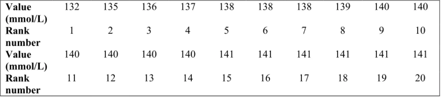
Table 4. Observed values, ranked from 1 to 20, for plasma sodium of the 20 healthy small size dogs present in the left tail of the distribution.
Table 5. Observed values, ranked from 1 to 20, for plasma sodium of the 20 healthy small size dogs present in the right tail of the distribution.
Table 6. Rank numbers of the 90% confidence interval of the 2.5th percentile for sample size between 119 to 1000 values. To obtain the corresponding rank number of the 97.5th percentile, subtract
the rank numbers in the table from (n+1). n, sample size. From the International Federation of Clinical Chemistry, Expert Panel on Theory of Reference Values: Approved recommendation on the theory of reference values: part 5. Statistical treatment of reference values. J Clin Chem Biochem 1987;25:650.
Table 7. Studies about the determination of reference intervals in healthy dogs and cats according to the International Federation of Clinical Chemistry and the Clinical and Laboratory Standards Institute recommendations in clinical pathology.
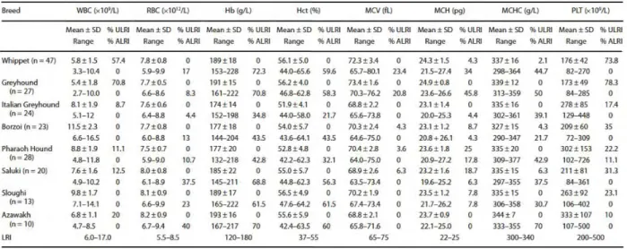
Table 9. Hematological values of eight sighthound breeds. LRI, laboratory reference interval; % ALRI,
% of dogs above LRI; % ULRI, % of dogs below LRI; Hob, hemoglobin; Hot, hematocrit; MCH, mean corpuscular hemoglobin; MCHC, mean corpuscular hemoglobin concentration; MCV, mean corpuscular volume; PLT, platelet count; RBC, red blood cell count; WBC, white blood cell count. From Uhríková et al. Hematological and biochemical variations among eight sighthound breeds. Aust Vet J 2013;91:452-459.
Table 10. Pairwise comparison between breeds indicating significant differences (X). AM, Alaskan
malamute; SH, Siberian Huskies; ES, English Setters; GR, Golden retrievers. From Sharkey et al., Breed-associated variability in serum biochemical analytes in four large-breed dogs. Vet Clin Pathol 2009;38:375-380.
Table 11. Studies about radiographic evaluation of heart in healthy dogs. CKCS, Cavalier King
Charles Spaniel; SD, Standard deviation.
Table 12. Studies about electrocardiography in healthy dogs. CKCS, Cavalier King Charles Spaniel;
SD, Standard deviation.
Table 13. Studies about cardiac biomarkers in healthy dogs. CKCS, Cavalier King Charles Spaniel;
SD, Standard deviation.
Table 14. Studies about echocardiography in healthy dogs. CKCS, Cavalier King Charles Spaniel; SD,
Standard deviation; ST, speckle tracking imaging; TDI, tissue Doppler imaging; 2D, bidimensional.
Table 15. Method of analysis for plasma urea, creatinine, total proteins, albumin and liver enzyme activities and results of the between-run coefficient of variations for control solutions.
Table 17. Reference ranges proposed for the conventional and pulsed wave Doppler modes variables assessed in the present study. From Chetboul et al. Use of quantitative two-dimensional color tissue
Doppler imaging for assessment of left ventricular radial and longitudinal myocardial velocities in dogs. Am J Vet Res 2005;66:953-961.
Table 18. Equation used to predict the reference ranges for the 6 body-weight dependent M-mode variables assessed in the present study (i.e., end-diastolic and end-systolic left ventricular wall thicknesses and internal diameters).From Gonçalves et al. Linear, logarithmic, and polynomial models of M-mode echocardiographic measurements in dogs. Am J Vet Res 2002;63:994-999.
Table 19. Reference intervals established from the 154 reference individuals and comparison with the laboratory’s reference intervals. BCG, Gaussian after Box-Cox transformation; CI, confidence
interval; LL, lower limit; NG, non-Gaussian; UL, upper limit.
Table 20. Predicted regression-based reference intervals (lower and upper limits) according to body weight for the six M-mode variables with a significant body weight effect assessed in a population of healthy adult Cavalier King Charles Spaniels (n=134). LVDd and LVDs, end-diastolic and end-systolic
left ventricular diameters, respectively; LVFWd and LVFWs, end-diastolic and end-systolic left ventricular free wall thicknesses, respectively; IVSd and IVSs, end-diastolic and end-systolic interventricular septum thicknesses, respectively.
Table 21. M-mode echocardiographic reference intervals (2.5th and 97.5th percentiles) from 134 healthy adult Cavalier King Charles Spaniels determined according to the Clinical and Laboratory Standards Institute (CLSI, 2008) statistical procedures (A), and predictive reference intervals calculated in the same population using Cornell’s formula (Cornell et al., 2004; B). See Table 20 for
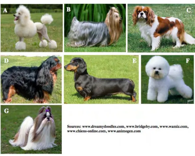
Figure 1. Photographs showing the 7 most common small size breeds in France. A, Miniature Poodle;
B, Yorkshire Terrier; C, Cavalier King Charles Spaniel; D, King Charles Spaniel; E, Dachshund; F, Bichon Frisé; G, Shih-Tzu.
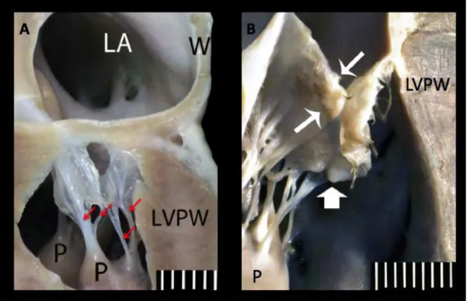
Figure 2. (A) Sagittal section through the left atrium (LA) and the left ventricle of a dog with a normal mitral valve apparatus. The two mitral leaflets are attached at the junction of atrial and
ventricular myocardium. The leaflets are thin, clear and translucent and the chordae tendineae are smooth and symmetric (arrows). (B) Close up view of a degenerative mitral valve. The leaflet edges are rounded and thickened at their contact point (thin arrows) and nodular thickening (broad arrow) is present along the distal leaflet segments. LVPW, left ventricular posterior wall; P, papillary muscle; W, LA posterior wall. Scale in mm. Photograph and legend: Fox PR. Pathology of myxomatous mitral valve disease in the dog. J Vet Cardiol 2012;14:103-126.
Figure 3. Bidimensional right parasternal long axis 4-chamber view showing echocardiographic images of a normal (A) and a degenerative mitral valve (B). Note the thickening of the mitral leaflet
(B), especially the distal part of the anterior mitral leaflet (AML). LA, left atrium; LV, left ventricle; PML, posterior mitral leaflet; RA, right atrium; RV, right ventricle. Photograph: Cardiology Unit of Alfort.
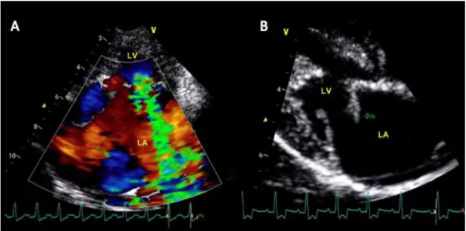
Figure 4. (A) Left parasternal long axis 4-chamber view using the color Doppler mode showing a severe mitral regurgitation. (B) Bidimensional right parasternal long axis 4-chamber view showing a chordae tendineae rupture and a prolapse of the mitral anterior leaflet (arrow). LA, left atrium; LV,
peptide (NT-proBNP) obtained from 46 dogs with symptomatic degenerative mitral valve disease:
ISACHC class 2 with NT-proBNP <1265 pmol/L (n= 13, solid line) and >1265 pmol/L (n=10, dashed line); ISACHC class 3 with NT-proBNP <2700 pmol/L (n= 13, dotted line) and >2700 pmol/L (n= 10, alternate dotted and dashed line). n, number of dog. From Serres et al., Plasma N-terminal pro-B-type natriuretic peptide concentration helps to predict survival in dogs with symptomatic degenerative mitral valve disease regardless of and in combination with the initial clinical status at admission. J Vet Cardiol 2009;11:103-121.
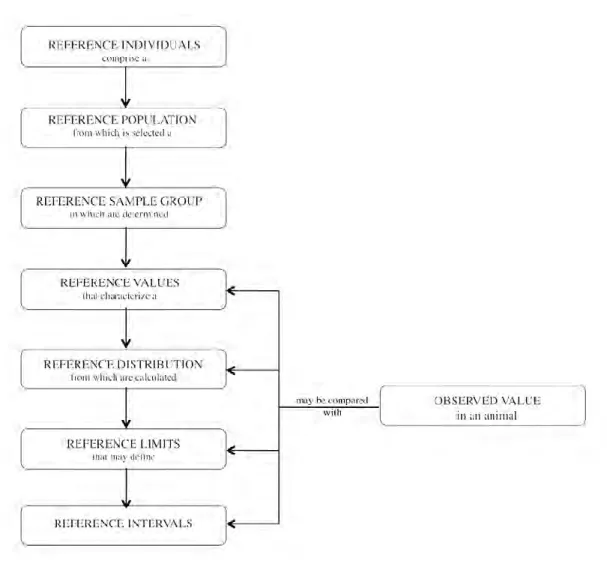
Figure 6. Relationship between the terms defined in the International Federation of Clinical Chemistry and the Clinical and Laboratory Standards Institute recommendations. From Geffré et al.
Reference values: a review. Vet Clin Pathol 2009;38:290.
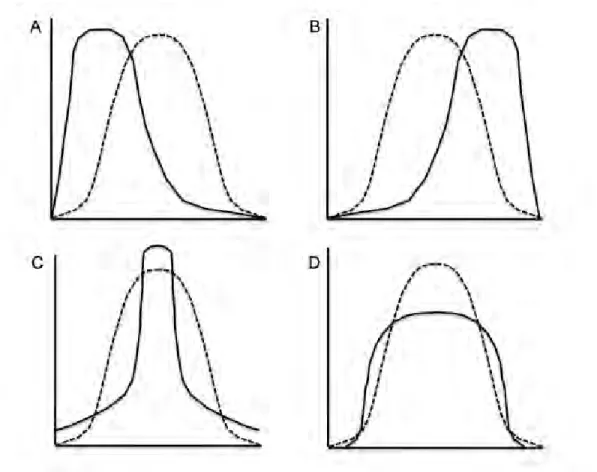
Figure 7. Curves showing different types of distribution: bell shape Gaussian distribution (dotted curve) as well as skewness and kurtosis (continuous curves) with the two upper figures showing asymmetric distributions (A and B, positive and negative skewnesses, respectively) and the two lower figures showing distributions with non-Gaussian peakedness (C and D, positive and negative kurtosis, respectively). From Solberg. Establishment and use of reference values. In: Burtis CA,
Ashwood ER, eds. Tietz Textbook of Clinical Chemistry. 3rd ed. Philadelphia, PA: Saunders;1999:336-356.
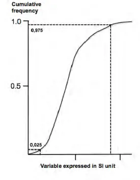
Figure 8. Graphic determination of the central 95% reference interval (2.5th and 97.5th percentiles) for a variable expressed in the “International System of Units” (SI) by plotting the cumulative distribution.
percentiles. The whiskers represent the 10th and 90th percentiles. The rounds represent extreme values. From Feeman et al. Serum creatinine concentrations in retired racing greyhounds. Vet Clin Pathol 2003;32:40-42.
Figure 10. Box-and-whisker plot depicting the serum troponin I concentrations in healthy Greyhound, non-Greyhound and Boxer dogs. The horizontal bar represents the median value and the
whiskers represent the 2.5% and 97.5 percentiles. From LaVecchio et al. Serum cardiac troponin I concentration in retired racing Greyhounds. J Vet Intern Med 2009;23:87-90.
Figure 11. Boxplots showing distribution of the natriuretic peptides pro-atrial-type (proANP 31-67, Figure 11A) and N-terminal pro-brain-type (NT-proBNP, Figure 11B) by breed. The top, bottom, and line through the middle of each box correspond to the 75th percentile (top quartile), the 25th percentile (bottom quartile) and the 50th percentile (median), respectively. The whiskers extend from the bottom 2.5th percentile to the top 97.5th percentile. Outliers are represented by black dots. Box, Boxer; BS, Belgian Shepherd; CKCS, Cavalier King Charles Spaniel; Dach, Dachshund; Dob, Doberman Pinscher; Fin L, Finnish Lapphund. From Sjöstrand et al. Breed differences in natriuretic peptides in healthy dogs. J Vet Intern Med 2014;28:451-457.
Figure 12. Linear regression plot for plasma N-terminal atrial-type natriuretic peptide (NT-ANP) concentrations in 17 healthy Cavalier King Charles Spaniels. The regression formula is Y = 184.9 +
25.4x with Y, the NT-ANP concentration and x, the age of the dogs. From Eriksson et al. Effect of age and body weight on neurohumoral variables in healthy Cavalier King Charles spaniels. Am J Vet Res 2001;62:1818-1824.
Figure 14. Right parasternal transaortic short-axis view in bidimensional mode showing positioning of calipers (double arrows) allowing calculation of the left atrium (LA) on aorta (AO) ratio. HR,
heart rate (in beat per minute); PA, pulmonary artery.
Figure 15. M-mode right parasternal left ventricular short-axis view. HR, heart rate (in beats per
minute); IVS: interventricular septum; LV, left ventricle; LVFW, left ventricular free wall; RV, right ventricle. Photograph: Cardiology Unit of Alfort.
Figure 16. Pulsed wave Doppler mode showing the maximal systolic aortic velocity, on the left apical long axis 4-chamber view (A) and the maximal systolic pulmonary velocity (B), on the right parasternal transaortic short-axis view. Photograph: Cardiology Unit of Alfort.
Figure 17. Material used for systemic arterial blood pressure measurement using the Doppler method. Source: www.dispomed.com
Figure 18. Distribution of plasma N-terminal-pro-brain-type natriuretic peptide (NT-proBNP) concentrations among the 154 healthy small size dog population. The thick horizontal bar represents
the median (i.e., 783 pmol/L) and the thin horizontal bars represent the first and third quartiles (i.e., 476 and 1232 pmol/L, respectively).
Figure 19. Comparison between plasma N-terminal pro-brain-type natriuretic peptide (NT-proBNP) concentrations assessed in the 11 outlier dogs at first sampling and 3 months later.
GENERAL INTRODUCTION
The dog was the first domesticated animal (Ovodov et al., 2011) and had been the most widely kept working, hunting, and pet animal in the whole human history. In France, there are more than 7.8 millions of dogs including 50% of purebred dogs. Additionally, no less than 25% of French households own at least one dog (Santé Vet, 2009). In the canine population, there are more than 300 different canine breeds from small to giant size, among which small size dogs (i.e., dogs with a body weight (BW) lower than 12 kg) are very popular, especially in urban areas (Société Centrale Canine, LOF Registration Statistics, 2012; American Kennel
Club 2011 Dog Registration Statistics). Among this small size dog population, 7 breeds
(Figure 1, Table 1), i.e., Cavalier King Charles Spaniel (CKCS), King Charles Spaniel
(KCS), Bichon (B), Dachshund (D), Miniature Poodle (MP), Shih-Tzu (ST) and Yorkshire Terrier (YT) represent almost 13% of all registrations to “Le livre des Origines Françaises”
Figure 1. Photographs showing the 7 most common small size breeds in France. A, Miniature
Poodle; B, Yorkshire Terrier; C, Cavalier King Charles Spaniel; D, King Charles Spaniel; E, Dachshund; F, Bichon Frisé; G, Shih-Tzu.
Breed Country of origin Coat Colour Weight (kg) Height (cm)
Miniature Poodle Germany
Corded or curly, generally groomed (continental, puppy, English saddle or
sporting clips)
Black, white, grey, brown, apricot - 28-35
Yorkshire Terrier England Long-haired (usually groomed) Black and tan < 3.2 -
Cavalier King Charles Spaniel
United Kingdom Long-haired Blenheim, tricolour, black and tan and ruby 5.4-8.2 30-33
King Charles Spaniel United Kingdom Long-haired Blenheim, tricolour, black and tan and ruby 3.6-6.3 23-28
Dachshund Germany Short, long and wire-haired
Single-colored, single-colored with spots (merle), single colored with tan points,
two-colored
4.0-9.0 -
Bichon Frisé France and Belgium Long-haired and curly White 3.0-7.0 23-30
Shih-Tzu China Long-haired
Various shades of gold, white, brown, and
Although these dogs are highly appreciated by people, they are unfortunately prone to develop degenerative mitral valve disease (DMVD), which is the most common heart disease in dogs. This acquired valvular disease consists of incomplete apposition of the two mitral valve leaflets resulting in chronic- mild to severe- mitral regurgitation (MR). During the course of DMVD, several neuro-hormonal mechanisms may be activated, in order to maintain adequate cardiac output, blood pressure, and tissue perfusion. As the disease worsens, those mechanisms may be deleterious, leading to fluid retention and therefore to the development of congestive heart failure (CHF) and CHF-related symptoms such as cough, dyspnea and exercise intolerance. Even if most of the small size dogs suffering from DMVD remain asymptomatic for years or even in their lifetime, severe complications may occur, such as CHF and alteration of other functions, such as renal function, leading to death or euthanasia because they no longer respond to medical treatments. The standard transthoracic echocardiography (TTE) is considered as the gold standard to assess mitral valve lesions and severity of MR, as well as its consequences on heart morphology and function. Moreover, TTE can be used in combination with evaluation of several blood markers, also called biomarkers, in order to assess severity and complications of DMVD. Additionally, biomarkers may help clinicians to make a prognosis and evaluate medical therapy efficiency. One of the most common biomarker used in veterinary cardiology is the plasma cardiac biomarker N-terminal pro-brain-type natriuretic peptide (NT-proBNP), a peptide synthesized by atrial and ventricular cardiomyocytes in response to increased filling pressures and myocardial wall stress.
The dog represents a unique animal model because there are more than 300 breeds with large phenotypic variations. The morphological differences between canine breeds can be significant, for example a BW may vary from less than 1 kg (Chihuahua dogs) to more than 100 kg (Mastiff dogs). Therefore, the effect of covariates such as breed and BW on blood and
Additionally, numerous other physiological effects have been pointed out, the most common being age and gender. Other effects include neutering status, exercise and body condition, since overweight and obesity increased dramatically in dogs as it does in humans. Owing to these observations, the major hypothesis of this work was that determination of specific reference interval (RI) in healthy small size dogs regarding the most common blood and echocardiographic variables used in the longitudinal evaluation of DMVD could be relevant.
The concept of RI is critical because it helps clinicians to make medical decisions about a patient’s test result. A RI is defined as an interval into which 95% of values of a reference group fall (Solberg et al., 1987a). In other words, 2.5% of the values are under the lower limit of this interval and the remaining 2.5% are above the upper limit of this interval, whatever the distribution of the reference values. In human medicine, experts from the International Federation of Clinical Chemistry (IFCC) and from the Clinical and Laboratory Standards Institute (CLSI) wrote guidelines providing definitions and specific recommendations about the determination of RI in clinical laboratory (Solberg, 1987a, 1987b, 1988; Solberg and
Stamm, 1991; Petit Clerc and Solberg, 1987; Dybkaér 1987; CLSI 2008). In veterinary
medicine, the American Society for Veterinary Clinical Pathology (ASVCP) advised adherence to the IFCC-CLSI guidelines while including recommendations for the determination of population-based RI in animals (ASVCP, 2012; Friedrichs et al., 2012). However, in veterinary medicine, there is a lack of studies focusing on RI determination with respect to these guidelines, especially in dogs, for which the quality of healthcare improves daily while remaining similar to human healthcare.
For this purpose, this work includes three studies, whose main objective was to determine RI according to the IFCC-CLSI guidelines in healthy small size dogs regarding plasma
echocardiographic and conventional Doppler variables.
The present manuscript is divided into chapters as follows:
Chapter I presents the epidemiological, clinical, pathophysiological and echocardiographic features of canine DMVD, as well as echocardiographic and blood variables commonly used in the assessment of disease severity and prognosis.
Chapter II describes the recommendations for RI determination in clinical pathology according to the IFCC-CLSI and the ASVCP guidelines.
Chapter III summarizes the RI previously determined in studies regarding veterinary clinical pathology and cardiology. Additionally, the effect of covariates such as breed, BW, age and gender on clinical pathology and cardiology variables in dogs will also be addressed.
Chapter IV presents the scientific aim of this work.
Chapter V describes the material and methods used in the present work to carry the studies.
Chapter VI summarizes the studies and provides the corresponding original articles.
CHAPTER I. THE DEGENERATIVE MITRAL VALVE
DISEASE
I.
INTRODUCTION
The DMVD, also previously called myxomatous mitral valve disease or mitral endocardiosis, is the most common acquired heart disease in dogs (Buchanan, 1977;
Thrusfield et al., 1985; Pedersen et al., 2000; Häggström et al., 2004; Kvart et al., 2005).
Degenerative lesions of the mitral valve leaflets and associated chordae tendineae lead to insufficient coaptation, and therefore a chronic regurgitation through the mitral valve. At physical examination, DMVD is characterized by a left apical systolic heart murmur of varying intensity, i.e., from grade 1/6 to 6/6 according to the Freeman and Levine’s scale
(1933). Progression of the disease is generally slow, especially in small size dogs, and the
pre-clinical phase (i.e., the asymptomatic period) may last for years and even for life. Nevertheless, progression is sometimes unpredictable and severe complications may occur, leading to the occurrence of clinical signs related to CHF, and ultimately death or euthanasia because of worsening or unresponsive clinical signs (Ettinger et al., 1998;
Pouchelon et al., 1999; Kvart et al., 2002; Atkins et al., 2007; Borgarelli et al., 2008; Häggström et al., 2008). Medical treatment of naturally occurring DMVD includes
numerous drugs or therapeutic classes, the most common being angiogensin-converting enzyme inhibitors (Pouchelon et al., 1999; Kvart et al., 2002; Amberger et al., 2004; Atkins
Häggström et al., 2008) and loop diuretics such as furosemide (Sisson and Kittleson, 1999; De Madron et al., 2011). More recently, surgical mitral valve repair including suture
annuloplasty and chordae tendineae replacement has been advocated (Mizuno et al., 2013).
In 2009, the American College of Veterinary Internal Medicine (ACVIM) consensus panel formulated guidelines for the diagnosis, classification (Table 2) and treatment of the DMVD (Atkins et al., 2009).
Table 2. American College of Veterinary Internal Medicine Consensus Statement for classification of dogs with degenerative mitral valve disease. Atkins et al. Guidelines for the
diagnosis and treatment of canine chronic valvular heart disease. J Vet Intern Med 2009;23:1142-1150.
STAGE A Patients at high risk for developing DMVD but that currently have no identifiable structural disorder of the heart (e.g., every Cavalier King
Charles Spaniel without a heart murmur).
STAGE B B1 Asymptomatic patients with DMVD having no radiographic or echocardiographic evidence of cardiac remodeling in response to DMVD.
B2 Asymptomatic patients with DMVD having radiographic or echocardiographic evidence of left-sided heart enlargement.
STAGE C Symptomatic patients with DMVD having past or current clinical signs of heart failure associated with structural heart disease.
STAGE D Symptomatic patients with end-stage DMVD with clinical signs of heart failure that are refractory to standard therapy.
Prevalence of DMVD is quite high in small size dogs and may attain 14% to 40% depending on the breed and even reach higher values in geriatric canine populations
(International Small Animal Cardiac Health Council 1999; Chetboul et al., 2004a; Serfass et al., 2006). Both prevalence and progression are believed to be higher and faster in males than
females (Beardow and Buchanan, 1993). The most common small size breeds known to be predisposed to DMVD include: CKCS, KCS, D, B, YT, ST, MP and Lhassa Apso (Thrusfield
et al., 1985; Pedersen et al., 1999a; Ettinger et al., 1998; Pouchelon et al., 1999; Pedersen et al., 2000; Kvart et al., 2002; Chetboul et al., 2004a; Serfass et al., 2006; Atkins et al., 2007; Häggström et al., 2008; Borgarelli et al., 2008). Within this small size dog population, the
CKCS show specific features, as they develop DMVD at a relatively young age with a high prevalence as compared to other small breed dogs. Nevertheless, the time course of their disease progression to CHF does not appear to be markedly different from that of other small size dogs except for the early age of onset (Häggström et al., 1992; Beardow and Buchanan,
1993; Chetboul et al., 2004a). Less commonly, large breed dogs such as German Shepherds
may also be affected by the disease and DMVD progression seems to be more rapid in this breed than that observed in small size dogs (Borgarelli et al., 2004 & 2007).
III. ETIOLOGY
Little is known about the signaling mechanisms that initiate the pathological process of DMVD in dogs. Such initiating mechanisms may include genetic, chemical and mechanical stimuli (Buchanan, 1977; Häggström et al., 2004; Madsen et al., 2011), among which the circulating serotonin concept has recently been studied with interest in dogs with naturally
et al., 2013; Cremer et al., 2014). Other examples of potential mechanisms include heritable
connective tissue disorders (Boudoulas, 2003), transforming growth factor beta (Disatian et
al., 2009; Aupperle et al., 2008; Obayashi et al., 2011), nitric oxyde (Moesgaard et al., 2007a) and angiotensin II (Mow et al., 1999).
IV. PATHOPHYSIOLOGY
The normal mitral valve apparatus (Figure 2) is composed of two leaflets (anterior or septal and posterior or mural), which are thin and transparent (Buchanan, 1977; Fox, 2012). Macroscopically, DMVD consist on degeneration of valve leaflets, mainly the anterior
(Tamura et al., 1995), and associated chordae tendineae (Trautwein et al., 1973; Liu et al., 1975; Kogure et al., 1980; Terzo et al., 2009). The degeneration is characterized by nodular
thickening, incomplete apposition of the valve leaflets during systole and secondary MR
(Kittleson et al., 2003; Muzzi et al., 2003; Gouni et al., 2007). Microscopically, canine
DMVD is characterized by increased cellularity in the different leaflet layers, valve structure disorganization, as well as differentiation of valve endothelial and interstitial cells, and glycosaminoglycan infiltration (Schneider et al., 1973; Buchanan, 1977; Rabkin et al., 2001;
Figure 2. (A) Sagittal section through the left atrium (LA) and the left ventricle of a dog with a normal mitral valve apparatus. The two mitral leaflets are attached at the junction of atrial and
ventricular myocardium. The leaflets are thin, clear and translucent and the chordae tendineae are smooth and symmetric (arrows). (B) Close up view of a degenerative mitral valve. The leaflet edges are rounded and thickened at their contact point (thin arrows) and nodular thickening (broad arrow) is present along the distal leaflet segments.LVPW, left ventricular posterior wall; P, papillary muscle; W, LA posterior wall. Scale in mm. Photograph and legend: Fox PR. Pathology of myxomatous mitral valve disease in the dog. J Vet Cardiol 2012;14:103-126.
Depending on MR severity, several potential hemodynamic consequences such as reduced forward cardiac output and increased intracardiac pressures may occur (Buchanan, 1977;
International Small Animal Cardiac Health Council, 1999; Borgarelli et al., 2008). In human
beings, such hemodynamic alterations may result in complex neurohormonal activation, especially adrenergic nervous and renin-angiotensin-aldosterone system (RAAS) activation, in order to maintain adequate cardiac output, blood pressure, and tissue perfusion (Franciset
activations have deleterious effects with adverse consequences at both cardiac and vascular levels (water and sodium retention, increased heart rate and blood pressure, myocardial and endothelial fibrosis, etc.), which contribute, inter alias, to CHF aggravation (Francis, 1998) and to occurrence of the cardiorenal syndrome. The cardiorenal syndrome (particularly type II) defines the decline of renal function in the setting of advanced CHF (Bongartz et al., 2005;
Ronco, 2012).
In dogs with DMVD, few studies have reported the activation of adrenergic system and RAAS, but results remain conflicting (Ware et al., 1990; Pedersen et al., 1995a; Fujii et al.,
Häggström et al., 1996 & 1997). Regarding the occurrence of type-II cardiorenal syndrome in dogs with naturally occurring DMVD, a study showed that the prevalence of azotemia was high (i.e., up to 50%), and increased with CHF severity (Nicolle et al., 2007). Moreover, the glomerular filtration rate decreased by 45% in advanced classes of CHF as compared with the early stages based on the New York Heart Association classification (Nicolle et al., 2007). More recently, another study reported a prevalence of azotemia of 29% and indicated that interlobar renal resistive index increased with CHF severity in 55 dogs suffering from DMVD
(Chetboul et al., 2012a).
In fine, chronic and severe MR causes enlargement of the cardiac chambers and leads to the
development of CHF-related symptoms including exercise intolerance, cough, and dyspnea caused by left-sided CHF (Häggström et al., 2004; Borgarelli et al., 2008; Atkins et al.,
2009), and ascitis or pleural effusion as signs of right-sided CHF secondary to pulmonary
V.
DIAGNOSIS
Although DMVD is characterized at cardiac auscultation by a left apical systolic heart murmur, whose grade is known to be well correlated with MR severity (Häggström et al.,
1995; Pedersen et al., 1999b), the standard TTE (including bidimensional, M-mode, as well
as color, pulsed and continuous wave Doppler) is currently considered as the non-invasive diagnostic method of choice for early detection of mitral valve lesions (Figure 3), evaluation of MR severity (Figure 4A) and complications (Figure 4B), and also for assessing its impact on cardiac remodeling, myocardial function, left ventricular filling pressures as well as pulmonary arterial pressure (Boon, 1983; Chetboul and Tissier, 2012). The ACVIM consensus statement recommends accordingly that standard TTE should be performed in every dog diagnosed with a left apical systolic heart murmur (Atkins et al., 2009). More recently, advanced echocardiographic techniques such as tissue Doppler imaging, speckle tracking imaging and three-dimensional echocardiography have been developed in veterinary cardiology and applied to dogs with DMVD in order to quantify global and regional myocardial function as well as cardiac chamber volumes (Ljungvall et al., 2011a; Tidholm et
images of a normal (A) and a degenerative mitral valve (B). Note the thickening of the mitral
leaflet (B), especially the distal part of the anterior mitral leaflet (AML). LA, left atrium; LV, left ventricle; PML, posterior mitral leaflet; RA, right atrium; RV, right ventricle. Photograph: Cardiology Unit of Alfort.
Figure 4. (A) Left parasternal long axis 4-chamber view using the color Doppler mode showing a severe mitral regurgitation. (B) Bidimensional right parasternal long axis 4-chamber view showing a chordae tendineae rupture and a prolapse of the mitral anterior leaflet (arrow). LA,
left atrium; LV, left ventricle. Photograph: Cardiology Unit of Alfort.
VI. PROGNOSTIC FACTORS
VI.1. CLINICAL AND EPIDEMIOLOGICAL FACTORS
Reported clinical and epidemiological risk factors in dogs with DMVD for negative progression of disease and/or death are numerous. A study found that age more than 8 years was associated with a reduced survival time, both when all and only cardiac causes of death
2 dogs from the International Small Animal Cardiac Health Council (ISACHC, 1999) classification (corresponding to stage C of the ACVIM classification) was significantly higher than that of ISACHC class 3 (corresponding to stage D of the ACVIM classification), i.e., 6 months versus 157 days (Serres et al., 2009a). A heart murmur grade of 3/6 or greater
(Borgarelli et al., 2008), as well as being a male (Beardow and Buchanan, 1993) is associated
with a higher risk of death. Other factors associated with a negative progression of the disease include presence of tachycardia (heart rate >140 bpm), arrhythmias and clinical findings such as syncope, dyspnea, or ascites (Buchanan, 1977; Egenvall et al., 2006; Serfass et al., 2006;
Serres et al., 2007; Borgarelli et al., 2008).
VI.2. RADIOGRAPHIC AND ECHOCARDIOGRAPHIC FACTORS
Several radiographic and echocardiographic variables are known to be associated with prognosis and outcomes in dogs with DMVD. The first proposed paraclinical marker of potential prognosis interest was heart size, assessed from thoracic radiographs (Hamlin et al.,
1968; Buchanan and Bücheler, 1995). Although rapidly replaced by echocardiography, the
usefulness of vertebral heart scale (VHS) to predict development of CHF has been reported
(Lord et al., 2011). There are lots of echocardiographic variables known to affect outcome in
canine DMVD. Dogs with a high degree of valve prolapse and severe valve lesions (thickness of the leaflets and chordae tendineae rupture) are at higher risk of decompensation and death
(Serres et al., 2007, Terzo et al., 2009). Moreover, degree of MR, assessed by using the color
mapping or the PISA (Proximal Isovelocity Surface Area) methods are useful prognostic factors in asymptomatic and symptomatic dogs with DMVD (Pedersen et al., 1999a; Olsen et
pressure are also commonly used for prediction of CHF and death. Among these variables, heart size (especially left atrium (LA) size, LA on aorta (Ao) ratio and left ventricular internal diameter at end-systole (LVES) and end-diastole (LVED)), fractional shortening (FS%), systolic pulmonary arterial pressure and mitral early (E) wave velocity are the most accurate
(Häggström et al., 1992; Serres et al., 2008; Borgarelli et al., 2008; Moonarmart et al., 2010; Reynolds et al., 2012; Hezzell et al., 2012a).
VI.3. CIRCULATING BIOMARKERS
During the last decade, the use of circulating biomarkers in combination with routine cardio-vascular examination has been advocated. A biomarker is "a characteristic (…) objectively
measured and evaluated as an indicator of normal biologic processes, pathogenic processes, or pharmacologic responses to a therapeutic intervention" (Atkinson et al., 2001). There are
many biomarkers in veterinary medicine (Boswood, 2009) but this paragraph will only focus on biomarkers with a demonstrated clinical usefulness in cardiology.
VI.3.1 NATRIURETIC PEPTIDES
To date, B-type natriuretic peptides, including brain natriuretic peptide (BNP) and its inactive aminoterminal portion of brain natriuretic peptide (NT-proBNP), are considered as being the most reliable neurohormonal markers of heart diseases in dog (Schober, 2005; Boswood et al.,
2008; Oyama, 2009; Sisson, 2009). Brain natriuretic peptide is secreted as an inactive
Inactive proBNP is then cleaved into the circulating biologically active BNP and the inactive NT-proBNP fragment (Schober, 2005). The NT-proBNP has a longer half-life and a better stability in a sample than BNP (Schober, 2005). Therefore, NT-proBNP is the most commonly used natriuretic peptide in veterinary cardiology.
Plasma NT-proBNP concentration is correlated with canine DMVD severity, and may be used in combination with clinical status to predict outcomes in both asymptomatic dogs and dogs with CHF (Chetboul et al., 2009; Serres et al., 2009a; Tarnow et al., 2009; Takemura et al.,
2009; Reynolds et al., 2012; Hezzell et al., 2012b). A study (Serres et al., 2009a) showed that
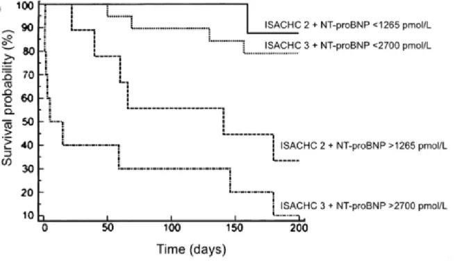
the median survival time of dogs from ISACHC class 3 with a plasma NT-proBNP concentration >2700 pmol/L was extremely short (5 days), whereas that of dogs from the same class but with NT-proBNP <2700 pmol/L was significantly higher (>6 months, P<0.0001, Figure 5). Additionally, several other studies showed that plasma NT-proBNP concentration decreased after initiation of medical treatment of CHF, although benefit on survival remains unclear (Atkinson et al., 2009; Schober et al., 2011; Wolf et al., 2012).
Figure 5. Kaplan-Meier survival curves according to both International Small Animal Cardiac Health Council (ISACHC) class and plasma concentration of N-terminal pro-brain-type natriuretic peptide (NT-proBNP) obtained from 46 dogs with symptomatic degenerative mitral valve disease. ISACHC class 2 with NT-proBNP <1265 pmol/L (n= 13, solid line) and >1265 pmol/L
(n=10, dashed line); ISACHC class 3 with NT-proBNP <2700 pmol/L (n= 13, dotted line) and >2700 pmol/L (n= 10, alternate dotted and dashed line). n, number of dog. From Serres et al., Plasma N-terminal pro-B-type natriuretic peptide concentration helps to predict survival in dogs with symptomatic degenerative mitral valve disease regardless of and in combination with the initial clinical status at admission. J Vet Cardiol 2009;11:103-121.
Another natriuretic peptide of interest is atrial natriuretic peptide (ANP). Even though this biomarker has been the first identified natriuretic peptide, it has been less extensively studied than NT-proBNP. Several studies demonstrated that plasma ANP concentration is significantly correlated with LA size, severity of disease, and may also help to predict outcomes in dogs with naturally occurring DMVD (Häggström et al., 1994 & 1997; Greco et
N-compared to 54 months in those with concentrations ≤1000 pmol/L (Eriksson et al., 2014).
VI.3.2 CARDIAC TROPONIN I
Cardiac troponin I is a protein exclusively present in cardiac muscle (Archer, 2003) and specifically used to detect acute myocardial infarction in humans, as well as marked myocardial damage (Khan et al., 1999). The presence of cardiac disease may lead to myocardial injury and release of troponin I in blood. Serum troponin I concentration is therefore proportional to the severity of myocardial damage (Boswood et al., 2009). In dogs with moderate to severe DMVD, significantly higher concentrations of plasma troponin I were found as compared to healthy controls, and severely affected DMVD dogs had significantly higher concentrations than do mildly and moderately affected dogs (Ljungvall et
al., 2010b; Noszczyk-Nowak, 2011). Moreover, a study showed that troponin I was
independently associated with survival (all-cause mortality) in dogs with DMVD (Hezzell et
al., 2012b).
VI.3.3 CIRCULATING NON-CARDIAC BIOMARKERS
Variables measured as part of standard laboratory investigations, such as hemoglobin (HGB), creatinine, albumin and sodium may help to predict outcomes in human patients with heart disease (Packer et al., 1987; Cowie et al., 2000; Horwich et al., 2002 & 2008). In one study including patients with CHF, the one-year survival was 66% in those with and 83% in those without hypoalbuminemia (Horwich at al., 2008). Similarly, low HGB concentration proved
heart failure with a low relative risk of 1.113 for each HGB decrease of 1 g/dL (Horowich et
al., 2002). Concentrations of some of these substances are known to change in response to
CHF development and its treatment in canine patients. Hence, dogs with CHF have significant lower, although normal, value of PCV (i.e., 42%) compared with healthy controls (i.e., 45%,
Farabaugh et al., 2004). Moreover, worsening of CHF is characterized by a significant fall in
serum sodium, potassium and chloride (Boswood and Murphy, 2006) and a significant increase in the serum urea and creatinine concentrations (Boswood and Murphy, 2006, Nicolle
et al., 2007; Boswood et al., 2009) in dogs with DMVD. In one previous study (Boswood and Musphy, 2006), asymptomatic dogs with DMVD had lower value of creatinine compared to
dogs with CHF (respectively, 92 and 127 μmol/L). To the contrary, a fall in serum sodium was found in dogs with advanced CHF compared to healthy dogs (i.e., 140 versus 147 mmol/L, respectively). Although these biomarkers are easy to evaluate and known to be correlate with CHF severity, further studies are warranted regarding their prognostic significance.
VII. CONCLUSION
The DMVD is the most common heart disease in small size dogs and has extensively been documented in the veterinary literature since decades. This acquired valvular disease may affect quality and duration of life of dogs to a more or less extend, depending of age of onset, breed, gender, cardiac and extra-cardiac complications and response to medical therapy. Reliable variables measured in routine are known to be correlated with disease severity, help to make a prognosis and monitor efficacy of medical treatment. These variables include echocardiographic variables (e.g., LA size) and blood variables such as creatinine and NT-proBNP.
CHAPTER II. THE CONCEPT OF REFERENCE INTERVAL
I. INTRODUCTION
The concept of RI consists in determining a set of values into which a specific percentage, most often 95%, of the values of a particular analyte in a population would fall (Solberg et al.,
1987a).It should be emphasized that RI are often established from general healthy populations. However, sets of values may also be determined from individuals with specific diseases or conditions (children, pregnant women, or smokers for example). These two types of RI are defined as health-related and disease-associated, respectively (Solberg et al., 1995a et 1995b). This RI is then used to help clinicians make the proper medical decisions about their patients’ test results. In human beings, the concept of ‘normal value’ was introduced by Grasbeck and Saris in 1969. Since then, because the term ‘normal’ leads to different meanings, i.e., ‘normal’ might describe a value common in a population, a value arising from a healthy population or a value which follows a Gaussian distribution in the population (Grasbeck and Fellman, 1968; Murphy,
1972; Vacha, 1978; Petit Clerc and Solberg, 1987), the term ‘normal value’ was replaced by the
term ‘reference value’ to avoid such confusions.
Soon after, the IFCC issued recommendations for the determination of RI that were internationally accepted in the “Approved Recommendation on the Theory of Reference Values”
(Solberg, 1987a & 1987b; Petit Clerc and Solberg, 1987; Dybkaér 1987; Solberg, 1988; Solberg and Stamm, 1991). Later, the former recommendations were adopted and updated in 2008 by the
CLSI (CLSI, 2008). In veterinary medicine, as early as in 1979, several authors applied the concept of RI in animals, such as dogs, horses, cattles and swines (Lumsden et al., 1979, 1980a &
1980b; Friendship et al., 1984). The first reported RI established according to the IFCC
recommendations was intended for plasma creatine kinase in a population of dogs (Aktas et al.,
1994). Additionally, the ASVCP recently advised adherence to the IFCC-CLSI guidelines and
provided recommendations for the determination of population-based reference RI in veterinary medicine (ASVCP, 2012; Friedrichs et al., 2012).
In the “Approved Recommendation on the Theory of Reference Values”, the IFCC provided the following general definitions (Solberg, 1987a & 1987b; Petit Clerc and Solberg, 1987; Dybkaér
1987; Solberg, 1988; Solberg and Stamm, 1991):
Reference individual: an individual selected for comparison by using defined criteria.
Reference value: the value obtained by measuring an analyte on a reference individual.
Observed value: the measured value for an analyte produced with the aim of making a
medical decision by comparison with the reference value. Reference population: all possible reference individuals.
Reference sample group: adequate number of reference individuals representing the
reference population.
Reference distribution: statistical distribution of reference values.
Reference limit: derived from the reference distribution and used for descriptive purposes.
inferior than or equal to the limit with a given probability, that is to say the 2.5th or 97.5th percentiles.
Reference interval: interval between and including two reference limits (should not be
named ‘reference range’ as it represents only one figure).
Decision limit: predetermined threshold that distinguishes 2 populations. Decision limits
are defined by consensus and based on investigations on individuals with and without a specific disease or condition.
The relationships between the terms defined above are illustrated in Figure 6 (Geffré et al.,
2009a).
After this introduction, the first part of this chapter will focus on the procedures recommended by the IFCC-CLSI (Solberg, 1987a & 1987b; Petit Clerc and Solberg, 1987; Dybkaér 1987;
Solberg, 1988; Solberg and Stamm, 1991; CLSI, 2008) for determination of population-based univariate reference values (i.e., the production, treatment and use of separate reference values
for one ore more analyte (s), leading to one or more set (s) of univariate reference values).
Multivariate reference values, defined as results of two or more analytes obtained from the same
reference population that are treated in combination (Solberg, 1995c; Concordet et al., 2008), will not be discussed in the present work since this method is not routinely used in veterinary medicine.
Figure 6. Relationship between the terms defined in the International Federation of Clinical Chemistry and the Clinical and Laboratory Standards Institute recommendations. From Geffré et al.
II. GENERAL PROCEDURES FOR THE DETERMINATION OF
REFERENCE INTERVALS
The IFCC-CLSI (Solberg, 1987a & 1987b; Petit Clerc and Solberg, 1987; Dybkaér 1987;
Solberg, 1988; Solberg and Stamm, 1991; CLSI 2008) recommendations imply following a
step-by-step sequence of operations for RI determination (Table 3).
Table 3. Procedures recommended by the International Federation of Clinical Chemistry and the Clinical and Laboratory Standards Institute for reference interval determination.
1. Establish a list of analytical interferences and source of biological variability
2. Establish selection and partition criteria
3. Execute a written consent
4. Categorize the potential reference individuals (questionnaires)
5. Exclude individuals (exclusion criteria)
6. Decide on appropriate number of reference individuals
7. Prepare the selected subject for specimen collection
8. Collect and handle the biological specimens
9. Collect the reference values by analyzing the specimens
10. Inspect the reference values data distribution and prepare a histogram
11. Identify possible data errors and/or outliers
12. Analyze the reference values
III.
ANALYTICAL INTERFERENCE AND SOURCE OF
BIOLOGICAL VARIABILITY
An important preliminary investigation is to define the analyte (s) for which the RI is being determined, as well as the main reason of its measurement (Solberg, 1999; CLSI, 2008; Jones
and Barker, 2008). Secondly, biological variability should be investigated. Variation may occur
at the level of the individual (i.e., intra-individual variability), between individuals (inter-individual variability) and as a result of pre-analytical and analytical imprecision. In dogs, this variability has been studied for biochemical (Ruaux et al., 2012), hematological (Jensen et al.,
1998) and hemostatic (Wiinberg et al., 2007) variables by calculating intra-individual,
inter-individual, and analytical coefficients of variation (CV). Results of these studies indicate that high individuality (reflected by the index of individuality, which is the ratio of within-subject to between-subject biologic variability) is present for many routinely measured analytes in dogs. Hence, index of individuality values were between 0.9 and 3.4, 1.03 and 1.40, 0.5 and 0.8 for respectively 14 biochemical, 4 hematological and 6 hemostatic variables, confirming that population-based RI are of significant value in the clinical setting.
By investigating on these potential sources of interference, clinicians can determine specifications regarding:
The population selection (i.e., inclusion and exclusion criteria); The need to separate RI according to criteria (i.e., partitioning); The procedure of collection and handling of samples.