THÈSE
En vue de l’obtention du
DOCTORAT DE L’UNIVERSITÉ DE TOULOUSE
Délivré par l'Université Toulouse 3 - Paul Sabatier
Cotutelle internationale : Université Libanaise-Ecole Doctorale des Sciences et Technologie (EDST)
Présentée et soutenue par
Sandy AL HAYEK
Le 25 octobre 2019
Etude du rôle du gène ovo/svb dans le maintien et la
différentiation des cellules souches intestinales chez la
Drosophile
Ecole doctorale : BSB - Biologie, Santé, Biotechnologies Spécialité : GENETIQUE MOLECULAIRE
Unité de recherche :
CBD - Centre de Biologie du Développement
Thèse dirigée par
François PAYRE et Dani Osman
Jury
M. Brian OLIVER, Rapporteur Mme Julia CORDERO, Rapporteure
M. Georges NEMER, Rapporteur M. Ziad ABDEL-RAZZAK, Examinateur
M. David CRIBBS, Examinateur Mme Laure EL CHAMY, Examinatrice
M. Dani OSMAN, Directeur de thèse M. François PAYRE, Directeur de thèse
1
« Je dédie ce travail à mon amour Khalil, mon
père, ma mère, mes frères, ma sœur et ma
belle-famille, pour leur bienveillance, soutien
sans faille et surtout pour leur confiance en
moi »
2
«
A mind that is stretched by a new experience
can never go back to its old dimensions. »
(Oliver Wendell Holmes)3
Acknowledgment
First, I would like to thank my jury members: Brian Oliver, Julia Cordero and
Georges Nemer for accepting to evaluate my thesis manuscript.
I also thank Laure EL Chamy, David Cribbs and Ziad-Abdel-Razzak for accepting to participate to my jury. In addition, I am so grateful to Dr
Ziad-Abdel-Razzak for his valuable help at the beginning of my thesis.
Je tiens également à remercier les membres de mon comité de suivi de thèse :
François Leullier et Allison Bardin pour leurs conseils et leurs encouragements.
C’était un grand plaisir pour moi de pouvoir discuter avec vous de mon projet de thèse.
Un énorme merci à François : mon directeur de thèse. Je te remercie du plus profond de mon cœur de m’avoir accueillie dans ton labo ainsi que pour ton aide et ton support tout au long de ma thèse. Merci pour toutes les discussions interminables, les réunions (longues mais productives). Tu m’as aidée à acquérir une maturité scientifique et surtout à croire en mes capacités ! Tu étais là pour les hauts comme pour les bas. Tu m’as vue craquer, rigoler (et râler). Tu m’as appris à être patiente, même si c’était un peu dur parfois ! Je suis également fascinée par ta façon de tout gérer et je vais prendre exemple sur toi pour la suite de ma carrière !
Dani, mon co-directeur de thèse, tu étais là dès le début pendant mes recherches
de thèse et tu n’as jamais lâché l’affaire ! Si j’en suis là aujourd’hui c’est en grande partie grâce à toi. Tu m’as confié un projet qui t’est très cher ! Tu m’as poussée à aller plus loin. J’étais ta première étudiante après ton retour définitif au Liban et j’ai eu l’occasion de participer (un tout petit peu) à tes enseignements. Tu m’as appris à bien réfléchir et à bien analyser mes données (sans sur-interpréter les résultats). Choukran !!!
Dani et François je vous remercie infiniment de m’avoir donné la chance de
présenter mon projet au meeting Américain de Recherche en Drosophile. C’était une opportunité en or que je n’oublierai jamais.
4 Je tiens également à remercier les membres de l’équipe sans qui ces quatre années de thèses n’auraient pas été si merveilleuses.
Un vif remerciement à ma meilleure amie française, ma voisine de bureau et ma complice Alex (ça risque d’être un peu long). Je ne me souviens pas comment nous sommes devenues si proches l’une et l’autre mais crois-moi sur paroles que ça a bouleversé ma vie de doctorante, en mode stress continu ! Merci d’être toujours là pour me remonter le moral, pour essuyer mes larmes et de faire de ton mieux pour me faire sourire ! Bon, passons aux choses rigolotes !! Merci Alex de m’avoir aidée à améliorer mon Français en ayant le courage et surtout la patience de me reprendre (gentiment). Tu as toujours apprécié ma volonté à vouloir apprendre encore plus. Et d’ailleurs ce fameux bouquin d’expressions françaises que tu m’avais conseillé m’a permis de bien briller en société ! Je n’oublierai jamais les soirées cinéma (les lundi soir ces derniers temps), les petites escapades et pleins d’autres belles aventures !!! Bon courage pour la suite de ta carrière ma star en Bio-informatique !!
Cédric, merci d’avoir été mon « confident expérimental » au début de ma thèse.
Je te remercie aussi pour tes conseils, ta bonne humeur et ta positivité. Merci également pour les journal clubs rigolos et la bonne ambiance au bureau !!
PhiPhi, la star de la génétique, merci pour ton humour et tes blagues sans cesse,
qui m’ont permis de déstresser !! Je ne serais malheureusement pas là pour ta soutenance de thèse mais je suis sûre que ça va être un énorme succès ! et « Ce qui est fait, ce n’est plus à faire ;) »
Hélène, merci pour ton encouragement « je veux voir des plus partout les
jeunes » ! Merci pour toutes les discussions aux mouches : « pas plus que deux gosses Sandy ;) » ; Pour tes retours sur mon article ainsi que pour tes commentaires constructifs sur tous mes topos !
Damien, le doctorant le plus jeune du labo, je ne me souviens pas d’un seul instant
où je t’ai vu fâché ou même énervé ! Merci pour ta positivité, ta gentillesse et ton soutien et encore pour les expressions françaises que tu m’as apprises !! Je te souhaite un avenir aussi beau que tes cheveux bien coiffés et tes belles introductions ;) !!!
5
Maleaume, nous avons commencé nos thèses en même temps et tu m’as toujours
épaulée. Merci pour ta confiance en moi, les fou rires, les discussions (sérieuses et rigolotes), mais également pour les Jeudi Subway (et cookie Macadamia), les exercices de prononciations : « Les chaussettes de la duchesse sont-elles sèches, archi sèches ? » ainsi que pour ta patience « Sandy comprends super vite mais il faut qu’on lui explique très longtemps ». Merci énormément d’avoir chouchouté mes mouches pendant mes vacances et évidemment pour la « Super macro de la mort qui tue », d’ailleurs elle a rendu mes analyses beaucoup plus rapides. DyDy te souhaite un grand succès ! Tu vas y arriver !
Maylis, Je te remercie surtout pour ta positivité, ta bonne humeur et la bonne
ambiance ! Et contrairement à Damien, je trouve que t’as une très belle voix que tu pourrais en profiter plus tard !
Anne, j’ai toujours apprécié nos discussions scientifiques et personnelles ! Tu
voulais toujours prendre de mes nouvelles tout en ayant peur de poser des questions qui fâchent (du genre : « il est parti ton papier Sandy ? ») !! Merci pour tout ça. Anne, tu es adorable !
Jenny, toujours souriante, toujours joyeuse et surtout positive !! Merci pour ton
support et pour tes conseils et bonne chance dans tes recherches des petits peptides !!!!
Simon, Merci pour ton sourire et ta bonne humeur ! Tu répands une énergie
positive !!
Une pensée aux anciens membres de l’équipe : Serge, Azza, Ahmad et Jérôme !! Je vous remercie pour votre aide et pour vos encouragements !!
En plus d’une bonne équipe, avoir des bons amis c’est crucial ! Et pas que pour endurer une thèse !
Cyril, ta présence m’était toujours agréable à la pause déjeuner ! Merci d’avoir
fait l’effort de me souhaiter mon anniversaire en arabe et d’apprendre quelques mots (haddi aasabak :D) !! Bonne chance pour tes recherches de post-doc !
Rim (Alice), ma compatriote Libano-Française. A toi je dois mes remerciements
6 souvent, mais enfin c’est l’intention qui compte !). Tu étais toujours à l’écoute et tu as toujours réussi à me réconforter et à me rassurer que tout allait bien se passer ! Merci pour les discussions très intéressantes ! Merci infiniment d’avoir été là pour le choix de ma robe de mariée ! C’était une belle aventure et t’as bien assuré !!!!
Christelle, mes remerciements ne pourront jamais être à la hauteur de ton grand
cœur qui m’a apporté beaucoup de soutien au moment où j’en avais besoin ! Merci d’être présente à mes côtés !
Rhoda, merci de m’avoir accueillie et d’avoir fait le guide touristique au début de
mon séjour à Toulouse ! Encore merci pour les soirées (gâteaux, pizza…) entre filles ! Et bonne chance pour ta nouvelle aventure à Berlin (ou ailleurs).
Katia, mon amie d’enfance et mon futur voisine au Liban !!! Je me souviens
comment tes yeux brillaient à chaque fois que je te parlais de mon projet de thèse ! Même si on ne parle pas tous les jours je veux que tu saches que je pense toujours à toi ! Merci ma Katia d’amour pour ta tendresse et ta précieuse amitié !!! Je sais que ça va être un peu compliqué pour toi d’être en France pour ma soutenance de thèse mais je serais évidemment au Liban pour la tienne !!!
Raymonda, mon adorable cousine ! J'ai vécu des périodes si pénibles que plus d'une
fois j'ai cru que jamais je ne verrai le bout du tunnel. A chacun de ces instants, tu étais là pour me consoler et me rappeler que je suis forte et que je peux y arriver ! Sache que tu m’as aidé beaucoup plus que tu ne le penses et je ne te remercierai jamais assez pour tout ce que tu as fait pour moi !
7
Table of contents
8 Acknowledgment ... 3 Table of contents ... 7 Abbreviations ... 10 List of figures ... 13 Introduction ... 14
1 Stem cells in health and in diseases ... 15
1.1 Emergence of the stem cell concept ____________________________________ 15 1.2 Adult stem cells ____________________________________________________ 15 1.3 Cancer stem cells ___________________________________________________ 16 2 The fly gut as a model system of stem cell function and dysfunction ... 19
2.1 Structural and cellular organization of Drosophila adult digestive tract _______ 20 2.1.1 The structural organization of the adult fly intestine ... 20
2.1.2 The midgut cellular composition and molecular markers ... 22
2.1.3 The midgut development: origin of ISCs ... 23
2.1.4 The Drosophila adult midgut regionalization ... 25
2.1.5 The Drosophila adult Malpighian tubules ... 27
2.2 Molecular mechanisms governing ISC behavior in the adult midgut __________ 29 2.2.1 ISC differentiation by Notch and JAK/STAT signaling pathways ... 29
2.2.2 Implication of Transcription factors in cell lineage ... 32
2.2.3 Regulation of ISC proliferation and survival ... 34
2.2.4 Regulation of ISC behavior in stress conditions ... 40
2.2.5 The importance of EMT-inducing factors in ISCs ... 42
2.3 Evolutionary conservation between mammals small intestine and Drosophila adult digestive tract ______________________________________________________ 45 2.3.1 Comparison between structural and cellular organization ... 45
2.3.2 Cellular plasticity ... 48
2.3.3 Conservation of signaling pathways ... 48
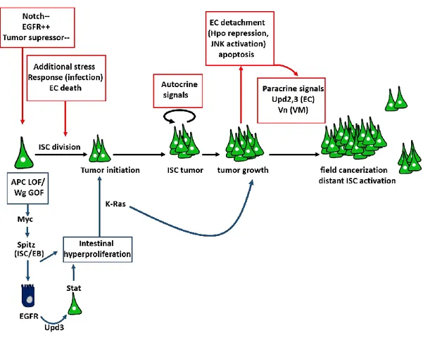
2.3.4 The deregulation of ISC function: intestinal cancer initiation and progression .... 50
3 OvoL/Shavenbaby factors and stem cells ... 53
9
3.2 Ovo functions in the germline _________________________________________ 55 3.3 Svb is required and sufficient for trichome formation ______________________ 57 3.4 Ovo/svb functions across animal species ________________________________ 57
3.5 The regulation of shavenbaby developmental expression __________________ 60 3.6 Svb governs the expression of trichome effector genes ____________________ 64 3.7 Pri temporally regulates Svb transcriptional activity _______________________ 64 3.8 The ecdysone hormone times epidermal differentiation by inducing pri expression
68 Results ... 71 Summary of paper 1... 72 Paper 1 ... 73 Summary of paper 2... 74 Paper 2 ... 75 Discussion ... 76
1. Shavenbaby as a regulatory hub ... 79
2. Ovo/Svb as an EMT regulatory factor ... 83
3. Hormonal control of stem cells behavior ... 86
10
Abbreviations
AMP : Adult midgut progenitors AMPs : Anti-microbial peptides APC : Adenomatous Polyposis Coli Arm : Armadillo
AS-C : Acheate-sense complex Ast : Allatostatine
bHLH : basic Helix-loop-Helix BMP : Bone morphogenic protein BrdU : Bromodesoxyuridin CBC : crypt base columnar cells Ck1α : casein kinase 1α
CNS : Central Nervous system CSCs : Cancer Stem cells Da : Daughterless
DH : Diuretic Hormones
DIAP1 : Death-associated inhibitor of apoptosis 1 dILP3 : drosophila insulin like peptide 3
Dl : Delta dlg : disc large
DNA : deoxyribonucleic acid Dome : Domeless
DSS : dextran sodium sulfate Dvl : Dishevelled
EB : Enteroblast ECs : Enterocytes
ECM : Extra Cellular Matrix EE : Enteroendocrine
EEPs : Enteroendocrine progenitors EcR : Ecdysone Receptor
EGFR : Epidermal Growth Factor Receptor EMT : Epithelial to Mesenchymal Transition esg : escargot
E(spl)-C : Enhancer of split FZD : frizzled
GSK3 : Glycogen Synthase Kinase 3 GOF : gain of function
Hes : Hairy Enhancer of Split Hh : Hedgehog
Hpo : Hippo
11 IIS : Insulin / IGF signaling pathway
ISCs : Intestinal Stem Cells
JAK/STAT : Janus Kinase/signal transducer and activator of transcription Kr : Krüppel
Lgr5 : Leucin-rich G protein-coupled receptor 5 LOF : loss of function
LRP6 : Lipoprotein Receptor related protein 6 mats : mob as tumor suppressor
MET : Mesenchymal to Epithelial Transition mi-RNA : micro-RNA
mRNA : messenger RNA MT : Malpighian Tubules
NICD : Notch Intra Cellular Domain Pan : Pangolin
PARP1 : Poly-(ADP-ribose) polymerase PC : Peripheral Cells
PC : Principle Cells Pdm1 : Pou domain 1 PGC : primordial germ cell
PGRP-LC : Peptidoglycan Recognition Protein-LC
PH3 : Histone 3 Phosphorylated at the serine on position 10 PM : Peritrophic Matrix
Pnt : Pointed pri : polished rice Pros : Prospero
PSF : Phenotypic Stability Factor RAL : RAL GTPase
RB : Renalblast Rpr : Reaper
RNA : Ribonucleic acid
RNSC : Renal Nephric Stem Cell
Robot2/leak : Roundabout receptor / leak receptor Sav : Salvador
SC : Stellate Cell scrib : scribble Sd : Scalopped
SMAD : Supressor of Mothers Against Decapentaplegic SmORF : small open reading frame
SoxN : SoxNeuro
Su(H) : Supressor of Hairless Svb : Shavenbaby
SvbAct : Shavenbaby Activator
SvbRep: Shavenbaby Repressor
12 TF : Transciption Factor
TGF-β : Transforming Growth Factor-β Ttk69 : Tramtrack 69
Ubr3 : E3-Ubiquitin protein ligase Upd : Unpaired like protein Usp : Ultraspiracle
UTR : Untranslated region VM : Visceral muscles Wnt/Wg : Wnt/Wingless Wts : Warts
Yki : Yorkie
13
List of figures
Figure 1: Adult Drosophila gut organization ... 21
Figure 2 : Adult midgut progenitors at the origin of ISC ... 24
Figure 3 : Drosophila adult midgut regionalization ... 26
Figure 4 : Notch signaling pathway in Drosophila adult midgut ... 31
Figure 5 : EE cell fate regulation ... 33
Figure 6 : Canonical Wnt signaling in Drosophila ... 35
Figure 7 : Canonical JAK/STAT signaling in Drosophila ... 35
Figure 8 : Overview of the Hippo pathway in Drosophila ... 39
Figure 9: esg TF regulates both stemness and ISC differentiation ... 43
Figure 10 : ISC regulation by neighboring cells and tissues ... 44
Figure 11 : The mammalian intestinal epithelium ... 47
Figure 12 : ISC implication in intestinal cancer ... 52
Figure 13: The ovo/shavenbaby locus encodes three different protein isoforms ... 54
Figure 14 : Svb is required for trichome formation in Drosophila ... 58
Figure 15: svb integrates multiple signaling pathways to define its spatial register... 61
Figure 16 : svb embryonic expression is governed by the collective activity of 7 enhancers ... 62
Figure 17 : Svb regulates the expression of effector genes for trichome formation ... 65
Figure 18 : Pri peptides induce proteasome-mediated processing of the Svb protein ... 67
Figure 19: Ecdysone regulates the temporality of trichome formation by activating the expression of polished-rice ... 70
Figure 20 : svb's shadow enhancers ensure the robustness of the trichome formation program in extreme temperature variations conditions ... 82
Figure 21 : Svb-Act is required for the maintenance of ISC/EB undifferentiated state ... 85
Figure 22 : svb target genes in ISC ... 85
14
Introduction
15
1 Stem cells in health and in diseases
1.1 Emergence of the stem cell concept
The term "stem cell" (German stammzelle) emerged with the German biologist Ernst Haeckel in 1868 to designate the origin of all cell types. After Darwin, who at the same time proposed his theory on the common origin of species, Haeckel used the term stem cells to refer to the fertilized egg that is the source of all the organism's cell types. In 1892, Theodor Boveri and Valentin Hacker expanded the definition of stem cells to include primordial germ cells (PGC), since germ cells maintain unaltered genetic material and transmit it to the next generation. At the same time, the hematologist Pappenheim used the word stem cell to denote common precursors that can generate both myeloid and lymphoid cells. Hematopoietic stem cells were then established as a prototype of stem cells according to two criteria: the capacity to proliferate indefinitely and the ability to generate differentiated cells (Ramalho-Santos and Willenbring, 2007).
1.2 Adult stem cells
Tissue homeostasis is the process by which a robust structural organization and function are maintained, in the face of varying internal and environmental conditions. This stability is accomplished by the activity of tissue-specific populations of non-differentiated cells, called adult stem cells (SCs), which have three distinguishing features. First, compared to the short-lived differentiated cells that insure tissue function, adult stem cells are long-short-lived to regenerate tissue damages throughout the adult life. Second, they divide to generate new SCs and maintain their pools. Third: they are able to differentiate into a limited range of cell types that build the tissue in which they reside; hence, adult stem cells are multipotent.
The rate of tissue renewal is very variable depending on the intrinsic characteristics of each organ. For instance, it is very high for blood cells (Seita and Weissman, 2010) or the gut
16 epithelium (Eastwood, 1977; Leblond, 1981) which is entirely renewed every week in humans. Some other tissues display a low rate of cell renewal, such as the brain (Ihunwo et al., 2016) , kidney (Little and McMahon, 2012) or heart (Doppler et al., 2017). In the case of lesions or other stresses (infections, injuries), these organs yet display increased regenerative capacities necessary to replace dead cells. In the heart, an organ less exposed to external aggressions, cell renewal can occur by the dedifferentiation of mature cells that will proliferate to repair the injury (Szibor et al., 2014). The study of adult stem cell behavior has become a major topic for the following reasons:
1. Given their potential to differentiate into various cell types, SCs have tremendous potentials for the treatment of human diseases (Kiskinis and Eggan, 2010).
2. Reduced SC function and/or homeostasis can trigger degenerative diseases and is involved in ageing (Clevers, 2011; Jones and Rando, 2011).
3. Deregulated growth of SCs may lead to tumors and adult stem cells share many similarities with the so-called cancer stem cells (CSCs) (Visvader and Lindeman, 2008).
1.3 Cancer stem cells
The discovery of cancer stem cells
The development of tumors and their metastatic potential depend on the activity of rare cancer stem cells (CSCs), or tumor-initiating cells. As for adult stem cells, CSCs have the capacity to proliferate in order to produce more CSCs and to give rise to committed malignant cells. The concept of CSCs has first emerged in the thirties, from the work of Furth and Kahn (Furth, 1935) showing that individual cells derived from mouse leukemia could generate new tumors when engrafted in a healthy recipient mouse. Lapidot et al later hypothesized that a population of CSCs sustains leukemic clones, since myeloid cancer cells have a restricted proliferative capacity (Lapidot et al., 1994). Cancer cells were sorted based on the differential expression of cell surface markers and then transplanted into immune deficient mice. Cells that are able to initiate new tumors in recipient mice were considered as CSCs (Lapidot et al., 1994). Furthermore, CSCs were also identified in solid tumors, such as in brain (Singh et al., 2004)
17 and breast cancers (Al-Hajj et al., 2003) using the same transplantation approach. Subsequently CSCs were identified in many solid cancers including colon, pancreas and lung among others (Tirino et al., 2013).
Origin of cancer stem cells
Despite numerous evidence of the importance of CSCs in tumors, their origin remains controversial. It was hypothesized that transformed cells can fuse with normal stem cells; the resulting fusion cells thus possess both self-renew and transformed abilities. The fusion may be accompanied by chromosomal loss further favoring CSC formation (Ogle et al., 2005). In addition, mutations in somatic cell’s DNA lead to apoptosis and DNA fragmentation. The fragmented DNA can be taken up by a recipient cell leading to nuclear reprogramming and tumor initiation (Camargo et al., 2004; O'Malley and Scott, 2004; Pomerantz and Blau, 2004). The resulted genomic instability such as chromosomal rearrangement and mutations within stem cells thus lead to their transformation to CSCs (Bergsmedh et al., 2001; Holmgren et al., 2002).
An alternative model suggests that CSC arise from tumor cells that undergo multiple changes that favor a stem-like phenotype, including loss of cell-cell adhesion molecules such as E-cadherin, alteration of apical basal polarity, and the gain of motility. This reversible process known as epithelial to mesenchymal transition (EMT) permits CSCs invasion to new sites where they seed micro tumors by reverse mesenchymal to epithelial transition (MET) (De Craene and Berx, 2013). In addition to generating CSCs and inducing metastasis, EMT governs multiple normal processes including tissue remodeling during embryogenesis, organ development and tissue regeneration (Thiery et al., 2009).
The EMT program is induced by a series of transcription factors (TFs) mainly Snail, Slug, Twist and Zinc finger E-box binding Homeobox 1 (Zeb1 and Zeb2) collectively called EMT-inducing factors. The expression of these TFs is induced by signaling cues emanating from tumor-associated stroma, including Transforming Growth Factor Beta (TGF-β), Notch and Wnt signaling (Peinado et al., 2007; Polyak and Weinberg, 2009).
Under normal conditions, micro-RNA composed of 20-22 nucleotides silences the expression of EMT-TF by binding to 3’UTR of target mRNA (Lewis et al., 2005). miR-200 and miR-205
18 act as EMT repressors by targeting Zeb1 and Zeb2 mRNA, thus their downregulation facilitates early steps of metastasis (Cano and Nieto, 2008). Moreover, the downregulation of Zeb1 in some cancer types lead to an upregulation of miR-200 expression, pointing to a negative feedback loop between Zeb and miR-200. miR-200 also acts in a zeb-independent manner, through the regulation of various stemness-related genes, and Zeb factors are regulated by additional micro-RNAs less characterized than miR-200. Another regulatory feedback loop also exists between miR-34, miR-203 and the EMT-inducing TF Snail. Conversely, both Zeb and Snail target 34 expression providing a tight crosstalk between the 200/Zeb and
miR-34/Snail networks (Diaz-Lopez et al., 2014).
Since the discovery of CSCs in solid tumors, many studies have focused on the mechanisms that induce EMT, based on the hypothesis that this process is at the origin of metastasis (Nieto
et al., 2016). In contrast, recent findings suggest that tumor cells that contain both mesenchymal
and epithelial features have the strongest metastatic potential. The screening for the factors that inhibit EMT have led to the identification of the transcription factor OvoL, as a crucial gatekeeper of the epithelial state (Roca et al., 2014; Watanabe et al., 2014). However, most studies that characterized OvoL functions in EMT-TF inhibition were performed using cultured cells. Thus, it is crucial to find a suitable in vivo model system to decipher OvoL mechanisms of action, in both physiological and pathogenic conditions.
19
2 The fly gut as a model system of stem cell function and
dysfunction
For over a century, the "fruit fly" Drosophila melanogaster has been a powerful model for many areas of research, such as genetics, development, evolution and diseases. Notably, six Nobel prizes were awarded to Drosophila researchers. Starting from 2006, Drosophila has now emerged as an attractive model system to study intestinal stem cells (ISCs) and gut regeneration. The intestine is one of the largest and most active organs of the adult body. Beside its function in food processing and nutrient absorption, the gut epithelium constitutes an efficient line of defense against a wide variety of ingested pathogens in both mammals and Drosophila (Buchon
et al., 2014; Chassaing et al., 2014). The gut is also a major source of hormones that modulate
the functions of other organs, such as pancreas and brain in mammals (Bewick, 2012). Thus the gut is not simply a passive digestive tract but it is a vital organ implicated in multiple physiological functions.
The Drosophila adult midgut epithelium is a simple structure that shares anatomical and functional similarities with its mammalian counterpart, the small intestine (Liu et al., 2017). Adult flies feed on decayed fruits and are therefore continually exposed to ingested pathogens, which can have detrimental impacts in the absence of an effective defense mechanism. To deal with these challenges, damaged cells are constantly replaced by new cells, through the activity of intestinal stem cells (ISCs) (Micchelli and Perrimon, 2006; Ohlstein and Spradling, 2006). In the following section, I will describe the structural and cellular organization of the fly digestive tract. I will focus on ISCs of the posterior midgut, the region of interest in my PhD work. In a second part, I will present key findings regarding the molecular mechanisms that regulate both ISC differentiation and proliferation in steady state and stress conditions. In the final part, I will summarize similarities between Drosophila and mammalian digestive tracts, in both structural and cellular organization, as well as the key role of signaling pathways in normal homeostasis and tumor development.
20
2.1 Structural and cellular organization of Drosophila adult digestive tract
2.1.1 The structural organization of the adult fly intestine
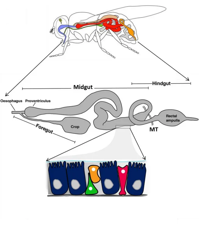
The adult Drosophila digestive system is subdivided into three main regions: the foregut, the midgut and the hindgut (Royet, 2011). The foregut originates from the ectoderm. It is a short tube-like structure composed of the esophagus, which connects to the salivary glands and to the crop, a bi-lobed structure responsible for temporary food storage before its transition into midgut lumen (For review see (Miguel-Aliaga et al., 2018)). The midgut represent the functional equivalent of the mammalian small intestine. It originates from the endoderm and is responsible for food digestion and absorption. The anterior midgut extremity, called proventriculus, is responsible for the initiation of the mechanical breakdown of the ingested food (King, 1988). The proventriculus synthesizes a chitin-containing membrane called peritrophic matrix (PM) that lines the midgut epithelium and separates it from the food bolus. The PM is thus analogous to mucous secretion in the mammalian intestine and constitutes a physical barrier that protects the epithelium from harmful microbes and food particles (Kuraishi et al., 2011). The midgut is enclosed by visceral muscles (VM), nerves, as well as trachea which communicate with the epithelium by signaling molecules regulating intestinal homeostasis. The hindgut derives from the ectoderm and is in charge of water and electrolytes uptake, before discharging the waste into the rectum. The boundary between the midgut and the hindgut is where branching tubules called Malpighian tubules (MTs) are connected. MTs the Drosophila renal system are responsible for adjustment of hemolymph ionic concentrations, through the absorption of electrolytes and their subsequent release into the hindgut. It is interesting to note that distinct populations of stem cells residing in each region maintain the overall integrity of the digestive tract throughout adult life (Zeng et al., 2013) (Figure 1).
21
Figure 1: Adult Drosophila gut organization
The Drosophila adult gut is folded into a 3D structure within the abdomen. It is composed of three mains regions: the foregut containing the esophagus, the proventriculus and the crop; the midgut, the main site of digestion and the hindgut. Malpighian tubules (MT) are attached to the midgut/hindgut boundary. The midgut epithelium is composed of Enterocytes (blue), Enteroendocrine cells (pink), intestinal stem cells (green) and enteroblast precursor cells (orange).
22
2.1.2 The midgut cellular composition and molecular markers
The Drosophila midgut epithelium is a cell monolayer, composed of two types of differentiated cells, responsible for its physiological functions, and stem cells and progenitors cells that ensure the replacement of damaged differentiated cells (Figure 2).
Enterocytes (ECs) are large polyploid absorptive cells that constitute 90% of the midgut epithelium. ECs are positive for the transcription factor Pdm1, also known as Nubbin (Beebe et al., 2010; Lee et al., 2009; Mathur et al., 2010; Tang et al., 2018), which is homolog to the mammalian Pou/Oct proteins (Holland et al., 2007; Tantin). Enteroendocrine cells (EE), are small secretory cells interspersed between ECs, and characterized by the expression of the pan-neural TF Prospero (Figure 2c). EE cells can be subdivided in four different classes, marked by the expression of specific neuropeptides named Allatostatine (Ast), Tachykinin (Tk) and diuretic hormones (DH) (Beehler-Evans and Micchelli, 2015). Class I (AstB+, AstC+) and class II (AstC+) EE cells are present in the anterior part of the posterior midgut, whereas classes III (AstA+, AstC+) and IV (Tk+, Dh+) are localized in the remaining part of the midgut.
Stem cells of the adult Drosophila midgut have initially been identified by the Perrimon and Spradling labs (Micchelli and Perrimon, 2006; Ohlstein and Spradling, 2006). In order to identify proliferating cells within the adult midgut, the researchers traced cells that incorporate Bromodesoxyuridine (BrdU) and thus synthetize DNA. They showed that both small diploid cells and large polyploid cells are BrdU-positive, but only diploid cells are positive for phosphorylated Histone 3 (on a serine at position 10, PH3), a specific marker of mitotic cells. Lineage tracing approaches demonstrated that PH3+ cells are able to both self-renew and give rise to differentiated intestine cells (Micchelli and Perrimon, 2006; Ohlstein and Spradling, 2006). The small diploid cells are thus intestinal stem cells (ISCs) and larger BrdU-cells represent their daughter post-mitotic progenitors, called enteroblasts (EBs).
ISC and EB are characterized by the expression of the zinc finger TF escargot (Esg), the
Drosophila counterpart of the mammalian genes Snail/Slug, and are found as duplets along the
basement membrane (Micchelli and Perrimon, 2006; Ohlstein and Spradling, 2006). In addition, ISCs specifically express Delta (Dl), a ligand of the Notch signaling pathway that binds to the Notch receptor present at the cellular surface of the newly formed EB. Consistently,
23 activation of Notch within EB cells is attested by the expression of the Su(H)-Gbe-LacZ reporter (Ohlstein and Spradling, 2007) (Figure 2c) .
2.1.3 The midgut development: origin of ISCs
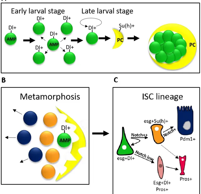
During embryonic development, the endodermal primordia from both sides of the embryo invaginate and some cells undergo epithelial to mesenchymal transition (EMT) to form loosely organized mesenchymal mass containing Adult Midgut Progenitors (AMPs) (Hartenstein et al., 1992). Mesenchymal cells then migrate toward each other’s and undergo reverse MET transition in order to produce EC. This is initiated when endodermal cells enter in contact with mesodermal cells. At this stage, the midgut is composed exclusively of ECs and AMPs closely associated to visceral muscles (Jiang and Edgar, 2009). The embryonic midgut is conserved until pupal stage. During metamorphosis, AMP daughter cells fuse together to form the adult midgut (Jiang and Edgar, 2009; Mathur et al., 2010). Lineage tracing demonstrated that AMPs give rise to all cells of the adult midgut, including ISCs, ECs and EEs (Mathur et al., 2010). During midgut development, AMPs undergo either symmetric or asymmetric division. During early larval stages, dispersed AMPs divide symmetrically to increase their number as the midgut increases in size. Later at mid-larval stage, AMPs undergo one asymmetric division generating a differentiated cell called Peripheral cell (PC), which wraps around clusters of AMP and inhibits their differentiation (Mathur et al., 2010). AMPs continue to divide within islets under the control of mitogenic signaling such as epidermal growth factor Receptor (EGFR) (Jiang and Edgar, 2009). AMPs behavior is also regulated by the ecdysone hormone during larval stages (Micchelli et al., 2011). During pupariation, PCs are lost by apoptosis and AMPs become free to differentiate (Mathur et al., 2010) (Figure 2a-b).
24
Figure 2 : Adult midgut progenitors at the origin of ISC
(A) During early larval development Adult Midgut Progenitors (AMPs) express the Notch ligand Delta (Dl) and divide symmetrically in order to increase their number. Later on, AMPs divide asymmetrically and give rise to Su (H)+ peripheral cells (PC), which serve as niche. PC cells inhibits AMP differentiation and induce their self-renewal.
(B) During metamorphosis, PC cells degenerate and AMPs start to differentiate.
(C) AMPs give rise to ISC that divide asymmetrically to a generate a new ISC and a committed EB (orange) or EEP (pink) progenitor, that will further differentiate into EE or EC cells, respectively.
25
2.1.4 The Drosophila adult midgut regionalization
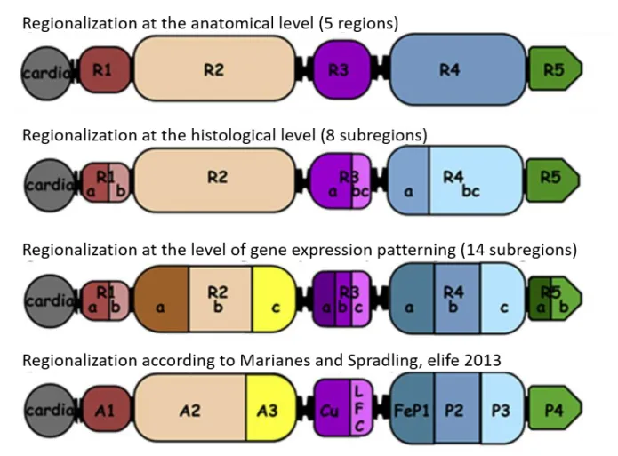
The midgut was initially subdivided into three regions namely the anterior, the middle and the posterior midgut (Demerec, 1950; Hakim et al., 2010). This subdivision was based on the discovery in the middle region of acid secreting cells called “copper cells” (Dubreuil, 2004). These cells confer a high acidity for the middle midgut, similarly to the mammal’s stomach. In contrast, the luminal content of both the anterior and the posterior midgut are mildly alkaline (Dubreuil, 2004).
Recently, work from two independent groups have resulted in the establishment of a nose-to-tail atlas of the Drosophila adult midgut compartmentalization (Buchon et al., 2013; Marianes and Spradling, 2013). Morphometric analyses conducted by Buchon et al. have revealed the presence of six constrictions that split the midgut into five main chambers named R1-R5. These domains were further subdivided into eight subregions based on EC morphology. Next, they performed transcriptome analysis that unveil an unprecedented compartment-specific gene expression and led to a fine-grained division of the midgut into 14 subregions. Interestingly, this high level of regionalization seems to be very robust since it remains intact independently of diet fluctuations or gut damage. This regionalization is governed by a complex regulatory network involving TFs that were used for midgut development during embryogenesis (Buchon et al., 2013). Marianne and Spradling also subdivided the midgut into 10 regions contained within the 14 regions identified by Buchon et al. They also revealed the existence of region-specific expression profiles by performing transcriptome analysis, region-region-specific microarrays and RNA-seq experiments (Buchon et al., 2013; Marianes and Spradling, 2013). Marianne and Spradling also uncovered a partial role for ISC in establishing midgut regionalization. They showed that ISC of different subregions have both different morphology and mitotic activity. ISCs of the posterior midgut region P1-P3 (R4) are the most rapidly dividing cells, with a division rate of once per day, the rate of division is less important in the anterior part. Importantly, ISC daughter cells residing near a region boundary always give rise to the same cell lineages and, even in rare cases where they cross the boundaries , they keep cellular memory of their region of origin (Marianes and Spradling, 2013) (Figure 3).
26
Figure 3 : Drosophila adult midgut regionalization
Based on anatomical features the midgut is divided into 5 different regions (R1-R5). These regions are further divided to 8 subregions containing EC cells with different morphologies (histological level). Gene expressing patterning has led to an additional level of regionalization (14 subregions ) (Buchon et al., 2013). Marianes and Spradling demonstrated the presence of 9 different regions contained within the 14 subregions found by Buchon et al. (Marianes and Spradling., 2013). (Adapted from Osman and Buchon., 2015)
27
2.1.5 The Drosophila adult Malpighian tubules
In addition to digestion and nutrient absorption, ionic concentrations should be tightly regulated in order to maintain a healthy organism. This function is achieved by the Malpighian tubules (MT) attached to the midgut/hindgut junction. MT are also renewed by adult stem cells called Renal Nephric Stem cells (RNSC). In the following section, I will describe MT structural and physiological organization with emphasize on RNSC origin and regulatory mechanisms. I have to note that the first paper of this manuscript concern RNSC maintenance.
2.1.5.1 Malpighian tubules structural organization
Each adult gut is attached to two pairs of MTs: one pair is projected toward the anterior part of the digestive tract whereas the second one is oriented posteriorly. Each MT is divided into two regions: the lower tubules attached to the midgut through the ureters, and the upper tubules distant from the gut and further divided into main, transitional and initial segments (Sozen et al., 1997). As for gut regionalization, MT subregions can be defined based on differential genes expression (Sozen et al., 1997). MT are composed of two types of polyploid differentiated cells: principal cells (PC) distributed all along the tubules, and stellate cells (SC) located in the upper MT (Denholm et al., 2003).
2.1.5.2 Malpighian tubules functions
MT are immerged in the hemolymph, the circulatory system of Drosophila. They ensure water reabsorption, ionic and acid/base equilibrium, as well as the elimination of metabolic waste. In addition, MT participate to innate immunity by acting as autoimmune sensor. A deprivation of nutrients and water (desiccation) induces the expression of the peptidoglycan recognition protein-LC (PGRP-LC). PGRP-LC is known for its role in bacterial detection and induction of the immune response, with an increased production of anti-microbial peptides (AMPs) in order to protect the animal from bacterial loads (Zheng et al., 2018).
2.1.5.3 Renal Nephric stem cells: origin and regulation
Similar to the midgut, MT are renewed by stem cells called Renal Nephric stem cells (RNSC) known as “tiny cells” due to their small size and to their diploid nuclei (Sozen et al., 1997). RNSC are located in the lower tubules and can divide to maintain their pool. They also give rise to committed progenitors called renalblasts (RBs) that further differentiate into PCs or SCs
28 (Singh et al., 2007). In contrast to differentiated cells, already present in the MT during embryogenesis, RNSC are specific to the adult stage. At the pupal stage, intestinal AMPs migrate from the midgut and form the ureter in order to attach the midgut to the MT (Takashima et al., 2013). The remaining undifferentiated AMPs then become RNSCs (Takashima et al., 2013). RNSCs conserve the expression of esg, already present in AMP but express an additional TF called Krüppel (Kr) (Denholm et al., 2003; Singh et al., 2007), required for normal MT formation during embryogenesis. Thus, migrating AMPs are reprogrammed within MTs to acquire novel characteristics different from those of ISC. In addition, RNSC are well individualized and their number is reduced compared to this of ISC (350 RSCs compared to 1000 ISCs approximatively).
Although RNSCs were identified about ten years ago, the mechanisms that regulate their activity remains poorly understood. Similarly to ISC, RNSC express Dl, which activates the Notch pathway in the RBs to initiate their differentiation into reabsorbing cells, CPs and SCs (Li et al., 2014). RNSCs proliferation is regulated by both EGFR and JAK/STAT signaling pathways (Li et al., 2015; Singh et al., 2007) .In contrast, Hippo pathway loss of function leads to an overproliferation of RNSCs (Doggett et al., 2011; Verghese et al., 2012; Yang et al., 2015; Zeng et al., 2010).
29
2.2 Molecular mechanisms governing ISC behavior in the adult midgut
Following the ISC discovery, many studies have focused on deciphering the molecular mechanisms that determine the choice between EC and EE fates, as well as the regulation of ISC proliferation. In the following paragraph, I will present our understanding of ISC regulation by both signaling pathways and transcription factors. I will next discuss recent findings concerning mitogenic signaling pathways required for ISC proliferation in both normal and stress conditions.2.2.1 ISC differentiation by Notch and JAK/STAT signaling pathways
The generation of absorptive enterocytes (EC) requires high levels of Notch activity within EBs, whereas weaker Notch activation leads to the production of EE cells (Ohlstein and Spradling, 2007). In the absence of Notch, ISC cannot differentiate and continue to proliferate leading to tumoral masses (Ohlstein and Spradling, 2007). Conversely, constitutive activation of the Notch pathway by expressing the Notch Intra cellular Domain (NICD) forces the differentiation of ISCs into ECs. Moreover, the activation of Notch within EB cells seems to be dependent of ISC-EB interaction mediated by E-Cadherin molecules, also required for cell shape maintenance (Maeda et al., 2008).
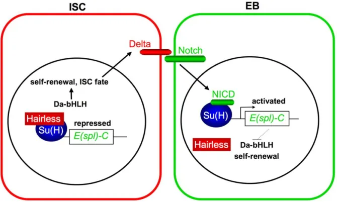
ISC identity is maintained, at least in part, by Daughterless (Da) a basic helix loop helix (bHLH) transcriptional activator that sustains Delta expression and self-renewal capacity (Bardin et al., 2010). In addition, Hairless (H) and Suppressor of Hairless (Su(H) act within ISCs to repress the expression of a main Notch target gene, Enhancer of split (E-(spl)-C, (Bardin et al., 2010)). Notch activation in EBs triggers the expression of E(spl)-C that, in turn, represses Da expression thus enabling cell cycle exit to generate an EC cell (Bardin et al., 2010) (Figure 4). Interestingly, the commitment of ISC to EB also requires the activation of Notch but at a very high level compared to level of Notch signaling required for terminal differentiation of EB to EC (Perdigoto et al., 2011). It was suggested that this differential level is required for the protection of ISCs from their loss through differentiation.
An alternative model for cell fate choice suggested that EE cells arise from another type of progenitors that do not activate Notch. This hypothesis is based on the fact that EE cell progenitors are negative of Su(H)-GBE reporter and that Notch mutant tumors still contain EE cells (Biteau and Jasper, 2014; Guo and Ohlstein, 2015; Zeng and Hou, 2015). This model is
30 further refined by recent observations demonstrating that EBs are able to produce a small population of class II EE cells (Beehler-Evans and Micchelli, 2015).
In addition to Notch, the signaling pathway Janus Kinase/signal transducer and activator of transcription (JAK/STAT) is also required for terminal differentiation of both ECs and EEs. JAK/STAT inhibition in progenitors leads to the accumulation of undifferentiated cells and Notch mutant clones lacking JAK/STAT components are devoid of EE cells (Beebe et al., 2010; Jiang et al., 2009). Epistasis genetic analyses revealed that Stat92E, the TF downstream of JAK/STAT, acts in parallel or downstream to Notch to induce both EC and EE specification (Beebe et al., 2010; Jiang et al., 2009). On the other hand, JAK/STAT induces Dl expression in ISCs (Buchon et al., 2009b; Jiang et al., 2009), suggesting that JAK/STAT can act both downstream and upstream of Notch. Additional functions for JAK/STAT signaling in the maintenance of the Drosophila midgut homeostasis are discussed below (section 2.2.3.)
31
Figure 4 : Notch signaling pathway in Drosophila adult midgut
In ISC Suppressor of Hairless (Su(H)) in association with Hairless bind to the regulatory region of Enhancer of split
(E(spl)-C) and inhibit its expression. In the absence of E(spl)-C the proneural factor Daughterless (Da) is expressed and induces the
expression of Delta, thus maintaining ISC properties. Delta binds to the Notch receptor expressed at the cellular membrane of EBs. Notch intra cellular domain (NICD) translocate to the nucleus where it destabilizes Hairless and binds to Su(H), which leads to E(spl)-C derepression, Da repression and EB differentiation (Bardin et al., 2010).
32
2.2.2 Implication of Transcription factors in cell lineage
Besides signaling pathways, a couple of transcriptional factors are known to determine the cell fate choice between EC and EE cells. In the following, I will describe the positive and the negative regulators of the EE cell fate determination.
2.2.2.1 Specific EE fate regulation program
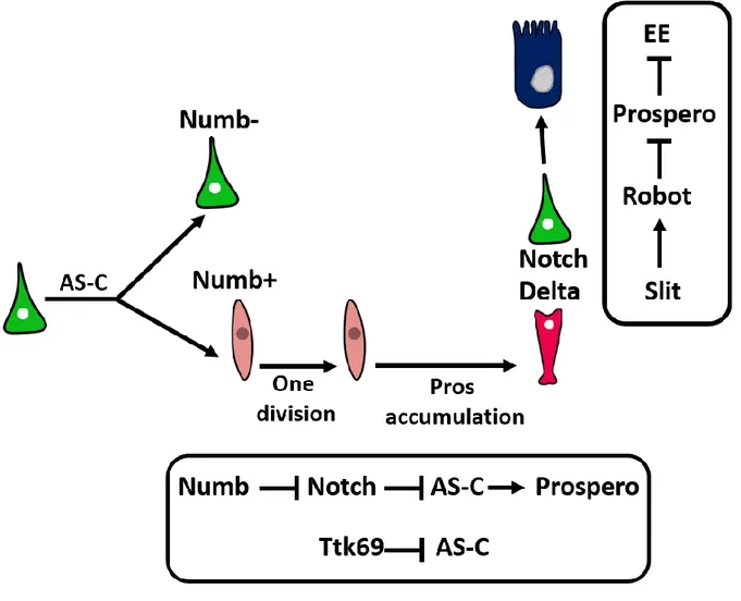
Bardin et al. have revealed a key role for the proneural bHLH factors of the Achaete-Scute complex (AS-C) in the generation of EEs. The deletion of AS-C leads to a complete depletion of EEs, whereas the overexpression of either Asense or Scute in ISC/EB is sufficient to expand the population of Prospero+ cells (Bardin et al., 2010). Moreover, it has been proposed that Scute, a mitogenic factor is transiently expressed in ISCs and leads to their asymmetric division producing a new ISC and an EE progenitor (EEP) (Chen et al., 2018). EEPs divide once before the accumulation of Prospero causes cell cycle exit and EE differentiation (Chen et al., 2018). Numb, an inhibitor of Notch signaling pathway, is asymmetrically dispatched between ISC daughter cells during mitosis (Salle et al., 2017). Notch signaling is inhibited in cells that inherits Numb, consequently Asense and Scute expression is derepressed and Prospero is expressed (Salle et al., 2017).
In addition to factors required for EE differentiation, negative regulators of this cell fate are also detected such as the transcriptional repressor Tramtrack 69 (Ttk69) that inhibits EE cell fate (Wang et al., 2015) and disrupts their hormone producing capacity (Wang and Xi, 2015). Interestingly EE cells are also able to regulate their number autonomously. Newly formed EE expresses the ligand Delta and activates Notch signaling in the apically located ISC. This activity blocks EE cell production and maintains ISC multipotency thus they revert to producing EC (Guo and Ohlstein, 2015). EE cells autoregulate their number by secreting a ligand called Slit. Slit is transduced by Roundabout receptor/leak receptor (Robot2/leak) present at the cell surface of intestinal precursor cells. Slit/robot pathway inhibits Prospero expression within ISC specifically and then inhibits the generation of new EE (Biteau and Jasper, 2014) (Figure 5).
33
Figure 5 : EE cell fate regulation
The achaete-scute complex (AS-C) required for prospero expression and subsequently EE cell production is transiently activated in dividing ISC (green) and leads to the generation of a new ISC and enteroendocrine progenitor cell (EEP)(light pink). EEP divide once before starting to accumulate prospero protein. In addition, the newly formed EEP inherits the protein Numb that inhibits Notch signaling and thus depresses AS-C genes.
EE cell generation is negatively regulated by the transcriptional repressor tramtrack69 (Ttk69) that inhibits AS-C in parallel to Notch. Newly formed EE (dark pink) cells express Delta ligand and activates Notch in the apically located ISC that switches to the production of EC cells rather than EE. EE autoregulate their production also by secreting the ligand Slit that activates Roundabout receptor/leak receptor (Robot2/leak) present at the surface of ISC leading to the inhibition of prospero expression.
34
2.2.3 Regulation of ISC proliferation and survival
In addition to a proper differentiation, midgut homeostasis requires a tight regulation of ISC proliferation in order to replace damaged cells, but also to prevent excessive ISC proliferation that can lead to tumor development.
In the following section, I will describe our current knowledge of the role of diverse signaling pathways in the regulation of ISC proliferation under both homeostatic and stress conditions.
2.2.3.1 The Wnt/Wingless signaling pathway in Drosophila adult midgut
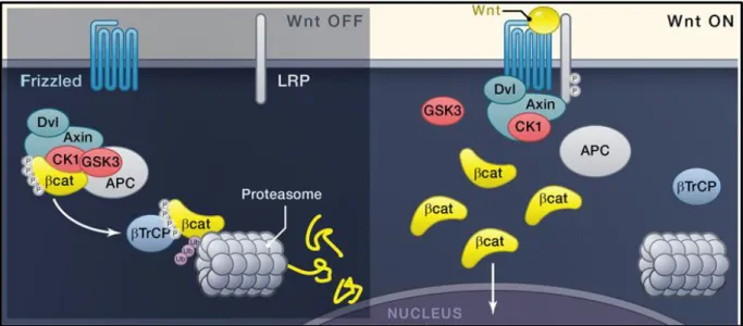
In Drosophila midgut (Figure 6, for the canonical Wnt pathway), Wingless (wg) is the only ligand that has a functional role in intestinal homeostasis (Cordero et al., 2012; Lin et al., 2008). The first evidence about a central role for Wnt signaling in regulating ISC behavior in the posterior midgut was reported by Lin et al. (Lin et al., 2008). wg expression was initially detected in the muscle layer surrounding the midgut. By generating clones lacking components of the Wnt pathway such as the receptor Frizzled, they observed a reduction of ISC proliferation rate that continue to produce EC and EE (Lin et al., 2008). Consistently the overexpression of
wg in esg+ cells was sufficient to induce an expansion of esg+ Dl+ cells. This study led to the
conclusion that Wnt signaling is necessary and sufficient to maintain ISC self-renewal and that the surrounding VM may serve as ISC niche (Lin et al., 2008).
Other studies brought different results about the source of Wnt ligands and the site of Wnt signaling activation. Takashima et al detected Wg in epithelial cells at the midgut hindgut junction (Takashima et al., 2008). Buchon et al. have observed an enrichment of Wnt signaling reporter genes expression at the different midgut boundaries (Buchon et al., 2013); this expression decreases within the different midgut compartments. Tian et al demonstrated that EC cells rather than ISC cells are the primary site of Wnt signaling activation within Drosophila adult midgut (Tian et al., 2016). This activity is required to inhibit growth factors release from EC such as Upd2 and Upd3 that act as paracrine signals and activate ISC proliferation (Tian et al., 2016).
35
Figure 7 : Canonical JAK/STAT signaling in Drosophila (Arbouzova and Zeidler, 2006).
Figure 6 : Canonical Wnt signaling in Drosophila
The β-catenin protein known as Armadillo (Arm) in Drosophila is a key effector in the Wnt signaling cascade. In normal conditions, β-catenin is kept in the cytoplasm where it is phosphorylated by a “destructive complex” composed of four kinases: Axin, Adenomatous polyposis Coli (APC), Glycogen Synthase Knase 3 (GSK3) and Casein Kinase 1α (Ck1α) (Kishida et al., 1998). This phosphorylation induces the ubiquitination of β-catenin that is then targeted to proteasome degradation. Upon Wnt binding to Frizzled (FZD) and lipoprotein receptor related protein 6 (LRP6), the receptor complex is endocytosed and the protein Dishevelled (Dvl) form polymers at the cytoplasmic domain of the receptors. Together, LRP6 FZD and Dvl form the Wnt signalosome that sequestrates and inactivates the destruction complex. β-cat thus reaches the nucleus where it acts in collaboration with Pangolin (a.k.a. TCF/LEF) to induce the expression of Wnt target genes (Korinek et al., 1997; Morin et al., 1997) (adapted from Clevers et al; 2012).
36
2.2.3.2 Epidermal Growth Factor Receptor (EGFR) pathway
Under homeostatic conditions, EGFR signaling is activated in ISC/EB by three redundantly active ligands released from different sources (Biteau and Jasper, 2011; Jiang et al., 2011; Xu et al., 2011). Visceral muscles produce the ligand Vein, Keren is released from ECs, and Spitz is expressed in ISCs and EBs. Clones mutant for EGFR components do not proliferate (Biteau and Jasper, 2011; Jiang et al., 2011; Xu et al., 2011). As for Wnt, EGFR inhibition does not alter the survival nor the differentiation of ISCs. In contrast, a gain of function of EGFR is sufficient to induce ISC overproliferation. Basal levels of EGFR signaling are maintained by different mechanisms avoiding tumor generation by excessive ISC proliferation: Jin et al have demonstrated that the MAP kinase cascade induced upon EGFR activation phosphorylates Capicua a major inhibitor of both cell cycle gene and Pointed, the transcription factor activated downstream of EGFR signaling pathway. Phosphorylated Capicua is then excluded from the nuclei, pnt and cell cycle genes repression is released and ISC proliferation is induced (Jin et al., 2015). Moreover, EGFR signaling levels are kept in check by chromatin modifiers called Kismet and Trr (Gervais et al., 2019). These two complexes cooperate to induce the expression of an EGFR regulatory gene called Cbl within ISCs. Trr and Kismet loss of function inhibits
Cbl expression and leads to abnormal ISC accumulation, associated with a significant increase
in EGFR activity.
2.2.3.3 The JAK/STAT signaling pathway
In Drosophila, the JAK/STAT signaling pathway is a major player of tissue homeostasis. It is required for the coordination of stem cell proliferation and differentiation in many tissues. In addition, it is required for testis niche maintenance and digestive tract development (Arbouzova and Zeidler, 2006).
JAK/STAT signaling is initiated by binding of glycosylated Unpaired (Upd1) (Harrison et al., 1998) and Upd-like proteins (Upd2, Upd3) (Agaisse et al., 2003; Gilbert et al., 2005; Hombria et al., 2005) to the receptor Domeless (Dome) (Brown et al., 2003; Chen et al., 2002). The signal is then transduced within the cells through the receptor associated JAK kinase Hopscotch (Hop) (Binari and Perrimon, 1994) and the transcription factor Stat92E (Hou et al., 2002; Yan et al., 1996). When activated, Stat92E dimerizes and translocates to the nucleus to induce the
37 expression of target genes, including Socs36E a negative regulator of JAK/STAT and Stat92E itself (Arbouzova and Zeidler, 2006) (Figure 7).
Upd ligands are released from multiple sources, but JAK/STAT activity is only detected in ISC/EBs (Beebe et al., 2010; Jiang et al., 2009; Liu et al., 2010). Osman et al. have reported that Upd1 is released from ISCs and acts in autocrine manner to induce ISC proliferation in baseline conditions (Osman et al., 2012). In ageing flies, ISC proliferate abnormally, this feature is partially mediated by both Upd2 and Upd3 ligands released from EBs and ECs. In addition, these two ligands are also overexpressed upon bacterial infection in order to induce rapid epithelial turnover (Osman et al., 2012). Upd ligands are also released from the inner circular muscles and the outer longitudinal muscles and activate JAK/STAT signaling within ISC/EB cells (Lin et al., 2010). Clones mutant for JAK/STAT components are less proliferative and lack mature cells, while elevated signaling levels accelerate the rate of ISC division. Hence, JAK/STAT pathway regulates ISC proliferation and differentiation in healthy and pathogenic conditions.
2.2.3.4 The Hippo pathway: autonomous and non-autonomous restriction of ISC proliferation
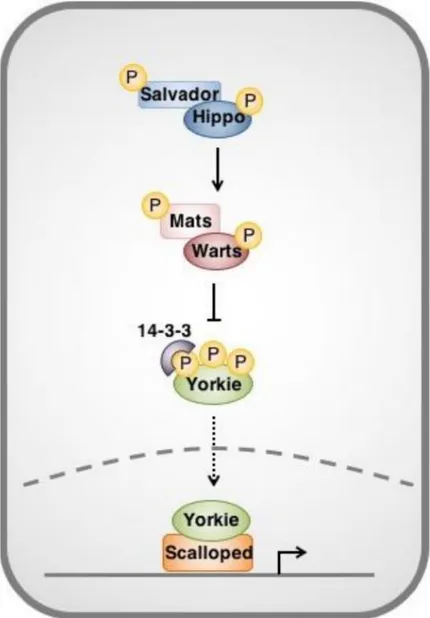
The Hippo pathway also plays a pivotal role in the regulation of ISC proliferation. Hippo pathway, which is regulated by mechanical forces applied on cells, ensure an adapted organ growth and an efficient tissue renewal by regulating both cell proliferation and apoptosis (Harvey et al., 2003; Pantalacci et al., 2003; Thompson and Cohen, 2006; Udan et al., 2003; Wu et al., 2003).
The Hippo complex is composed of four kinases: Warts (wts) (Justice et al., 1995; Xu et al., 1995), Hippo (hpo) (Harvey et al., 2003; Pantalacci et al., 2003; Udan et al., 2003; Wu et al., 2003), mob as tumor suppressor (mats) (Lai et al., 2005) and Salvador (Sav) (Kango-Singh et al., 2002; Tapon et al., 2002). Hpo phosphorylates Sav and thus stabilizes their physical interaction (Aerne et al., 2015; Wu et al., 2003). Hpo than phosphorylates Mats that interacts with Wts and potentiates its kinase activity (Justice et al., 1995; Wei et al., 2007; Wu et al., 2003). The kinase cascade inactivates a unique nuclear effector called Yorki (Yki) (Huang et al., 2005; Oh and Irvine, 2008; Ren et al., 2010b), a transcriptional coactivator that induces the
38 expression of antiapoptotic (DIAP1) and cell cycle genes (Cyclin E) (Dong et al., 2007; Huang et al., 2005).
Within ISCs, Hippo inhibits Yki expression required for ISC proliferation (Huang et al., 2005; Karpowicz et al., 2010; Ren et al., 2010a; Shaw et al., 2010). Yki gain of function (or wts loss of function) induces abnormal ISC proliferation by surexpression of DIAP1 and Cycline E (Shaw et al., 2010). Moreover, Yki is required for ISC proliferation following pharmacological or immune stress (Ren et al., 2010a). Yki activity is dependent of its physical association to two proteins: Scalloped (Sd) and Brahma. The complexe Yki/Sd/Brahma binds to Yki target genes and promotes ISC proliferation (Jin et al., 2013). Interestingly, in steady state conditions Hippo activates caspase proteins that induce programmed cell death. These caspases inactivate Brahma by inducing its proteolytic cleavage (Jin et al., 2013) (Figure 8).
In addition, Hippo signaling acts non-autonomously within EC cells in order to restrict ISC proliferation. In damaged ECs, Yki increases the expression of both Upd (Shaw et al., 2010; Staley and Irvine, 2010; Vodovar et al., 2005) and Keren (Ren et al., 2010a). Subsequently, both JAK/STAT and EGFR signaling are activated within ISCs, inducing their proliferation. Therefore, the Hippo signaling pathway in general -and Yki in particular- acts as a sensor of epithelia stress within ECs allowing them to signal to the ISC when damaged.
39
Figure 8 : Overview of the Hippo pathway in Drosophila Adapted from (Taha et al. ; 2018)
40
2.2.4 Regulation of ISC behavior in stress conditions
The intestine is an organ in continuous contact with the surrounding environment. Thus, in addition to the normal regeneration of the tissue, the gut has also to cope with external stresses like physical (lesions), chemical (oxidative stress) or infectious aggressions. To re-establish the integrity of the tissue and ensure a functional physiological state of the organ, ISCs are also actively solicited. A careful control of their proliferative potential is then indispensable and ensured by the same signaling pathways that are active in basal conditions. In the following section, I will describe some experimental approaches used to induce the fly midgut regeneration. In the second part, I will describe different signals that are communicated to ISC in stress conditions in order to activate mitogenic signaling pathways mainly EGFR, Wnt and JAK/STAT.
2.2.4.1 Experimental techniques used to study the midgut regeneration
In order to study ISC response to epithelial damage several approaches involving feeding flies different tissue-damage inducing agents have been used:
1. Oral infection of flies with different strains of pathogenic bacteria (Pseudomonas
entomophila, Erwinia carotovora (Ecc15), Pseudomonas aeruginosa, Serratia marcescens, Escherichia coli...) (Apidianakis et al., 2009; Buchon et al., 2009a;
Buchon et al., 2009b; Cronin et al., 2009; Vodovar et al., 2005).
2. Chemicals feeding with compounds like Hydrogen peroxide and Paraquat (Choi et al., 2008) that induce oxidative stress, dextran sodium sulfate (DSS) that disrupts the basement membrane, or Bleomycin, a cytotoxic antibiotic that causes damages to ECs (Amcheslavsky et al., 2009).
3. The targeted expression of proapoptotic genes such as reaper (Rpr) within EC cells inducing their death (Jiang et al., 2011).
2.2.4.2 Signaling pathways in midgut regeneration upon damages
Differentiated ECs are the first target of bacterial infections and cytotoxic agents since these cells are the first to encounter the stressors. Indeed, pathogenic bacterial infection or Bleomycin treatment induce EC apoptosis and removal from the intestinal epithelium (Amcheslavsky et
41 al., 2009; Apidianakis et al., 2009; Buchon et al., 2010; Jiang et al., 2009). ISCs respond to EC loss by increasing their proliferative capacity in order to replenish the midgut. Interestingly, apoptosis inhibition within ECs abolishes ISC proliferation, whereas the apoptosis induction by the expression of Rpr in ECs is sufficient to induce ISC proliferation, even in the absence of infection (Jiang et al., 2011). Thus, EC apoptosis is a main signal for the activation of ISC proliferation upon gut damages.
At the molecular level, damaged ECs secrete the Upd3 ligand that activates JAK/STAT signaling within ISCs, therefore inducing their proliferation (Buchon et al., 2009a; Cronin et al., 2009; Jiang and Edgar, 2009). Moreover, Upd3 released from damaged EC signals to the surrounding visceral muscles that transduce the JAK/STAT signaling and release Vein that also induces ISC proliferation (Buchon et al., 2010; Jiang et al., 2011). The other EGFR ligand Keren is also released from damaged EC and further potentiates ISC proliferation (Buchon et al., 2010; Jiang et al., 2011). Interestingly, EGFR signaling is also required for both damaged EC-detachment from the epithelium and their morphogenesis during regenerative episodes (Buchon et al., 2010). Indeed, EGFR inhibition within infected ECs blocks the disruption of cell-cell adhesion and their subsequent delamination from the epithelium, and abolishes ISC proliferation (Buchon et al., 2010). Recently, an additional function of EGFR within regenerating EC was also reported. In addition to ISC proliferation, endoreplication within EC provides a mechanism of infection-induced regeneration to compensate for lost cells. During regeneration, newly formed ECs inherit the activated EGFR signaling from EB, post-transcriptionally activate the translational factor E2F gene and undergo multiple rounds of endoreplication that lead to EC hypertrophy and higher ploidy (Xiang et al., 2017).
EBs also participate to the induction of ISC proliferation. Upon damage, EBs secrete the EGFR ligand Spitz and the JAK/STAT ligand Upd and activate these pathways within ISCs inducing their proliferation (Buchon et al., 2010; Jiang et al., 2011; Tian et al., 2015). Similarly, following infection or DSS treatment, Wg is secreted by EBs and activates Wnt signaling within ISCs via the activation of Myc expression (Cordero et al., 2012). It has been shown that the RAL GTPase proteins (RAL) are expressed in ISC/EB and are required for membrane receptor endocytosis within ISCs to properly induce Wnt signaling. RALA acts downstream of the Wg ligand and upstream of β-catenin and its overexpression is sufficient to induce Wnt activation
42 and ISC proliferation upon damages. Moreover, the same mechanism is conserved in murine intestine (Johansson et al., 2019).
Investigating the communication between signaling pathways, epistasis genetic analysis demonstrated that the Upd capacity to induce ISC proliferation upon infection is abolished in an EGFR mutant background. Thus, EGFR acts downstream of JAK/STAT in order to induce ISC proliferation upon acute damage (Buchon et al., 2010; Jiang et al., 2011; Xu et al., 2011). In the same line, Xu et al. showed that EGFR, Wnt and JAK/STAT act redundantly in the regulation of ISC proliferation in steady state condition and that simultaneous inhibition of the three signaling pathways induces ISC loss (Figure 10).
Despite the critical requirement for these signaling pathways in the regulation of ISC behavior, their activity is not exclusive to these cells. It is rather conserved in different type of organs among diverse species. Thus, it is important to look for intrinsic factors that specifically characterize adult stem cells. In the Drosophila adult midgut, escargot (esg) is expressed in ISC/EB and it is a hallmark of diploid cells including germline stem cells and cyst stem cells of the adult testis (Kiger et al., 2000; Voog et al., 2014) in addition to imaginal discs during development (Fuse et al., 1994).
2.2.5 The importance of EMT-inducing factors in ISCs
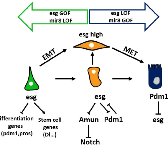
Clones lacking Esg or the zeb TF Zfh1 loose Dl-positive ISCs and they are solely composed of differentiated cells. It was shown that esg knockdown induces an upregulation of Pdm1 and Prospero, as well as other differentiation markers. Dam-ID profiling further showed that Esg represses the expression of differentiation genes by direct binding to their regulatory regions under basal conditions (Korzelius et al., 2014). In regenerating guts, when the production of new ECs is vital, Pdm1 represses the expression of esg in differentiating EBs thus ensuring a rapid turnover of lost cells (Korzelius et al., 2014; Tang et al., 2018). Within EBs, Esg is required at two different levels. First, it enables EBs to form long protrusions that establish contacts with differentiated cells. This mesenchymal-like phenotype allows EBs to sense environmental cues, pausing their differentiation until a signal is communicated from neighboring dying ECs (Antonello et al., 2015). Expression of miR-8, the fly homolog of the mammals miR200, represses the expression of esg and zfh1, the homologous gene of the EMT-TF Zeb, and induces final differentiation of EB into EC. On the other hand, esg plays a central
43 role in the EC fate decision by directly inhibiting the expression of a negative regulator of Notch called Amun (Loza-Coll et al., 2014).
Therefore, esg plays a pivotal role in both maintenance of stemness and the cell fate choice between EEs and ECs (Figure 9).
Figure 9: esg TF regulates both stemness and ISC differentiation
Within ISCs (green) and EBs (orange), Esg activates the transcription of stemness genes (Dl) and inhibits that of differentiation genes, Pdm1, Prospero (pros). High Esg levels in EBs leads to the formation of cellular protrusions that sense the external environment. The balance between Esg and miR8 triggers EC terminal differentiation. Within EBs, Esg also inhibits the expression of Amun, a negative repressor of Notch. Esg expression is inhibited by Pdm1 within ECs.
44
Figure 10 : ISC regulation by neighboring cells and tissues
ISC receive signals from both epithelial (EC : Enterocytes, EE: Enteroendocrine, EB : Enteroblast) and non epithelial sources ( VM : visceral muscles , basement membrane and enteric neurons). Some cells such as the EE regulate ISC activities indirectly via the VM.