HAL Id: tel-01124384
https://tel.archives-ouvertes.fr/tel-01124384
Submitted on 6 Mar 2015HAL is a multi-disciplinary open access archive for the deposit and dissemination of sci-entific research documents, whether they are pub-lished or not. The documents may come from teaching and research institutions in France or abroad, or from public or private research centers.
L’archive ouverte pluridisciplinaire HAL, est destinée au dépôt et à la diffusion de documents scientifiques de niveau recherche, publiés ou non, émanant des établissements d’enseignement et de recherche français ou étrangers, des laboratoires publics ou privés.
Deciphering ColQ induced mechanisms in the control of
AChR mRNA levels
Jennifer Karmouch
To cite this version:
Jennifer Karmouch. Deciphering ColQ induced mechanisms in the control of AChR mRNA levels. Genetics. Université René Descartes - Paris V, 2014. English. �NNT : 2014PA05T007�. �tel-01124384�
THÈSE DE DOCTORAT DE L’UNIVERSITÉ PARIS DESCARTES
Spécialité Génomes, épigénomes, et destin cellulaire
Ecole doctorale
Biochimie, Biothérapies, Biologie Moléculaire et Infectiologie-B3MI
Présentée par
Jennifer KARMOUCH
Pour obtenir le grade de
DOCTEUR de l’UNIVERSITE PARIS DESCARTES
Sujet de la thèse
DECIPHERING ColQ INDUCED MECHANISMS IN THE CONTROL OF
AChR mRNA LEVELS
Soutenance le 9 Avril 2014
Devant le jury composé de :
Pr. Jocelyn Côté
Rapporteur
Pr. Sophie Nicole
Rapporteur
Pr. Frédéric Charbonnier
Examinateur
Pr. Bernard Jasmin
Examinateur
Pr. Bruno Eymard
Examinateur
Pr. Claire Legay
Directeur de thèse
ACKLOWLEDGMENTS
I would like to express my deepest gratitude to my supervisor, Prof. Claire Legay, for her
guidance, encouragement and support throughout my graduate studies. It would not have been
possible to complete this work without her continuous assistance.
I would also like to extend my appreciation to Prof. Bernard Jasmin, for his effective supervision
and helpful discussions, both of which significantly contributed to the completion of this thesis.
I would like to thank the Association Française contre les Myopathies (AFM) for their financial
support.
My gratitude also extends to my jury members, comprising of Professors Jocelyn Côté, Sophie
Nicole, Bernard Jasmin, Bruno Eymard, and Frédéric Charbonnier, for their assessment of my
work, advice and constructive feedback.
I am delighted to have had the opportunity to work with exceptional colleagues and friends who
provided me with encouragement throughout my research. I would like to thank Julien Messéant,
Laure Strochlic, Perrine Delers, Francine Bourgeois, Alexandre Dobbertin, and Katia Kordeli
and all my lab mates at the ‘CeSEM Laboratory’ for their cooperation and advice. I would also
like to thank Aymeric Ravel-Chapuis, Guy Belanger, Tara Crawford, John Lunde, and the entire
team at the University of Ottawa for their continuous feedback and encouragement.
My thanks also extend to my brother, Eric Karmouch, for his help, guidance, and mentoring.
Without his assistance, I would have never been introduced to the world of science.
Words are not enough to thank my husband, Amjad, for his patience, love, and emotional
support which made it all possible.
Last but not least, my deepest thanks go to my parents for their on-going love, support, and
encouragement. Their positive influence provided me with the determination to persevere with
my studies. They have always been a source of inspiration throughout my life and their selfless
sacrifices shall never go unacknowledged.
TABLE OF CONTENTS
ACKLOWLEDGMENTS………i TABLE OF CONTENTS……….……iii LIST OF FIGURES……….vii LIST OF ABBREVIATIONS………..ixINTRODUCTION………... 1
I. THE CELLULAR COMPONENTS OF THE NMJ AND THEIR ORIGIN…... 4
A. The Birth of the Motor Neuron and its Specification………. 4
B. Schwann Cell Development………...
8
C. Skeletal Muscle Development………... 11
1. Somitogenesis and early myogenesis……….. 11
2. Myofiber formation………. 15
II. THE FORMATION OF THE NMJ………... 17
A. Making Synaptic Contact……….... 17
B. Early Events Following Synaptic Contact………... 18
1. Presynaptic differentiation……….. 18
2. Post-synaptic differentiation………... 19
a. AChR structure………... 23
b. Biosynthetic and secretory pathways of AChR………. 25
c. MuSK regulates AChR levels at the NMJ………. 28
i. MuSK structure and activity………. 28
ii. AChR clustering via MuSK and rapsyn………... 29
d. The control of AChR gene expression………
31
e. Pathologies linked to AChR deficiency………... 34
III. THE MATURATION AND MAINTENANCE OF THE NMJ……….. 36
A. Presynaptic Maturation………... 36
1. Synapse elimination……….... 36
2. Interplay between the motor neuron and the Schwann cells……….. 38
3. Pathway regulating presynaptic maturation………... 38
B. Post-Synaptic Maturation... 39
1. Transformation of AChR clusters during NMJ maturation……….... 39
2. The formation of junctional folds………... 40
3. The organization of the synaptic cleft………... 41
a. Laminins………. 41
b. Collagens……….
42
c. Perlecan……….. 42
C. NMJ Function: From an Action Potential to a Muscle Contraction………... 46
A. ColQ Structure and Function………. 51
1. The origin of ColQ……….. 51
2. The structure of ColQ………. 52
3. Consequences of ColQ deficiency for muscle activity………... 53
a. ColQ and congenital myasthenic syndromes………... 53
b. A mouse model of congenital myasthenic syndrome with AChE
deficiency……… 55
4. The roles of ColQ………... 55
a. ColQ and postsynaptic differentiation………. 55
b. ColQ regulates AChR expression……… 56
V.
HUR, A UBIQUITOUS POST-TRANSCRIPTIONAL REGULATOR……… 58
A. Post-transcriptional regulation in muscle…………...………... 58
1. RNA binding proteins………. 58
a. HuR………... 58
b. KSRP………... 59
c. Staufen………... 60
d. CUGBP1………... 61
e. Lin-28……… 62
f. TTP……… 62
B. Hu/Elav Family………... 62
C. HuR Function………... 63
1. mRNA degradation pathways……….………... 63
2. HuR Binding to ARE cis-elements……… 64
D. HuR Expression……….. 65
1. Transcriptional regulation of HuR………. 65
2. The post-transcriptional regulation of HuR………... 65
a. Autoregulation………... 65
b. MicroRNA……… 65
3. Post-translational modifications of HuR regulate its localization... 66
a. HuR shuttling……….………... 66
c. Post-translational modifications regulate HuR localization and
function………..
66
E. HuR at the NMJ……….……….. 70
1. HuR stabilises key players of the NMJ………... 70
2. HuR is a therapeutic target of NMJ diseases……..……… 70
VI. OBJECTIVES OF THE THESIS – DECIPHERING ColQ INDUCED
MECHANISMS IN THE CONTROL OF AChR mRNA LEVELS…………..….
72
A. Identifying ColQ’s Phenotypic and Molecular Effects in a Model
of CMS with AChE Deficiency………...
72
B. Deciphering the Mechanism by which ColQ Regulates AChR Subunit mRNA
at NMJ………
73
C. Providing an Updated and Integrative View of ColQ’s Functions….………
74
I. DECIPHERING THE MECHANISM BY WHICH ColQ REGULATES
AChR SUBUNIT mRNA AT NMJ………...
76
A. Introduction...………. 76
B. Primary Results………... 78
C. Conclusions and Perspectives………...…….. 80
D. Article 1………... 109
II. PROVIDING AN UPDATED AND INTEGRATIVE VIEW OF ColQ’S
FUNCTIONS………... 138
A. Introduction………. 138
B. Article 2………... 143
GENERAL DISCUSSION……….. 144
I. FURTHER DECIPHERING THE CASCADE LINKING ColQ TO AChR
mRNA LEVELS………
147
A. How is ColQ regulating p38 phosphorylation?... 147
B. Activation of p38, a stress response?... 147
C. What is upstream of p38 activation?... 148
1. The MuSK signaling platform... 148
2. The disruption of the ECM …………...………. 149
i. MuSK Acts as an “ECM sensor”……….. 149
ii. Partial denervation... 150
II. THE MOLECULAR SIGNATURE OF CMS WITH AChE DEFICIENCY:
SPECIFIC AND COMMON TRAITS WITH OTHER CMS, SIGNIFICANCE
FOR THERAPEUTIC APPROACH……….
152
A. ECM gene mutations………... 152
B. Therapeutic strategies………. 152
REFERENCES………
154
ANNEX A: IDENTIFYING ColQ’S PHENOTYPIC AND MOLECULAR
EFFECTS IN A MODEL OF CMS WITH AChE DEFICIENCY….
169
A. Introduction………. 170
B. Primary Results………... 171
C. Conclusions and Perspectives………. 174
D. Article 3………... 175
LIST OF FIGURES
Introduction
Figure 1. The neuromuscular junction. (p3)
Figure 2. Specification of motor neurons in the vertebrate spinal cord. (p6)
Figure 3. Neural crest cell destiny. (p10)
Figure 4. Myogenesis. (p14)
Figure 5.
Hierarchy of transcription factors regulating progression through the myogenic
lineage. (p16)
Figure 6. The organization of the maturing post-synaptic membrane. (p21)
Figure 7. The structure of AChR. (p24)
Figure 8. Dynamics of nicotinic acetylcholine receptors at the neuromuscular junction. (p26)
Figure 9. The working hypothesis ACh activates Cdk5 in a manner dependent on calpain. (p30)
Figure 10. Model for stabilization of synaptic gene expression through stabilization of MuSK
expression by agrin from motor nerve terminal. (p33)
Figure 11. Synapse elimination. (p37)
Figure 12. The postnatal maturation of the mouse NMJ. (p44)
Figure 13. Schematic of the SNARE/SM protein cycle mediating fusion and the role of
synaptotagmin and complexin in Ca
2+triggering of fusion. (p48)
Figure 14. Schematic representation of ColQ and its gene mutations leading to CMS with AChE
deficiency. (p57)
Figure 15. Regulation of HuR expression localization and function. (p68)
Results
Figure 16. The transcriptional activity of the AChR δ subunit in WT and ColQ-/- myotubes
at T2. (p83)
Figure 18.
HuR is capable of binding endogenous AChR subunit mRNA in C2C12 myotubes. (p90)Figure 19. HuR interacts with the conserved ARE motif in the AChR γ 3’UTR. (p93)
Figure 20. HuR interacts with the AChR α 3’UTR. (p94)
Figure 21. The AChR 3’-UTR and its expression of a reporter construct in human
embryonic kidney cells. (p97)
Figure 22. HuR localization in WT (MLCL +/+) and ColQ-deficient muscle
cells (MLCL -/-). (p100)
Figure 23. The inhibition of JNK does not alter HuR in the absence of ColQ. (p102)
Figure 24. The inhibition of ERK1/2 prevents the upregulation of HuR in the absence of ColQ.
(p104)
Figure 25. ColQ regulates AChR mRNA levels at adult NMJs. (p106)
Figure 26. The absence of ColQ does not alter HuR protein levels in vivo. (p108)
Figure 27. Synaptic phenotype of ColQ mutant diaphragm at E14. (p141)
LIST OF ABBREVIATIONS
A
ACh :
Acetylcholine
AChR:
Acetylcholine receptor
ARE:
AU rich element
AUF1:
AU-rich binding factor
B
BD:
Boundary Cap
BChE:
Butyrylcholinesterase
bHLH:
Basic Helix Loop Helix
BMP:
Bone Morphogenetic Protein
C
CARM1:
Coactivator-associated Arginine Methyltransferase 1
CAST:
CAZ-associated Structural Protein
Cdc:
Cell division control protein
Cdk:
Cyclin-dependent kinase
ChK:
Checkpoint kinase
CMAP:
Compound Muscle Action Potential
CMS:
Congenital Myasthenic Syndrome
CNS:
Central Nervous System
ColQ:
Collagen Q
CRM1:
Chromosome Region Maintenance 1
CT:
Column of Terni
CUGBP1:
CUG-Binding Protein 1
D
DM:
Dermomyotome
DM1:
Myotonic Dystrophy Type 1
DMD:
Duchenne Muscular Dystrophy
DML:
Dorsomedial
DMPK:
Dystrophia Myotonica Protien Kinase
Dok7:
Docking protein 7
DUSP:
Dual Specificity phosphatase
E
Elav:
Embryonic lethal abnormal vision
ErbB (EGFR): Epidermal Growth Factor Receptor
ECM:
Extracelullar Matrix
ERK:
Extracellular signal-Regulated Kinase
ETS:
E26 Transformation Specific
F
FGF:
Fibroblast Growth Factor
G
GABP:
GA Binding Protein
GFP:
Green Fluorescence Protein
H
HBS:
Heparin-Binding Site
HNS:
Nucleocytoplasmic Shuttling Domain
Hox:
Homeobox
HuR:
Human antigen R
I
IgG:
Immunoglobulin
IL4:
Interleukin-4
IMZ:
Involuting Marginal Zone
J
JNK:
c-jun NH
2-terminal kinase
K
KSRP:
KH-domain-Splicing Regulatory Protein
L
Lfng:
Lunaticfringe
Lmc:
Lateral Motor Column
LRP4:
Liprotien Related Protein 4
M
MAPK:
Mitogen-Activated Protein
MAP2K:
MAPK Kinase Kinase
MKK:
MAPK-Activated Protein Kinase
MKP:
MAPK Phosphatase
MY:
Myotome
Myf:
Myogenic Factor
MyoD:
Myogenic Differentiation antigen
MRF:
Myogenic Regulatory Factors
MuSK:
Muscle Specific Kinase
N
nAChR:
nicotinic Acetylcholine Receptor
NC:
notochord
NF-κB:
Nuclear Factor kappa-light-chain-enhancer of activated B cells
NMJ:
Neuromuscular Junction
NPC:
Nuclear Pore Complex
NRG:
Neuregulin
NT:
Neural Tube
P
Pax:
Paired box
PH:
Pleckstrin-Homology
PI3K:
Phosphatidyl Inositol-3 kinase
PKC:
Protein Kinase C
PNS:
Peripheral Nervous System
PRAD:
Proline-Rich Attachment Domain
PRiMA:
Proline-Rich Membrane Anchor
PSM
Presomitic mesoderm
PTB:
Phosphotyrosine Binding Domain
Q
R
RA:
Retinoic Acid
RBP:
RNA Binding Protein
RNA:
Ribonucleic Acid
RRM:
RNA Recognition Motif
S
SC:
Sclerotome
SE:
Surface Ectoderm
Shh:
Sonic hedgehog
SIRPS:
Signal Regulatory Protiens
Six:
Sine oculis homeobox
SMA:
Spinal Muscle Atrophy
SMN:
Survival Motor Neuron
SNAP:
Synaptosomal-Associated Protein
SNARE:
SNAP Receptor
Src:
Sarcoma, proto-oncogenic tyrosine kinase
SRY:
Sex determining region Y
Sauf:
Staufen
T
TGFβ:
Transforming Growth Factor β
TIA-1:
Intracellular Antigen-1
TLDA:
Taqman Low Density Array
Trm:
Trimerization domain
TRP:
Tetratricopeptide Repeats
TTP:
TTR-RBP Tristetraprolin
U
UTR:
Untranslated Region
V
VDCC:
Voltage-Dependent Calcium Channel
VLL:
Ventrolateral
Six hundred different skeletal muscle types are responsible for the posture, voluntary movement, and locomotion of the human body. The hundreds of fibers within the skeletal muscle are innervated by axons of the motor neuron forming active synapses called the neuromuscular junction (NMJ) at their site of contact. Each motor neuron can innervate thousands of muscle fibers within the same muscle, but only one terminal axonal branch from the motor neuron will innervate a single muscle fiber.
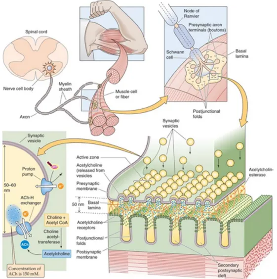
The NMJ is a synapse that activates muscle contraction, a function that relies on cholinergic neurotransmission in mammals, and its integrity is indispensable for survival. Its formation requires the functional interaction of three cell types: a motor neuron, a Schwann cell and a skeletal muscle fiber. Because of its rather simple organization and accessibility, the NMJ is the best-characterized model synapse in terms of physiology and development. The stimulation of the motor neuron evokes the release of Acetylcholine (ACh) into the synaptic cleft where it binds and activates the acetylcholine receptors (AChR) clustered on the post synaptic membrane (Sanes and Lichtman, 2001). The binding of ACh to AChR results in the depolarization of the muscle membrane inducing the opening of Na-voltage dependant channels. Consequently to the activation, an action potential is generated and quickly spreads throughout the muscle membrane, triggering a muscle contraction (figure 1).
The NMJ has contributed greatly to the understanding of the general principles of synaptogenesis and of the pathophysiological mechanisms underlying a number of neuromuscular disorders.
Figure 1. The neuromuscular junction. The motor neuron extends its axon from the motor from the
spinal cord where it is guided by its growth cone to the central region of each skeletal muscle fiber within a bundle. The interplay between the Motor axon Schwann cells, and skeletal muscle fibers regulate muscle contractions vital for the posture, voluntary movement and locomotion of the human body (Boron and Boulpaep, 2012).
I. THE CELLULAR COMPONENTS OF THE NMJ AND THEIR
ORIGIN
A. The Birth of the Motor Neuron and its Specification
Motor neurons are specific neurons located in the central nervous system (CNS) that control muscle movement. They relay signals from their soma in the spinal cord to the skeletal muscle via their axons, which project outside the CNS. Motor neurons can be classified into three groups: 1) Somatic motor neurons that innervate skeletal muscles, 2) Special visceral motor neurons that innervate bronchial muscles and 3) General visceral motor neurons that indirectly innervate cardiac muscle (Sanes et al., 2011).
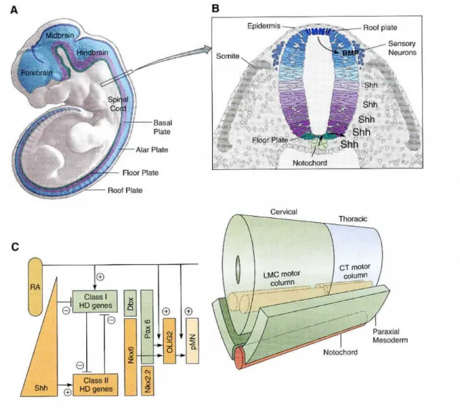
Motor neurons are derived from the neuroectoderm of the developing embryo. As a newly fertilized egg develops it endures multiple cleavage and division events to form the blastula which through the process of gastrulation forms the three layers of the embryo: ectoderm, mesoderm, and endoderm. The ectodermal layer gives rise to the neuroectoderm in a process which begins upon the differentiation of a group of cells at the surface of the gastrula. These cells form the neural plate. Involuting cells underneath the neural plate come together to form the notochord. At the same time, the neural plate folds and dissociate from the flanking ectoderm to form the neural tube. The epidermis and notochord send signals to the neural tube which polarise the neural tube by defining the roof plate and floor plate respectivey. The rostral region of the neural tube will give rise to the forebrain, mid brain, and hind brain while the remaining caudal part gives rise to the spinal cord (Figures 2A and B).
Motor neurons are derived from the ventral axis of the neural tube. The notochord and floor plate release sonic hedgehog (Shh), a secreted morphogen that establishes a signalling gradient into the ventral neural tube. The roof plate releases Bone Morphogenic Protein (BMP) to counteract the Shh gradient (Figures 2A and B). Together, the BMP morphogens, Shh and retinoic acid (RA; expressed by the paraxial mesoderm) form opposing gradients
that initiate a complex pattern of transcription factors, defining the fate of cellular regions. As a result, the ventral progenitor cells destined for a motor neuron fate will express Class I homedomain genes, specifically Pax6, Nkx6.1 and Nkx6.2, SSh and RA (Figure 2C).
The activation of the motor neuron differentiation pathway is in part induced by Nkx6.1 through its activation of the basic Helix-Loop-Helix (bHLH) transcription factors OLIG2. OLIG2 targets motor neuron specific transcription factors that initiate spinal progenitor cells down the motor neuron pathway (Figure 2C). Once the motor neurons are formed, Shh and RA gradients will define the general characteristics of motor neurons, such as projections of the axons and the release of acetylcholine (ACh) as the primary neurotransmitter. In the mouse, these generic characteristics are commenced at embryonic day 9.5 (Jessell, 2000; Sanes et al., 2011).
Figure 2. Specification of motor neurons in the vertebrate spinal cord
A. The neural tube, shown here for a mouse, is subdivided into four longitudinal domains: the floorplate, basal plate, alar plate, and roof plate. Motor neurons are derived from the basal plate.
B. Schemtatic cross section of the neural tube. The notochord, which is located underneath the floorplate, releases sonic hedgehog (Shh) which induces the floorplate to release its own Shh. This forms a gradient in the neural tube with high concentrations ventrally and low concentrations dorsally. BMP molecules released from the dorsal epidermis and roof plate form an opposing gradient.
C. (left) A gradient of Shh emanates from the notochord and floorplate; threshold levels of Shh turn on Class II HD genes. Retinoic acid (RA) expressed by the paraxial mesoderm induces the expression of Class I HD genes that are turned off more ventrally by threshold levels of Shh. Class I and Class II HD transcription factors cross-repress each other, creating sharp definitive boundaries at different dorso-ventral levels of the cord. Thus the boundary between Dbx and Nkx6 is more dorsal that the boundary between Pax6 and NKx2.2. Between these boundaries, the OLIG2 bHLH transcription factor necessary for motor neuron specification is turned on by the concerted action of RA, Nkx6, and Pax6. OLIG2 and RA are necessary for the expression of motor neuron differentiation genes of the pMN family. (Below) A gradient of FGF8 emanates from the mesoderm. High levels of FGF8 turn on more caudal HoxC genes, whereas RA and low levels of FGF8 turn on rostral HoxC genes. Rostral and cadal HoxC transcription factors cross-repress each other, creating sharp definitive boundaries at different rostrocaudal levels in the cord. The boundary between Hoxc6 and Hoxc9 establishes the boundary between the LMC of the cervical cord and the CT of the thoracic cord (Sanes et al., 2011).
Further maturation of the motor neurons is focused on their diversification into functional groups in the ventral spinal cord. The motor neurons in the spinal cord are organized into two functional columns that project to different muscle groups along the anterior to posterior axis. The first column is called the Lateral Motor Column (LMC) and is located in the cervical region and innervates forelimb muscles. The second is the Column of Terni (CT), located in the mid-thoracic region and innervates the sympathetic chain (Sanes et al., 2011).
The anterior to posterior differentiation of the motor neurons into their specific columns is accomplished by Fibroblast growth factor (FGF8) gradients secreted by the paraxial mesoderm (High FGF8 posterior expression, low FGF8 anterior). This gradient establishes domains of different Hox gene expression, with the posterior domain inhibiting the expression of anterior Hox genes and vice versa. The co-repression of Hox genes creates boundaries for the development of different motor columns of the ventral spinal cord. Motor columns are further subdivided into classes that innervate groups of muscle based on different combinations of the LIM homeodomain (LIM-HD) family of transcription factors. Lastly, these classes are then divided into motor pools that innervate specific muscles. Motor pools are defined by the E26 transformation-specific or E-twenty-six (ETS) family of transcription factors (Sanes et al., 2011).
B. Schwann Cell Development
Schwann cells of the peripheral nervous system (PNS) are important for the survival and maintenance of the motor axon. Throughout their development, Schwann cells aid in such processes as conduction of impulses along axons, nerve development and regeneration, trophic support, participation to the production of the nerve extracellular matrix, and the modulation of neuromuscular synaptic activity.
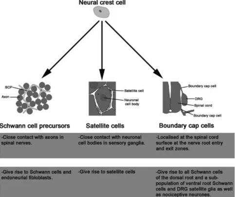
Schwann cells are derived from the neural crest cells, a transient stem cell population located at the most dorsal axis of the neural tube in the “trunk” region where the neuroectodermal folds fuse to form the neural tube. Once at the trunk, crest cells destined for Schwann cell development migrate along the neural tube ventrally (Figure 3).
Schwann cell development passes through three stages: (1) The formation of neural crest cells present in the immature connective tissue, (2) The generation of Schwann precursor cells associated with axons and (3) Immature Schwann cells which persist till birth. Initially, immature Schwann cells interact with a bundle of axons but quickly initiate radial sorting isolating large diameter axons. Depending on the axon they are associated with, immature Schwann cells will form either non myelinating or myelinating Schwann cells (Jessen and Mirsky, 2005; Sanes et al., 2011; Woodhoo and Sommer, 2008).
In the mouse, these three stages of Schwann cell development occur from embryonic day 10 to the neonatal period. The factors involved in the early stages of Schwann cell maturation are still unclear, however the activation of the transcription factor SOX10 (SRY-related HMG-box 10) is required for the crest cell to glial lineage transition. Axon derived neuregulin 1 (NRG1) and Notch have both been found to promote Schwann cell survival and proliferation and may help accelerate the Schwann cell precursor-Schwann cell transition.
Figure 3. Neural crest cell destiny. Neural crest cells give rise to various types of peripheral glia
distinguished by their localization, specific marker expression, factor responsiveness, and function. Schwann cells along peripheral nerves, satellite cells in peripheral ganglia, and boundary cap cell-derived Schwann cells in nerve roots represent distinct peripheral glia subtypes generated from the neural crest (Woodhoo and Sommer, 2008).
C. Skeletal Muscle Development
Skeletal muscle is a dense, fibrous contractile tissue which exists throughout the entire body, and functions to allow body movement by applying force to bones and joints, via contraction. Its formation occurs between embryonic day 7.5 to 14 in the mouse (Buckingham and Rigby, 2014). An overview of skeletal muscle development in the embryo is shown in Figure 4. Figure 5 provides insight into the hierarchy of transcription factors regulating myogenesis.
1. Somitogenesis and early myogenesis
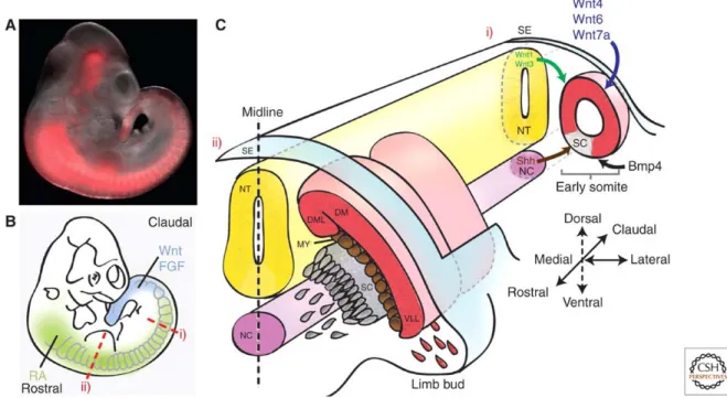
Somites are groups of cells that form sequentially from the paraxial mesoderm also known as the presomitic mesoderm (PSM), flanking the neural tube. Somite formation (Somitogenesis) has been hypothesised to follow a “clock and wavefront” model (Gilbert and Gilbert, 2010). The clock incorporates FGF, Wnt and Notch Pathways, with the FGF and Wnt pathways interacting to activate the Notch pathway. Notch, the transcription factor Hes7, a general repressor of the BHLH family, and Lunaticfringe (Lfng) act as the clock for somitogenesis, through the oscillation in their gene expression (Gilbert and Gilbert, 2010; Saga, 2012). The mechanism involved in the wavefront is still unclear, but it has been proposed to include FGF8, Wnt, and retinoic acid (Gilbert and Gilbert, 2010; Naiche et al., 2011). Both Wnt and FGF maintain the PSM population in an undifferentiated state, by upregulating each other’s expression. Furthermore, FGF has been shown to independently inhibit somitogenesis and thus as the embryo elongates, the PSM are displaced further away from the highly concentrated posterior source of FGF (Naiche et al., 2011). The point of segmentation is defined when FGF signalling is reduced below a threshold and the oscillating clock is in the on state. The boundrary between somites results from the intersection of the caudal-rostral gradiant, with the rostral-caudal gradient formed by FGF8, Wnt and retinoic acid respectively. Once somites have been cleaved, they immediately undergo epithelialization
(Gilbert and Gilbert, 2010). The newly formed somite is a mesenchymal mass which must be defined into an epithelium with an internal mesenchyme.
Somites are surrounded by the neural tube, notochord, epidermis, and the lateral mesoderm. These surroundings tissues secrete paracrine factors, which depending on their distance, will commit regions of the somite to specific cell types. The ventral medial epithelial cells of the somite closest to the neural tube, which secretes Noggin (Nog) and Shh, form what is called the sclerotome, which contains cells destined for the bones and cartilage. Nog and Shh induce the epithelial cells to be reverted back to mesenchymal cells through the activation of the transcription factor Pax1. Pax1 is also required for the differentiation of cells of the scleretome into cartilage. Noggin and Shh also induce the expression of Imf (inhibitor of MyoD family), an inhbitior of a group of myogenic transcription factors (Chen et al., 1996; Gilbert and Gilbert, 2010).
The remaining dorsal epithial cells form the dermomytome, which having a high expression of Pax3 and Pax7 will go on to form the myotome and the central dermomyotome (Bentzinger et al., 2012). The Myotome is formed from the two lateral “lips” of the dermomyotome. These two lips are the closest and furthest away from the neural tube. Cells of the myotome then migrate underneath the dermomyotome and form a layer of two types of myoblast. The myoblasts furthest away from the neural tube form hypaxial muscles (muscles of limbs and tongue) and the myoblasts closest to the neural tube differentiate into the epaxial muscles (intercostal muscle and vertebrate muscle) (Bentzinger et al., 2012; Gilbert and Gilbert, 2010).
In order for myoblasts to be formed in the somite, Wnt signalling must be present and BMP must be absent (Reshef et al., 1998). Interestingly, Shh and Nog have been shown to inhibit BMP4, ensuring the myoblast state (Marcelle et al., 1997). Thus, the formation of epaxial myoblasts are dependent on the neural tube secretion of high levels of Wnt1 and Wnt3a and
low levels of Shh, while the hypaxial mesoderm is dependent on the secretion of Wnts from the epidermis (Gilbert and Gilbert, 2010).
The myogenic program is induced by the Shh dependent high expression of two bHLH family members, MyoD and Myf5 (Bentzinger et al., 2012). These proteins bind the E-box in the promoter region to control the expression of their target genes. MyoD and Myf5 are part of a group of transcription factors called the Myogenic Regulatory factors (MRF’s). MRFs activate the transcriptional cascade needed for the differentiation of skeletal muscle (Bentzinger et al., 2012; Gilbert and Gilbert, 2010). Mrf4 is the third member of the MRF family and has been recently shown to act similar to MyoD and Myf5 as a determining factor for a myogenic cell fate (Kassar-Duchossoy et al., 2004).
The central region of the dermomyotome undergoes an epithelial mesenchymal transition, a process induced by neurotrophin-3 and Wnt1, both of which are secreted from the neural tube. As these newly formed mesenchymal cells undergo mitosis, their daughter cells will either enter into the myotome or relocate dorsally to become part of the dermis (Bentzinger et al., 2012; Gilbert and Gilbert, 2010). Those cells that enter into the myotome will differeniate into myoblast or remain in their undifferentiated state as satellite cells to be used in post-natal muscle growth (Gilbert and Gilbert, 2010).
Figure 4. Myogenesis. (A) Embryonic day 10.5 (E10.5) mouse embryo carrying an Myf5 lineage
tracer that induces irreversible expression of a red fluorescent protein. Expression can be observed in the presomitic mesoderm, the somites, and in several head structures. (B) Illustration of the morphogen gradients along the rostral– caudal axis of the embryo. (C) Schematic of transverse sections through the embryo at early (i) and late (ii) stages of somitogenesis. (Ci)Morphogens secreted from various domains in the embryo specify the early somite to form the sclerotome (SC) and dermomyotome (DM). Wnts secreted from the dorsal neural tube (NT) and surface ectoderm (SE) along with bone morphogenetic protein (BMP) from the lateral plate mesoderm maintain the undifferentiated state of the somite, whereas Sonic hedgehog (Shh) signals from the neural tube floor plate and notochord (NC) to induce the formation of the sclerotome. (Cii) As the sclerotome segregates, muscle progenitor cells (MPCs) from the dorsomedial (DML) and ventrolateral (VLL) lips of the dermomyotome mature to give rise to the myotome (MY). At the level of the limb bud, Pax3-dependent migrating MPCs delaminate from the ventrolateral lips to later give rise to limb muscles (Bentzinger et al., 2012).
2. Myofiber formation
Myoblast fusion into myofiber occurs in four main steps (Gilbert and Gilbert, 2010): a) Cell Cycle Exiting – Proliferating myoblasts are maintained by FGFs and thus its
removal induces the myoblasts to secrete fibronectin into its extracellular matrix. Once in the ECM, fibronectin binds the α5β1 integrin, halts cell proliferation and instructs the myoblasts to differentiate.
b) Alignment of Myoblasts - Cell membrane glycoproteins, such as Cadherins and cell adhesion molecules (CAM), align myoblasts in a chain-like formation. Cytoplasms are also re-arranged by actin to allow for accurate cell contact.
c) Cell Fusion - Myoblast fusion into myofibers is dependent on Calcium ions and is mediated by the metalo proteinases called metrins. Once myoblasts are induced to fuse, they activate the βHLH transcription factor myogenin, which goes on to activate the expression of muscle specific genes. Myogenin is the last member of the MRF family of transcription factors and is expressed in the myoblast to aid in myoblast fusion and myofiber formation (Bentzinger et al., 2012).
d) The “Healing Step” – Opposing membranes of the fused myoblasts are re-sealed with the help of myoferlin and dysferlin, which have been known to stabilise phospholipids. Newly fused myofibers secrete interleukin-4 (IL4) which recruits other myoblasts to fuse with the tubes. After birth, the expression of the TGF-β protein myostatin inhibits the growth of myofibers and their number.
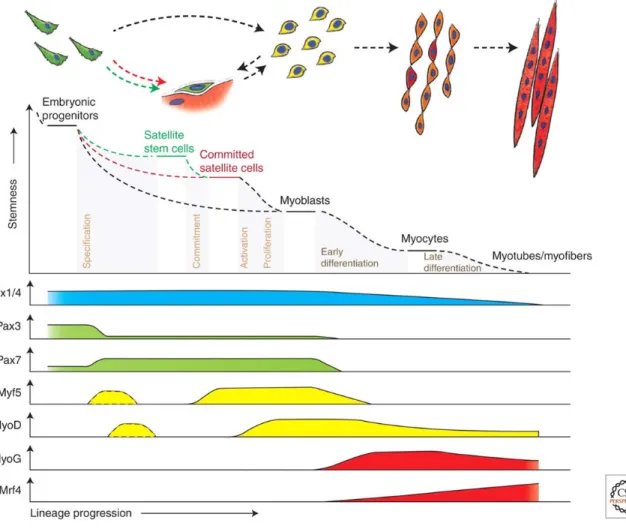
Figure 5. Hierarchy of transcription factors regulating progression through the myogenic lineage.
Muscle progenitors that are involved in embryonic muscle differentiation skip the quiescent satellite cell stage and directly become myoblasts. Some progenitors remain as satellite cells in postnatal muscle and form a heterogeneous population of stem and committed cells. Activated committed satellite cells (Myoblasts) can eventually return to the quiescent state. Six1/4 and Pax3/7 are master regulators of early lineage specification, whereas Myf5 and MyoD commit cells to the myogenic program. Expression of the terminal differentiation genes, required for the fusion of myocytes and the formation of myofibers, are performed by both myogenin (MyoG) and MRF4 (Bentzinger et al., 2012).
II THE FORMATION OF THE NMJ
A. Making Synaptic Contact
The formation of the neuromuscular junction (NMJ) requires the precise opposition of a pre and post synaptic domain through a complex interchange of information between the pre- and postsynaptic partners, including activity dependent and independent processes. In vertebrates, synaptogenesis occurs shortly after myoblast fusion, initiating on embryonic days E13–E14 (Witzemann, 2006). Synaptogenesis follows the extension of the axon from the motor neuron, which is guided by its growth cone, to the central region of the muscle fiber, making initial contact on day E13/14 (Sanes and Lichtman, 2001; Witzemann, 2006).
The extention of the axon of the motor neuron from the neural tube occurs from its ventral exit point and depends on a combination of repulsive and attractive signals from the floor plate and midline. Signals from the surrounding mesenchyme then induce these axons to exit the neuroepithelium. Once at the ventral exit point motor axons are surrounded by a subtype of neural crest cells called boundary cap (BD) cells that ensure axons exiting but prevents the motor neuron cell body from leaving the CNS. Once exited, motor axons grow towards the muscle. The axons then take a ventral, dorsal or axial path depending on the muscle which they will innverate. Once at their respective muscle, individual growth cones are guided by environmental cues, ensuring their positioning at the correct muscle region (Bonanomi and Pfaff, 2010).
It has been postulated that the central domain in the mammalian muscle fiber contains aggregate of macromolecular complexes, including the Acetylcholine Receptors (AChR), that aid in guiding the innervating motor neuron and confirming its synaptic location (Sanes and Lichtman, 1999). This muscle “pre-patterning” is dependent on the post-synaptic protein muscle specific tyrosine kinase (MuSK) and its co-receptor, the low density lipoprotein related protein 4 (LRP4), which forms a complex with MuSK, stimulating MuSK kinase
activity (Lin et al., 2011; Yang et al., 2001). MuSK is expressed primarily in the central region of the muscle prior to innervation and is believed to dictate the accumulation (clustering) of AChR in the middle of the muscle, followed by the terminal phase of axon guidance to the muscle (Kim and Burden, 2008) (Figure 6A).
Once the motor neuron has made contact with the muscle, it releases the neuronal form of the
proteoglycan agrin, which binds LRP4, stimulating further association between LRP4 and MuSK, and increases MuSK kinase activity (Kim et al., 2008b; Zhang et al., 2008). This increase in MuSK activity leads to an increase in AChR clustering and density, resulting from a transcriptional and non-transcriptional mechanism. (Gautam et al., 1996; Kishi et al., 2005). It has been hypothesized that LRP4 is capable of acting bi-directionally, stimulating presynaptic differentiation (Yumoto et al., 2012) independently of agrin, through its binding to the motor neuron axons and inducing the clustering of synaptic-vesicles and active zone proteins.
B. Early Events Following Synaptic Contact
1. Presynaptic differentiation
When the motor neuron first makes contact with the muscle fiber, it resembles a bulb-like enlargement. However, as the presynaptic terminal differentiates, the fillopodia of the growth cone retract, the synaptic vesicle carrying the neurotransmitter Acetylcholine (ACh) increase and accumulate at the nerve terminal active zone and are subsequently released as the nerve terminal becomes polarized (Sanes and Lichtman, 1999).
Scientists using electron microcopy have identified the active zone as an electron dense thickening of the presynaptic motor terminal, where synaptic vesicles accumulate and dock. The active zone plays a central role in synaptic transmission and is formed directly opposing the postsynaptic muscle fiber (Couteaux and Pecot-Dechavassine, 1970; Harlow et
al., 2001; Hirokawa and Heuser, 1982; Nagwaney et al., 2009). The active zone is where synaptic vesicles carrying neurotransmitter ACh are released (Propst and Ko, 1987).
The extracellular matrix (ECM) plays a large role in active zone formation and presynaptic differentiation with collagen α1/2 (IV) chains, directing the initial differentiation of the presynaptic terminal (Fox et al., 2007). Laminin β2, an ECM protein secreted by the muscle, has been found to organize active zone formation through its binding to the presynaptic voltage-dependent calcium channels (VDCCs) on one end and Dystrophin on the other. Laminin β2 anchors VDCCs and accurately positions them in the active zone allowing for the recruitment of active zone specific proteins including Bassoon and CAST/ERc2/ELKS2α to VDCCs’ intracellular domain (Nishimune, 2012). VDCCs function as scaffolding proteins that link the extracellular proteins to cytosolic active zone proteins (Nishimune, 2012). Other factors involved in presynaptic differentiation include the TGF β family of proteins. Mutations in genes that encode TGF β family proteins have been found to disrupt T-bars and presynaptic high density areas where synaptic vesicles accumulate (Wu et al., 2010). Furthermore, Fibroblast Growth Factors (FGFs) and Signal Regulatory Proteins (SIRPS) have been demonstrated to induce synaptic vesicle clustering at the nerve terminal (Fox et al., 2007).
2. Post-synaptic differentiation
A defining feature of post-synaptic differentiation is the re-distribution of muscle AChRs following innervation, with the formation of high-density AChR clusters at the postsynaptic membrane and their dispersion outside the NMJ (Sanes and Lichtman, 2001). Early in vertebrate myogenesis, around embryonic day E13, AChR subunit gene expression and assembly takes place throughout the myofiber. AChR is inserted into the membrane of the myofiber in clusters that are formed and regulated by the transmembrane tyrosine kinase
MuSK (Figure 6B). The importance of the accurate control of AChR expression, insertion
and clustering at the post-synaptic membrane is highlighted by the fact that alterations in their membrane density and kinetics are associated with congenital myasthenic syndromes.
A.
B.
Figure 6. The organization of the maturing post-synaptic membrane
A. The mammalian muscle fiber contains an increase in the expression of groups of macromolecular transmembrane complexes including AChR (blue) along with their clustering in the central region of the muscle fiber (red); a process independent of innervation. The formation of this central domain requires LRP4 and MuSK. The motor axons are thought to approach this region of the muscle, marked by nerve terminals juxtaposed to AChRs. (Adapted from Burden snapshot)
B. Postsynaptic maturation is regulated by positive and negative signaling pathways for AChR stabilization. Positive signaling pathways – including those activated by the MuSK receptor, the dystroglycan–glyoprotein complex, and ErbB and TrkB receptors – act on the assembly and maintenance of AChR clusters. CaMKII, a serine/threonine kinase activated by electrical activity, promotes insertion of AChRs into synaptic sites. Synapse-specific gene transcription, such as that mediated by the Ets transcription factor Erm, is important for AChR maintenance. By contrast, ephexin1-mediated upregulation of RhoA and activation of PKC and the actin modulator ADF/cofilin regulate AChR removal during plaque-to-pretzel transition. Cdk5-dependent dispersal of AChR clusters, enclosed in a dotted square, is a signaling pathway that has only been demonstrated in embryonic stages. In addition, dashed arrows indicate regulatory pathways established in vitro or in systems other than the NMJ. Abbreviations: Abl, v-abl Abelson murine leukemia viral oncogene homolog; ACh, acetylcholine; AChE, acetylcholinesterase; AChR, acetylcholine receptor; ADF, actin depolymerizing factor; APC, adenomatous polyposis coli; CaMKII, Ca2+/calmodulin-dependent kinase II; Cdk5, cyclin-dependent kinase 5; CK2, casein kinase 2; ColQ, collagen Q; DB, dystrobrevin; DG, dystroglycan; Dvl, dishevelled; Eph, erythropoietin-producing human hepatocellular carcinoma; ErbB, erythroblastic leukemia viral oncogene homolog; Erm, Ets-related molecule; GABP, growth-associated binding protein; GGT, geranylgeranyltransferase; JNK, c-Jun N-terminal kinase; LRP4, low-density lipoprotein receptor-related protein 4; MuSK, muscle-specific kinase; nNOS, neuronal nitric oxide synthase; NRG, neuregulin; NT-4, neurotrophin-4; NSF, N-ethylmaleimide sensitive factor; PDZRN, PDZ-domain-containing RING finger 3; PKC, protein kinase C; PTP, protein tyrosine phosphatase; SFKs, Src-family kinases; Tid1, tumorous imaginal disc 1; Trk, tropomyosin receptor kinase (Shi et al., 2012).
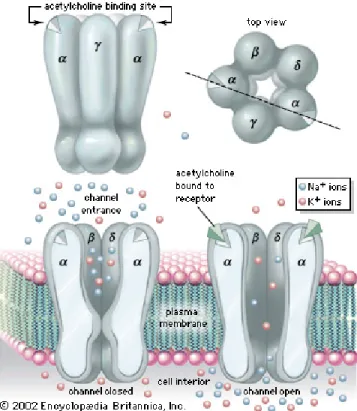
a. The AChR structure
The nicotinic AChR receptor ion channel is a pentamer composed of four subunits; 2 α’s, 1 β, 1 γ or 1 ε, and 1 δ, each encoded by separate genes. During the latter stages of maturation, (after birth), the AChR receptors replace their γ subunit with the ε subunit, with the replacement of the γ subunit by ε seen as a gradual transcriptional decrease and increase respectively (Sanes and Lichtman, 1999). The functional importance of this exchange has been proposed to be related to the kinetics of AChR and/or for the structural maturation of the synapse (Sanes and Lichtman, 1999) (Figure 7).
There are currently two models that explain how these subunits form the ion channel:
1) The first model (“heterodimer” model) postulates that the subunits first post-translationally fold into their tertiary structures. Once folded, two α subunits each associate with γ or δ to assemble α/γor α/δ heterodimers. Finally, the heterodimers come together with the β subunit to structure the α/β/γ/δ pentamer (Green, 1999).
2) In the second model (“sequential” model”), the β, and γ subunits are assumed to post-translationally fold into their tertiary structures and quickly assemble into a heterotrimer with an unfolded α subunit. The α subunit fold into its tertiary structure only after it has been incorporated into the heterotrimer. Once α has obtained its correct tertiary structure, the δ subunit joins the complex to make the α/β/γ/δ tetramers. The proper orientation of the tetramer exposes the ACh binding site on the α subunit recruiting the second α subunit to make α/β/γ/δ pentamers. The second ACh site forms on the pentamer shortly after (Green, 1999).
Figure 7. The structure of AChR. The AChR is a pentamer composed of two α subunits, a β subunit,
a δ subunit and an embryonic γ subunit which is replaced shortly after birth by the adult ε subunit. The binding of two ACh molecules to their sites present on the N-terminal domain of the α subunits AChR, the channel formed by the hydrophobic transmembrane region of the five AChR subunits opens to let in sodium and calcium ions and potassium ions out.
b. Biosynethic and secretory pathways of AChR
AChR subunit folding and pentamer formation occurs with the help of multiple chaperones in the endoplasmic reticulum (ER). Once inserted in the ER membrane, AChR pentamers are recruited to the ER exit site where they pass a quality control checkpoint. These checkpoints recognise specific retention and retrieval signals visible in misfolded subunits or improperly formed pentamers and target them for degradation. For example, each AChR subunit has a retention signal conserved in its transmembrane domain. This retention signal is not detectable if the subunits are properly assembled into a pentamer. The α subunit also contains a di-basic retrieval signal in its cytoplasmic loop which is exposed in unassembled pentamers. In accurately formed pentamers or when the subunit is bound to chaperones this sequence is no longer visible. Once properly oriented, AChR pentamers pass the ER checkpoint, then are transported by COPII vesicles to the Golgi and on to the muscle membrane (Colombo et al., 2013).
Once in the muscle membrane, AChR turnover is dependent on synaptic activity, as AChR membrane half-life is drastically decreased from 10-14 days to a few hours upon synaptic blockage. The stabilisation effect of synaptic activity on AChR has been proposed to be post-translationally regulated by the increases in calcium influx and cAMP (Pires-Oliveira et al., 2013).
AChR turnover at the membrane is primarily regulated by endocytosis via clathrin dependent and non-clathrin dependent mechanisms. However, the details of this mechanism are still unknown. Once internalized in the early endosome, AChR is either targeted to the lysosome where it is degraded via the ubiquitin-proteasome system or recycled back to the muscle membrane in an activity dependent manner (Pires-Oliveira et al., 2013). The dynamics of AChR at the NMJ discussed above is reviewed in figure 8.
Figure 8. Dynamics of nicotinic acetylcholine receptors at the neuromuscular junction.
Nicotinic acetylcholine receptors (nAChR) subunits are synthesized in the ER and exported to
the muscle plasma membrane. From the ER, instead of being targeted to the cell surface,
most nAChR subunits are degraded by the ER-associated ubiquitin-proteasome degradation
pathway. In the postsynaptic membrane, there is significant lateral diffusion between the
synaptic and perisynaptic membrane spaces. Lateral diffusion of nAChRs from the
perisynapse into the NMJ contributes significantly to maintain the synaptic receptor density.
there is significant internalization of nAChRs into endosomal compartments. Trafficking
through the endosomal pathway, a fraction of internalized nAChRs is targeted for
degradation. However, a significant portion of those nAChRs actually recycle back into the
synaptic membrane, contributing to the maintenance of the synaptic nAChR pool. Most of
these dynamics are tightly regulated in the NMJ by several stimuli, such as synaptic activity
c. MuSK regulates AChR levels at the NMJ i. MuSK structure and activity
MuSK is indispensable for the formation of the NMJ as it serves as a multifunctional hub orchestrating the successive steps of synapse formation (Glass et al., 1996; Kim and Burden, 2008; Weatherbee et al., 2006). Its extracellular domain contains three immunoglobulin-like motifs and a cystein-rich region called C6 (DeChiara et al., 1996). The C6 region is homologous to a sequence present in the Frizzled receptors and known to bind Wnt proteins. The extracellular domain of MuSK also forms a macrocomplex with LRP4, via its first immunoglobulin-like domain (Kim et al., 2008b; Zhang et al., 2008). The intracellular domain of MuSK contains a juxtamembrane domain that autophosphorylates in the presence of agrin, and a kinase domain. MuSK contains nineteen possible phosphorylations sites (tyrosine residues) and it has been shown that six of these sites must be phosphorylated in order for MuSK to be active (Watty et al., 2000)
MuSK intracellular co-receptor, Dok7 is both a substrate and activator of MuSK. Dok7 is an adaptor protein that contains an N-terminal pleckstrin-homology (PH) domain, a phosphotyrosine binding (PTB) domain and a C-terminal region containing sites of tyrosine phosphorylation (Beeson et al., 2006; Cai et al., 2003). The autophosphorylation of MuSK at Ty553 located in an NPXY sequence motif induces its binding to Dok7 through Dok7’s PTB domain (Okada et al., 2006).
MuSK is activated by agrin binding to its extracellular domain, which helps to strengthen the LRP4/MuSK interaction. Agrin signalling is passed through the membrane to the intracellular domain where the MuSK/Dok7 interaction results in the phosphorylation of MuSK. The phosphorylation of MuSK results in its endocytosis via clathrin coated vessicles with Dok7 localizing MuSK in the caveolin-specifics structures. The internalization of MuSK is required for AChR clustering (Luiskandl et al., 2013).Together the phosphorylation and endocytosis
of MuSK results in its activation and further induction of an amplitude of downstream signaling pathways, and interactions with scaffolding proteins and transcription of postsynaptic proteins (Wu et al., 2010).
ii. AChR clustering via MuSK and rapsyn
The agrin dependent activation of MuSK induces AChR clustering through a signaling cascade leading to the phosphorylation of the AChR β subunit at its Tyrosine Y390 and the recruitment of the cytoplasmic membrane associated protein, Rapsyn (Borges and Ferns, 2001; Borges et al., 2008). Rapsyn contains a myristoylation site necessary for its submembrane localization, seven tetratricopeptide repeats (TRPs), a putative coiled-coil domain, and a RING-H2 domain (Ramarao et al., 2001). Rapsyn is capable of interacting with itself through its TRP domain, with AChR by its coiled-coil domain, and to the actin cytoskeleton through its RING-H2 domain. Once at the post-synaptic membrane, rapsyn clusters phosphorylates AChR receptors through the interaction with the intracellular loop of AChR and the formation of small rapsyn bridges, with each receptor capable of binding three bridges (Ramarao et al., 2001; Zuber and Unwin, 2013).
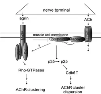
The agrin/MuSK signaling pathway stabilizes the AChR clusters by inhibiting the ACh induced dispersion of the AChR clusters. It has been shown that ACh negatively regulates AChR cluster size through the activation of the cytoplasmic serine/threonine kinase cdk5 (Fu et al., 2005; Lin et al., 2005). Furthermore, in the absence of agrin, cholinergic stimulation activates calpain, a member of the calcium-activated intracellular cysteine protease family. Calpain cleaves p35 to p25 which goes on to activate cdk5. The presence of agrin inhibits the cholinergic dependent activation of calpain by inducing its interaction with rapsyn (figure 9) (Chen et al., 2007).
Figure 9. The Working Hypothesis ACh activates Cdk5 in a manner dependent on calpain.
Activated Cdk5 disperse AChR clusters. Agrin increases the interaction of rapsyn with
calpain and thus inhibits calpain activity to stabilize AChR clusters at synapse (Chen et al.,
d. The control of AChR gene expression
Synapse formation induces an increase in the levels of AChR and other post-synaptic proteins. Post-synaptic gene expression is thought to be induced by the mitogen-activated protein kinase (MAPK), c-jun NH2-terminal kinase (JNK) and the Phosphatidyl Inositol-3 kinase (PI3K)
signaling pathways. These pathways converge to phosphorylate and activate the Ets family transcription factor, GA binding protein (GABP) in vitro. Phosphorylated GABP binds an N-box (CCGGAA) in the promoters of synaptic genes inducing their transcription (Schaeffer et al., 2001). Another member of the Ets family, Erm, has also been involved in the regulation of subsynaptic gene expression in vivo (Hippenmeyer et al., 2007) but only GABP was been shown to bind an N-box in the AChR subunit promoters, inducing their transcription in synaptic nuclei (figure 10) (Schaeffer et al., 2001; Schaeffer et al., 1998).
It has been well accepted that AChR gene expression is triggered by the previously described agrin/MuSK complex and by the neuregulin/Erb complex. At the NMJ two isoforms of Neuroregulins (NRGs) are present, nrg-1 present in both the motor neuron and muscle fiber and nrg-2 present in the terminal Schwann cells. NRGs activate the receptor tyrosine kinase ErbB family. These include ERbB2, ERbB3, and ERbB4. ERbB4 binds and activates NRG. ERbB2 cannot bind to NRG but possess tyrosine kinase activity while ERbB3 binds NRG but lacks catalytic activity. Therefore, ERbB2 and ERbB3 can only be NRG activated as heterodimers with other ERbBs (Rimer, 2007). Muscle fibers express all three ERbB receptors while terminal Schwann cells express only ERbB2 and ERbB3 (Rimer, 2007). NRG has been widely accepted to bind ErbB receptors and stimulate synaptic gene transcription. However, these conclusions were all based on in vitro studies because knock-out mice died before birth (Chu et al., 1995; Falls et al., 1993; Gramolini et al., 1999). Since 2005, two groups using conditional knock-out mice have shown that NMJs can form in the
absence of muscle specific ErbB2 and ErbB4 and even went further to demonstrate that post-synaptic differentiation is induced by agrin (Escher et al., 2005). In 2006, Jaworski and Burden inactivated nrg-1 in the motor neuron and skeletal muscle first separately then simultaneously, and found that the AChR clusters still formed at the synapse and that synapse-specific transcription was normal (Jaworski and Burden, 2006). Thus the role of NRGs in synaptic gene transcription has yet to be clarified in vivo versus in vitro.
Figure 10. Model for stabilization of synaptic gene expression through stabilization of musk expression by agrin from motor nerve terminal. Agrin secreted from nerve terminal
activates preexisting MuSK to induce expression of musk via its N-box (i) by organizing an
NRG/ErbB pathway, involving MuSK-induced recruitment of ErbB receptors and of
muscle-derived NRG and (ii) by MuSK-induced activation of JNK (via Rac/Cdc42). With musk
expression stabilized, the same pathways are used for AChR and erbB expression. Expression
may be strengthened by NRG-1 secreted from nerve terminal. Complete inhibition of
Agrin-induced musk transcription in C2C12 cells by overexpression of inactive ErbBs (HER2KM
and HER4KM) and by dominant-negative JNK suggests that the two pathways are connected.
The model is based on present data (solid arrows) and references cited (broken arrows)
e. Pathologies linked to AChR deficiency
Congenital myasthenic syndromes (CMS) correspond to a class of human pathologies resulting from mutations in genes coding for proteins involved in NMJ activity, structure, or development. Mutations in AChR subunits were the first to be studied in patients with CMS. These studies focused on subunit mutations that impaired channel gating or that decreased AChR levels at the membrane. Scientists were able to separate 2 classes of heterozygous recessive mutations: those that alter the kinetic properties of AChR and those that reduce the expression of AChR. The latter group of mutations can be further classified into “slow channel” mutations and “fast channel” mutations (Engel and Sine, 2005).
Slow channel mutations are dominant gain of function mutations that increase ACh response resulting in the slow decay of synaptic current. This gain of function mutation can occur via two mechanisms. The first being the abnormally slow closure of activated receptor channels and the second being ACh bound receptor channels having an increased probability of opening. Thus both mechanisms increase the probability of open AChR channels (Engel and Sine, 2005).
Contrary to slow channel mutations, the fast channel mutations decrease synaptic response to ACh by decreasing the probability that AChR channels will be open, resulting in an abnormally fast decay of synaptic response. The reduced activation of AChR results from mutations which cause the channel to close abnormally fast and those that result in ACh bound channels closing abnormally slow. Fast channel mutations cause the synapse to have brief current pulses followed by prolonged intervals between pulses (Engel and Sine, 2005). The incomplete atomic structure of AChR has made it difficult to understand how mutations in the AChR subunits affect its kinetics. However, the recent crystallization of ACh structure provided insight into the characteristics of its ligand binding site on AChR (Brejc et al., 2001). This was then fused together with the atomic structure of the pore from the torpedo
and a recent density electron map providing almost the whole AChR receptor (Bouzat et al., 2004; Miyazawa et al., 2003; Unwin, 2005). This has allowed for the mapping of the CMS mutations from which they have determined that mutations in the ACh ligand binding site alter the channel kinetics, while mutations in the pore domain alter the gating of AChR. CMS with AChR deficiency is not only encountered upon mutations in AChR subunits but also has been found to be induced by mutations in Rapsyn, MuSK and Dok7 all of which reduce AChR clustering at the post-synaptic membrane (Engel and Sine, 2005).
2.
THE MATURATION AND MAINTENANCE OF THE NMJ
A. Presynaptic Maturation
1. Synapse elimination
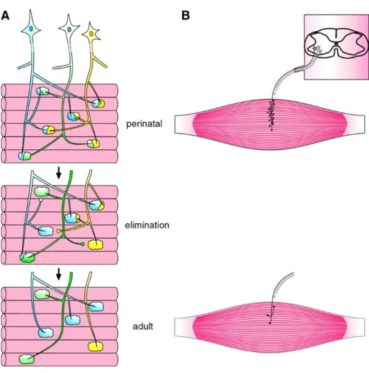
In neonatal animals, each muscle fiber is innervated by multiple motor axons, whereas in the adult only one motor axon is present at the mature NMJ (Figure 11). This developmental phenomenon is known as synapse elimination and its occurrence is comprised in rodents between the time of birth and postnatal day 15 (P15), with a few days’ variation in specific muscles. The number of motor neurons in the spinal cord remains constant during this developmental period. Synapse elimination is therefore a peripheral process that only involves motor nerve endings and their axonal branch, occurring over several days, with a percentage of fibers becoming singly innervated each post-natal day.
Synapse elimination occurs in 4 steps: axonal atrophy, axonal detachment and the formation of the retraction bulb, and axonal withdrawal from the endplate. Through synapse elimination and post-natal presynaptic maturation, each motor unit (the muscle fibers and their innervating motor neuron) undergoes a change from a large size, overlapping with other units, to a much smaller, non-overlapping one, which is retained as a permanent feature throughout adult life (Buffelli, 2004).
Figure 11. Synapse elimination. Neonatal synapse elimination represented as the loss of
2. The interplay between the motor neuron and the Schwann cells
Motor neuron maturation depends highly on that of the maturation of Schwann cell and vice versa. Schwann cells have been known to generate diffusible signals such as TGFβ, which promote the development and function of the motor neuron. Schwann cells also engulf and clean up axons that become fragmented during synapse elimination.
Equally, Schwann cell survival is dependent on signaling from the motor neuron, specifically, the release of neutrophin 3, which regulates the number of Schwann cells and myelinating glial cells present at the NMJ. Further, the motor neuron also releases NRG 1 which promotes Schwann cell survival and development (Wu et al., 2010).
3. Pathways regulating presynaptic maturation
Most of the key players in the NMJ formation are found on the post-synaptic muscle and the pathways used to organize the post-synaptic differentiation have been largely studied. It seems as though these post-synaptic molecules also act in retrograde to control presynaptic differentiation and maturation, as independently knocking out MuSK, LRP4, or Agrin results in the loss of presynaptic differentiation (DeChiara et al., 1996; Kim et al., 2008b; Zhang et al., 2008).
What we do know is that the accuracy of protein turnover appears to be important in presynaptic differentiation. Ubiquitin specific peptidase 14 (Usp14) is a proteasomal deubiquitinating enzyme, which when knocked out, results in the accumulation of phosphorylated neurofilaments and nerve terminal sprouting (Chen et al., 2009). Furthermore, ubiquitin carboxyl-terminal hydrolase L1 (UCH-L1), also a deubiquitinating enzyme, is needed for accurate synaptic vesicle accumulation and the turnover of tubulovesicular structures at the presynaptic terminal (Chen et al., 2010). Therefore, it