6091
Comparison of the performances and validation of three
methods for Yersinia spp. detection from animals stools
samples in Abidjan: a non-endemic and a tropical area
Saraka N.D1,3 , Koffi.K.E1, Koffi K.S2, Kouassi.K.S1 ,Ehuie1.P, Faye-Kette1,2,3 and Dosso M1,2,3 1 : Institut Pasteur, Côte d’Ivoire, Département Environnement et Santé2 : Université Félix Houphouët Boigny Abidjan, UFR des Sciences médicales, Département de Microbiologie 3 : Université Félix Houphouët Boigny, UFR des Sciences médicales, Département de biologie humaine tropicale Corresponding author: Email address: [email protected] /[email protected] ; Phone: +225 07240218; Postal address: 01 BP 490 Abidjan 01
Original submitted in on 21st November 2013 Published online at www.m.elewa.org on 28th February 2014. http://dx.doi.org/10.4314/jab.v74i1.13
ABSTRACT
Objective: Animals are the main reservoir for human enteropathogenics Yersinia. The aim of this survey was to compare the performances through three methods for detection of Yersinia enterocolitica 2/O: 9 and 1A/O: 14 bioserotypes in animals faecal samples contaminated in an artificial way and to validate the most efficient on animals samples from a tropical area.
Methods and results: The compared methods were the direct plating of samples on selective agar CIN, (M1) the cold enrichment at +4°C) in PBS(Phosphate-buffered saline), (M2) and the prior-enrichment at 25°C in a BCC broth containing 2.5 mg / l of novobiocin followed by an enrichment modified PBS supplemented with 1% mannitol, 0.15% of bile salts, 0.5% of soy peptone (M3). These methods respectively had a sensitivity of 0.66, 0.73 and 0.88 on the non-pathogenic strain 1A/O: 14 and 0.54, 0.59 and 0.78 on the pathogenic strain 2/O: 9. The negative predictive value was 0.39, 0.45 and 0.65 respectively. The limit of detection of the two enrichment methods was 102 CFU ml-1 versus 103 CFU ml-1 for the direct plating. When detection rates of these methods were compared, it was observed that a significant difference on the one hand between the direct plating and the PBS modified and on the other hand between the enrichment in the cold weather and the PBS modified. No significant difference has been observed between the direct plating and the cold enrichment in Phosphate-buffered saline. It has been observed that no method encouraged significantly the detection of one of the two reference strains.The validation of the enrichment methods out of 496 samples showed that only the method of M3 enrichment permitted isolation of two strains of Yersinia intermedia, biotype 4, serotype 7,8-8-8,19 in piglets’ faeces.
Conclusion: It appears from this study thatthe prior-enrichmentat 25 °C in aBrain Heart Infusion broth containing 2.5 mg/ l novobiocinfollowed by amodified PBS (Phosphate-buffered saline) enrichment are appropriate methods for monitoring the epidemiology of enteropathogenics Yersinia in the animal faecal samples.
Key words: Yersinia enterocolitica, Livestock animals, faecal samples, Methods of detection, sensitivity, limit of detection.
Journal of Applied Biosciences 74:6091– 6098
ISSN 1997–5902Saraka et al. J. Appl. Biosci. 2014. Comparison and validation of methods of Yersinia spp detection from animal stool samples in Abidjan.
6092 INTRODUCTION
Yersinia enterocolitica and Yersinia pseudotuberculosis are zoonotics pathogenic bacteria responsible for gastroenteritis named yersiniosis. This infection represents the third food origin zoonosis frequently met in the European Union after the campylobacteriosis and the salmonellosis [EFSA 2007]. The human infections in addition to diarrhoeas, fever and abdominal pains, provoke some aftermath, especially the reactional arthritis, after some weeks to years in 10% of the patients [Bottone et al., 1997]. Although these two bacterial species are a frequent and important causative agents of human illness in the developed countries, especially in the temperate areas; ,human infections dues to enteropathogenics Yersinia have also been met these last years in Africa continent in the south of Sahara [Okwori et al. , 2009; Frickmann et al. 2013].These bacteria are isolated the most often from pigs and pork products [Boyapalle et al., 2001; Von Altrock et al ., 2006; Lambertz et al., 2007; Hudson et al ., 2008; Billiards et al., 2009] The animals don't develop any clinical sign, but they carry the bacteria in the oral cavity, notably on the tongue and the tonsils and in the lymph nodes, and they excrete them by their faeces [Thibodeau et al., 1999; Nesbakken et al. ., 2003].The Yersinia are psychotropic enterobacteria able to grow to low temperature and the method of enrichment to cold weather at +4°C in a phosphate buffered saline exploits this capacity [Schiemann and Olson, 1984] but the long time of incubation of 21 days doesn't allow for the investigations on the epidemics where fast detection is necessary [Fredriksson-Ahomaa Korkeala and, 2003]. Furthermore, PCR methods developed for the rapid detection of Yersinia in clinical and environmental samples do not seem suitable for an understanding of the epidemiology of enteropathogenic Yersinia [Savin et al .,2008]. For all these reasons, other methods have been described for the isolation of these bacteria stains for example the direct plating on selective medium
and the methods of selective enrichment [Fredriksson-Ahomaa 2003]. Besides an alkali treatment with potassium hydroxide (KOH) is useful to increase the selectivity of these isolation methods [Aulisio et al. 1980]. Because These pathogens are essentially transmitted to human through the consumption of contaminated food and the water [Huovinen et al. . 2010], two methods have been standardized to search for enteropathogenic Yersinia enterocolitica in order to guarantee the microbiological quality of food on an international level. These are the protocols of the International Standardization Organization [ISO 10273, 2003] and the NCFA 117 of the Nordic Committee of Food Analysis [NCFA 117, 1996] who describes their own procedure. But these protocols are very coercive and are often modified for the epidemiological studies in human and veterinary medicine. In the countries with high impact of yersiniosis, the livestock (sheep, cattle, goat and especially pigs) and rodent constitute potential reservoirs of Yersinia sp strains transferable to the man. In a non-endemic area as the Cote d’Ivoire very little information on the circulation of Yersinia sp strain are available whereas the possible reservoirs exist and cohabit with the human populations. It can be hypothesized that a transmission of enteropathogenics Yersinia strains could exist in subjects at risk among children and immuno-depressed people. In Sub-Saharan African country with limited resources such as Côte d’Ivoire, it is therefore important to formalize a fast, reliable and non-coercive microbiological diagnostic method for a future epidemiological study among animals and human population. The objective of this survey was to compare the performances of three methods of detection of enteropathogenics and non-pathogenic strains of Yersinia enterocolitica in animals faecal samples contaminated in an artificial way and validate the most effective method on natural samples.
6093 MATERIALS AND METHODS
Sampling: The comparison of the performances used faecal samples of animals (sheep, cattle and especially pigs) which are widely consumed. The samples constituted. Ten milligram (10mg) of the fresh faecal samples put in a suspension of 10 ml PBS (phosphate buffered saline). Samples were then sent to laboratory in an icebox at +4°C. The validation of the methods is used samples of pork from livestock farms and slaughter houses (tonsils, tongue swabs, faeces), of intestinal content of rodent captured in livestock farms (Rattus ratus and Rattus norvegecus), of faeces of aulacode (Thryonomys swinderianus ) and digestive tracts of gigantic snails (Achatina fulica).
Bacterial strains: Two strains of Yersinia enterocolitica from the collection of Institut Pasteur of Paris were used in the comparison of the methods of detection of Yersinia sp from animal faecal samples. These were the Y.enterocolitica strains belonging to the pathogenic bioserotype 2/O: 9 (IP 383) and the non pathogenic biosérotype 1A/O: 14 (IP 480).
Preparation of bacterial inocula in animal’s faecal samples: The reference strains were revived in Muller Hinton broth (Scharlau) for 24 hours at 30°C. From a pure culture of 18-24 hours on non selective agar, a bacterial suspension in physiological serum was prepared at Mac-Farhland 0.5 equivalents of 108 CFU/ ml with the help of a DENSIMAT (VITEK, Biomérieux®). Each bacterial suspension of reference strain was subjected to a successive decimal dilution until 10 CFU / ml in physiological serum. The samples of faecal samples were contaminated with the bacterial suspensions in a ratio of 1: 10 (1 ml of bacterial suspension + 9 ml of faeces) with all concentration bacterial suspensions.
Microbiological analysis: Before microbiological analysis, the contaminated faecal samples were homogenated with a votex mixer (Stuart) at 1800 rpm. These samples were analyzed by following three methods:
- M1: Direct plating of 10 µ l of sample was streaked on selective CIN agar (Cefsulodin Irgasan Novobiocin, Yersinia Selective Agar Base and Yersinia Supplement, Liofilchem)
- M2: Cold enrichment at +4°C during 21 days of 1 ml of samples in 9 ml of PBS(Phosphate Buffered saline) . An alkali treatment of 0 ,5 ml of enriched sample in 4,5 ml of potassium hydroxide (KOH) solution at 0,25% for 20 s was performed before 10 µl was streaked on CIN agar.
- M3: A prior-enrichment at 25°C during 24 hours of 1ml of sample in 9 ml of a Brain Heart Infusion broth containing 2.5 mg / l novobiocin was performed. That was followed by enrichment during 7 and 14 days at +4°C in a modified PBS supplemented with 1% mannitol, 0.15% of bile salts, 0.5% of soy peptone. An alkali treatment of 0.5 ml of enriched sample in 4.5 ml of potassium hydroxide (KOH) solution at 0.25% for 20 s was also performed before 10 µl was streaked on CIN agar. The CIN agar plates were incubated at 30 °C for 24 h or at room temperature for 48 h according Martínez et al. 2010. Tests of presumption on the typical colonies were achieved : the Test of the urease, the test of TDA, (tryptophane désaminase) ,Gram Coloration, the test of mobility at 30°C and 37°C, the reduced Leminor tests. The strain with Urease positive, TDA -, Bg -, motile at 30°C and Immotile at 37°C, Fermenting glucose without gas, H2S , lactose , LDA -, LDC - was confirmed with Api 20E reagent (Biomérieux®).The isolated strains in the setting of the validation of the methods have been confirmed by the Centre National de la peste et autres yersiniose of Institut Pasteur of Paris
Analysis of performance criteria: Analysis of performance criteria was performed according Soria et al., 2012. The detection limit of the methods was considered and it was defined as the lowest concentration (CFU/ml) of the Yersinia enterocolitica strains inoculums that could be recovered. The AC, Se, SP, PPV, and NPV were calculated for each method. The assumption was that all non-contaminated samples were negative for Yersinia sp and only those samples that were contaminated with Yersinia enterocolitica were true positives (TP). Samples with typical colonies on the CIN agar plate were considered positive. Based on this, the AC, Se,SP, PPV, and NPV rates were obtained by using the following definitions and equations:
True positive (TP): Y.enterocolitica was detected in a sample where Y.enterocolitica had been added. True negative (TN): Y.enterocolitica was not detected in a sample where Y.enterocolitica had not been added. False positive (FP): Y.enterocolitica was detected in a sample where Y.enterocolitica had not been added. False negative (FN): Y.enterocolitica was not detected in a sample where Y.enterocolitica had been added. Accuracy is a measure for the ability of a method to correctly classify samples containing Y.enterocolitica as positive for Yersinia and samples not containing Y.enterocolitica as negative for Yersinia:
Saraka et al. J. Appl. Biosci. 2014. Comparison and validation of methods of Yersinia spp detection from animal stool samples in Abidjan.
6094 E = (TP+TN) / (TP+FP+TN+FN).
Sensitivity is a measure for the ability of a method to classify a sample containing Salmonella as positive for Yersinia:
SE =TP/(TP +FN)
Specificity is a measure for the ability of a method to classify a sample not containing Salmonella as negative for Yersinia:
SP =TN/(TN+ FP)
Positive predictive value is a measure for the probability of the samples with positive test results for Yersinia sp is correctly determined:
PPV =TP/(TP +FP)
Negative predictive value is a measure for the probability of the samples with negative test results for
Yersinia sp that are correctly determined: NPV=TN(TN+FN)
Statistical analysis: To compare the results of all assays, a hypothesis test for a difference of proportions was made. The Se, AC, PPV, and NPV of the test were reported at the shortest confident intervals, under the assumption that all values are equally probable. The statistical analysis was performed using the software STATISTICA version 7.1. The values reported define the boundaries of an interval that, with 95% certainty, contains the true value of AC, Se, PPV, or NPV. The detection rates the 3 methods were treated as raters and the Khi2–test was used to test the statistical significance. The results were only considered statistically different at P < 0.05.
RESULTS
Methods detection comparison: A total 306 samples were analyzed. For each Yersinia enterocolitica strain three batches of 42 samples inoculated with 10 to 107 CFU and 27 uninoculated samples corresponding to
the three livestock animals (pig, sheep, cattle) were set up. Also for each bacteria concentration 18 batches and 27 negative controls were set up and for each method, 102 samples were analysed. (Table 1) Table 1: Methods and detection rates of each Yersinia enterocolitica strains in faecal samples
Method 1(M1) Method 2 (M2) Method 3(M3) Pig Cattle Sheep Pig Cattle Sheep Pig Cattle Sheep
IP 480 Y.enterocolitica 1A/O:14 0 0/3 0/3 0/3 0/3 0/3 0/3 0/3 0/3 0/3 10 0/2 0/2 0/2 0/2 0/2 0/2 0/2 1/2 0/2 102 0/2 0/2 0/2 1/2 0/2 0/2 2/2 2/2 2/2 103 1/2 2/2 1/2 2/2 2/2 2/2 2/2 2/2 2/2 104 2/2 2/2 2/2 2/2 2/2 2/2 2/2 2/2 2/2 105 2/2 2/2 2/2 2/2 2/2 2/2 2/2 2/2 2/2 106 2/2 2/2 2/2 2/2 2/2 2/2 2/2 2/2 2/2 107 2/2 2/2 2/2 2/2 2/2 2/2 2/2 2/2 2/2
IP383 Y.enterocolitica 2/O:9
0 0/3 0/3 0/3 0/3 0/3 0/3 0/3 0/3 0/3 10 0/2 0/2 0/2 0/2 0/2 0/2 0/2 0/2 0/2 102 0/2 0/2 0/2 0/2 0/2 0/2 0/2 1/2 2/2 103 0/2 1/2 0/2 0/2 1/2 1/2 2/2 2/2 2/2 104 1/2 2/2 1/2 1/2 2/2 2/2 2/2 2/2 2/2 105 2/2 2/2 2/2 2/2 2/2 2/2 2/2 2/2 2/2 106 2/2 2/2 2/2 2/2 2/2 2/2 2/2 2/2 2/2 107 2/2 2/2 2/2 2/2 2/2 2/2 2/2 2/2 2/2
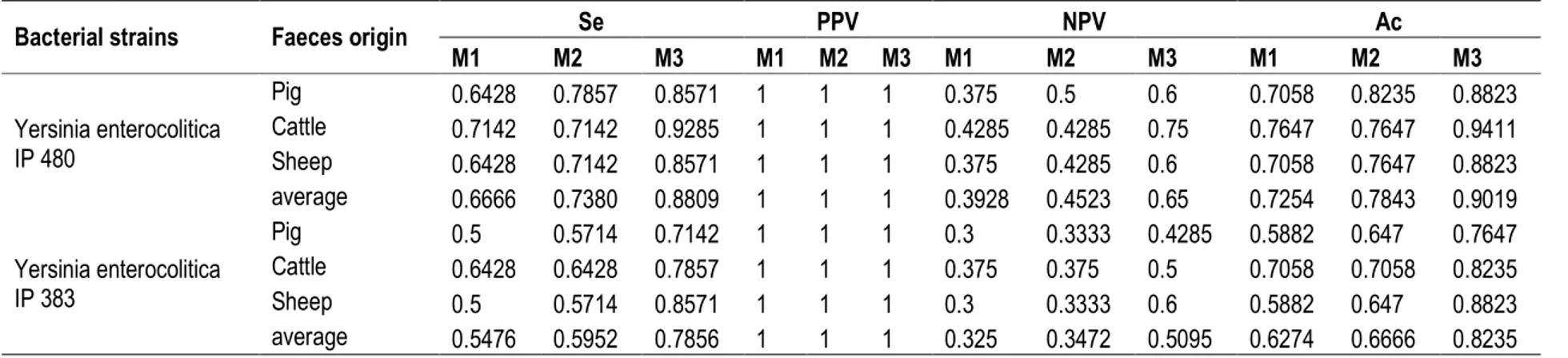
The average sensitivities of direct plating method( M1), Cold enrichment method (M2) and modified PBS method (M3) o were respectively 0,66, 0,73 and 0.88 for the non pathogenic strain belong to biosérotype
1A/O:14 (IP480). For the pathogenic strain IP 383 belong to biosérotype 2/O: 9 the average sensitivities were respectively 0.54, 0.59 and 0.78 (Table 2)
6095
Table 2: Sensitivity (Se), accuracy (AC), and negative predictive value (NPV) of methods for each Yersinia enterocolitica strain in artificially contaminated animal faecal samples
Bacterial strains Faeces origin Se PPV NPV Ac
M1 M2 M3 M1 M2 M3 M1 M2 M3 M1 M2 M3 Yersinia enterocolitica IP 480 Pig 0.6428 0.7857 0.8571 1 1 1 0.375 0.5 0.6 0.7058 0.8235 0.8823 Cattle 0.7142 0.7142 0.9285 1 1 1 0.4285 0.4285 0.75 0.7647 0.7647 0.9411 Sheep 0.6428 0.7142 0.8571 1 1 1 0.375 0.4285 0.6 0.7058 0.7647 0.8823 average 0.6666 0.7380 0.8809 1 1 1 0.3928 0.4523 0.65 0.7254 0.7843 0.9019 Yersinia enterocolitica IP 383 Pig 0.5 0.5714 0.7142 1 1 1 0.3 0.3333 0.4285 0.5882 0.647 0.7647 Cattle 0.6428 0.6428 0.7857 1 1 1 0.375 0.375 0.5 0.7058 0.7058 0.8235 Sheep 0.5 0.5714 0.8571 1 1 1 0.3 0.3333 0.6 0.5882 0.647 0.8823 average 0.5476 0.5952 0.7856 1 1 1 0.325 0.3472 0.5095 0.6274 0.6666 0.8235
Table 3 : Prevalence of Yersinia sp in animal samples
Sampling Sites Samples Method 2 (M2) Method 3 (M3)
Numbers Pos % Numbers Pos %
Pig Slaughter
Tonsils 53 0 0 53 0 0
Faeces 70 0 0 70 0 0
Lymph nodes 34 0 0 34 0 0
Tongue swabs 74 0 0 74 0 0
Animals farms Pig Faeces 97 0 0 97 2 2.06
Aulacode faeces 62 0 0 62 0 0
Other Animals in Farms Rodent intestinal contents 71 0 0 71 0 0
Saraka et al. J. Appl. Biosci. 2014. Comparison and validation of methods of Yersinia spp detection from animal stool samples in Abidjan.
6096 In this survey, no false positive result was observed, so the specificities and the positive predictive values of the three methods were 1 (100%). Their average values negative predictive values were respectively 0,39, 0,45 and 0,65 for the biosérotype 1A/O:14 and respectively 0,32 , 0,34 and 0,50 for biosérotype 2/O:9. The Limit of detection of the Enrichment methods (M2 and M3) was 102 CFU/ml whereas the one of the direct plating was of 103 CFU / ml. In cattle faecal samples, this limit detection was the lowest in method M3 (10 CFU/ml for the biosérotype 1A/O: 14 and 102 CFU/ml for the biosérotype 2/O: 9.) When the detection rates of the methods were compared, it was observed a significance difference between the one hand the direct plating (M1) and the PBS modified (M3) on and on the other hand between the enrichment cold enrichment method (M2) and M3. No significant difference was observed between the direct plating and the cold enrichment methods. Also when the detection rates of these two strains were compared, no method was
encouraged in preferential treatment for the detection one of these strains.
Validation of the most efficient on animals samples : A total 496 samples were analyzed by the methods of M2 enrichment and M3 for the research of Yersinia spp considering their good performance (Table 3). Three hundred and twenty eight (328) pigs samples from the farm and slaughter of (53 tonsils, 74 tongue swabs, 34 lymphatic nods and 167 faeces). Also 71 rodent intestinal contents (Rattus ratus and Rattus norvegecus) of the livestock farms, 62 pools of three (3) aulacode faeces (Thryonomys swinderianus) and 35 digestive tract of achatine (Achatina fulica) were analyzed. In total five (5) farms and 47 pigs, enclosures and only one farm of 30 enclosures of aulacode were sampled. The samples were constituted of 231 (46.57%) samples from the slaughterhouse and 265 (53.42%) from livestock farms. Only the method M3 permitted to isolate two strains of Yersinia intermedia, biotype 4, sérotype 7,8-8-8,19 in fatting piglets faecal samples.
DISCUSSIONS
The objective of this survey was to provide a simple, fast and reproducible, non coercive and an efficient isolation method for the surveillance of yersiniosis due to the enteropathogenics Yersinia in human and animal sample stools.
In this survey the intrinsic performances of three methods of isolation and detection of Yersinia enterocolitica were compared using animals faecal samples contaminated artificially according to the same criteria used by Soria et al., 2012. Two pathogenic Yersinia enterocolitica strains belonging to the pathogenic biosérotype 2/O: 9 (IP 383) and the non pathogenic biosérotype 1A/O: 14 (IP 480) were used. This survey showed that the PBS modified is the most sensitive method for the detection of Yersinia sp in the animal faecal samples. This sensitivity was 0.8809 (88.09%) for the non pathogenic bioserotype and 0.7856 (78.56%) for the pathogenic biosérotype. These results agree with those of Laukkanen et al. 2010 that coupled this method to a selective enrichment to the ITC (Irgasan Ticarcilline and potassium chlorate) broth. This method is currently used by the DFEH (Department Food of and Environmental Hygiene of the Faculty of Veterinary medicine) from University of Helsinki in Finland. Before Savin et al. in 2008 had found that the enrichment to the cold enrichment at +4°C tended to select non-pathogenic strains. This method was considered to be the most efficient for the detection of Yersinia enteropathogenic strains in human
and pork faecal stools [Kontiainen et al. 1994; Nowak et al. 2006] and in pigs tonsils [Nesbakken 1985; Nesbakken and Kapperud 1985; Nowak. et al. 2006]. But in this study, this tendency was not observed and besides the enrichment the cold enrichment method although detection rate lower than the PBS modified method had the same limit detection with this last method. Nevertheless the method M3 permitted a more precocious detection in cattle faecal samples for the pathogenic strain IP 383 that the non pathogenic strain IP 480. All time, the method of direct plating showed the lowest sensitivity (p < 0.05). Besides its limit detection was the highest at 103 CFU / ml and those of the other methods was situated at 102 CFU/ml. But it is less coercive and less expensive character can make it a very useful for the screening of enteropathogenic Yersinia in human diarrheal samples stools. This survey also permitted to confirm that pork was the major reservoir of it and that the breeding activity could constitute a source of human contamination in Abidjan. Because of the slower division time comparatively to the other enterobactéria, the detection of enteropathogenics Yersinia from contaminated samples is very difficult in faecal stool. The enrichment methods which were advised by Savin et al. in 2008 facilitated the detection of two strains Yersinia intermedia of biotype 4. The alkali treatment in potassium hydroxide (KOH) at 0.25% permitted to reduce background flora and the detection of these strains. The prevalence of
6097 Yersinia sp was 0.40% in all animal species, 1.19% in the pig faecal sample. These results were lower than those of Bhaduri et al, 2005 in USA in this same breeding animal who is of 4.08%. These two results
without being identical show that the prevalence of the Yersinia in the samples of faecal matters at reservoirs adult stays weak.
CONCLUSION
It appears fromthis study thattheenrichment method and especiallytheprior-enrichmentat 25 °Cin aBrain Heart Infusion broth containing 2.5 mg / l novobiocin followed by a modified PBS (Phosphate-buffered
saline) enrichment are appropriate methods for monitoring epidemiological of enteropathogenics
Yersiniaintheanimal faecalsamples
ACKNOWLEDGEMENTS
We thank Dr. Elisabeth CARNIEL (Centre National des Yersinia, Institut Pasteur, Paris, France) for reference strains and her kind cooperation, her generous advice
and for confirming of strains characteristic (biotype, serotype and phagetype) of the Yersinia strains.
REFERENCES
Aulisio CCG, Mehlman IJ, Sanders AC, 1980. Alkali method for rapid recovery of Yersinia enterocolitica and Yersinia pseudotuberculosis from foods. Applied and Environmental Microbiology 39: 135-140.
Bhaduri S., Wesley I.V., Bush E.J. (2005). Prevalence of Pathogenic Yersinia enterocolitica strains in pigs in the United States. Applied and Environmental Microbiology, 71(11): 7117-7121.
[EFSA] European Food Safety Agency). (2009) The community summary report on trends and sources of zoonoses, zoonotic agents in the European Union in 2007. EFSA J 2009; 223:223–226.
Fredriksson-Ahomaa M., Korkeala H. (2003). Low occurrence of pathogenic Yersinia enterocolitica in clinical, food and environmental samples: a methodological problem. Clinical Microbiology Reviews, 16(2): 220-229.
Frickmann.H , Dekker .D ,Boahen.K , Acquah .S , Sarpong .N,Adu-Sarkodie Y , Schwarz .G.N ,May .J, Marks.F ,Poppert .S, Wiemer.F.D,, Hagen .M.R .2013.Increased detection of invasive enteropathogenic bacteria in pre-incubated blood culture materials by real-time PCR in comparison with automated incubation in Sub-Saharan Africa: Scandinavian Journal of Infectious Diseases,; Informal Healthcare, Early Online: 1–7
Huovinen E, et al. 2010. Symptoms and sources of Yersinia enterocolitica infection: a case-control study. BMC Infect. Dis. 10:122.
International Organization for Standardization (2003) Microbiology of food and animal feeding stuffs – Horizontal method for the detection of presumptive pathogenic Yersinia enterocolitica. ISO 10273: 2003, 2nd edn. 1– 33.
Kontiainen, S., Sivonen, A. and Renkonen, O.V. 1994 Increased yields of pathogenic Yersinia enterocolitica strains by cold enrichment. Scand J Infect Dis 26, 685–691.
Laukkanen R, Martinez PO, Siekkinen KM, Ranta J, Maijala R, Korkeala H, 2009. Contamination of carcasses with human pathogenic Yersinia enterocolitica 4/O: 3 originate from pigs infected on farms. Foodborne pathogens and Disease 6: 681-688.
Laukkanen;R, Hakkinen M, Lunden, Fredriksson-Ahomaa M., Johansson T and Korkeala H.2010: Evaluation of isolation methods for pathogenic Yersinia enterocolitica from pig intestinal content. Journal of Applied Microbiology 108 : 956–964
Lambertz, S.T., Granath, K., Fredriksson-Ahomaa, M., Johansson, K.E., Danielsson-Tham, M.L., 2007. Evaluation of a combined culture and PCR method for detection of presumptive pathogenic Yersinia enterocolitica in pork products. J.Food Prot. 70, 335–340.
Nesbakken, T., Eckner, K., Hoidal, H.K., Rotterud, O.J., 2003. Occurrence of Yersinia enterocolitica and Campylobacter spp. in slaughter pigs and consequences for meat inspection, slaughtering, and dressing procedures. Int. J. Food Microbiol. 80, 231–240.
Saraka et al. J. Appl. Biosci. 2014. Comparison and validation of methods of Yersinia spp detection from animal stool samples in Abidjan.
6098 Nesbakken, T. 1985 Comparison of sampling and
isolation procedures for recovery of Yersinia enterocolitica serotype O: 3 from the oral cavity of slaughter pigs. Acta Vet Scand 26, 127–135.
Nesbakken, T. and Kapperud, G. 1985 Yersinia enterocolitica and Yersinia enterocolitica-like bacteria in Norwegian slaughter pigs. Int J Food Microbiol 1, 301–309.
[NCFA] Nordic Committee of Food Analysis (1996) Yersinia enterocolitica Detection in foods 117, 3rd,edn,1-12
Nowak, B., Mueffling, T.V., Caspari, K. and Hartung, J. 2006 Validation of a method for the detection of virulent Yersinia enterocolitica and their distribution in slaughter pigs from conventional et al. tentative housing systems. Vet Microbiol 117, 219–228.
Savin C., Carniel, E., 2008. Les diarrhées d'origine bactérienne : le cas de Yersinia enterocolitica. Rev. Francophone Lab. 400, 49–58.
Schiemann, D.A., 1982. Development of a two-step enrichment procedure for recovery of Yersinia enterocolitica from food. Appl. Environ. Meth. 43, 14–27.
Soria M. C. ,. Soria M. A and D. J. Bueno: 2012 Comparison of 2 culture methods and PCR assays for Salmonella detection in poultry faeces .Poultry Science 91:616–626
Thibodeau, V., Frost, E.H., Chénier, S., Quessy, S., 1999. Presence of Yersinia enterocolitica in tissues of orally inoculated pigs and the tonsils and faeces of pigs at slaughter. Can. J. Vet. Res. 63, 96–100.