Université de Montréal
Upstream Mechanisms Responsible for H202-induced Activation of
MAPK and
PKBin Vascular Smooth Muscle Cells
par Zeina Azar
Département des Sciences Biomédicales Faculté de Médecine
Mémoire présenté à la Faculté des études supérieures en vue de l’obtention du grade de Maîtrise en Sciences (M.Sc.)
en Sciences Biomédicales
Août 2006
n
Direction des bibliothèques
AVIS
L’auteur a autorisé l’Université de Montréal à reproduire et diffuser, en totalité ou en partie, par quelque moyen que ce soit et sur quelque support que ce soit, et exclusivement à des fins non lucratives d’enseignement et de recherche, des copies de ce mémoire ou de cette thèse.
L’auteur et les coauteurs le cas échéant conservent la propriété du droit d’auteur et des droits moraux qui protègent ce document. Ni la thèse ou le mémoire, ni des extraits substantiels de ce document, ne doivent être imprimés ou autrement reproduits sans l’autorisation de l’auteur.
Afin de se conformer à la Loi canadienne sur la protection des renseignements personnels, quelques formulaires secondaires, coordonnées ou signatures intégrées au texte ont pu être enlevés de ce document. Bien que cela ait pu affecter la pagination, il n’y e aucun contenu manquant.
NOTICE
The author of this thesis or dissertation has gtanted a nonexciusive license allowing Université de Montréal to reproduce and publish the document, in part or in whole, and in any format, solely for noncommercial educationat and research purposes.
The author and co-authors if applicable retain copyright ownership and moral rights in this document. Neither the whole thesis or dissertation, nor substantiel extracts from it, may be printed or otherwise reproduced wïthout the author’s permission.
In compliance with the Canadien Privacy Act some supporting forms, contact
information or signatures may have been removed from the document. White this may affect the document page count, it does flot represent any loss of content from the document.
Université de Montréal Faculté des études supérieures
Ce mémoire intitulé:
« Upstream Mechanisms Responsible for H202-induced Activation
of
MAPK and PKB in Vascular Smooth Muscle Celis»
Présenté par: Zeina Azar
a été évaluée par un jury composé des personnes suivantes:
Président-rapporteur : Dr Pangala Bhat
Directeur de recherche D Ashok K. Snvastava Membre du jury : D’ Angelino Calderone
RÉSUMÉ
Les espèces réactives oxygénées (ROS) sont produites de façon naturelle dans les
cellules. Cependant, une production excessive ou une insuffisance dans le processus de leur élimination est associée à différentes maladies. Ainsi, les dernières années ont vu les
ROS émerger comme des joueurs importants dans la pathogenèse des maladies
cardiovasculaires, comme l’hypertension, l’athérosclérose et l’hypertrophie cardiaque,
ainsi que les complications vasculaires du diabète. Il est connu que les ROS peuvent
oxyder certains constituants cellulaires et qu’elles activent les voies de signalisation impliquées dans la croissance cellulaire, l’hypertrophie, la prolifération et la migration,
ainsi que la survie, qui sont des événements caractéristiques du remodelage cardiovasculaire. Les voies de signalisation les plus importantes sont celles des MAPK et
de la phosphatidylinositol-3-kinase (PT3-K)/protéine kinase B (PKB). Cependant, les
mécanismes moléculaires par lesquels les ROS déclenchent l’activation de ces voies restent à être clarifiés. Bien qu’un rôle des récepteurs et non récepteurs protéines tyrosines kinases
(«
PTKs»)
dans la médiation de la phosphorylation des ERKI/2 par le H202 soit suggéré, une implication possible des tyrosines kinases dans l’activation de PKB et de Pyk2 par le stress oxydatif n’a pas encore été clarifié dans les cellules dumuscle lisse vasculaire
(«
VSMC»).
Donc, l’objectif principal de notre étude était d’examiner le rôle des récepteurs et non récepteurs PTK dans l’activation de la PKB parle H202 dans la lignée cellulaire de «VSMC
»
AlO. Ces cellules sont isolées de l’aortethoracique de l’embryon de rat.
La phosphorylation de laPKB induite par le H202 a été complètement inhibée par
le prétraitement des cellules avec l’AG1024, un inhibiteur pharmacologique spécifique du IGF-1R-PTK
(«
insulin-like growth factor typel receptor PTK»), alors que J’AG1478,un inhibiteur spécifique du EGFR-PTK
(«
epidermal growth factor receptor PTK»)
n’apas eu d’effet inhibiteur sur la phosphorylation de la PKB. De même, la phosphorylation de Pyk2, Src et ERK1/2 induites par le H202 ont été complètement inhibées par AG1024
iv
et non par AG1478. L’implication du «IGF-1R> dans la signalisation induite par F1202 a aussi été démontrée par le fait que le H202 augmente la phosphorylation en tyrosine des sous unités
f3
du «IGF-1R » et que le prétraitement au AG1024 a inhibé cette phosphorylation. De plus, l’inhibition pharmacologique de Src a diminué de façon significative la phosphorylation de la PKB et de Pyk2 par le H202. Tout comme l’effet de l’AG1478, l’AG1295, un inhibiteur spécifique du PDGFR-PTK («platelet-derived growth factor receptor PTKx’) n’a pas inhibé la phosphorylation de PKB ni de ERK1/2 induite par le H202, alors qu’il a supprimé complètement la réponse au «PDGF». En conclusion,nos résultats suggèrent que le «IGF-1R>, et non pas le «EGFR» ni le «PDGFR» joue un
rôle critique dans la médiation des évènements induits par le H202 et qui sont responsables de l’hypertrophie et de la croissance des «VSMC ».
Mots clés: ROS, H202, MAPK, PKB, Src, Pyk2, récepteurs et non récepteurs PTK, IGF
ABSTRACT
Reactive oxygen species (ROS) are produced normally in the ceils, but their excessive production or a Jack in their elimination process is associated with various dïseases. Thus, ROS have emerged in the recent years as important players in the pathogenesis of cardiovascular disorders, such as hypertension, atherosclerosis and cardiac hypertrophy as well as in the vascular complications of diabetes. ROS are known
to modify the celi components by oxidation and to activate signaling pathways
responsible of celJ growth, hypertrophy, proliferation, and migration as well as survival, which are characteristic events in cardiovascular remodeling. The most important of these pathways are the mitogen-activated protein kinases (MAPKs) and the
phosphatidylinositol-3 -kinase (P13-K)/protein kinase B (PKB) pathway. However, the precise molecular mechanisms by which ROS trigger the activation of these pathways remain to be clarified. AJthough a role for receptor and non-receptor protein tyrosine kinases (PTKs) in mediating H202-induced ERK1/2 phosphorylation has been suggested, a possible involvement of tyrosÏne kinases in PKB and Pyk2 activation by oxidative stress has not yet been clanfied in VSMC. Therefore, the main objective of our studies
was to investigate the role of receptor and non-receptor tyrosine kinases in H202-induced PKB activation in AlO VSMC. These ceils are obtained from rat embryonic thoracic
aorta.
H202-inducedPKB phosphorylation was completely abolished by pretreatment of
the ceils with AG1024, a specific phannacological inhibitor of the insulin-like growth
factor type 1 receptor PTK (IGF-1R-PTK), whereas AG1478, the specific inhibitor of the epidermal growth factor receptor PTK (EGFR-PTK) did flot have any inhibitory effect on
PKB phosphorylation under the same conditions. Similarly, H202-induced phosphorylation of Pyk2, Src and ERK1/2 phosphorylation were completely blocked by AG1024 but not by AG1478. The involvement of the IGF-IR in H202-induced signaling
vi
was also demonstrated by the resuits showing that H202 enhanced the tyrosine
phosphorylation of the r3-subunit of IGF-IR and that AG1024 pretreatment markedly inhibited this phosphorylation. Moreover, pharmacological inhibition of Src significantly decreased H202-induced PKB and Pyk2 phosphorylation. Similar to the effect of AG1478, AG1295, the specific inhibitor of the platelet-denved growth factor receptor PTK (PDGFR-PTK) failed to inhibit H202-induced PKB and ERKI/2 phosphorylation, whïle it abrogated the PDGF-induced response under the same conditions. Taken together, our data suggests that IOF-1R, but flot EGFR nor PDGFR plays a critical role in mediating H202-induced signaling events which are important mediators for hypertrophy and growth in VSMC.
Key words: ROS, H70,, MAPK, PKB, Src, Pyk2, receptor and non-receptor PTK, IGF 1R, VSMC.
TABLE 0f CONTENTS
Résumé iii
Abstract y
Table ofcoiztents vii
List offigures x
List ofabbreviations xiii
Acknowtedgements xvi
CHAPTER 1: INTRODUCTION 1
1.1 The multiple sources of ROS 2
1.2 TheNAD(P)Hoxidase 4
1.3 The ROS scavenging systems 4
1.4 Regulation of ROS production by vasoactive peptides and growth factors.7
1.5 ROSinvasculardiseases 9
1.5.1 ROS in genetic and experimental hypertension 9 1.5.2 ROS in human hypertension 11
1.5.3 ROSinheartfailure 11
1.5.4 ROS in vascular complications of diabetes 12
1.6 The intracellular effects ofROS 13
1.7 The MAPK pathway 14
1.7.1 Activation of MAPK pathway by ROS 17
1.7.2 MAPK activation in cardiovascular diseases 17 1.8 The phosphatidylinositol-3-kinase (P13-K)/protein kinase B (PKB)
pathway 18
viii
1.8.2 Activation ofPKB by ROS .20
1.9 The epidermal growth factor receptor (EGFR) 22
1.9.1 Transactivation of the EGFR 22
1.10 The insulin-like growth factor type 1 receptor (IGF-1R) 24
1.10.1 Transactivation of the IGF-1R 25
1.11 The platelet-denved growth factor receptor (PDGFR) 26
1.11.1 Transactivation of the PDGFR 27
1.12 The Src-family tyrosine kinases (SFKs) 27
1.12.1 Activation of SFKs and its implication in signal
transduction pathways 28
1.13 The Proline-rich tyrosine kinase 2 (Pyk2) 29 1.13.1 Activation of Pyk2 and its implication in signal
transduction pathways 30
1.13.2 Activation of Pyk2 and its implication in
cardiovascular diseases 31
1.14 General Conclusion 32
CHAPTER 2: ARTICLE 34
Abstract 36
Introduction 37
Materials and Methods 40
Resuits 43
Discussion 47
Figure Legends 52
Figures 55
CHAPTER 3: GENERAL DISCUSSION .70
CHAPTER 4: CONCLUSION 79
X
LIST 0F FIGURES
CHAPTER 1: INTRODUCTION
figure 1.1 : Simplified scheme showing key steps in the production of reactive
oxygen species (ROS) 3
Figure 1.2: Production and elimination of 02 and H202 6
figure 1.3: Regulation of ROS generation by vasoactive peptides $
Figure 1.4: Schematic mode! showing the signa!ing cascade induced by
receptor tyrosine kinases leading to ERK1/2 activation 16
Figure 1.5: Schematic mode! showing the signa!ing cascade induced by
receptor tyrosine kinases leading to PKB activation 21
Figure 1.6: Scheme summarizing the major ROS-induced signaling pathways
that are responsib!e of their pathologica! effects 33
CHAPTER 2: ARTICLE
Figure 1(A): Effect of AG1478 on H202-inducedPKB phosphorylation
inAIOVSMC 55
Figure 1(B): Effect of AG1024 on H202-induced PKB phosphory!ation
Figure 2: Effect of H202 on IGF-lRf3 tyrosine phosphorylation in AlO VSMC 57
Figure 3: Effect of PP2 on H202-induced PKB phosphorylation in AlO VSMC....58
Figure 4: Effect of AG1478 and AG1024 on H202-induced c-Src phosphorylation
inA1OVSMC 59
Figure 5(A): Effect of PP2 on H202-induced Pyk2 phosphorylation
inA1OVSMC 60
Figure 5(B) : Effect of AG1478, AG1024 and PP2 on H202-induced
Pyk2 phosphorylation 61
Figure 6: Schematic model showing the signaling cascade induced byH202
inA1OVSMC 62
CHAPTER 3: GENERAL DISCUSSION
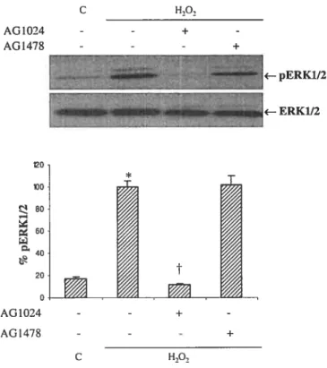
Figure 3.7: Effect of AG1478 and AG1024 on H202-induced ERK1/2
phosphorylation in AlO VSMC 76
Figure 3.8(A) : Effect of AG1295 H202-induced PKB phosphorylation
inA1OVSMC 77
Figure 3.8(B) : Effect of AG1295 H202-induced ERK1/2 phosphorylation
xii
CHAPTER 4: CONCLUSION
Figure 4.9: Schematic, hypothetical mode! showing key steps in
LIST 0F ABREVIATIONS
ACE : Angiotensin converting enzyme
Angil : Angiotensin II
BAD : Bcl-2-associated death CAK : Ceil adhesion kinase beta
UNA : Deoxyribonucleic acid
DOCA-sait : Deoxycorticosterone acetate-sait
EGf : Epidermal growth factor
EGFR : Epidermal growth factor receptor
ERK1/2 : Extracellular signal-regulated kinases 1 and 2
ET-1 : Endothelin-1
fAK : Focal-adhesion kinase
FKHR : Forkhead transcription factor Gabi : Grb2-associated binding 1 GPCR : G protein-coupled receptor
Grb2 : Growth factor receptor binding protein 2
GSH : Glutathione
GSK-3p Glycogène synthase kinase 3 beta GSSG : Glutathione disulfide
xiv
HB-EGF : Heparin-binding epidermal growth factor
H202 Hydrogen peroxide
IGF-1R : Insulin-like growth factor type ireceptor
IL-lp Interleukin-1 beta
IRS : Insulin receptor substrate JNK : c-Jun NH2-terminal kinase LDL : Low density lipoprotein
MAPK : Mitogen-activated protein kinase MAPKK/MEK : Mitogen-activated protein kinase kinase
MAPKKKJRaf : Mitogen-activated protein kinase kinase kinase mTOR : mammalian target of Rapamycin
NADP÷ : Nicotinamide adenine dinucleotide phosphate, oxidizedfonn NADPH : Nicotinamide adenine dinucleotide phosphate, reduced form NF-id3 : Nuclear factor-kappaB
NO : Nitric oxide
02 : Molecular oxygen
02 Superoxide anion
OH : Hydroxyl radical
ONOO : Peroxynitrite
PDGF : Platelet-derived growth factor
PDK1/2 : Phosphoinositide-dependent kinases 1 and 2
PI : Phosphatidylinositol
P13-K : Phosphatidylinositol-3-kinase
PIP2 : Phosphatidylinositol-4,5-biphosphate PIP3 : Phosphatidylinositol-3 ,4,5-triphosphate
PKB t Protein kinase B
PL : Phospholipase C
PTK : Protein tyrosine kinase Pyk2 : Proline-rich tyrosine kinase
RAfTK : Related adhesion focal tyrosine kinase ROS : Reactive oxygen species
SFKs : Src family tyrosine kinases
5112 : Src homology 2
SHP-2 : Src Homology 2 domain-containing tyrosine phosphatase SHR Spontaneously hypertensive rat
SOD : Superoxide dismutase
Sos : Son of sevenless
TGF-betal : Transforming growth factor beta 1 TNF-Π: Tumor necrosis factor alpha VSMC : Vascular smooth muscle celi
xvi
ACKNOWLEDGEMENTS
I would like to express my sincere gratitude to my supervisor D’ Ashok K. Srivastava for offering me the opportunity to upgrade myseif in research, for ‘avoir confiance en my capabilities, for bis advïce, his patience, and bis friendshïp. His help in so many ways was important for me to achieve this work.
I express reverence and sincere thanks to Dt Lise Coderre for her help, advice and encouragement during my study.
I am thankful to my colleagues for their help and tbeir fnendship, Ah Bouallegue, George Vardatsikos and Demiana Ekladous. Special thanks and immense gratitude to Dr Mohammed Mehdi for bis suggestions, bis cnticism and bis help in accomplishing my task (tough task).
I will aiways be (reconnaissante) for my parents for their love and support dunng ail my life.
Last but not the least, my thanks to my husband Walid for bis love and his support during cntical times.
2
Evidence generated during the recent years has suggested a crucial role for reactive oxygen species (ROS) in the pathophysiology of cardiovascular disorders including hypertension, atherosclerosis and restenosis after angioplasty [1-3] as well as in diabetes (reviewed in [4]) and cancer (reviewed in [5]). Therefore, there is a lot of interest to understand the mechanism of ROS generation, its elimination and identification of its cellular targets. Thus, the objective of this section is to provide a brief overview on these aspects of ROS and its relationship in the pathophysiology of cardiovascular disease.
1.1 Tlie multiple sources of ROS
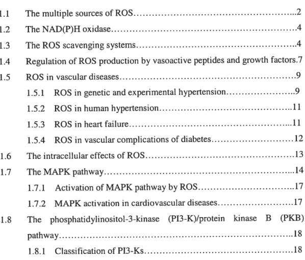
ROS are formed as intermediates in redox reactions, leading from molecular oxygen (02) to water (H20) [6]. They are small, quickly diffusibie and highly reactive molecules [7] and are classified into superoxide anion (02), hydroxyl radical (OH), hydrogen peroxide (H202) and peroxynitrite (0N00) [8]. Their major intracellular source is the mitochondna which converts 1-2% of consumed 02into 02 [7]. 02 andlor H202 can also be derived from NAD(P)H oxidase, xanthine oxidase, cyclooxygenase, lipoxygenase, heme oxygenases, peroxidases, as well as hemoproteins such as heme and hematin (reviewed in [9]). The univalent reduction of 02 leads to 02, which is relatively unstable and short-lived because of its unpaired electron [10] (fig. 1.1).
011
e-
Metals
02
02
11202
NAD(P)H
SOD
Catalase
oxidase
figure 1.1: Simplified scheme showing key steps in the production of reactive oxygen species (ROS). The most important ways of ROS generation are shown here. One electron from NAD(P)H is transfened to molecular Oxygen (02) by NAD(P)H oxidase to generate superoxide anion ÇOj), which can be converted to hydrogen peroxide (H202) either spontaneously or by superoxide dismutase (SOD). In presence of metals, H202 can be converted to Hydroxyl anion ÇOH). (Adapted from Ref. [li]).
4
1.2 The NAD(P)ll oxidase
The major source of ROS in the vascular walI is the NAD(P)H oxidase [12j, which is a complex enzyme system cornposed of many subunits including p22phox, p47phox, the GTPase Rac and the recently identified Noxi and Nox4 [12-15]. The NAD(P)H oxidase catalyzes 02 production by the one electron reduction of 02 where
NAD(P)H is the electron donor:
202+ NAD(P)H—* 202 +NAD(P) +
W
[16].NAD(P)H oxidase is activated in hypertensive animais [17;18] as weli as in human hypertensive subjects [19;20] and it is believed to act as a major player in the deveiopment of atherosclerosis [21]. Particuiarly, p22phox was shown to play an important role in agonist-induced ROS generation and gene expression in VSMC in response to Angiotensinil (Angil), Tumor necrosis factor Π(TNF-a) and thrombin
[12;22-24]. Moreover, Noxi seems to be upregulated by proliferative stimuli, such as Angiotensinli (Angil) and plateiet-derived growth factor (PDGf) in VSMC [25].
1.3 The ROS scavenging systems
Under physiological conditions, 02 undergoes dismutation either spontaneously
or by a reaction catalyzed by superoxide dismutase (SOD) to produce H202. Dismutation of 02 by SOD ïs favored at low concentrations of 02 and at high concentrations of
SOD, which happens under physiologicai conditions. H202 is much more stable than 02,
can cross celi membranes and lias a longer haif-life. Normaiiy it is scavenged by catalase and giutathione peroxidase to produce H20 [101. In the presence of metal-containing moiecules such as Fe2, H202 can also be reduced to generate the extremeÏy active
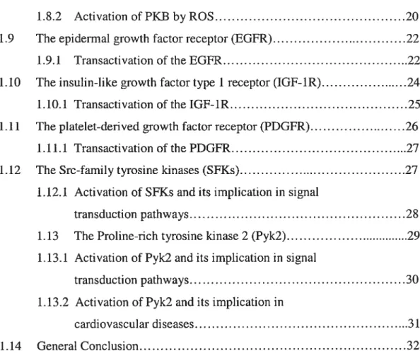
hydroxyl radical ÇOH) that causes damage to the ceil components [6]. In the glutathione peroxidase reaction, glutathione (GSH) is oxidized to glutathione disulfide (GSSG), which can be converted back to GSH by glutathione reductase in a NADPH-consuming process (fig. 1.2). Several forms of SOD are known: copper-zinc SOD (Cu/Zn SOD), mitochondrial or manganese SOD (Mn SOD) and iron-containing SOD (Fe SOD)
[26;27].
Normally, the rate of ROS production is balanced by the rate of their elimination. However, in pathological conditions, a disequilibrium between ROS generation and elimination resuits in increased ROS bioavailability leading to oxidative stress [2$]. Furthermore, in case of excessive production, 02 reacts with nitric oxide (NO) to produce the very hamiful ONOO [29].
6
NADH
o.
NAD(P)HElectron transport chain NAD(P)H oxidase Xanthine oxidase, etc....
NADNADP+ NO
0N00
H70
GSSG
NAD(P)H
Figure 1.2: Production and elimination of 02 and H202. 02 can undergo dismutation by superoxide dismutase (SOD) to produce H202. H202 is scavenged by catalase or Glutathione peroxidase to produce water (H20) whule in the sarne reaction, Glutathione (GSH) is converted to Glutathione disulfide (GSSG), which can be converted back to GSH by Glutathione peroxidase. In presence of metals, H20, can be converted to Hydroxyl anion (‘oH). It is noteworthy that high levels of ‘02 may lead to the generation of peroxynitrite anion (0N00) since 02 can react with nitric oxide (NO).
OH
1.4 Re%ulation of ROS production by vasoactive peptides and growth factors
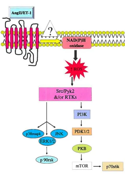
An increasing number of studies have demonstrated that vasoactive peptides mediate their responses through the generation of ROS. The potent vasoconstrictor endothelin-1 (ET-I) bas been shown to activate NAD(P)H oxidase resulting in ROS generation in endothelial cells [30] and to increase H202 levels via its subtype A receptor (ETA receptor) in pulmonary smooth muscle cells [31]. Similarly, a role for ROS generation in mediating ET-1-induced activation of various signaling pathways, such as ERK1/2, PKB and Pyk2 [32] as well as JNK and p3$mapk [33] has also been demonstrated. Moreover, ET-1, through ETA receptor, bas also been implicated in the increased vascular levels of 02 in both low-renin hypertension and chronic ET-1-infused rats models of hypertension [34;35].
Another important vasoactive peptide, Angli is also known to induce ROS generation in many ccli types including cardiomyocytes [36], endothelial cells [37] and VSMC [12]. In hypertension, Angil activates the NAD(P)H oxidase thereby enhancing the ROS generation [12;38]. Furthermore, H202 appears to play a direct role in AngII induced vascuÏature hypertrophy [39;40] (Fig. 1.3).
Growth factors such as the epidermal growth factor (EGF) [41], the PDGF [42] and cytokines were also shown to induce the generation of ROS such as 02 and H202 in nonphagocytic celis [43]. Transforming growth factor-betal (TGF-betal), which is abundantly expressed in pulmonary hypertension, induced expression of Nox4 and likewise increased production of ROS, in freshly isolated human pulmonary artery smooth muscle celis [44]; this is believed to be an important mechanism in pulmonary vascular remodeling.
8 SrcIPyk2 &IorRTKs
I
PI3KI
1
mTOR —, p7Os6kFigure 1.3: Regulation of ROS generation by vasoactive peptides. The vasoactive peptides Angil and ET-1 are known to increase the ROS generation in many ccli types, particuiarly in VSMC, by activatÏng the NAD(P)H oxidase through an as yet unknown mechanism. Endogenously produced ROS activate many protein tyrosine kinases, such as c-Src, Pyk2 and receptor tyrosine kinases (RTKs) that act as upstream regulators of MAPK as weii as P13-KIPKB pathway.
1.5 ROS in vascular diseases
There is increasing evidence that oxidative stress is involved in the initiation and progression of cardiovascular disease. An enhanced ROS generation is observed at sites of vascular injury and has been related to the development of restenosis/ atherosclerosis [45]. A key role for vascular NAD(P)H oxidases in the development of human atherosclerosis was reported recently [46-4$]. Originally, it was thought that ROS are involved in low density lipoproteins (LDL) oxidation, a key step in the initiation and progression of atherosclerosis [49]. More recently, studies have shown that ROS are also implicated in endothelial dysfunction, increased contractility, VSMC migration, growth and apoptosis, inflammation and increased depositions of extracellular matnx proteins, whïch are important factors in hypertensive damage [50].
The decrease in NO bioavailability bas been correlated to an ïncrease in in hypertension and hypercholesterolemia (reviewed in [51]). O2 reacts with NO and forms 0N00, leading to a decreased NO bioavailability. Elevation in O2 levels contributes to impaired endothelial function associated with atherosclerotic disease [52].
1.5.1 ROS in genetic and experimental hypertension
A considerable number of studies have shown that hypertension is associated with an elevated level of ROS and also with an impairment of endogenous antioxidant defense mechanisms (reviewed in [53]). ROS, such as H2O2 and O2 are increased in vessels, heart and lddneys of the spontaneously hypertensive rats (SHR) [16;54], the obesity prone and the Dahi salt-induced sensitive rats [55;56]. In SHR, the increased NAD(P)H induced 0j generation appears to be associated with NAD(P)H oxidase subunit Nox4
10
overexpression and enhanced oxidase activity [17;57]. Polymorphisms in the promoter region of the p22phox gene have also been identified in SHR [58]. Renal NAD(P)H oxidase upregulation has been reported in young SHR [17J. Vascular O2 and oxidant markers are also increased in aortas of mice with deoxycorticosterone acetate-sait (DOCA-salt)-induced hypertension [18] as well as in hypertensive models created by infusion of ET-1 [34;59;60]. In a study performed on Noxi-deficient mice, Matsuno et al. demonstrated that Noxi- derived ROS are involved in AngII-induced hypertension and this occurred by reducing the bioavailability of NO [61]. Recently, Callera et al. demonstrated that ET-1-induced oxidative stress in DOCA-sait hypertension not only involves NAD(P)H oxidase, but also mitochondria-derived ROS [62].
Experimentally, generators of O2 were shown to abolish endothelium-dependent relaxation of aortic rings isolated from SHR [63], induce contraction of mesentenc beds [64] and to increase chlonde reabsorption by isolated thick ascending limbs of the loop of Henle [65]. Makino et al. demonstrated that infusion of H202 in the renal medulla of whole rats increased blood pressure and decreased urine flow as well as sodium excretion [66]. In accord with this study, Kopkan et al. demonstrated that enhanced O2 generation in AngII-induced hypertensive rats modulates renal hemodynamic and tubular reabsorptive function which leads possibly to sodium retention [671. It was also shown that H202 as well as Oj regulated vascular contraction by increasing intracellular calcium concentration ([Ca2]) in pig coronary artery SMC [6$;69] and in rat mesenteric arteries with greater effects in SHR versus normotensive Wistar Kyoto (WKY) rats [69;70].
1.5.2 ROS in human hypertension
Recent clinical studies have demonstrated increased oxidative stress and reduced antioxidant status in patients with essential hypertension, renovascular hypertension and malignant hypertension. In these studïes, the leveis of plasma thiobarbituric acid-reactive substances and $-epi-isoprostames, biomarkers of iipïd peroxidation and oxidative stress, were shown to be increased [19;20;71;72]. In mild-to-moderate hypertension, iipid peroxidation and oxidative stress are not increased [73], suggesting that ROS may flot be cntical in the early stages of human hypertension, but could be more important in severe hypertension. Activation of the renin-angiotensin system has been proposed as a mediator of NAD(P)H oxidase activation and ROS production [16;74-76]. In fact, some of the therapeutic blood pressure-Iowering effects of AT1 receptor blockers and angiotensin converting enzyme (ACE) inhibitors have been attnbuted to NAD(P)H oxidase inhibition and decreased ROS production [77;7$J. An association between a p22phox gene polymorphism and NAD(P)H oxidase-mediated 02 production in the vascular wall of patients with hypertension and atherosclerosis bas aiso been descnbed [79].
1.5.3 ROS in heart failure
Experimental and clinical studies have suggested an increased production of ROS in animais and in patients with acute and chronic heart failure (reviewed in [$0]). ROS are released during ischemia and ischemic reperfusion (reviewed in [$1]). Moreover, oxidants production is increased by Angil and catechoiamine, two endocrine factors known to ïnduce cardiac remodeling [$2;83]. The possible sources are many and include xanthine and NAD(P)H oxidoreductases, cyclooxygenases, mitochondn ai electron
12
transport chain and activated neutrophïls. Excessive NO denved from NO Synthase seems to be implicated in the pathogenesis of chronic heart failure: NO reacts with O2 to form ONOO, a reactïve oxidant which impairs cardiac function. Increased oxidative and nitrosative stress activates also the nuclear enzyme poly-ADP-nbose polymerase which is known to contribute to pathogenesis of cardiac and endothelial dysfunction associated with myocardial infarction, congestive heart failure, hypertension, atherosclerosis and diabetes (reviewed in [$0]).
1.5.4 ROS in vascular complications of diabetes
Oxidative stress can induce abnormal changes in intracellular signaling and result in chronic inflammation and insulin resistance. It is known that hyperglycemia as well as free fatty acids stimulate ROS production [84]. In diabetes, plasma levels of ROS are significantly elevated [85]. Inflammation and oxidative stress have been linked to insulin resistance in vivo, not only in type 2 diabetes, but also in obese, nondiabetic individuals and in patients with metabolic syndrome (reviewed in [4]). Moreover, NAD(P)H oxidase components are upregulated in vascular tissues of animal models and patients suffering from diabetes and obesity (reviewed in [86]). High levels of ROS trigger the activation of senne/threonine kinase cascades such as c-Jun N-terminal kinase, nuclear factor-kappaB (NE-KB), and others that in tum phosphorylate multiple targets, including the insulin receptor and the insulin receptor substrate (IRS) proteins. Increased serine phosphorylation of IRS reduces its ability to undergo tyrosine phosphorylation and may accelerate the degradation of IRS-1 leading to an aberrant insulin signaling and insulin resistance (reviewed in [4]).
Studies performed in vitro could also explain in part the molecular mechanism by which ROS can contribute to insulin resistance. H202 as well as a ROS inducer, diamide was found to inhibit insulin signaling in cultured rat VSMC by blocking the insulin induced protein kinase B (PKB) phosphorylation on Senne 473 (Ser473) [87]. In addition, H202 pretreatment also attenuated the insulin receptor bïnding and insulin receptor autophosphorylation in this study [87]. The negative regulation of PKB could contribute to insulin resistance because its activation by insulin is critical in stimulating glucose transport, glycogen synthase, protein synthesis, antilipolysis and suppression of hepatic gluconeogenesis [88]. Moreover, studies have suggested that insulin might protect VSMC from remodeling by inhibiting apoptosis [$9] and migration [90]. Taken together, it is possible that ROS, through their effects on the insulin signaling pathway, may contribute to cardiovascular disease.
1.6 The ïntracellular effects of ROS
Ceils respond to oxidant injury with the activation of multiple signal transduction pathways that serve to coordinate the cellular response and determine the outcome. High levels of ROS resuit in severe damage to the celi components (lipids, proteins, carbohydrates and DNA). ROS-induced oxidative modification of proteins and lipids can modify the structure and function of proteins and lipid bilayers resulting in cellular dysfunction. In the vasculature, 02 and H202 are particularly important since they act as inter- and intracellular signaling molecules. OH induces local damage, whereas 02 and H202 can travel some distance from their site of generation (reviewed in [9]). The biochemical events by which ROS mediate cellular dysfunction and contnbute to
14
pathogenesis of vascular diseases include changes in gene expression and celi signaling pathways. Two major signaling pathways, the mitogen-activated protein kinase (MAPK) and the phosphatidylinositol-3-kinase (P13-K)/protein kinase B (PKB) pathways, have been identified as important targets of ROS (Fig. 1.3). Aberrant activation of these pathways in the pathogenesis of vascular diseases has been suggested (Fig. 1.6).
1.7 The MAPK pathway
MAPK are a family of ubiquitous serine/threonine protein kinases, classicaÏly associated with celi growth, differentiation and death [91]. MAPK are activated in response to a variety of extemal stimuli such as growth factors, hormones and stress, as well as vasoactive peptides and ROS (reviewed in [92;93]). 0f the major mammalian MAP kinases, extracellular signal-regulated kinases (ERK1/2), p38MAP kinase (p38mapk), c-Jun N-terminal kinases (JNK) and ERK5 are the best characterized.
MAPK are activated by MAPK kinase (MAPKK also known as MEK) which, in tum are activated by MAPKK kinase (MAPKKK also known as MEKK or RaO. The MAPKKKIRaf are serine/threonine kinases and are activated by phosphorylation andlor by their interaction with small GTP-binding protein of Ras/Rho family through adaptor proteins Son of sevenless/Growth factor receptor binding protein 2 ((Sos)IGrb2)). Activated MAPKKKIRaf phosphorylate and activate MAPKKJMEK, which then phosphorylate MAPK on Thr and Tyr residues located in the activation ioop of the kinases (Fig. 1.4). Activated MAPKs phosphorylate target substrates on Ser or Thr residues followed by a proline, such as p9Orsk, and transcription factors such as c-Jun, CHOP, CREB and MEF-2 (reviewed in [94]). ERK1/2, phosphorylated by MEK1/2
(MAP/ERK kinase), is a major growth signaÏing kinase, whereas p38mapk and JNK, phosphorylated by IVIEK3/6 and MEK4/7 respectively, influence celi survival, apoptosis, differentiation and inflammation [91]. ERK5, regulated by MEK5, is invoÏved in protein synthesis, celi cycle progression and ceil proliferation [95-97].
16 MEK
I
—- Genes transcription ERK1/2 4ansriptio%j
______________ factors ‘Protein synthesis, ccli growth, proliferation, differentiation,
inflammation...
Figure 1.4: Schematic model showing the signaling cascade induced by receptor tyrosine kinases leading to ERK1/2 activation. kisulin-like growth factor (IGF-1), by binding to its receptor (IGF-1R) enhances tyrosine phosphorylation of the insulin receptor substrates (TRSs). Phosphorylated IRSs recruit Src homology 2 (SH2) domain containing proteins, such as Grb2ISos. Ibis complex formation triggers the activation of
RasIRafJMEKIERK1/2 signaling pathway, which is important for celi growth,
proliferation and differentiation. Similarly, epidermal growth factor (EGF) and platelet derived growth factor (PDGF) activate ERK1/2 signaling cascade. However, in this case, Grb2/Sos interacts directly with tyrosine phosphorylated EGFR or PDGFR, triggering thereby the activation of ERKÏ/2 pathway.
ÈGF[.
I,
1.7.1 Activation of MAPK pathway by ROS
In the Iast few years, many reports have shown that ROS activate the MAPK pathway in different celi types, including Rat-2 fibroblasts [98], rabbit renal proximal tubular celis [99], Chinese hamster ovary (CHO) cells [1001, rat cardiomyocytes and heart fibroblasts [101] as well as VSMC [11;102;103]. Moreover, ROS generation was shown to be cntical in the activation of MAPK by Angli [104;105] and ET-1 [32;105] in VSMC. Despite an overwhelming evidence supporting an involvement of ROS in vasoactive peptide-induced ERK1/2 sïgnaling, ROS-independent mechanism of this response has also been documented in some ccli types [106].
1.7.2 MAPK activation in cardiovascular diseases
Several unes of evidence have indicated that an aberrant activation of the MAPK is often associated with vascular remodeling in cardiovascular diseases. For example, the enhanced activation of vascular MAPK has been demonstrated in different models of hypertension [107;108]. Activation of MAPK by Angli and ET-1 in VSMC was shown to be invoived in vascular changes associated with hypertension [109;110J and a role of MAPK in VSMC contraction and migration has also been suggested [111;112]. Thus, a heightened activation of MAPK may be responsible for aberrant vascular remodeiing and muscle contractility, which is a haiimark of vascular disease.
18
1.8 The phosphatidylinositol-3-kinase (P13-K)/protein kinase B (PKB) pathway Another key cellular signaling pathway that plays an important role in ceil growth, survival, proliferation and gene expression is P13-KJPKB pathway. P13-K is a heterodimeric lipid kinase, which is divided into three classes that are different in structure and mechanism of regulation [1131.
1.8.1 Classification of P13-Ks
Class I P13-K are activated by receptor tyrosine kinases and G protein-coupled receptors (GPCRs).They catalyze the phosphorylation of phosphatidylinositol (PI), PI 4-phosphate and PI 4,5-bi4-phosphate in the 3’ position of the inositol ring. This reaction promotes the generation of PI 3’-P, PI 3, 4-biphosphate (PI-3,4-P2) and PI-3, 4, 5-triphosphate (PIP3) [114]. Class II P13-K possesses a lipid binding domain and generates PI 3’-P as well as PI-3,4-P2 whereas class III P13-K generates only PI 3’-P [114]. Class I P13-Ks represent the dominant form in cardiovascular tissues [115] and are divided into class lA and lB. The llOkDa catalytic subunit of class TA exists in three isoforms: pi lOa, p11013 and pi 106. Similarly, the 85kDa regulatory subunit of class TA exists in three isoforms: p$5Œ, p8513 and p55y, which are products of three different genes [116]. Class TA of PI3-Ks are typically activated in response to tyrosine kinase-coupled stimuli [115]. The p85 subunit contains the Src homology-2 (SH-2) domain and is able to interact with phosphorylated tyrosine residues on receptor or other docking proteins, leading to the stimulation of the 110 kDa catalytic subunit, then pi 10 catalyzes the phosphorylation of PI substrates into PI 3’-P, PI-3’,4-P2 and PI-3,4,5-P3 (PW3). PW3 binds to Pleckstrin homology (PH) domain of several proteins which are downstream
targets of P13-K, such as PKB. In contrast, class lB consist of a catalytic subunit (pi lOy) and a regulatory subunit (p101) and are usually activated by GPCR activation [115].
PKB, also known as Akt, which mediates several effects of P13-K, exists as three isoforms: PKBa/Akti, PKBf3/Akt2 and PKBy/Akt3; each isoform possesses an amino terminal PH domain, a kinase domain and a carboxy-terminal regulatory domain. PKB is rapidly activated in response to insulin [117;118J, AngIl [119], ET-i [32] and many other growth factors [120-123] in a phosphatidylinositol-3-kinase (P13 -K)-dependent manner.
Binding of PW3 to PKB recmits it to the plasma membrane for phosphorylation by phosphoinositide-dependent kinases 1 (PDK-1) and 2 (PDK-2). PDK-1 phosphorylates PKB at Threonine 30$ (Thr308) in the catalytic domain whule putative PDK-2 phosphorylates it at Senne 473 (Ser473) in the C-terminal regulatory domain of PKB (reviewed in [124]). Activated PKB phosphorylates several downstream substrates, such as glycogen synthase kinase-313 (GSK-3f3), Forkhead transcription factor (FKHR), Bcl-2-associated death (BAD), Caspase 9, mammalian target of rapamycin (mTOR), Mdm2, nuclear factor-icB (NF-KB) and endothelial nitric oxide synthase (eNOS) (reviewed in [124;125]). Phosphorylated form of these substrates regulates diverse cellular functions, such as glucose transport, cell growth, gene expression, celI survival and death as well as protein synthesis [119](reviewed in [126]) (Fig. 1.5).
20
1.8.1 Activation of PKB by ROS
Exogenously added H202 was shown to enhance the phosphorylation of PKB on Ser473 in many celi types including NIH3T3, MCF-7 and malignant HeLa ceils [127], Rat-2 fibroblasts [98], CHO celis [100], renal celis [99], cardiomyocytes and heart fibroblasts [101] as well as VSMC [11;102]. Moreover, an intermediary role of ROS in mediating PKB activation by AnglI [128] and ET-1 [32] in VSMC has also been demonstrated.
21
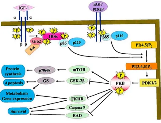
Figure 1.5: Schematic modet showing the signaling cascade induced by receptor tyrosine kinases leading to PKB activation. Insulin-like growth factor (IGF-l), by binding to its receptor (IGF-1R) enhances tyrosine phosphorylation of the insulin receptor substrates (IRSs). Phosphorylated IRSs recruit Src homo[ogy 2 (SH2) domain containing proteins, such as Grb2/Sos and p85 subunit of the lipid kinase P13-K, leading
to activation of P13-K. Activated P13-K catalyzes the phosphorylation of PW2 into PW3. PW3 binds to Pleckstrin homology (PH) domain of PKB and recruits it to the plasma
membrane for phosphorylation by PDKÏ/2. Actïvated PKB phosphorylates several
targets sucli as mTOR, GSK-3f3, FKHR, BA and Caspase that regulate metabolism, gene
expression and ceil survival. Similarly, epidermal growth factor (EGF) and platelet
derived growth factor (PDGF) activate P13-KIPKB signaling cascade. However, in this case, p85 subunit interacts directly with EGFR or PDGFR, triggering thereby the
activation of the pathway.
Πci
EGF/
f F
Protein 4— p7Os6k 4— mTOR PI(3,4,5)P3
Apoptosis GsK3
i—
(
PKB — PDK1/2ene expression K
a
22
The precise upstream mechanisms by which ROS activate MAPK and P13-KJPKB pathways remain unclear. However, a potential role for both growth factor receptors [99; 101; 103; 127; 129], and non-receptor protein tyrosine kinases [98-100; 103] as signal transducers of H202-induced responses bas been suggested.
1.9 The epidermal %rowth factor receptor (EGfR)
The EGFR is a receptor tyrosine kinase that is ubiquitously expressed in avariety of celi types, with the most abundant expression in epithelial ceils and many cancer celis [130-132]. It contains an extracellular ligand binding domain, a single transmembrane domain and a cytoplasmic tyrosine kinase autophosphorylation and regulatory domain (reviewed in [133]). EGFR belongs to a family containing three other members (ErbB2, ErbB3 and ErbB4) that undergo homodimenzation or heterodimerization to induce autophosphorylation and receptor tyrosine kinase activation in response to ligand binding [131;134J. Dimerization activates the intrinsic tyrosine kinase activity of the intracellular domain at different residues, resulting in the recruitment of the SH-containing domain proteins, which trigger downstream events. The phosphorylation of EGFR on tyrosine 1068 (Tyr1068) is followed by recruitment of the adaptor protein Grb2, leading to the activation of RasJERK1/2 pathway (Fig. 1.4).
1.9.1 Transactivation of the EGFR
The EGFR can also undergo phosphorylation in a ligand-independent manner by a process called transactivation and tngger the response of many agonists, such as Angli, ET-1, thrombin, lysophosphatidic acid, H202 and others. Thus, EGFR was identified as
an essential link in AngII-mediated activation of MAPK [135;136] andPKB [137;138J in VSMC. In the same context, Voisin et al. have reported that the activation of EGFR by Angil is necessary for increasing protein synthesis in vitro in cultured aortic smooth muscle celis and in vivo in the rat aorta as well as in small resistance arteries [139]. Similarly, ERK1/2 activation by ET-1 appears to be dependent on transactivation of EGFR in Rat-1 fibroblasts [140], VSMC [141], cardiomyocytes [142] and rat mesangial cells [143].
H202 has been shown to enhance tyrosine phosphorylation of EGFR in endothelial ceils [144], renal cells [99], HeLa cells [127] as well as in VSMC [129;145;146]. Whether H202 directly activates the intnnsic tyrosine kinase activity of EGFR or modulates signaling molecules that transactivate the receptor is stiil unclear. There are some reports proposing that ROS may exert their effects through targeting the cysteine regions of the active sites of tyrosine phosphatases, which in tum activates tyrosine kinases [147]. In fact, H202 was shown to inhibit the dephosphorylation of the EGFR by inhibiting a tyrosine phosphatase [148]. Some other reports suggest that ROS may activate receptor tyrosine kinases by generating growth factors, such as heparin binding EGF-like growth factor (HB-EGF) through metalloprotease-dependent cleavage [129]. ROS production [149] and metalloprotease-dependent RB-EGF generation are also implicated in EGFR transactivation by different GPCR agonists in many ceil types [150;151]. Frank et al. have demonstrated that both mechanisms are necessary for EGFR transactivation by AnglI in VSMC [152;153].
The tyrosine phosphorylation of the EGFR by ROS is accompanied by its association with Grb2 leading to activation of ERK1/2, which appears to be an important
24
mechanism in triggering MAPK activation induced by H202 in vanous celi types [99;1O1;103;145;154J. On the other hand, studies on the involvement of EGFR transactivation in H202-induced PKB phosphorylation gave discrepant resuits: for example, EGFR transactivation was implicated in H202-induced PKB phosphorylation in HeLa cells [127], but flot in renal cells [99] or in PC12 ceils [155]. However, no attempts have been made to investigate a possible role for EGFR in mediating the H202-induced phosphorylation of PKB in VSMC.
1.10 The insulïn-like growth factor type 1 receptor (IGF-1R)
The IGF-1R is a transmembrane protein tyrosine kinase that shares structural and functional homology with the insulin receptor and is abundantly expressed in VSMC. The mature receptor is a tetramer consisting of 2 extracellular a-chains and 2 intracellular 3-chains [156]. The 3-chains include an intracellular tyrosine kinase domain that is believed to be essential for most of the receptor’s biologic effects [157]. Binding of IGF 1 or Insulin (at very high, unphysiological concentrations) induces the activation of PTK domain of IGF-1R13 subunit which in tum activates the autophosphorylation of the receptor (reviewed in [158]).
One of the earliest steps in signal transduction initiated by the IOF-1R is the phosphorylation of adaptor/docking proteins such as insulin receptor substrate (WS-1 or IRS-2), Shc and Grb2 [159;160]. WS-1, an important substrate for both the insulin and the IGF-Ï receptor, contains multiple tyrosine phosphorylation sites that recognize and bind SH2-containing signaling molecules, such as Grb2, Nck, the p85 subunit of P13-K and the SH2 domain-containing tyrosine phosphatase-2 (SHP-2) [159]. 0f these, the
binding of Grb2ISos to tyrosine-phosphorylated WS-1 activates Ras, which then stimulates the Raf-1/MAPK cascade [161]. Shc can also interact directly with IGF-1R [162]. After tyrosine phosphorylation of Shc, it recruits the Grb2/Sos complex and activates the Ras/Raf-1/MEKIERK pathway [161] (Fig. 1.4). The activated IGF-1R also triggers the P13-K and its downstream targets PKB and p7Os6k [163;164J (Fig. 1.5). The Grb2-associated binding 1 (Gabl) can also function as an adaptor protein in mediating IGF-W-induced signaling events [165] by interacting with Grb2, p85 subunit of P13-K and SHP-2 [166]. As stated earlier, activation of MAPK pathway is critical for ceil proliferation, whereas the P13-K pathway mediates the metabolic and antiapoptotic response of IGF-1.
1.10.1 Transactivation of the IGF-1R
An important role for IGF-YR transactivation in triggenng AngII-induced responses in VSMC was reported recently [167;168]. Pharmacological inhibition of IGF 1R kinase was found to markedly inhibit AngII-induced ERKI/2 phosphorylation in rat V$MC [167] and P13-KIPKB activation in VSMC denved from the left anterior descending coronary artery of porcine hearts [168]. It was also demonstrated that Angli enhances the phosphorylation of the IGF-lRf3 subunit [16$] and that this phosphorylation was prevented by AG53$ and AG1024, two selective inhibitors of IGF-1R tyrosine kinase [169;170]. Interestingly, AngII-induced activation of MAPK appeared to be IGF 1R independent [168]. It should also be noted that the IGF-1R expression in VSMC is regulated by several factors, including AngIl [171] and ROS [172]. Moreover, the up
26
regulation of IOF-ÏR expression by Angil and basic fibrobÏast growth factor in VSMC appears to be a critical determinant for their mitogenic effects [171].
Recently, Tabet et aï. have demonstrated that H202-induced ERK1/2 was blocked by AG1024 in cultured VSMC from mesentenc arteries, suggesting a role for IGF-YR transactivation in mediating the H202 response [103]. Whether H202 enhances the tyrosine phosphorylation of IGF-1R or utilizes this receptor to activate other downstream signaling components in VSMC has flot yet been investigated.
1.11 The ylatelet-derived growth factor receptor (PDGFR)
The PDGFR is a receptor tyrosine kinase expressed in many celi types including VSMC [173] and its expression can be stimulated by AngIl [174]. It exists in two isoforms: PDGFRŒ and PDGFRf3 that display affinity for the different isoforms of PDGF family PDGF-AA, PDGF-AB, PDGF-BB, PDGF-CC and PDGF-DD. PDGFRŒ can be activated by PDGF-AA, PDGF-AB, PDGF-BB and PDGF-CC, while PDGF-BB and PDGF-DD bind and activate PDGFR (reviewed in [175]). Tissue culture and in vivo mouse models studies have suggested that PDGFRŒ and PDGFRI3 activate distinct signaling pathways. Downstream targets of the PDGFRF3 include ERK, PKB and smaïï G proteins including Rho and Rac-1, which ultimately mediate PDGF-induced responses such as celi cycle progression, migration, and survival (reviewed in [176]). Similar to EGF, binding of PDGF to its receptor on the celi surface induces its dimenzation and autophosphorylation of the tyrosine kinase domain which in tum recruits and activates SH2 domain containing proteins such as Grb2, Src, p85 subunit of P13-K and phospholipase C (PLCy). The Tyr75’ in the kinase domain of PDGFRf3 is the docking
site for P13-K. Tyr74° is also important for the activation of P13-K by PDGFRf3 while PDGF-stimulated PLCy signaling is dependent on the phosphorylation at the two sites Tyr’°°9 and Tyr’°2’ (reviewed in [175]) (Fig. 1.4 and 1.5).
1.11.1 Transactivation of the PDGFR
Several studies have shown that PDGFR undergoes tyrosine phosphorylation in response to AngIl [177] as well as ROS in different celi types such as MCF1OA [9$], NIH3T3 fibroblasts [17$] and VSMC prepared from the thoracic aorta of Sprague Dawley rats [146]. H202 was found to enhance the Tyr’°2’ phosphorylation of PDGFRf3 in a $rc- and PKOE-dependent manner [146]. On the other hand, unlike the EGFR transactivation by ROS, PDGFR transactivation was shown to be independent of metalloproteases effect [146]. An implication of PDGFR in ROS-induced activation of MAPK pathway in VSMC derived from mesentenc arteries has been studied by using AG1295, a selective inhibitor of PDGFR kinase, and it was found that this inhibitor blocked only the phosphorylation of ERK1/2 but had no effect on p3$mapk phosphorylation in response to H2O2 [103]. However, a similar involvement of PDGFR in H202-induced phosphorylation ofPKE bas flot been investigated to date.
1.12 The Src-family tyrosine kinases (SFKs)
The SFKs, a family of non-receptor tyrosine kinases, consists of nine structurally related peptide members: c-Src, Fyn and Yes are widely expressed in most tissues, whereas the others (Lck, Lyn, Hck, Fgr, Blk, Yrk) have a more restricted distribution (reviewed in [179]). SFK are 52—62 kDa proteins composed of six distinct functional
28
regions: the SH4 domain, the unique region, the SH3 domain, the SH2 domain, the catalytic domain (SH1), and a short C-terminal tau containing a negative regulatory tyrosine residue (Tyr3° in humans) (reviewed in [179]). The SH2 and SH3 domains are important for molecular interactions that regulate Src catalytic activity, Src localization, and recruitment of substrates, whereas the SH1 kinase domain contains autophosphorylation site, which is important for regulation of its catalytic activity. This site of phosphorylation corresponds to Tyr416 in mouse c-Src and Tyr419 in human’s, which is flot phosphorylated in inactive wild type Src, but is constitutively phosphorylated in activated Src (reviewed in [179]).
1.12.1 Activation of SFKs and its implication in signal transduction pathways SFKs can be activated by various extra cellular stimuli, such as antigens, growth factors, integnns [179], AngIl [180], ET-1 [181] and oxidative stress [99;100;182] in vanous ceil types (Fig. 1.3). Among the SFKs, c-Src is the major isoform in the vascular wall [183] and appears to be involved in contraction [184], proliferation [185], growth [186] and cytoskeletal reorganization [187].
In the last few years, much attention has been given to the critical role of c-Src as a mediator of GPCR- and H202-induced responses: transactivation of EGFR by GPCR agonists [140;188;189] as well as by H202 [99] in different cell types were shown to be c Src-dependent. Similarly, PDGFR transactivation by ROS seems to be Src-dependent in VSMC [146] and in NIH3T3 fibroblasts [178].
Src is not only ïnvolved in GPCR-induced transactivation of RTK, but also in tnggering the activation of MAPK and P13-KJPKB pathways in many celI types. Several
studies reported that GPCR-mediated activation of the Ras/MAPK pathway involves Src function [190-192]. Particularly in VSMC, AngII-induced activation of ERKÏ/2 pathway, which is a potential contributor pathway to vascular remodeling, appears to be mediated by Src [186]. Similarly, Src was shown to be a critical link in ET-1-induced ERK1/2 activation in rat myometnal cells [1811 and in ET-1-induced cardiomyocyte hypertrophy [193].
Pharmacological approaches have suggested a role of c-Src in mediating H202-induced ERK1/2 phosphorylation in mesenteric arteries VSMC [103], in CHO-W celis [100] and in renal celis [99;154J. Moreover, c-Src-PTK activity appears to be required in mediating H202-induced PKB phosphorylation in different ceil types such as CHO-W [100], renal ceils [99] and in Rat-2 fibroblasts [98]. In contrast, diethylmaleate, which is known to induce an increase in intracellular ROS levels, bas enhanced ERK1/2 phosphorylation in a Src-independent fashion in Rat-2 fibroblasts [98]. Conversely, a recent report has shown that H202 markedly inactivates Src in vivo, but flot in vitro and that a reduced phosphorylation of the tyrosine residue in the activation ioop is responsible for the inactivation in vivo [194]. Therefore, it appears that H202 exerts both activation and inhibitory effect on Src and triggers the responses of several agonists in a celi specific manner and differently in vitro and in vivo.
1.13 The Proline rich tyrosine kinase 2 (Pyk2)
Pyk2 is a non-receptor tyrosine kinase, also known as related adhesion focal tyrosine kinase (RAFFK) [195] or celi adhesion kinase beta (CAK) [196] or calcium dependent tyrosine kinase [197]. It is a member of a family of tyrosine kinases that also
30
includes p125 focal-adhesion kinase (FAK). Pyk2 and FAK are structurally related and are both implicated as important integrating molecules in signal transduction cascades. Pyk2 is expressed mainly in the nervous system, while FAK is widely expressed in vanous tissues [198;199]. Pyk2 contains a central catalytic domain and two non-catalytic domains, the N- and the C-terminal domains. The N-terminal domain contains a tyrosine autophosphorylation site at Tyr402, which serves as a docking site for SFKs to bind via their SH2 domains (reviewed in [199]). Studies have shown that Pyk2 plays a key role in ceil signaling in many cell types, including rat pheochromocytoma PC12 cells [155;190;19$], embryonic mouse fibroblasts [188], clone 9 (C9) hepatic celis [200] and VSMC [32;201-205].
1.13.1 Activation of Pyk2 and its implication in signal transduction pathways Pyk2 is activated by phosphorylation on tyrosine residues (Tyr402’ 580 and 881) (reviewed in [199]) in a Ca2t and PKC-dependent fashion in PC12 [198] and VSMC [201] in response to stimulation by GPCR agonists. Moreover, Pyk2 is known to interact physically with SH2 domain containing molecules such as Src in PC12 cells [190;198;206J, mouse embryonic fibroblasts [188], and VSMC [201]. This complex formation between Pyk2 and Src is important in mediating GPCR-induced activation of many downstream signaling, such as EGFR transactivation in mouse fibroblasts [18$] and VSMC [207].
Several reports have shown that activated Pyk2 signals through the MAPK family, such as ERK1/2 [19$], c-Jun amino-temiinal kinase (JNK) [190] or p3$mapk [208] in PC12 cells. In VSMC, Rocic et al. have shown that Pyk2 acts as an upstream
modulator of multiple signaling pathways involved in AngII-induced VSMC growth: Angli ïnduced the formation of a complex between Pyk2 and the ERK1/2 regulators Shc and Grb2 [204]. A role for Pyk2 in AngII-induced activation of P13-K and p70s6 kinase has also been reported [204] (Fig. 1.3). Later on, the same group demonstrated by using genetic approach the involvement of Pyk2 in AngII-induced protein synthesis in VSMC [209]. Another vasoactive peptide, ET-1, was shown to induce tyrosine phosphorylation of Pyk2 in cardiomyocytes [142;210] and rabbit carotid artery VSMC [211] as well as in embryonic VSMC of rat aorta [32]. In cardiomyocytes, Pyk2 activation by ET-1 is dependent on Src and PKC epsilon (PKC) [212], as well as Ca2 [142;210;212] and is followed by ROS generation [210]. Kodama et al. have reported that Pyk2 was involved in ET-1-induced ERK activation [142] and that Pyk2 activation as well as Pyk2/Src association was required for ET-1-induced JNK activation in cardiomyocytes [213]. Furthermore, Pyk2 activation appears to be cntical in ET-1-induced cardiomyocyte hypertrophy [210].
1.13.2 Activation of Pyk2 and its implication in cardiovascular diseases Direct stimulation with exogenous F1202 was shown to enhance the tyrosine
phosphorylation of Pyk2 in many cell types, such as PC12 [155] and VSMC [203]. Recent studies have suggested that Pyk2 may represent a key signaling molecule involved in vascular diseases because an upregulation in the basal phosphorylation of Pyk2 in VSMC from SHR as compared with VSMC from WKY has been documented recently. Moreover, AngII-enhanced phosphorylation of Pyk2 appeared to be more rapid and more potent in SHR in these studies [214]. More recently, an involvement of Pyk2 in
32
AngII-induced VSMC migration via JNK activation has also been reported, further strengthening an important role for Pyk2 in VSMC functioning [215].
1.14 General Conclusion
In summary, oxidative stress is believed to be involved in vascular abnormalities, such as hypertension and atherosclerosis. A number of studies have shown that ROS activate key components of growth promoting and proliferative signaling pathways, such as ERK1/2 and PKB in VSMC. An important role for receptor and non-receptor tyrosine kinases in triggering some of the H202-evoked responses has been demonstrated in many ceil types, including VSMC. Several studies carried out in VSMC isolated from different vessel types have revealed that many tyrosine kinases act as upstream components in H202-induced ERKI/2 phosphorylation. These tyrosine kinases include both growth factor receptors and non-receptor tyrosine kinases. Most of these studies on ROS-induced signaling in VSMC have focused on MAPK pathways and the intermediary role of EGFR transactivation in this process (fig. 1.6). However, the potential contribution of other growth factor receptors has flot been investigated in any detail and not much is known on the mechanisms by which ROS activate PKB signaling in VSMC. Therefore, the studies presented in this thesis were undertaken to elucidate a possible role for receptor and non receptor tyrosine kinases on PKB, Pyk2 and ERKÏ/2 phosphorylation by H202 in VSMC. These studies have used standard protocols of cellular and molecular biology such as ceil culture, immunoprecipitation and western blotting.
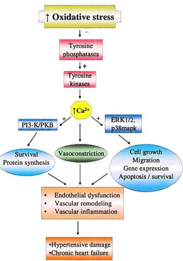
33 Oxidative stress Tyrosine phosphatascs Tyr&sinê kinases
I
1Ca2 +7 -J11KBZ’ Vasoconstriction rthProteïn synthesis—----iiai Migration
I
• Endothelial dysfunction • Vascular remodeling • Vascular inflammationI
•Hypertensive damage •Chronic heart failureFigure 1.6: Scheme summarizing the major ROS-induced signalïng pathways that are responsible of their pathological effects. High levels of ROS or oxidative stress, by inhibiting protein tyrosine phosphatases and activating thereby protein tyrosine kinases, trigger the activation of two major signaling pathways, the MAPK and the P13-KIPKB pathway in a Ca2-dependent fashion. Aberrant activation of these pathways enhances protein synthesis, celi growth, hypertrophy, migration as well as survival responses, which are critical events in vascular remodeling, associated with cardiovascular
disorders. Increased Ca2 levels may also have a direct effect on vascular disorder by modifying the contractile response of the vascular smooth muscle cells.
34
CHAPTER 2
ARTICLE
Activation ofinsuÏin-like growthfactor type-] receptor is required
for H202-induced PKB phosphorylation in vascular smooth muscle
celis
(Published in Canadian Journal of Physiology and Pharmacology
Activation of insulin-like growth factor type-1 receptor is
required for H202-induced PKB pliosphorylation in vascular
smooth muscle ceils
Zeina M. Azar, Mohamad Z. Mehdi and Ashok K. Snvastava
Laboratory of Ceil Signaling, Research Centre
Centre hospitalier de l’Université de Montréal (C1{UM)—Hôtel-Dieu
and Department of Medicine, Université de Montréal, Montreal
Q
uebec, CanadaShort titte: IGF-IR mediates H202—induced PKB phosphorylation
Address for correspondence: Ashok K. Srivastava, Ph.D.
Research Centre, CHUM- Hôtel-Dieu
3850, St. Urbain Street, Rm 7-13 5 Montreal (Quebec) H2W 1T7 Canada
Tel.: (514) $90 8000 ext. 12917 Fax: (514) 412 7152
36
ABSTRACT
Evidence accumulated in recent years lias revealed a potential role of reactive oxygen species (ROS) in the pathophysiology of cardiovascular diseases. However, the precise mechanisms by which ROS contribute to the development of these diseases are flot fuÏly identified. Previous work from our laboratory lias indicated tliat exogenous liydrogen peroxide (H202) activates several signaling protein kinases, such as extracellular signal regulated kinasel and 2 (ERK1/2) and protein kinase B (PKB) in AlO vascular smootli muscle cells (VSMC). However, the upstream elements responsible for this activation remain to be clarified. Although an important role of epidermal growth factor receptor (EGFR) protein tyrosine kinase (PTK) in H202-induced ERK1/2 signaling lias been suggested, tlie contribution of this PTK or other receptor or non-receptor PTK in PKB activation is not well defined in VSMC. In tlie present study, we liave investigated tlie role of receptor and Src-family-PTKs in H202-induced PKB pliosphorylation using pliarmacological inhibitors. AG1478, a specific inliibitor of EGFR, failed to attenuate tlie H202-induced increase in PKB Ser473 phospliorylation, whereas AG1024, an inhibitor of insulin-like growth factor typel receptor (IGF-1R)-PTK, almost completely blocked tliis response. H202 treatment also enlianced tyrosine pliospliorylation of the IGF-lRf3 subunit, wliich was significantly inliibited by AG1024 pretreatment of cells. Furtliermore, pharmacological inliibition of Src by PP2 (4-amino-5-(4-clilorophenyl)-7-(t-butyl)pyrazole[3,4-d] pyrimidine) decreased PKB pliospliorylation. Moreover, H202-induced PKB pliosphorylation was associated witli increased tyrosine phospliorylation of c-Src and Pyk2 in an AG1024 and PP2-inliibitable manner. In conclusion, tliese data provide evidence for the contribution of IGF-1R-PTK in initiating H202-evoked PKB pliosphorylation in AlO VSMC witli an intermediary role of c-Src and Pyk2 in tliis process.
Key words: Protein kinase B, Hydrogen peroxide, Growth factor receptor protein tyrosine kinase, Src-family protein tyrosine kinase, Proline-ricli tyrosine kinase, Vascular smootli muscle cells.
Introduction
Reactive oxygen species (ROS) are believed to play a critical role in the pathophysiology of cardiovascular diseases such as hypertension, atherosclerosis and restenosis after angioplasty (Dhalla et al. 2000; Droge 2002; Fortuno et al. 2005; Pollock and Pollock 2005), as well as in vascular complications associated with diabetes (Ceriello and Motz 2004). Two key vasoactive peptides, angiotensin II (Angil) and endothelin-1 (ET-1), that are also important growth factors for vascular smooth muscle cells (VSMC) (Battistinï et al. 1993; Daou and Srivastava 2004; Touyz et al. 2004), and are believed to contribute to vascular diseases (Touyz and Schiffrin 2003; Pollock and Pollock 2005; Sainani et al. 2005; Sun 2005; Tostes, and Muscara 2005) mediate their vasoactive responses through ROS generation (Zafan et al. 199$; Fei et al. 2000; Wedgwood. et al. 2001; Daou and Srivastava 2004). An important source of ROS in VSMC is NAD(P)H oxidase, which catalyzes the generation of superoxide anion (Oj) from molecular oxygen (02) (Ushio Fukai et al. 1996; Touyz et al. 2002; Niemiec and Zak 2005). 02 is a relatively unstable molecule and is rapidly converted by a dismutation reaction to hydrogen peroxide (H202) (Bovens and Chance 1973).
Recent studies have demonstrated an exaggerated production of ROS such as 02 and H202 in many animal models of hypertension (Guo et al. 2003; Li et al. 2003; Sedeek et al. 2003). Moreover, a direct role of H202 in AngII-induced vasculature hypertrophy has been suggested recently (Zhang et al. 2005). Exogenous H202 activates several signaling protein kinases, such as mitogen-activated protein kinases (MAPK) and protein kinase B (PKB) (Wang et al. 2000; Blanc et al. 2003; Blanc et al. 2004; Mehdi et al. 2004; Zhuang and Schnellmann 2004), which have been proposed to play key roles in mediating the hypertrophie response in VSMC (Daigle et al. 2004). Although the precise mechanism
38
and intermediary steps by which H202 activates these signaling pathways remain poorly characterized, the involvement of receptor and non-receptor protein tyrosine kinases (PTK) as potential transducers of H202-evoked responses lias been suggested (Frank et al. 2003; Melidi et al. 2004; Zhuang and Sclinellmann 2004; Purdom and Clien 2005; Tabet et al. 2005). Specifically, a prominent contribution of epidermal growth factor receptor (EGFR) transactivation in mediating H202-induced activation of extracellular signal regulated kinase 1 and 2 (ERK1/2) lias been documented in many ceil types (Zhuang and Schnellmann 2004; Purdom and Chen 2005). However, a precise role of growtli factor receptor and Src-family PTKs in mediating H202-induced effects on PKB signaling in VSMC bas not yet been elucidated.
PKB, also known as Akt, exists as 3 isoforms, a,
f3
and y (Aktl, Akt2 and Akt3, respectively); eacli isoform possesses an amino-terminal pleckstrin liomology (PH) domain, a kinase domain and a carboxy-terminal regulatory domain. PKB is rapidly activated in response to insulin (Farese et al. 2005; van der Heide et al. 2005), AngIl (Saward and Zahradka 1997), ET-1 (Daou and Srivastava 2004), and many other growtli factors (Duan et al. 2000; Cenni et al. 2003; Dolloff et al. 2005; Markadieu et al. 2005; Zaka et al. 2005) in a pliospliatidylinositol 3-kinase (PI3K)-dependent manner. PI3K is a lipid kinase that promotes the generation of 3’-phosphoinositides, such as phosphatidyl inositol 3,4,5 triphosphate (PW3). PW3 binds to the PH domain ofPKB and recruits it to the plasma membrane for phosphorylation by phosplioinositide-dependent kinases 1 (PDK-1) and 2 (PDK-2). PDK-1 phosphorylates PKB at threonine 308 in the catalytic domain whereas putative PDK-2 phosphorylates it at serine 473 in the C-terminal regulatory domain of PKB (Whiteman et al. 2002). Activated PKB phosphorylatesseveral downstream substrates, such as glycogen synthase kinase-3f3, Forkhead transcription factor, Bcl-2-associated death, 1kB kinase, mammalian target of rapamycin, Mdm2, Caspase 9 and endothelial nitnc oxide synthase (Whiteman et al. 2002; Song et al. 2005). Phosphorylated forms of these substrates regulate diverse cellular functions, such as glucose transport, celi growth, gene expression, celi survival and death as well as protein synthesis (Saward and Zahradka 1997; Datta et al. 1999).
In the present study, by using a series of pharmacological and celi biological approaches, we have examined the involvement of receptor and non-receptor PTKs as potential upstream modulators of H202-induced PKB phosphorylation and activation in AlO VSMC. AlO VSMC, denved from the rat embryonic thoracic aorta, which exhibit characteristics similar to those of normal VSMC (Kimes and Brandt 1976) and have been extensively accepted as a model for the investigation of vascular cellular processes (Hashim et al. 2004, Daou and Snvastava 2004, Chen et al. 2006).
40
Materials and Methods
MateriaïsH202 was purchased from Sigma Chemical Co. (St. Louis, MO, U.S.A.). Phospho specific antibodies to PKB (Ser473) and Pyk2 (Tyr402), total PKB antibody and anti rabbit secondary antibody were from New England BioLabs (Beverly, MA, U.S.A.). Phospho-specific c-Src antibody (Tyr418) was from Biosource (Camarillo, CA, U.S.A.). Antiphosphotyrosine antibody (PY99), total c-Src antibody, total insulin-like growth factor receptor-ibeta (IGF-1R13) subunit antibody and anti-mouse secondary antibody were from Santa Cruz Biotechnology (Santa Cruz, CA, U.S.A.). Total Pyk2 antibody was from Upstate Biotechnology (Lake Placid, NY, U.S.A.). Epidermal growth factor (EGF) and ail pharmacological inhibitors were from Calbïochem (San Diego, CA, U.S.A.). Human IGF-1 was from PeproTech Inc. (Rocky Hill, NJ, U.S.A.). Ail cell culture materials were from Invitrogen Corp. (Grand Island, NY, U.S.A.). Protein A Sepharose beads and enhanced chemiluminescence (ECL) detection kits were from Amersham Pharmacia Biotech (Baie d’Urfé, Quebec, Canada).
Ce!! Culture
AlO VSMC, obtained from the American Type Culture Collection (Manassas, VA, U.S.A.), were maintained in Dulbecco Modified Eagle Medium (DMEM) containing 10% of fetal bovine serum and 1% of penicillin + streptomycin in a humidified
atmosphere of 5% C02 exchange at 37°C as described earlier (Srivastava and Pandey 2000). They were passaged twice a week by harvesting with TrypsinIEDTA.