Université de Montréal
Exploration of the Cerebral Dysfunctions Induced by
Arterial Rigidity and/or the Overexpression of TGFβ in a
Mouse Model
par Sherri Bloch
Département de Pharmacologie et Physiologie Faculté de Médecine
Mémoire présenté
en vue de l’obtention du grade de Maîtrise en Pharmacologie
Juin 2017
Résumé
Exploration des dysfonctions cérébrales induites par la rigidité artérielle et / ou la surexpression de TGFβ chez la souris
Introduction: Les déficits cérébrovasculaires au cours de la maladie d'Alzheimer (MA) et la démence vasculaire (DV) sont multiples et ne se limitent pas à la pathologie amyloïde-β (Aß). Les modifications comprennent des modifications de structure des vaisseaux sanguins telles que reproduites dans les souris transgéniques qui surexpriment le facteur TGF-β1, une cytokine accrue dans le cerveau avec démence vasculaire et les patients avec la MA. Une circulation cérébrale compromise de façon chronique comme on le voit chez les souris surexprimant la TGF-β1 peut ainsi précipiter les troubles cognitifs lorsqu'elle est combinée avec des facteurs de risque pour les démences, telle que la rigidité artérielle. Objectif: Déterminer si la rigidité artérielle induite par calcification va aggraver les dysfonctions vasculaires cérébrales chez les souris surexprimant la TGF-β1 et déclencher des troubles cognitifs. Méthodologie: Nous avons testé si la rigidité artérielle induite par une chirurgie servant à calcifier des artères carotides pourrait induire les troubles de la mémoire et d’apprentissage chez les souris de type sauvage ou souris surexprimant la TGF-β1. L’apprentissage, la mémoire spatiale et la consolidation de la mémoire ont été étudiées avec la Piscine de Morris et le test de reconnaissance des objets nouveaux (RON) à un, deux et quatre mois après la chirurgie. Résultats: Aucune différence significative n'a été observée entre tous les groupes dans le test de RON spatiales (1, 2 et 4 mois post-chirurgie). Cependant, à 4 mois post-opératoire, les souris de type TGFβ dont la carotide a été calcifiée ont montré une mauvaise consolidation de la mémoire par rapport à des souris de type TGFbeta dont la carotide n’a pas été calcifiée (p <0,05). Conclusion: Ces résultats suggèrent que la calcification artérielle et la surexpression de TGF-β1 peuvent agir en synergie pour aggraver les troubles de la mémoire. Par conséquent, ces deux voies pourraient constituer des cibles thérapeutiques pour prévenir les démences.
Mots-clés : TGFβ, rigidité artérielle, démence, Alzheimer, démence vasculaire, piscine de Morris, calcification, dysfunction cérébrale, unité neurovasculaire
Abstract
Exploration of the Cerebral Dysfunctions Induced by Arterial Rigidity and/or the Overexpression of TGFβ in a Mouse Model
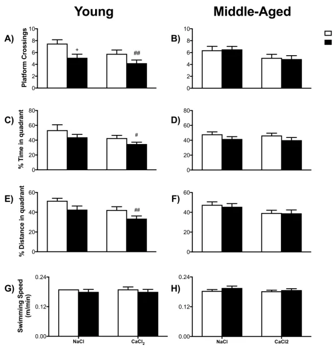
Introduction: Alzhemer’s disease (AD) and vascular dementia (VaD) is multifaceted with multiple cerebrovascular deficits and not limited to the amyloidβ (Aβ) pathology. Cerebral modifications include changes to vascular structure, which can be reproduced in transgenic mice overexpressing transforming growth factor β1 (TGFβ1), a cytokine found in increased quantities in brains of AD and VaD patients. The mouse model overexpressing TGFβ demonstrates a chronically compromised cerebral circulation, which may precipitate cognitive deficits if combined with other risk factors of cognitive decline, such as arterial rigidity. Objective: To determine whether arterial rigidity induced by calcification will provoke cerebrovascular dysfunctions in the TGFβ mouse and trigger cogitive deficits. Methodology: We tested whether arterial rigidity induced by calcification of the right carotid artery trigger deficits in memory and learning in mice overexpressing TGFβ1 and their wild type litermates in a set of young and a set of aged mice. Spatial memory and memory consolidation were studied via the Morris water maze (MWM) and novel object recognition (NOR) at 1-, 2- and 4-months followin surgery to induce arterial calcification. Results: No significant difference was observed between all groups in the NOR test or the spatial learning of MWM (1-, 2- and 4-months post-surgery). For the memory consolidation tests of the MWM, young TGF mice with calcification showed significance at 4-months post-surgery (p <0,05) while TGF mice without calcification failed to demonstrate cognitive deficits. Conclusion: These results suggest that arterial calcification and overexpression of TGFβ1 may act in a synergistic manner to trigger cognitive deficits. Therefore, these two pathways may constitute possible therapeutic targets for prevention of dementia.
Keywords : TGFβ, arterial rigidity, dementia, Alzheimer, vascular dementia, Morris water maze, calcification, cerebral dysfunction, neurovascular unit
Table of Contents
RESUME ... I
TABLE OF CONTENTS ... III
LIST OF TABLES ... VI
LIST OF FIGURES ... VII
LIST OF ABBREVIATIONS ... VIII
ACKNOWLEDGEMENTS ... XII
INTRODUCTION ... 1
LITERATURE REVIEW ... 3
1. DEMENTIA ... 3
1.2 Vascular Dementia ... 4
1.2.1 Neuropathological features of VaD ... 5
1.2.2 Brain-‐related structures of VaD ... 6
1.3 AD and VaD Comorbidity ... 7
1.3.1 Current hypotheses linking AD and VaD ... 9
1.3.2 Mouse model ... 10
2. NEUROVASCULAR COUPLING ... 11
2.1 Vasculature of the CNS, Aging and Dementia ... 11
2.1.1 Macrovasculature ... 12
2.1.2 Microvasculature ... 14
2.1.3 Pericytes ... 17
2.2 The Neurovascular Unit, Aging and Dementia ... 19
2.2.1 Neurons ... 21 2.2.2 Glial Cells ... 22 2.2.2.1 Astrocytes ... 22 2.2.2.2 Microglia ... 24 2.2.2.3 Oligodendrocytes ... 25 2.3 TGFβ-‐associated pathologies ... 25
2.3.1 Physiological use of TGFβ in Brain ... 26
2.3.2 Impact of TGFβ-‐associated vascular pathologies on NVC ... 27
3. ARTERIAL RIGIDITY ... 28
3.1 Layers of the vasculature ... 29
3.1.2 Vascular Compliance and distensibility ... 32
3.2 Arterial Rigidity, Aging and Dementia ... 32
3.2.1 Arterial Stiffness model ... 34
3.2.1.1 Vascular significance in the mouse model ... 34
3.2.1.2 Neurological significance in the mouse model ... 36
4. NEUROLOGICAL INSULTS ... 37
4.1 Oxidative stress ... 37
4.1.1 Free Radicals: Formation and Biological Uses ... 38
4.1.2 Markers of Oxidative stress ... 39
4.2 Gliosis ... 40
4.2.1 Microglia: Activation and labeling ... 41
4.2.2 Astrocytes: astrogliosis and labeling ... 42
5. BEHAVIORAL TESTING ... 43
5.1 Morris Water Maze ... 43
5.1.1 Brain-‐related areas ... 44
5.1.2 Limitations ... 45
5.1.3 MWM and TGF mice ... 45
5.2 Novel Object Recognition ... 46
5.2.1 Brain-‐related areas ... 46
5.2.2 Limitations ... 47
5.2.3 NOR and TGF mice ... 47
HYPOTHESIS AND OBJECTIVES ... 48
METHODS ... 50
RESULTS ... 59
DISCUSSION ... 87
PART I – BAHAVIORAL TESTING ... 87
Morris Water Maze ... 87
Novel Object Recognition ... 89
PART II – VASCULAR ASSESSMENTS ... 91
Circumferential Strain ... 91
Gliosis ... 93
Oxidative Stress ... 95
PART IV – LIMITATIONS ... 97
CONCLUSION ... 97
List of Tables
TABLE I VAD RISK FACTORS [MODIFIED FROM (12)] ... 5
List of figures
FIGURE 1 VASCULAR DEMENTIA BRAIN TARGETS AND EFFECTS (MODIFIED FROM(13)) ... 7
FIGURE 2 MACROVASCULATURE AND MICROVASCULATURE ... 14
FIGURE 3 COMPONENTS OF THE NEUROVASCULAR UNIT ... 20
FIGURE 4 LAYERS OF THE VASCULATURE ... 30
FIGURE 5 MWM ESCAPE LATENCIES OF YOUNG AND OLDER MICE THROUGH TIME. ... 59
FIGURE 6 MWM PROBES OF YOUNG AND OLD MICE CONDUCTED 1 MONTH POST-‐SURGERY. ... 61
FIGURE 7 MWM PROBES OF YOUNG AND MIDDLE-‐AGED MICE CONDUCTED 2 MONTHS POST-‐SURGERY. ... 63
FIGURE 8 MWM PROBES OF YOUNG AND MIDDLE AGE MICE CONDUCTED 4 MONTHS POST-‐SURGERY. ... 65
FIGURE 9 NOVEL OBJECT RECOGNITION OF YOUNG AND OLDER MICE. ... 67
FIGURE 10 CIRCUMFERENTIAL STRAIN DETECTED IN OLDER MICE. ... 69
FIGURE 11 CEREBRAL BLOOD FLOW OF OLDER MICE 5 MONTHS POST-‐SURGERY. ... 71
FIGURE 12 PERCENTAGE OF AREA STAINED FOR GFAP OR IBA1 IN THE CORTEX OF YOUNG MICE. ... 73
FIGURE 13 CORTEX OF MIDDLE AGE MICE, COMPARING DAB AND FLUORESCENT STAINING. ... 75
FIGURE 14 HIPPOCAMPAL CA1 REGION OF AGED MICE, COMPARING DAB AND FLUORESCENT STAINING. ... 77
FIGURE 15 HIPPOCAMPAL CA3 REGION OF AGED MICE, COMPARING DAB AND FLUORESCENT STAINING. ... 79
FIGURE 16 HIPPOCAMPAL DG REGION OF AGED MICE, COMPARING DAB AND FLUORESCENT STAINING. ... 81
FIGURE 17 4-‐HYDROXYNONENAL (HNE) ELISA ASSESSING OXIDATIVE STRESS IN THE CORTEX AND HIPPOCAMPUS OF YOUNG AND AGED MICE. ... 83
List of abbreviations
3-NT 3-Nitrotyrosine AD Alzheimer’s Disease
Aβ Amyloid β
APP Amyloid precursor protein ATP Adenosine triphosphate BBB Blood brain barrier Ca2+ Calcium ion
CA1 Cornus Ammonis 1 of the hippocampus CA3 Cornus Ammonis 3 of the hippocampus CAA Cerebral amyloid angiopathy
CaCl2 Calcium chloride CBF Cerebral bloof flow COX-1 Cyclooxygenase-1 CNS Central nervous system
DG Dentate gyrus of the hippocampus DHE Dihydroethidium
E East
EC Endothelial cell ECM Extracellular matrix
EDHF Endothelium derived hyperpolarizing factor eNOS Endothelial nitric oxide synthase
GFAP Glial fibrillary acidic protein HNE 4-Hydroxynonenal
H2O2 Hydrogen peroxide HOO Hydroperoxy radical
Iba1 Ionized calcium-binding adapter protein molecule 1 IFN-γ Interferon γ
iNOS Inducible nitric oxide synthase L Lipid radical
LO Lipid hydroxyl radical LOO Lipid peroxyl radical LOOH Lipid hydroperoxide LPS Lipopolysaccharide MWM Morris Water Maze
N North
nNOS Neuronal nitric oxide synthase
NADPH Nicotinamide adenine dinucleotide phosphate NMDA N-methyl-D-aspartate
NO Nitric Oxide
NO2 Nitrogen dioxide
NO2 Nitrogen dioxide radical NOR Novel object recognition test NVC Neurovascular coupling NVU Neurovascular unit O2- Superoxide anion OH Hydroxyl radical ONOO- Peroxynitrite
OPC Oligodendrite precursor cell PFA Paraformaldehyde
PUFA Poly-unsaturated fatty acids PWV Pulse wave velocity
RNS Reactive nitrogen species ROH Peroxyl radical
ROS Reactive oxygen species α-SMA α Smooth muscle actin
S South
SOD Superoxide dismutase SVD Small vessel disease
TGFβ1 Transforming growth factor β1 TNFα Tumor necrosis factor α
VaD Vascular dementia
Acknowledgements
First I would like to thank my principal investigator, Dr. Girouard, for believing in me and offering her continuous support. No matter how stressed I was, she always managed to put a smile on my face. Next I would like to thank Dr. Hamel who allowed me the use of her lab, her ever-helpful consultations and always inspiring me to work harder.
From the Girouard laboratory, I would like to thank Diane for her assistance in my experiments as well as easy-going advice and guidance. Furthermore, I would like to thank Kelly Xuewei Wang for always letting me know when I was headed in the wrong direction and making me laugh, Dima Obari for her awesome companionship and Gervais Muhire for his guidance and wisdom, and Florencia Iulita for imparting her vast knowledge. I also would like to thank all other members of the Girouard Lab for their friendship and support: Lin Li, Maryam Tabatabaei, Stéphane Lique, Adrian Noriega, Steven Kammi and Atef Badji.
From the Hamel laboratory, I would like to thank Clotilde Lecrux, Lianne Trigani, Maria La Calle Aurioles and Diego Fernandes for all their help, guidance and companionship during my time at their lab. I would like to especially thank Jessika Royea and Xinkang Tong for their guidance and help.
Finally, I would like to thank my family for their continued support throughout my life, which contributed to who I am today. Thanks to my parents, Tamar and Yoram Bloch; my grandparents, Violet and David Bensamuel and Roza and Leon Bloch; my brother, Jonathan Bernard Bloch; my aunts Dalia and Nora and their beautiful families. I would also like to extend my thanks to Jonathan K. Lai who always tells me to finish what I start and believe in myself and to Alia Yousif who gave me the strength to apply for a master’s degree.
Introduction
One of the major growing health issues is dementia affecting approximately 47.5 million worldwide with an expected increase of 7.7million every year. Of these numbers, Alzheimer’s disease is the cause of 60 to 80% while vascular dementia comes in next with approximately 10% of cases. Other causes of dementia include dementia with lewy bodies, Parkinson’s disease, frontotemporal dementia, Creutzfeldt Jakob disease, normal pressure hydrocephalus, Huntington’s disease and Wernicke-Korsakoff syndrome. Interestingly, in the Memory and Aging project study, 94% of patients demonstrating dementia were diagnosed with AD, and of those, 54% displayed a coexisting pathology (1). The AD exhibits multiple pathophysiological characteristics outside of the well defined Aβ pathology. Overexpression of TGFβ cytokine has been identified as an etiology of the cerebrovascular pathology of AD. TGFβ is central to fibrotic responses and is elevated in brain tissue, cerebrospinal fluid, blood and cerebral blood vessels of AD patients. Vascular fibrosis, thickened vessel walls, degenerating capillaries (also known as string vessel pathology) and hypoperfusion are all pathological events found in AD and mimicked in transgenic mice overexpressing the TGFβ1 cytokine.
Another suggested predictor of cognitive decline in AD patients is the increase in pulse-wave velocity caused by arterial stiffness. Recent studies have shown a significant correlation between increasing pulse-wave velocity and cognitive decline from normal cognitive function to impaired cognition to AD to vascular dementia. Whether the relationship between arterial stiffness and cognitive degeneration is causal, additive or synergistic has yet to be classified.
This memoir aims to elucidate the relationship between dementia, arterial stiffness and the cerebrovascular pathology found in AD and VaD. To achieve this objective, we combine a surgically altered mouse model of arterial stiffness and the TGFβ mouse model of AD. Following surgery, mice undergo behavioural testing via the Morris Water Maze and Novel Object Recognition. Cerebral blood flow is observed and brain slices are immunostained to
reveal astrocytic and microglial gliosis. Finally, lipid peroxidation is quantified by ELISA assay to reveal reactive oxygen species in the cortex and hippocampus.
Literature Review
1. Dementia
Dementia is an overall term for a decline in mental ability severe enough to affect daily activities. Symptoms may include impairment of memory, communication and language, focus and attention, reasoning and visual perception. Alzheimer’s Disease accounts for 60 to 80% of cases while Vascular dementia is the second principal type of dementia attributing to approximately 10% of cases(1, 2). To elucidate the defining characteristics of the two leading causes of dementia, significant behavioral and post mortem data have been accumulated regarding neuropathological features and changes in brain-related structures of the diseases. These findings have brought a greater awareness of overlapping AD and VaD characteristics in up to 50% of demented elder patients (3, 4). Following AD and VaD, dementia with Lewy bodies has been found to account for 10 to 25% of dementia cases, while other causes of dementia such as Creutzfeldt-Jakob disease, frontotemporal dementia, huntington’s disease and others are rare (1).
1.1 Alzheimer’s Disease
Alzheimer’s disease is a progressive neurodegenerative disorder with classic clinical characteristics such as language deterioration, visuospatial deficits and amnestic-type memory impairment (5, 6). As the leading cause of dementia world wide, much investigation has been conducted on AD forecasting future social and economical issues. In 2015, the International
Alzheimer’s Disease Report estimated 47 million people around the world to be living with AD and an anticipated increase of 131 million people by 2050 (4). To determine targets for treatment, multiple features in the AD brain have been examined.
Typical neuropathological features of AD include amyloid beta deposits into senile plaques (6, 7), hyperphosphorylated tau proteins forming neurofibrillary tangles, neuronal degeneration, loss of synapses and gliosis (4, 6). Furthermore, AD is associated with a cerebrovascular pathology where Aβ deposits are observed within the cerebrovasculature, described as
cerebral amyloid angiopathy, and an increase in transforming growth factor β1 (TGFβ1) brain levels (8).
These characteristics of AD are accompanied by alterations in brain structure and functions. Indeed, cognitive and behavioral deficits observed in AD patients have been correlated with neuronal loss and atrophy detected mainly in the hippocampus and neocortex (6). The hippocampus, which is critical for the encoding and storage of information in memory (9), is of particular interest. Experiments using MRI in AD patients have shown severe and progressive hippocampal atrophy throughout the advancement of AD as well as a decrease in the volume of the hippocampus (10, 11).
1.2Vascular Dementia
cognitive abilities. Patients are prone to suffer from delayed thinking, forgetfulness, depression and anxiety, disorientation, loss of executive functions such as problem solving, working memory, thinking, reasoning, judgment, planning and execution of tasks, and task performance declining with increased complexity (4, 12, 13). Vascular risk factors such as hypertension, hyperlipidemia, diabetes, smoking and aging have been tied to the alteration of vessels in end organs, including the brain (14) (Table I). Depending on the neuropathological features displayed, VaD has been broken into multiple subtypes and different brain-related structures may be affected.
Table I VaD Risk Factors [modified from (12)]
R
ISKF
ACTORR
EFERENCEAdiponectin (15, 16)
Apolipoprotein E4 (17, 18)
Atherosclerosis (19, 20)
Blood-brain barrier deficiency (21, 22)
Diabetes (4, 23)
Metabolic syndrome (14, 24, 25)
Hypertension (26, 27)
Aging (4, 14, 28)
Late-life depression (29, 30)
Caspase-cleaved Tau protein (31)
Stroke (32, 33)
1.2.1 Neuropathological Features of VaD
There are multiple subtypes of VaD that may or may not be due to stroke. A common neuropathological characteristic among VaD patients is cerebrovascular alterations due to vascular remodeling and disrupted blood vessel integrity caused by disturbances of the macro-
and microvasculature (3, 12). Hemodynamic abnormalities caused by vascular risk factors lead to a variety of possible lesions. The most common subtypes are multi-infarct dementia resulting from multiple small strokes, single-infarct dementia caused by a single stroke inciting hippocampal damage, small vessel disease (SVD) and mixed dementia which is based on a combination of AD and VaD pathologies (12, 13, 34).
1.2.2 Brain-Related Structures of VaD
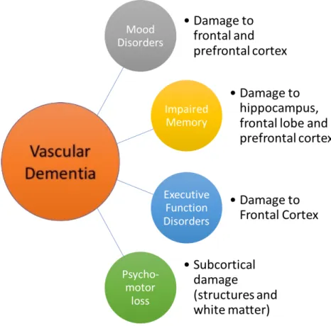
While in the case of AD specific brain-related structures have been identified, those of VaD are highly variable and dependent upon the size and location of brain injury (35, 36). However, a popular hypothesis in the field of VaD research claims that the prefrontal cortical-basal ganglia networks, periventricular white matter and the hippocampus are the locations of initial hemodynamic-based insult resulting in various pathological consequences (12). Experimental data with VaD patients show that neurodegeneration in prefrontal and frontal-subcortical circuits may be responsible for the impairment of elaboration and undertaking of memory retrieval strategies (9). A summary of possible locations of damage and the resulting behavioral features is shown in figure 1.
Figure 1 Vascular Dementia brain targets and effects (Modified from(13))
1.3 AD and VaD Comorbidity
Brain autopsies of elder patients who displayed AD dementia exhibited a spectrum of conditions, suggesting AD dementia in aged patients to be multifactorial (4). Interestingly, several studies have shown that of all mixed brain pathologies, the type most commonly demonstrated in aging community-based populations is AD and VaD (37-39). In fact, the mixed AD VaD pathology has been displayed in up to 50% of dementia cases (14). Given this information, it is important to note that though these two diseases are distinct, they can share clinically similar symptoms [see table II], which leads to confusion (12, 13). Therefore,
following the question of overlaps, another question that arises is the relationship these two diseases might share.
In the case of AD, one of the primary targets of mainstream research is misfolded amyloid-β for prevention and treatment (35). In AD patients, Aβ accumulates to form senile plaques, attach to blood vessels, which causes cerebral amyloid angiopathy (CAA), and free-float in its soluble form (4, 6, 8); however, Aβ abnormalities are also present in VaD. In fact, multiple sources in the literature define CAA as a vascular pathology of VaD (4, 40, 41). Furthermore, recent research comparing CAA found in autopsied AD, VaD and mixed dementia patients showed that the mixed dementia patients showed a higher severity of CAA and were older than the other patients (42). Additionally, multi-infarct VaD patients show an increase in amyloid precursor protein and Aβ1-42 in brain (12) as well as amyloid precursor protein mRNA increase in the periphery following stroke (12, 40).
Multiple vascular pathologies have been associated with aged AD patients, such as tortuous vessels, venous collagenosis, SVD, decreased microvascular density and microembolic brain injury (43). Again, SVD is a subtype of VaD (12, 13) and can be found in demented patients without signs of AD, such as patients with degenerative dementia like Parkinson’s Disease (44, 45).
All these similarities have lead researchers to speculate about the connection these two separate but coinciding diseases might share.
Table II Common pathological lesions in AD and VaD [data from (41)]
PATHOLOGICAL CHARACTERISTIC AD(%) VAD(%)
Cardiovascular disease 77 60
Microvascular degeneration 100 30
Cerebral Amyloid Angiopathy 98 30
Total Infarcts 36 100
Micro-infarcts 31 65
Intracerebral hemorrhage 7 15
Loss of cholinergic neurons 70 40
White matter lesions 35 70
1.3.1 Current Hypotheses Linking AD and VaD
Two main hypothesis relating AD and VaD exist in the literature: 1) the two diseases are independent but additive; 2) one disease gives rise to the other, also known as the two-hit theory. Chui et al explored the relationship between the two diseases using cognitive data from autopsied patients. According to their investigation, the cognitive deficits caused by microinfarcts were not exacerbated by the presence of AD, leading them to conclude that the two pathologies were independent but additive (35). It is interesting to note however that another author declared that microinfarcts in the cortex could give rise to the acceleration of AD (36). In the case of the two-hit theory, there are multiple models being studied in the literature. One model posits vascular damage to cause deficits in Aβ clearance and thus causing Aβ accumulation and aggregation in brain (46). Another model suggests that the chronic cerebral hypoperfusion due to vascular lesions triggers cascades leading to Aβ overproduction and tau hyperphosphorylation (47).
Due to the confusion caused by these coinciding diseases, the elucidation of the effects of the vascular pathologies seen in AD is of utmost importance. Furthermore, an important question that arises is how the vascular pathologies of AD might make the brain more vulnerable to the defects of the vasculature of the periphery.
1.3.2 Mouse Model
An important mouse model investigating the vascular pathologies displayed in AD is the TGFβ mouse. The TGFβ mouse is a transgenic mouse overexpressing a constitutively active form of TGFβ1, driven by GFAP promoter (8, 48). Pathological features present in this mouse model are vascular fibrosis, SVD, cerebral microhemorrages, thickened vessel walls, chronic hypoperfusion as well as compromised vascular dilation and contraction (8). Most of these pathologies are seen in AD patients (43). For further detail concerning the TGFβ mouse model, please refer to section 2.2.
2. Neurovascular Coupling
Though the brain only makes up 2% of total body mass, it requires up to 20% of total cardiac output to conduct all its required activities, thus making it one of the most highly perfused organs in the body (49, 50). A delicately balanced homeostasis is required in brain environment through functional hyperaemia, also known as neurovascular coupling (51) in which the communicating mechanisms among neurons, astrocytes and vessels of the neurovascular unit function together to spatially and temporally adjust cerebral blood flow (CBF) probably to ensure a continuous supply of essential nutrients needed by activated neurons (52). In fact, any kind of imbalance in homeostasis arising from the inability of cerebral blood vessels to respond to neurons, also known as neurovascular uncoupling, is associated with several cerebral diseases leading to neurodegeneration and dementia (51, 53). To be precise, neuron protein synthesis is inhibited at only 80% of the normal CBF flow rate, followed by modified glucose uptake, energy and neurotransmitter production and anoxic depolarization at 20% of normal CBF (54). Given these facts, one can only imagine the delicate nature of the neuron and the importance of a reliable homeostatic environment.
2.1 Vasculature of the CNS, Aging and Dementia
Compared to other organs, the brain has a particularly high metabolic demand for nutrients delivered by the microvasculature of the CNS (55). The vascular network of the CNS responds to the high metabolic demands of activated neurons. It is responsible for the homeostatic maintenance of essential nutrients, careful pressure conservation as well as clearance of
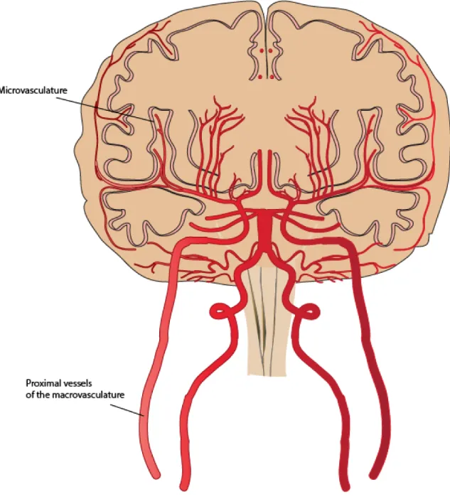
metabolic waste (14, 52). The vascular network of the CNS is divided into the macrovasculature comprised of the larger proximal vessels, and the microvasculature defined by the small penetrating capillaries (55, 56).
2.1.1 Macrovasculature
Blood is delivered to the brain by the two carotid arteries (each contributing roughly 40% of total brain perfusion) and the two vertebral arteries that combine to form the basilar artery. Finally, the carotid arteries and the basilar artery are linked to form the circle of Willis (CW) (57). The CW’s primary role is believed to be the control of pressure reaching the capillaries (58). Interestingly, the vascular system of the brain branches out into smaller and smaller vessels that are progressively more vulnerable to blood pressure; however, these bifurcations cause an increase in resistance, which dampens pressure (58). To protect the most distal vessels from the high blood pressure, the large proximal cerebral arteries are responsible for up to 40% of complete cerebrovascular resistance (55, 58). The intrinsic myogenic response of smooth muscle cells (SMC) to changes in blood pressure is fundamental to CBF autoregulation (55). The importance of the resistance created by arterioles penetrating the parenchyma is a point of debate among authors. Capillaries provide minimal damping of the pulsatile pressure and flow, therefore, intact function of the large proximal vessels as well as the CW is necessary for maintenance of physiologic pressure (59). Other studies have observed that the large proximal arteries together with the intracranial pial arteries and arterioles contribute ~50% of cerebrovascular resistance (60, 61).
While age-related dysfunction in the myogenic responses to pressure have been found in rodents (62), different research groups exploring dynamic myogenic responses yielded mixed results when comparing aged and young control patients (55). Regardless, diminished dilatation of aged proximal cerebral arteries responsible for resistance might increase the risk of ischemia during hypotensive conditions (55).
Figure 2 Macrovasculature and microvasculature
2.1.2 Microvasculature
At the distal end of cerebral circulation, delicate regulation of tissue perfusion must be maintained through finely tuned signals released by neurons and communicated through
astrocytes to the nearby vessels. This regulation may be done through metabolic, myogenic and neurogenic mechanisms (58, 63). Approximately 90% of the arteriolar surface penetrating the brain parenchyma is covered with astrocytic endfeet, which emit signals for CBF maintenance. It has been shown that impairing astrocytic signaling of arterioles (in vivo) can cause approximately a 50% decrease of CBF(64). The capillaries within brain parenchyma are abundant in inter-endothelial tight junctions formed through interactions between endothelial cells (EC), pericytes and astrocytes. Therefore, the tight junctions, which are a fundamental component of microvascular permeability regulation, are dependent upon the integrity of the neurovascular unit (NVU) (63). Another interesting point about the pericytes and the ECs of capillaries is that they both secrete extracellular matrix which forms the basal lamina of capillaries (65).
Altogether, the tight junctions, basal lamina, ECs, pericytes and astrocyte endfeet function together form the blood brain barrier (BBB) (63). The BBB is responsible for metabolic homeostasis of the CNS and the passage of essential nutrients into the CNS environment; BBB disruption is linked with pathogenesis of multiple CNS diseases (66). The main component of the BBB is ECs, therefore, proper function of the BBB is highly dependent on EC integrity.
EC are also highly involved in CBF through multiple pathways. A first proposed way is through cyclo-oxygenase-1 (COX-1), a rate limiting enzyme produced in most cells responsible for the production of prostanoids by arachidonic acid (67). Experimental evidence demonstrates that COX-1 is capable of influencing resting CBF as well as vasodilation induced by certain endothelium-dependent vasodilators or hypercapnea (67). However,
COX-1 has no impact on CBF increase induced by vibrissal stimulation to the whisker-barrel cortex (67). A second recent model posits that pulse pressure plays a central role in the optimal function of cerebrovascular endothelium and eNOS coupling (68). When eNOS coupling is engaged, eNOS produces nitric oxide, a vasodilator; however, when eNOS uncoupling is engaged, eNOS instead produces reactive oxygen species (ROS) such as H2O2 which functions to signal dilation of the vessel as well as the free radial O2- (68, 69). Experimentation with mouse cerebral arteries demonstrated that physiological pulse pressure enhanced sensitization of EC to shear stress as well as myogenic contractions and prompted the production of NO by supporting eNOS coupling. Furthermore, experimental evidence suggested that mechanical flow signal was transferred via NOX2 as it was necessary for stimulating both the coupled and uncoupled eNOS pathways (68). NOX2 is a superoxide generating enzyme and one of the isoforms of the catalytic subunit of NADPH oxidase (70).
Modifications in functions of pericytes (2.1.3) and astrocytes (2.2.2.1) due to aging will be discussed later in this memoir; here we will focus on the age-related endothelial dysfunction. A proposed theory is that accumulation of ROS in brain due to aging is responsible for decreased production of NO by ECs (55). Superoxide, in particular, readily reacts with NO to form peroxynitrite and decrease bioavailability of NO (71). It has been proposed that decreased NO production is a universal effect of aging due to an increase in arginase production leading to decreases in L-arginine necessary for production of NO (72). Therefore, since endothelium-derived NO is involved in vascular tone, it is likely that the decreased production of the dilator NO will cause chronic hypoperfusion giving rise to cerebral dysfunction and dementia (55). It is important to note that NO is also involved in platelet
aggregation inhibition, SMC proliferation and exerts anti-inflammatory, anti-apoptotic and pro-angiogenic effects. Altogether, this information suggests that age-related decrease in NO influences neuronal, astrocytic, microglial and cerebrovascular functions. Therefore, mounting evidence points to the association of dysfunctional production of NO by ECs with augmented amyloid precursor protein and Aβ, as well as increased microglial activation leading to degeneration of cognitive function (73). Interestingly, it has been proposed that changes in cerebrovascular reactivity provoked by APP and Aβ which result in increased vasoconstriction may be mediated by ROS. Experimental evidence has demonstrated that exogenous free radical scavenger superoxide dismutase (SOD) as well as the overexpression of SOD1 rescued endothelial dysfunction in APP mice (74). Overall, ROS may play an important role in mediating age-related changes in the vasculature as well as APP and Aβ-related changes seen in AD.
2.1.3 Pericytes
In the CNS, pericytes are a type of contractile cell found embedded within the basal lamina, that may participate in the regulation of CBF (66). In fact, smooth muscle cells responsible for contraction of blood vessels are not present in capillaries, leaving pericytes and EC responsible for fine regulation of blood supply through their capacity for contractility (66). The main contracting protein in pericytes is α-SMA, which can be upregulated by TGFβ (75). Interestingly, different levels of α-SMA have been observed depending on the location of pericytes, with the highest concentration of α-SMA in the arteriolar side of capillaries (66). As
mentioned before, arterioles are an important contributor of vascular resistance in the CNS and have a strong impact on CBF (64). In NVC, pericytes receive signaling information from astrocytes, which are also capable of secreting TGFβ (63, 76).
In the aging brain, the number of pericytes as well as the secretion of TGFβ by astrocytes decreases, although α-SMA within pericytes increase (63, 77). Experiments conducted in pericyte-deficient mice demonstrated that a reduction in pericytes induces increased permeability of the microvasculature (78). Furthermore, it is suggested that the accumulation of toxic blood-derived proteins around the vessels may take advantage of the resulting permeability to cross the BBB with consequential neurodegeneration (77-79). Although TGFβ production by astrocytes decreases with age, the increased production of TGFβ seen in AD patients is capable of inducing high levels of α-SMC in pericytes (66, 80). Other alterations seen in senescent pericytes include vacuoles and inclusions, which have been suspected to contribute to the inability of pericytes to deposit the contents back through capillaries; possible pericyte sclerosis which may lower the ability of pericytes to control CBF. Furthermore, the increased oxidative stress seen in older patients leads to the heightened intensity of pericyte constriction and subsequent significant hypoperfusion. Due to these pathological changes, it has been suggested that age-dependent modifications to pericytes may contribute to vascular changes that have been shown to foreshadow the neurodegeneration and neuroinflammation. Finally, these age related changes may make the brain more vulnerable to vascular lesions such as ischemic and reperfusion injury (66).
2.2 The Neurovascular Unit, Aging and Dementia
The acting players in NVC are the neurons, glial cells and vascular cells, which make up the NVU (63, 81). Working together as a finely tuned orchestra to maintain optimal conditions for neurons, the smallest discord or dysfunction can lead to pathogenic circumstances. Furthermore, the natural process of aging is capable of manifesting senile alterations to the components of the NVU (56, 63). Here, we will discuss the instruments of the NVU within the context of age-related impairments leading to cognitive impairments and dementia.
2.2.1 Neurons
Neurons make up the basic unit of the central nervous system, their cells composed of two parts, the cell body (or soma) and the processes. The soma contains the genetic material and machinery required for protein synthesis while the processes are long extensions connecting neurons to other cells (82). Neurons are constantly at work and have a very high metabolic demand. However, they are not capable of storing large amounts of energy and are constantly releasing large amounts of metabolic products (52, 63). Due to these conditions, it is up to the proper function of the local microvasculature and other cells of the NVU to maintain homeostasis.
Among the different populations of cells in brain, neurons are among the most vulnerable to age-related dysfunction. Neurons are highly active and therefore require large amounts of energy, which are generated by mitochondria. Another thing the mitochondria are responsible for is the production of large amounts of toxic ROS and oxidative stress in the cell. ROS attach to and cause the disruption of nucleic acids (the building blocks of DNA), proteins and lipoproteins. As time goes by, the endogenous DNA repair and ROS clearance functions decrease, allowing an accumulation of damaged DNA, modified proteins and peroxidated lipoproteins. It is to be noted that the mitochondria is itself severely affected over time due to its unprotected DNA’s proximity to the electron transport chain leading to mitochondrial dysfunction. Overall, the accumulation of toxic conditions in the neuron due to aging make the cell even more dependent on other members of the NVU. For this reason, the system of an
aged NVU is vulnerable to brain injuries leading to cognitive impairments which progress to dementia. (63)
2.2.2 Glial Cells
There exist three types of glial cells in brain: astrocytes, microglia and oligodendrocytes, all of which are vulnerable to the aging process. There is mounting evidence indicating that impaired function of any of the glial cells leads to neurovascular uncoupling. (63)
2.2.2.1 Astrocytes
As the most abundant cell type in brain, astrocytes have been found to be responsible for numerous roles (80). In the human brain, astrocytes form highly complex networks: a single astrocyte contacts thousands of neuronal synapses while the astrocytic endfeet cover approximately 99% of blood vessel surface (83). The astrocyte maintains ion and metabolic homeostasis for the neuronal cells as well as control of glutamate signaling in the synapse through Ca2+ oscillations (63, 80). In fact, increasing evidence suggests astrocytes are used for modulation of microglial response (84) and inter-astrocytic tight gap junctions have an active role in information processing (83, 85) and long-distance synaptic homeostasis (63); however, for the purpose of this memoir, we will focus on the role of astrocytes in NVC. As members of the NVU, astrocytes serve to communicate with neurons and conduct cross talk between the neuroglial segment and cerebral vessels in proximity to the astrocytic endfeet (63). Depending
on the level of neuronal activity, the astrocyte is responsible for modulation of cerebral blood flow and capillary permeability to adjust the supply of glucose and oxygen (63, 86, 87).
As the number of astrocytes does not change much with the aging of the organism, astrocytes are thought to be the type of glial cell least affected by aging; however, senescent astrocytes do display a decrease in their supportive functions (63, 80). To begin, the abnormal protein accumulation in senescent astrocytes affects their capacity for neuronal metabolic support (88). Another change is the reduction of inter-astrocytic gap junctions, leading to impairment of inter-astrocytic cross-talk and synaptic function (63). Next, chronic stress during aging causes the secretory profile of astrocytes to become inflammatory by the release of soluble mediators such as ROS, leading to inflammation in the senile brain (63, 80). Finally, the increase TGFβ production seen in the elder, especially aged AD patients (8), causes an increase in glial fibrillary acidic protein (GFAP) expression in astrocytes (80). Not only is the increased GFAP associated with senescent morphology (80), but also the repression of the supportive capacity of astrocytes (89), one being glutamate clearance leading to neuronal cytotoxicity and another the reduction in neurotransmitter-induced Ca2+ signaling (63). Finally, it has been proposed that these age-related alterations to astrocytes may be a contributing factor to cognitive degeneration as a result of loss of function and neuroinflammation (90).
2.2.2.2 Microglia
Microglia are mobile glial cells that patrol the CNS environment, responsible for CNS pathologies as well as homeostasis maintenance (91). As the innate immune cells of the CNS, microglia are the first line of defense against invading microbes leading to microglial activation and release of protective substances (92). Except for micro-organisms, microglia sense the neural environment to detect neuronal health. Mounting evidence shows microglia function in synaptic pruning (92, 93), pruning of axonal collaterals (92, 94), phagocytosis of apoptotic neurons (92, 95), facilitation of axonal sprouting (92, 96), and remyelination of central axons (92, 97). Therefore, microglial function is necessary for neuronal health and brain homeostasis.
Interestingly, although microglia increase in numbers in the aged brain, there is accumulating evidence suggesting the decline of microglial function due to decreased mobility and efficiency (63). Impaired capacity of senescent microglia also extends to their ability to respond to regulatory signals such as TGFβ (98) capable of inhibiting microglial activation. The senile microglia have impaired mobility (99), loss of microbial elimination (100) and metabolite elimination (101) such as Aβ. However, though senescent microglia have a decrease in functions, they tend to shift to the M1 phenotype, which has elevated production of pro-inflammatory molecules (63). Interestingly, there is mounting evidence suggesting that TGFβ signaling in the brain is in part responsible for the development into adult microglia (102, 103). Mounting evidence links age-related changes in microglial function to neurodegenerative disorders (92).
2.2.2.3 Oligodendrocytes
Neural axons are sheathed with lipid-enriched myelin produced by proximal oligodendrocytes, which speed up signal conduction and allow higher velocity (63, 82, 104). Not all oligodendrocytes in the CNS act as supporting cells: some remain free-floating and act as oligodendrocyte precursor cells (OPC). Under normal conditions, degenerated myelin and exposed axons stimulate the proliferation of OPCs and remyelination of axons (63).
As seen in other glial cells, the aging process impairs oligodendrocyte function. In the aged brain, recruitment of OPCs and remyelination is compromised potentially leading to broad and irreversible demyelination in the event of myelin injury (63, 105). Furthermore, myelin sheaths formed by senile oligodendrocytes are thin with short inter-nodal lengths resulting in lower conduction velocity and brain dysfunction (14, 106).
2.3 TGFβ-Associated Pathologies
As previously mentioned, vascular pathologies displayed in AD patients with increased of all TGFβ isoforms comprise vascular fibrosis, SVD, cerebral microhemorrages, thickened vessel walls, chronic hypoperfusion as well as compromised vascular dilation and contraction: abnormalities mirrored in TGFβ mice (8). Because, of the very similar cerebrovascular pathology compared with AD, transgenic mice overexpressing the TGFβ1 isoform are
instrumental in the elucidation of the effects of increased this cytokine in AD patients. However, it is important to note that the TGFβ mouse is not a model of AD neuropathology per se although they display multiple abnormalities found in AD (52). For instance, TGFβ mice show no signs of Aβ or tau accumulations, though it has been shown that an increase in TGFβ1 aids in the microglial clearance of Aβ (107). To be specific, the TGFβ mouse is a model of cerebrovascular dysfunction associated with AD. However, it has to be noted that a decrease in neuronal TGFβ signaling has also been associated with neurodegeneration (108).
2.3.1 Physiological use of TGFβ in Brain
To understand the abnormalities provoked by pathological over-production of TGFβ1, it is necessary to comprehend the physiological function of TGFβ1 in the healthy brain. Under normal conditions, TGFβ1 is a cytokine central to injury response in the CNS and is responsible for the initiation of fibrotic response and wound healing (48, 109, 110). Upon injury, TGFβ is released by a multitude of cells including platelets, microglia and astrocytes (111, 112). Through experimental data with mice, we know that TGFβ causes overexpression of ECM proteins for wound closure and scarring as well as contraction of the ECM (109, 110). Under pathological conditions, upregulation of TGFβ levels in transgenic mice induces thickened ECM and contracted vasculature. This evidence leads researchers to believe that TGFβ upregulation is responsible for the vascular pathogenesis in AD patients.
2.3.2 Impact of TGFβ-associated vascular pathologies on NVC
Through use of transgenic TGFβ mouse, researchers could unravel the mystery surrounding the effects of TGFβ upregulation on NVC. In vitro experiments showed impaired responses to dilatory substances and inability to contract in response to contractile agents (52, 113, 114). These dysfunctional responses to signaling lead to hypoperfusion at baseline in limbic brain regions such as the hippocampus (8) of TGFβ mice with up to 33% decrease when compared to wild type (WT) controls (52, 115). There was a 23% reduction in CBF response to neuronal activation (52, 116). Another important impairment is resting glucose uptake by brain structures, which showed a 15 to 20% decrease in transgenic mice (52, 117). Furthermore, postmortem tissues from AD patients as well as TGFβ mice display astrogliosis (116), which has been associated with demyelination, degeneration of neuronal axons and white matter dysfunction (34). Overall, TGFβ transgenic mice present both neurovascular and neurometabolic deficits mirroring effects seen in AD patients. With age, the increased deposition of profibrotic proteins, namely fibronectin, perlecans, connective tissue growth factor and collagen I and IV which lead to fibrosis cause stiffening and damage to the cerebral blood vessels, as well as capillary degeneration (SVD) (8). However, it is important to note that despite these changes, there is no marked cognitive deficits in young TGFβ mice, while senescent TGFβ transgenic mice showed only insignificant cognitive impairment (52).
2.3.3 Linking TGFβ Mouse and AD Patients
The involvement of TGFβ1 in pathological situations such as AD, Parkinson’s disease, stroke, brain tumors, ischemia and abscess is marked by an increase in cerebrospinal fluid TGFβ1 levels (48). Tremendous amounts of research have been conducted targeting multiple facets of AD. However, drugs that were found to be successful on transgenic TGF mice failed when tried on humans (8). To this extent, double mutants combining the Aβ pathology with TGF vascular pathology (APP/TGF mice) were used to emulate neuronal, cerebrovascular and cognitive impairments seen in AD patients (8). However, treatments that were successful on an isolated mutation had no effect on the double mutant suggesting different patterns between cerebrovascular dysfunctions induced by these two parameters (118, 119). Given these facts, it is clear that other factors involved in AD must be traced and treated. A possible culprit for the vascular contribution to dementia in the elder is arterial stiffness.
3. Arterial Rigidity
Arterial rigidity has long been shown to be an independent predictor of cognitive decline in the elderly (27, 120-123). Multiple studies have been conducted in seniors measuring pulse wave velocity, that is the rate at which pressure waves move down the vessel, between the femoral and carotid arteries (cfPWV). The results of these studies pointed to a positive correlation between the increase in cfPWV and deterioration of cognitive abilities in the elder
(27, 124, 125). To fully appreciate the effects of arterial rigidity, we must first appreciate the peripheral vasculature and its mechanics.
3.1 Layers of the Vasculature
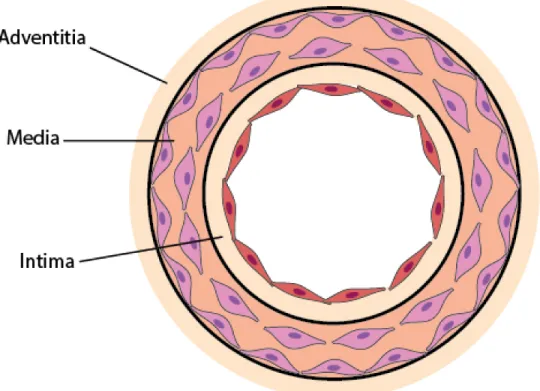
The network of tubes responsible for the transportation of essential nutrients throughout the body is composed of three layers: the abluminal layer known as the adventitia, the medial layer known as the media and the luminal layer named the intima (82, 126). Each layer is responsible for supplying proper amounts of nutrients in the case of active hyperemia where an organ has increased metabolic demands.
Figure 4 Layers of the Vasculature
Beginning with the outermost layer in contact with peripheral tissue, the adventitia functions in the transfer of nutrients and waste substances between the blood and the environment. This layer is looser and thinner than the media from which it is separated by a thin layer of elastin called the external elastic lamina. It is composed of collagen fibres and fibroblasts that produce collagen as well as nets of elastic fibre (126, 127).
Next, the media is the thickest layer and is composed of vascular smooth muscle cells, elastin and collagen as connective tissue, and polysaccharides. The smooth muscle cells relax and contract in response to chemical (NO) and mechanical (blood flow) stimuli to regulate the diameter of the arteries and blood flow. The large arteries closest to the heart, such as the
carotid artery have much higher levels of elastin, and therefore are more compliant. On the other hand, arteries further away from the heart (arterioles) around the muscular tissue have a lower amount of elastin compared to collagen. Collagen protein found in connective tissue is very stiff (126).
Finally, The intima layer of the artery is described as a thin monolayer of ECs attached to a layer of elastin called the internal elastic lamina, spanning approximately 10µm in thickness (126). The endothelial layer presents a large interface (350m2) with the passing blood of the lumen and creates an excellent location for the exchange of substances. In addition to nutrient exchange, ECs serve a multitude of purposes such as being a barrier for large molecules, secreting antithrombotic and anticoagulant molecules, modulating vascular tone, inflammation and angiogenesis.
The adhesive structures between ECs control vascular permeability and are regulated in coordination with the organ through which the vessel passes. This is important in connection with vessels that pass through brain tissue. In physiopathological conditions, permeability is compromised. Another function of ECs is the secretion of antithrombotic and anticoagulant molecules to maintain blood fluidity. The ECs also regulate vascular tone through the secretion of the free radical gas NO, generated by NOS isoforms (eNOS and iNOS) found in the cellular cytoplasm which respond to forces on the cell wall as well as prostaglandins and the endothelium derived hyperpolarizing factor (EDHF). The ECs of the intima are also involved in inflammatory reactions through their response to cytokines and signalling to
leukocytes during leukocyte extravasation (128). Finally, the ECs are responsible for neoangiogenesis, the formation of new blood vessels (128).
3.1.2 Vascular Compliance and Distensibility
One of the important functions of the vascular wall is the compliance, or distensibility, of the elastic arteries, which depends on the ratio of elastin fibres in proportion to collagen fibres. This gives the vessel the ability to dampen the pulsatile intensity of the heart. If the intensity were too high, downstream capillaries in end organs, such as the brain, would undergo micro haemorrhaging. It is the elastin together with the smooth muscle cells in the vascular wall that account for the flexibility and resilience of the vessel wall. It is the larger vessels closest to the heart, such as the aorta and carotid arteries, which have relatively higher amounts of elastin, compared to the smaller downstream vessels (58, 129). It has been shown that stiffening of the vessel is inversely related to pulse wave velocity; that is, an increase of PWV is associated with decreased distensibility (27, 130). Therefore, measurement of propagation of the pulse wave or cfPWV allows for a robust and reproducible method to measure arterial stiffness (27, 131).
3.2 Arterial Rigidity, Aging and Dementia
Aging is the cause of various changes in the physiology of large arteries in the periphery responsible for stiffness. To begin, the principal structural change in arteries is due to the
fatigue and fracture of the media layer of vessels. Given that media is accountable for mechanical properties of the vessel, the fragmentation of elastin and fracture of muscle attachment due to age leads to arterial stiffening (132). Next, there are increases in factors of the arterial ECM including collagen, fibronectin, proteoglycans and vascular smooth muscle cells (27, 132). A decrease in the elastin to collagen ratio in the media decreases distensibility of large arteries such as the aorta and the carotid artery (125, 133). An important change to the arterial wall due to aging is the increase in calcium deposits after the 5th decade (27, 134). While aging induces the production of matrix metaloproteinases (MMPs), which degrade elastin, vessel calcification has also been associated with concomitant elastin degradation (134, 135). Further changes due to aging are impaired vascular EC function, which has been previously discussed in section 2.1.4.2. EC dysfunction affects vascular smooth muscle response, which is suggested to modify vascular tone (27).
Hardening of the vessel wall leads to numerous mechanical changes that have multifactorial effects on the body. Impaired function to diminish the pulse wave emanating from the heart is associated with microvascular remodeling and dysfunctional autoregulation, which ultimately leads to end organ injury (124, 133). Furthermore, stiffness of the aorta is the cause for increased workload on the heart via increased reflected waves, which result in decreased stroke volume and coronary artery perfusion (133). Interestingly, various studies have linked increased pulse wave reflection and cardiovascular disease (136). Indeed, arterial stiffness has been associated with multiple risk factors of cardiovascular disease such as hypertension (137), atherosclerosis (138), kidney disease (139) and metabolic syndrome (123, 140). Experimental data studying the effects of cfPWV on brain using magnetic resonance imaging
noted an increased risk of silent subcortical infarcts, higher white matter hyperintensity volume and a decline in grey matter, white matter and whole brain volumes (141). It is important to note that due to the multifactorial influences of arterial stiffness on the body, isolating the effects of arterial stiffness on brain is very difficult. Therefore, we will continue to elucidate the effects of arterial stiffness on brain using a mouse model of arterial stiffness developed by Girouard’s Lab (124).
3.2.1 Arterial Stiffness Model
Here we will discuss the published data of the arterial stiffness model developed by Sadekova
et al. (124). The carotid calcification model has a significant advantage over past models of
arterial stiffness: the ability to isolate the effects of arterial stiffness on brain without affecting any other organs or systems in the body, which may then affect the CNS. As the blood journeying from the carotid artery goes directly toward the circle of Willis and the brain, this model ensures that only the brain will be affected by surgical calcification of the right carotid artery.
3.2.1.1 Vascular Significance in the Mouse Model
The effects of periarterial application of calcium chloride coincide with the literature describing the effects of calcium deposits on arteries. However, it is important to note that all arterial changes occurring due to the periarterial application of CaCl2 remain localized.
Two weeks post-surgery, the artery displays fragmentation of elastin, an increase in collagen and media thickness with accompanying decrease in distensibility of the carotid artery and the resulting increase in cerebral blood flow pulsatility. Furthermore, all of these changes manifested without altering systemic blood pressure or vessel radius. Therefore, the model is capable of examining the effect of arterial stiffness on brain and dementia without affecting the systemic networks. Reporting on the short-term effects of carotid calcification, Sadekova noted the cerebral blood flow pulsatility increased from 17.2±5.2% to 23.1±5.1% and 15.1±0.8% to 18.7±1.0% in mice with a calcified carotid in medium-sized arteries (diameter of 50 to 95 µm) and large arteries (>95µm), respectively. Importantly, the treated right side of the brain had significantly higher blood velocity, changing from 16.9±4.3% in the sham group to 23.5±5.7% in the group with the calcified carotid, while the blood speed of the left brain depicted only a non significant increase. These changes in cerebral blood flow pulsatility and blood speed seen in the mouse model give indices of the effects of arterial stiffness on the vasculature and hemodynamics in humans.
Previously, we described the distal microvasculature of the brain to be characterized by the inability to dampen increased pulsatile load. It has been proposed that a protective measure in the brain to intercept the increased load is hypertrophic remodeling (142). Although this response would initially function to limit penetration of the pulsatile load, it will eventually lead to impaired vasoreactivity, hypoperfusion and chronic ischemia: symptoms present in vascular dementia (125).
Given the effects of the model on the vascular bed, Sadekova proceeded with the exploration of the factors most vulnerable to fluctuations in vascular integrity: neurodegeneration and oxidative stress.
3.2.1.2 Neurological Significance in the Mouse Model
As mentioned before, neuronal maintenance is dependent upon the ability of local capillaries to provide proper amounts of nutrients and oxygen. A lack of proper oxygenation would lead to hypoxia and the resulting toxic oxidative stress, which would cause neurodegeneration.
To investigate the possibility of oxidative stress, Sadekova performed dihydroethidium staining which allowed for the revelation as well as the localization of the production of superoxide anion, a reactive oxygen species. Staining revealed a 1.2-fold increase of the superoxide anion in the cornu ammonis 1 and 3 (CA1 and CA3 respectively) of the hippocampus as well as the dentate gyrus (DG). Interestingly, although only the right carotid artery was calcified, no inter-hemispheric difference in superoxide anion production was observed.
For the purpose of examining the manifestation of neurodegeneration, Sadekova used Fluorojade B staining, which also had the added benefit of localizing neurodegeneration in the brain. Realization of the experiment lead to the discovery that the area of neurodegeneration was specific to the lacunosum moleculare in the CA1 region of the hippocampus with a
treated group. The lacunosum moleculare is the layer of the hippocampus that serves as the connection between the entorhinal cortex and the CA1 of the hippocampus(143). The fact that this specific layer is targeted is an important finding to the field of AD: the first sign of neurodegeneration in AD is the group of neurons in the entorhinal cortex that project into the CA1 (144). Therefore, due to Sadekova’s model of arterial stiffness, a clear liaison has been mapped between arterial rigidity, VaD, AD and ultimately dementia.
4. Neurological Insults
Until now, this memoir has discussed several perturbations of the neuronal environment, which have been linked with dementia. Here, we will discuss these symptoms in greater detail.
4.1 Oxidative Stress
Oxidative stress has been widely defined as an imbalance between the production of reactive oxygen species and the ability to counteract through antioxidants defenses (145). The hypoxic conditions that arise from the VaD facet of mixed AD/VaD dementia as well as aging are responsible for the injurious increase in ROS (12, 63). Mitochondrial respiration through the electron transport chain (ETC) is responsible for 90% of both cellular oxygen consumption and ROS production (12, 63); also, mitochondria are very susceptible to age-related impairments (63, 146).
4.1.1 Free Radicals: Formation and Biological Uses
Physiological as well as pathological systems yield free radicals. Free radicals have been defined as molecules with a single unpaired electron in search of its pair (147). It is the unpaired electron in the outer orbit that causes the molecule to be unstable and highly reactive, attaching itself to proteins, lipids and DNA, thus altering their normal structure and function (148, 149). Among these reactive species, there are also non-radical molecules such as H2O2 and ONOO− and which do not have an unpaired electron (147). There exist three types of reactive species: ROS, reactive nitrogen species (RNS) and reactive chlorine species, although during brain injury, ROS and RNS are the major sources of oxidative stress (149, 150). ROS include peroxyl radicals ROH, hydroxyl radicals OH, superoxide O2− and H2O2 while RNS contain ONOO−, nitrogen dioxide radical NO2 and nitric oxide NO.
The superoxide O2− is very reactive and is involved in the formation of other highly reactive ROS. Its production accounted for the development of many pathologies. Enzymes responsible for the generation of O2− are the electron transport chain of mitochondria, NADPH oxidase (NOX family subunits), xanthine oxidase as well as NOS in the case of its uncoupling (68, 147).
Multiple significant members of RNS exist. To begin, NO is an important signaling molecule
involved in inflammation, nerve transmission, vasodilation and perfusion, as seen earlier (147). It is formed by members of the NOS family, which include iNOS (inducible NOS), nNOS (neuronal NOS) and eNOS (endothelial NOS), all of which require L-arginine,
molecular oxygen and NADPH. eNOS and nNOs are constitutively actives whereas iNOS is inducible. An important cofactor also involved in NOS activity is BH4: when the source of BH4 is exhausted, NOS becomes uncoupled and produces O2− and H2O2 (147). A second
important RNS is the non-radical ONOO−, produced when superoxide reacts with NO.
ONOO− has been proposed to have anti-microbial features, although it is mainly known to be deleterious to biomolecules (147, 149). A third RNS is generated when ONOO− undergoes homolytic cleavage to compose OH and NO2 (147).
Therefore, although free radicals have been shown to be an important component of physiological functions through signal transduction, regulation of enzymatic activity and gene transduction, they are injurious to cells at pathologic concentrations (149).
4.1.2 Markers of Oxidative Stress
In an attempt to pair their unpaired electrons, free radicals attach to proteins, lipids and DNA, thus altering their structures and functions. The resulting modifications can be used to trace the level of oxidative stress. As oxidative stress has been linked to aging, protein nitration and lipid peroxidation products have been recognized as age-related modifications (63, 151).
A possible protein marker is 3-NT, which is generated when ONOO− undergoes homolytic cleavage to compose OH and NO2 , leaving the NO2 to attach to a nearby tyrosine molecule
(152). Concentrations of 3-NT above physiological levels denote an environment with elevated quantities of RNS. Interestingly, if RNS imbalance occurs under neurodegenerative
![Table I VaD Risk Factors [modified from (12)]](https://thumb-eu.123doks.com/thumbv2/123doknet/2042978.4866/18.918.111.816.492.792/table-vad-risk-factors-modified-from.webp)
![Table II Common pathological lesions in AD and VaD [data from (41)]](https://thumb-eu.123doks.com/thumbv2/123doknet/2042978.4866/22.918.114.809.156.375/table-ii-common-pathological-lesions-vad-data-from.webp)