• Aucun résultat trouvé

Risk Factors for Cervical HPV Infection in Montreal University Students

N/A
N/A
Protected

Academic year: 2021

Partager "Risk Factors for Cervical HPV Infection in Montreal University Students"

Copied!
184
0
0

Texte intégral

(1)

Université du Québec

Thesis presented to the Institut Armand-Frappier in partial fulfillment of the requirements for the degree of

M.Sc. (lmmuno-Virology)

by

Harriet Richardson

Risk Factors for Cervical HPV Infection in

Montreal University Students

(2)
(3)

TABLE OF CONTENTS

TABLE OF CONTENTS ... iii

LIST OF TABLES ... vii

LIST OF FIGURES ... ix LIST OF APPENDICES ... x ABSTRA CT ... xi ; ; RESUME ... xii ABBREVIATI 0 NS ... xvi INTRODUCTION ... ! REVIEW OF THE LITERA TURE ... 4

1 The Evidence for a Relationship Between HPV and Cervical Cancer ..•...•... 5

1.1 Epidemwlogy of cervical cancer ....... 5

1.2 Etiology of cervical cancer ... 7

1.3 HPV and cervical carcinogenesis ... 11

1.4 Screeningfor cervical cancer •... 14

2 Characterization of HPV ... 17 2.1 2.2

2.3

2.4 HPV structure ... 17 HPV classifiCation ...................................... 18 HPV genome organization ........ 19

2.3.1 Open reading frames (ORF) ... l9 2.3.2 Long control region .............................................. 22

2.3.3 Caps id assembly .... 23

HPV DNA detection ... 23

2.4.1 2.4.2 DNA hybridization techniques ......... 23

Polymerase chain reaction (PCR) ...... 25

3 Pathogenesis of Cervical HPV Infection ... 26

3.1

3.2

3.3

3.4

HPV type and cancer risk ... 26

Viral entry ... 28

Tissue datnage ... 28

(4)

3.5 Treatment ...... 3 3

3.5.1 Current therapies ..................... 33

3.5.2 Prospects for the future ...... 34

4 Epidemiology of Cervical HPV Infection ... 35

4.1 Prevalence of cervical HPV infection ...... 35

4.2 Risk factors for HPV infection ... 39

OBJECTIVES ... 42

MA TE RIALS AND METH 0 DS ... 44

1 Study Design ... 45

1.1 Subjects ... 45

1.2 Data collection ... 45

1.3 Sample collection ... 45

2 La bora tory Methods ... 46

2.1 Specimens ... 46 2.1.1 Clinicat samples ..................... 46 2.1.2 Controls ... 47 2.1.3 Cell cultures ...................... 48 2.2 HPV ampl'ifi.cation ...... 48 2.2.1 PCR ... 48

2.2.2 Refining the PCR protocol ........ 51

2.3 Gel electrophoresis ... 57

2.4 Southern transfer ...... 57

2.5 Dot blot transfer ...... 58

2.6 Preparation of probes ... 58

2.6.1 First generation of generic probes ..................... 58

2.6.2 Second generation of generic probes ... 60

2.6.3 {3-globin probe .................. 60

(5)

2. 7 Hybrid.ization ...... 62

2.7.1 Hybridization with gene rie HPV probe ... 62

2.7.2 Hybridization with /3-globin probe ..................... 63

2. 7.3 Hybridization with type-specifie oligonucleotides ............... 63

2. 7.4 Re-use of dot blot nylon membranes ........................... 63

2. 8 Cloning ......... 65

2.8.1 Cloning of the 450 base pair amplicon ............................ 65

2.8.2 Analysis of clones ... 66

2.8.3 Typing by sequencing .... 66

3 Statistical Analysis .......... 67

RESUL TS ........ 69

1 Laboratory results ........... 70

2 Epidemiological profile

··~···

·

··

··

···

·

··

···

···

7 4 2.1 Prevalence of cervical HPV infection .............. 77

2.1.1 Prevalence and sociodemographics ...... 77

2.1.2 Prevalence and sexual activity ...... 78

2.1.3 Prevalence and contraceptive history .... 79

2.1.4 Prevalence and hygiene and medical history ... 84

2.2 Univariate analysis ... 84

2.2.1 Risk associated with sociodemographics ................... 84

2.2.2 Risk associated with sexual behaviour ... 90

2.2.3 Risk associated with contraceptive history ................................ 91

2.2.4 Risk associated with hygiene and medical his tory .... 9

2. 3 M ultivarWte analysis ·~··. 11 • • • • • • • • • • • • • • • • • • • • • • • • • • • 11 • • 11 • • • • • • • • • • • • • • • • 11 ·~·· 11 ·~·· 1 • • • 1 • ••• • •• 1 • • 96 2.4 HPV infection as a risk factor for squamous intraepithelial lesions···~~····~····~···~··· 98

DISCUSSION ... 100

1 Estima ting HPV pre valence ... 101

(6)

1.2 Disadvantages ... 101

1.2.1 Problems related to biological sampling ... 102

1.2.2 Inter laboratory variability ... 104

1.2.3 Variability within and between the populations studied ... 105

2 Determinants of cervical HPV infection ... 105

2.1 Sociodemographics ... 105

2.2 Sexual behaviour and contraceptive use ... 106

2.2.1 Sexual activity ... 106 2.2.2 Oral contraceptives ... 112 2.2.3 Barrier contraceptives ... 113 2.3 Hygiene ... 115 2.4 Cytology ... 116 CONCLUSION ... 118 A CKN 0 WLEDG MENTS ... 120 BIBLIOGRAPHY ... 123 APPENDICES ... 141

(7)

LIST OF TABLES

REVIEW OF THE LITERA TURE

Table I Table II Table III Table IV Table V

Historical outline of epidemiological research on cancer ... 8

Biological evidence for a carcinogenic role of HPV ... 13

Bethesda classification of epithelial cell abnormalities ... 16

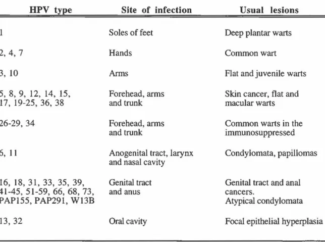
HPV types, sites of infection and clinical manifestations ... 27

HPV DNA detection rates by PCR among cytologically normal women ... 37

MATERIALS AND METHODS Table VI RESULTS Table VII Table VIII Table IX Table X Table XI Table XII Table XIII Table XIV Table XV Table XVI Table XVII Table XVIII Table XIX Parameters tested to refine MY09/MY11 PCR protocol.. ... 52

Comparison of non-respondents and respondents with respect to age, cytological morphology and HPV pre valence ... 71

Comparison of 13-globin negative and 13-globin positive subjects with respect to age, ethnicity and selected sexual characteristics ... 72

Prevalence rates of overall HPV infection according to oncogenic risk ... 73

Distribution and prevalence of individual HPV types ... 75

Prevalence of HPV infection by demographie factors: overall and according to oncogenic risk ... 78

Prevalence of HPV infection by reproductive and sexual factors: overall and according to oncogenic risk ... 80

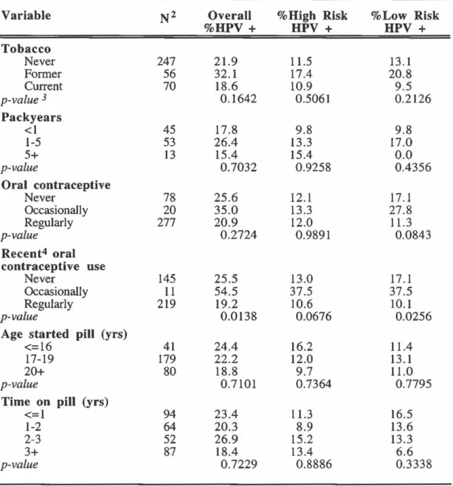
Prevalence of HPV infection by smoking and contraceptive use: overall and according to oncogenic risk ... 82

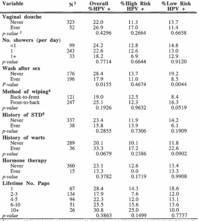
Prevalence of HPV infection by hygiene and history of STD's: overall and according to oncogenic risk ... 85

Odds ratios of overall, high-risk and low-risk HPV infection according to demographie factors ... 86

Odds ratios of overall, high-risk and low-risk HPV infection according to reproductive and sexual activity ... 87

Odds ratios of overall, high-risk and low-risk HPV infection according to smoking and contraceptive use ... 92

Odds ratios of overall, high-risk and low-risk HPV infection according to hygiene habits ... 94

Odds ratios of overall, high-risk and low-risk HPV infection according to medical history ... 95

(8)

LIST OF TABLES (Continued)

Table XX Adjusted odds ratios of overall, high-risk and low-risk HPV

infection according to selected risk factors ... 97 Table XXI Association between HPV infection by oncogenic risk and

cervical cytological abnormalities ... 99 DISCUSSION

Table XXII Consistancy of the evidence of an association between sexual

(9)

LIST OF FIGURES

Figure 1 Incidence rates of cervical cancer worldwide ... 6

Figure 2 PCR amplification strate gy ... 49

Figure 3 Oligonucleotide primers for PCR ... 53

Figure 4 PCR protocol re finement (1) ... 54

Figure 5 PCR protocol re finement (II) ... 55

Figure 6 Detection limits of HPV PCR products ... 56

Figure 7 Sequences of type-specifie oligonucleotides ... 61

(10)

LIST OF APPENDICES

Appendix 1 Persona! Data Sheet ... 141

Appendix 2 HPV Study- Consent Form ... 143

Appendix 3 Women's Health Study Questionnaire ... 146

Appendix 4 Distribution of Socio-Demographic Characteristics of

the Study Population ... 160

Appendix 5 Distribution of Reproductive and Sexual Characteristics of

the Study Population ... 162

Appendix 6 Distribution of Smoking and Contraceptive Characteristics of

the Study Population ... 165

Appendix 7 Distribution of Hygienic and Health Characteristics of

(11)

ABSTRACT

The objectives of this study were to estimate the point prevalence of cervical HPV infection in asymptomatic women and to identify risk factors for overall HPV infection and HPV infection by type. The study was a cross sectional survey at the McGill university student health clinic. Endocervical and ectocervical cell scrapings were collected from students presenting themselves at the clinic for a routine pap smear. Cytology results from the clinic were provided and epidemiological data on potential risk factors including sexual behaviour was obtained from a detailed, self-administered questionnaire. The presence of HPV DNA was detected in specimens by polymerase chain reaction (PCR) using consensus primers (MY09/MY Il) targe ting a 450 base pair segment in the L 1 gene. Amplified products were hybridized with generic and type-specifie probes using Southern blot and dot blot techniques, respectively, to determine overall HPV prevalence and HPV prevalence by type.

A total of 489 women agreed to participate in the study, of which 375 were eligible for final analysis. Overall cervical HPV prevalence was 21.8%. Among those women infected with HPV, 6.2% bad a low-risk HPV infection, 11.8% had a high-risk HPV infection, 7.1% had an unknown HPV type infection and 2.7% had a multiple type infection.

Two profiles emerged for sexual activity and risk of HPV infection according to oncogenic risk after multivariate analysis. Sexual activity (frequency of sex and lifetime number of oral sex partners) was associated with high oncogenic-risk HPV infections. However, HPV infection with low oncogenic-risk types were mostly invariant with respect to markers of sexual activity, suggesting that there may be differences in epidemiological correlates of transmission between low-risk and high-risk HPV types on the basis of their oncogenicity. In view of our findings, we suggest that low oncogenic-risk HPV types may be less mucosotropic and therefore be transmitted by modes other than sexual activity.

(12)

RÉSUMÉ

Les études épidémiologiques effectuées au cours des trente dernières années ont invariablement démontré que les marqueurs d'activité sexuelle étaient les principaux déterminants du risque de cancer du col de l'utérus, suggérant ainsi qu'un agent microbien transmissible sexuellement pourrait causer ce type de cancer [zur Hausen, 1976 #191]. Les données biologiques et épidémiologiques accumulées montrent de façon convaincante que 1' infection du col utérin par certains types de HPV est un événement précurseur dans la genèse des néoplasmes cervicaux [zur Hausen, 1991 #50; Schiffman, 1993 #94; Mufioz, 1992 #146; Franco, 1995 #260]. Par conséquent, l'activité sexuelle devrait pouvoir prédire encore plus effectivement les infections par le HPV, étant donné le rôle étiologique démontré du HPV dans la genèse du cancer du col utérin.

Ceci n'a pas toujours été facile à démontrer, principalement à cause de techniques de laboratoire insuffisamment sensibles ou spécifiques pour la détection des infections latentes au HPV qui sont présentes dans la population générale. Avec l'avènement du PCR et de l'union interdisciplinaire de la biologie moléculaire et de l'épidémiologie, un certain nombre d'études récentes d'épidémiologie moléculaire utilisant le PCR pour la détection du virus ont révélé que l'infection du col utérin par le HPV était transmise sexuellement [Ley, 1991 #9; Bauer, 1993 #162] Cependant, ces résultats n'ont pas été reproduits uniformément par toutes les études utilisant le PCR réalisées dans des populations différentes. Selon les études, l'association entre l'activité sexuelle et la prévalence du HPV peut être forte [Ley, 1991 #9; Bauer, 1993 #162], modérée [Franco, 1995 #260; Wheeler, 1993 #8], faible [Rohan, 1991 #7; Hildesheim, 1993 #262] ou même absente [Kjaer, 1993 #144]. De nouvelles données suggèrent une explication de cette disparité des observations épidémiologiques : la répartition entre divers types de HPV ayant une transmissibilité sexuelle inégale varierait d'une population à une autre [Franco, 1995 #260]. Le risque relié

(13)

à l'infection au HPV semble être influencé de façon indépendante par des facteurs tel que la parité, l'usage de contraceptifs oraux et le tabagisme [Bauer, 1993 #162]. Toutefois, le principal déterminant du risque d'infection au HPV est l'âge, la plupart des études ayant rapporté une forte diminution de la prévalence chez les femmes âgées de plus de trente ans [Bauer, 1993 #162; Wheeler, 1993 #8].

Le présent travail visait à estimer le taux de prévalence des infections au HPV du col utérin chez des étudiantes universitaires de Montréal et à identifier les facteurs de risque d'infection au HPV généraux, ainsi que les facteurs de risque propores aux infections à faible risque oncogène et des infections à risque oncogène élevé. Cette recherche consistait en une étude épidémiologique transversale d'étudiantes consultant la clinique médicale de l'université McGill. Des cellules endocervicales et ectocervicales ont été prélevées par frottis vaginal chez des femmes asymptomatiques se présentant à la clinique pour un prélèvement vaginal ou « test de Pap » de routine. La clinique nous a fourni les résultats

cytologiques. Nous avons obtenu les données épidémiologiques à partir d'un questionnaire auto-administré portant sur l'âge, le statut socio-économique, le niveau de scolarité, l'usage du tabac, les habitudes sexuelles, l'usage de contraceptifs, les antécédents médicaux et l'hygiène personnelle des participantes.

Les échantillons étaient d'abord amplifiés par PCR à l'aide de la paue d'amorce GH20/PC04. Cette amplification visait une région de 268 paires de bases du gène de la b-globine, qui servait d'étalon interne dans le but d'assurer une quantité et qualité adéquates d'ADN. La présence de l'ADN du HPV était également détectée par amplification au PCR en utilisant les amorces consensus MY09/MY Il, ces amorces s'hybridant à un fragment d'ADN de 450 paires de bases dans la région Ll du génome viral [Ley, 1991 #9]. Les produits d'amplification, après un transfert de type Southern, étaient hybridés à une sonde générique pour déterminer la prévalence globale du HPV. Les échantillons positifs étaient

(14)

ensuite déposés sur une membrane de nylon et hybridés avec des sondes types spécifiques pour identifier les différents types de HPV. Les ADN viraux identifiables par électrophorèse mais ne réagissant pas avec la sonde générique ont été clonés et séquencés. Des 489 femmes participant à l'étude, 411 (84%) ont complété et retourné le questionnaire. Les échantillons cellulaires de 40 (8,2%) de ces femmes n'ont pu être amplifiés pour la b-globine. Des infections au HPV à un ou plusieurs types ont été détectées chez 98 femmes (21,8%). Parmi ces femmes infectées, 6,2% avaient un HPV à faible risque oncogène, 11,8% avaient un HPV à risque oncogène élevé, 7,1% avaient un HPV de type indéterminé et 2,7% avaient une infection mixte. Les types de HPV les plus prévalents étaient les types indéterminés (7,1 %), le HPV-16 (4,7%), le HPV-51 (2,2%), le PAP-155 (2,0%), le HPV-66 (1,6%), les HPV-6,11,31,33,et 58 {1,1% chacun), le HPV-18 (0,9%), le HPV-53 (0,9%), le HPV-56 (0,4%) et les HPV-35,68,W13B et 238A (0,2% chacun). Les résultats de cytologie ont révélé que 55 des 449 femmes éligibles {12,2%) présentaient des «test de Pap >>anormaux. Parmi celles-ci, 37 (67,3%) avaient des cellules squameuses atypiques de signification indéterminée ("ASCUS") et 18 (32,7%) avaient de faibles ou de fortes lésions squameuses intraépithéliales (SIL).

Les différences brutes de prévalence du HPV par catégorie d'exposition ont été évaluées par le test du chi-carré de Pearson. Les rapports de cotes ou "odds ratios" (OR) bruts et ajustés pour l'âge ont été calculés pour estimer le risque d'infection au HPV selon les groupes démographiques et les habitudes de comportement. Les types de HPV ont été regroupés selon leur potentiel oncogène. Le OR a été utilisé pour quantifier le degré d'association entre l'exposition et chacune des différentes variables. Suivant des critères de sélection prédéterminés précis, les variables identifiées comme facteurs de risque et variables confondantes potentiels dans les analyses univariées ont été intégrées à un modèle de régression logistique multivariée. Des modèles multivariés ont été élaborés pour les

(15)

infections globales, et séparément pour les infections au HPV à faible risque oncogène et à risque élevé. Les résultats cytologiques constituaient la variable dépendante, tandis que l'infection au HPV constituait la variable prédictive de résultats cytologiques anormaux après ajustement pour l'âge et pour différents indicateurs d'activité sexuelle.

L'association entre l'infection au HPV et les anomalies cytologiques était statistiquement significative, que ce soit pour les infections à risque oncogène élevé ou à risque faible. Cependant, l'infection au HPV prédisait mieux les lésions SIL (OR=IO) que la présence de cellules ASCUS (0R=2.5). De plus, le risque de présenter une anomalie cytologique ASCUS ou SIL ne variait pas de façon tangible en fonction du type de HPV.

Les analyses multivariées ont montré que l'association entre le profil d'activité sexuelle et le risque d'infection au HPV était modifiée selon que le risque cancérogène de l'infection était faible ou élevé. Le risque d'infection au HPV était associé avec le degré d'activité sexuelle -la fréquence des rapports sexuels et le nombre total de partenaires de sexe oral dans le passé- si le risque oncogène était élevé. Par contre, le risque d'infection au HPV ne variait pas en fonction de l'activité sexuelle lorsque le risque oncogène était faible. En outre, tandis que l'usage du condom semblait prévenir les infections à risque oncogène élevé, il était au contraire associé avec le risque d'infection au HPV à faible risque oncogène. Enfin, le lavage des parties génitales après les rapports sexuels semblait prévenir les infections au HPV à faible risque oncogène.

Nos résultats suggèrent que les facteurs épidémiologiques de transmission des infections au HPV différent selon que le type d'infection est à risque oncogène élevé ou à faible risque oncogène. À la lumière de ces résultats, nous avançons une hypothèse suivant laquelle les types de HPV comportant un plus faible risque oncogène seraient moins mucosotropiques et que, par conséquent, leur transmission se ferait davantage par d'autres voies que par l'activité sexuelle.

(16)

ABBREVIATIONS AP-l: ASCUS: bp: CIN: EGF: FCS: HG-SIL: HPV: HSV: lU: LCR: LES: LG-SIL: NFA: octl: OR: PCR:

pRB:

PVF-1: SIL: SSC: SSPE: STD: TE: Tm: URR: VLP: Ubiquitous Factor

Atypical Squamouus Cells of Undetermined Significance Base Pair

Cervical Intraepithelial Neoplasia Epithelial Growth Factor

Fetal Calf Serum

High Grade - Squamous Intraepithelial Lesions Human Papillomavirus

Herpes Simplex Virus International Units Long Control Region Lower Economie Status

Low Grade - Squamous Intraepithelial Lesions Novel Factor

octl Factor Odds Ratio

Polymerase Chain Reaction Retinoblastoma Gene Novel Transcription Factor Squamous Intraepithelial Lesions Standard Saline Citrate

Standard Salt Phosphate EDT A Sexually Transmittted Disease Tris-HCl & EDTA

Metting Temperature Upper Regulatory Region Virus-Like Protein

(17)
(18)

INTRODUCTION

Cervical cancer is still one of the most common neoplastic diseases affecting women,

with a combined worldwide incidence that is second only to breast cancer, despite the advent

of cytological screening. Epidemiologie research conducted over the last three decades bas

consistently shown that markers of sexual activity are the most important determinants of a

woman's risk of developing cervical cancer, suggesting a sexually-transmissible microbial

agent as the cause (zur Hausen, 1976). There is now overwhelming evidence, both biologie and epidemiologie, that cervical infection by certain human papillomavirus (HPV) types is a precursor event in the genesis of cervical cancer (Franco et al., 1995; Mufioz et al., 1992; Schiffman et al., 1993; zur Hausen, 1991).

Clinical and subclinical HPV infections are the most common sexually-transmitted diseases today (von Krogh, 1991). There are over 70 different HPV types defined on the

basis of DNA homology. Two major groups are defined according to their epithelial affinity:

those infecting the dry skin ( cutaneotropic) and those infecting the moist mucosal areas of the body (mucosotropic). Genital types are typically divided into three groups based on the frequency of association with malignant tumours and are thus ranked according to oncogenic potential (i.e. low oncogenic-risk, intermediate oncogenic-risk and high oncogenic-risk) (Bauer et al., 1993; de Villiers, 1994).

Since sexual activity and cervical HPV infection are two of the strongest predictors of cervical cancer, it follows then, that sexual activity should be an even stronger predictor of cervical HPV infection, considering HPV's apparently strong etiological role in cervical carcinogenesis. In the past, however, it bas not been easy to demonstrate this, in large part because laboratory techniques were not sufficiently sensitive or specifie enough to detect latent HPV infections within the general population. With the advent of the polymerase chain reaction (PCR) and the interdisciplinary marriage of molecular biology and epidemiology, a

(19)

number of more recent molecular epidemiological surveys using PCR to detect the virus have unveiled the sexually transmitted profile of cervical HPV infection (Bauer et al., 1993; Ley et al., 1991 ). However, not ali PCR based studies conducted in different populations have been able to reproduce these results uniformly (Kjaer et al., 1993; Rohan et al., 1991). There is sorne new evidence to suggest that the variation within the Iiterature might be caused by differences across populations in the relative prevalence of HPV types with greater or lesser transmissibility by the sexual route (Franco et al., 1995).

In an attempt to clarify the role of sexual behaviour and the transmission of HPV infection, this study aimed to determine the point prevalence of cervical HPV infection in young, asymptomatic women attending university in Montreal and to identify risk factors for low oncogenic-risk HPV infection and high oncogenic-risk HPV infection.

(20)
(21)

REVIEW OF THE LITERATURE

1 The Evidence for a Relationship Between HPV and Cervical Cancer 1. 1 Epidemiology of cervical cancer

Cervical cancer is one of the most conunon neoplastic diseases affecting women, with a combined worldwide incidence (437,000 new cases annually), that is second only to breast cancer (Pisani et al., 1993). While cervical cancer ranks as the tenth most common malignant disease in Western developed nations, it is the most important cancer in developing countries, even when considering both sexes. Y earl y incidence rates may be as low as 3-4 new cases per 100,000 women, as in Israel, or as high as 80 new cases per 100,000 women, as in Recife, Brazil, (which can be equated to a lifetime risk of approximately 8%) (Franco, 1991a). In Canada, an estimated 1,500 new cases are diagnosed annually, and with a survival rate that is far worse than that for breast cancer, over 400 deaths per year are

attributed to cervical cancer (Canadian Centre for Health Information, 1992) (Figure 1).

The past severa! decades have seen a decrease in morbidity and mortality due to

cervical cancer in the industrialized nations. Improved perceptions about health and greater

knowledge of associated risks have undoubtedly contributed to this observed decline, but it is the introduction of the Papanicolaou (Pap) test and the adoption of large-scale cytological

screening that has contributed most significantly to the decline. Unfortunately, this decline

has not been observed in most Third World countries where availability of Pap smear screening is limited to a small portion of the population (Franco, 1993). Consequent! y, the cervical cancer mortality rate is still increasing every year in non-industrialized nations, with 203,000 deaths presently reported annually (Pisani et al., 1993). Alarmingly, this trend is also observed in sorne undeserved sub-populations in more developed countries. For ex ample, while the incidence of cervical cancer for most of the Canadian female population is less theo 3%, both the Inuit and Indian female population have much higher incidence rates

(22)

Australia, South

Brazil, Recife

Canada, Ontario

lh•l'f•!•i'HI

China,

Shanghai~##~~~~~~~~

Colombia, Cali

~TnmnTrr-r~~ft.ftO~~Wr~~

Costa Rica

~.,u..u..LLU.II.4.il-ll..l.l.l.l..u.l.l.'""""~

Denmark

HongKong

lndia, Bangalore

~~---~

Israel, Jews

ltaly, Varese

Japan, Miyagi

Martinique

a""'-<...,

Norwaylj!![

Philipines, Rizal

~~~~---..,

Singapore, lndian

~~~~~~

U.K., Birmingham

U.S.A., Connecticut

0

2

4

6

Lifetime Risk 0-7 4 yrs (%)

Figure 1:

Cumulative Risk of Cervical Cancer

8

Age-standardized incidence rates of invasive cervical cancer in selected countries or areas served by population-based registries. Rates are expressed as lifetime (0-74 years) cumulative risk of acquiring disease. (Adapted from Muir et al., 1987)

(23)

of 15% and 29% respectively (Canadian Centre for Health Information, 1991). In fact, incidence of cervical cancer is more frequent than incidence of breast cancer in both these populations, which is a very different epidemiological profile then that observed in most North American women, where breast cancer is by far the most common malignancy. lmproved health education and screening participation in these high risk groups for cervical cancer could undoubtedly help reduce the distressingly high incidence rates (McNicol and Young, 1995).

1. 2

Etiology of cervical cancer

As early as 1743 a French eighteenth century physician by the name of Jean Astruc hypothesized that uterine cancer (including cancer of the cervix) may be a sexually transrnitted disease, postulating that a putative cause included "injection of semen tainted with lues" and "venereal virus" (Skrabanek, 1988). Two hundred years later, Gagnon observed that cervical cancer was extremely rare in virgins and nuns (Gagnon, 1950). Other early studies looking at environmental factors of cervical cancer, observed that women with cervical cancer had lower socio-economic status, were married earlier and bad their first sexual encounter at an earlier age than controls (Boyd and Doll, 1964; Jones et al., 1958;

Wynder et al., 1954). Since then, the association of cervical cancer and sexual activity has

been the most consistent epidemiological finding, irrespective of study design and methods of analysis. In the last two decades, with better-controlled studies, the effect of sexual activity has been targeted to two critical determinants of risk: age at first intercourse and lifetime number of sexual partners (Brinton et al., 1987; Brinton, 1992; Brinton et al., 1989;

Brisson et al., 1994; Brisson et al., 1988; Kess1er et al., 1974; La Vecchia et al., 1986a;

Martin, 1967; Pridan and Lilienfeld, 1971; Rotkin, 1967; Terris et al., 1967) (Table 1).

(24)

Table 1

Historical Outline of Epidemiological Research on Cervical Cancer

Before 1980 Sexual activity

Astruc, 1743:

Gagnon, 1950:

Rotkin , 1967:

Kessler et al., 1976:

Harris et al., 1980:

----·~ Microbial agent ----•~ Cervical cancer

Putative causes of uterine cancer included "injection of semen tainted with lues" and "venereal virus"

Cervical cancer rare in nuns

Adolescent coitus associated with cervical cancer

ls cervical cancer a venereal disease? Male partner is investigated

Number of male sex partners associated with cervical

dysplasia and carcinomas

1980-1987: Use of modern epidemiological study designs

Sexual activity Viral agent ----·~ Cervical cancer

Brinton & Fraumeni, HSV*, HPV putative candidates as infectious agent 1986:

La Vecchia et al., 1986a: HSV and warts associated with CIN**, age first coitus and number of sex partners associated with cervical cancer

Brinton et al., 1987: Age first coitus and number of sex partners associated with cervical cancer

zur Hausen, 1989: HPV associated with cervical cancer

1988-1990: Use of DNA hybridization techniques to detect HPV Sexual activity HPV ----·~ Cervical cancer

Villa and Franco, 1989: Sexual activity not associated with HPV infection in Brazilian asymptomatic women

Brinton et al., 1989: Husbands of cases and controls tested for HPV. HPV positivity not associated with risk of cervical cancer.

Kjaer et al., 1990: Sexual activity not associated with HPV infection in Danish

and Greenlandic asymptomatic women.

(25)

Table 1

Historical Outline of Epidemiological Research on Cervical Cancer (Continued)

1991-1995: Use of PCR techniques to detect HPV in epidemiology studies

Sexual activity

Ley et al., 1991:

Schiffman 1992:

Bauer et al., 1993: Wheeler et al., 1993: Hildesheim et al., 1993: Franco et al., 1995: HPV

-~~-~

Cervical cancer

Number of sexual partners strongly associated with HPV infection. Decreased age, oral contraceptives and ethnicity moderately associated with HPV infection

Failure of association between sexual activity and HPV infection

Number of lifetime sex partners strongly associated with

HPV infection. Low economie status moderately associated

with HPV infection

Number of lifetime sex partners moderately associated with HPV infection

Number of recent sex partners strongly associated with HPV infection. Number of lifetime sex partners mildly associated

with HPV infection. Current use of oral contraceptives

associated with HPV infection

Number of sexual partners strongly associated with high-risk HPV infection

(26)

increase in the risk of cervical cancer with the number of sexual partners reported by the male partner/husband. In all of these studies, the husbands of cases reported significantly more sexual partners than husbands of controls (Bosch et al., 1995; Brinton et al., 1989; Buckley

et al., 1981; Kjaer et al., 1991; Zunzunegui et al., 1986). Poor hygiene of the male partner has also been thought to play a role in the etiology of cervical cancer, with special attention given to the effects of circumcision. Despite early reports of a protective effect associated

with circumcision of the partner (Terris and Oalmann, 1960; Wynder et al., 1954) most

studies have shown no substantial difference between case and control husbands (Boyd and Doll, 1964; Brinton et al., 1989; Jones et al., 1958; Rotkin, 1967). However, in light of a

recent investigation that showed wives of circumcised men were at a significantly low relative risk (0.3) (Kjaer et al., 1991), coupled with the consistently low incidence rates of cervical cancer in countries that practice circumcision, (Middle East), the question still warrants further exarnination. Finally, mortality and morbidity data have revealed strong associations between cervical and penile cancer in correlation studies (Franco et al., 1988; Li et al., 1982) and there is evidence from cohort studies showing wives of patients with penile cancer at an

increased risk of developing cervical cancer later in life (Graham and Schotz, 1979; Smith et

al., 1980).

In recent years, cigarette smoking has also emerged as a potential etiological factor. However, there is still much debate as to the role of tobacco in cervical carcinogenesis. Nicotine derivatives have been found in the cervical mucus of smokers (Brinton, 1990;

Schiffman et al., 1987) and earl y investigations have found that the proportion of smokers

among patients with cervical cancer was greater than that among the control subjects (Naguib

et al., 1966; Thomas, 1972). However, smoking has often been associated with sexual behaviour, and while an independent role for tobacco smoking in the risk of cervical cancer bas been found in sorne studies that attempted to control for the confounding effect of age at

(27)

al., 1982; Peters et al., 1986) other researchers have not observed such an in dependent effect

(Celentano et al., 1987; Stellman et al., 1980). A further need in evaluating smoking effects

is consideration of confounding by dietary factors (Brinton, 1990). Studies suggest that diets low in either carotenoids or vitanùn C may predispose to high risk of cervical cancer and smokers have been shown to have lower levels of plasma P-carotene than nonsmokers (Brock et al., 1988; La Vecchia et al., 1986b; Verreault et al., 1989). Thus, future

investigations of smoking and cervical cancer need to carefully evaluate both confounding

and effect modifying effects of nutritional factors.

Other risk factors for cervical cancer include use of oral contraceptives, conditions leading to immunosuppression and infrequent Pap smear testing (Brinton, 1992; Franco, 1991 a; Muiioz et al., 1992). The re is also growing evidence that multiparity may significantly relate to cervical cancer risk (Brinton et al., 1987; Brinton, 1992; Brinton et al.,

1989; Parazzini et al., 1989). A four-fold excess risk of cervical cancer was found associated with 12 or more births compared to 0-1 births in Latin America, even after adjustment for a variety of socio-econonùc and sexual factors (Brinton et al., 1989). Possible

explanations for the association include cervical trauma during parturition and hormonal or nutritional influences of pregnancy (Brinton, 1992). A selection of epiderrùological research on cervical cancer is summarized in Table 1.

1. 3 HPV and cervical carcinogenesis

The consistently strong association between cervical neoplasia and sexual behaviour prompted the suggestion that a sexually transrrùtted, infectious agent may have an etiological role in cervical carcinogenesis. Although the possible chenùcal carcinogenicity of semen and

sperm were proposed as possible etiological agents of cervical cancer (Coppleson, 1970;

Malhotra, 1971), along with many nùcro-organisms including bacteria (Treponema pallidum, Neisseria gonorrhoe, Chlamydia trachomatis), and protozoa (Trichomonas vaginalis), very

(28)

little evidence was generated to support these putative agents. Focus quickly shifted to herpes simplex virus type 2 (HSV-2) and to human papillomavirus (HPV) as potential viral etiological agents. HSV was proven to be carcinogenic in vitro and in vivo, however, clinical studies eventually demonstrated that only a fraction of cervical carcinomas contained traces (viral DNA) of HSV infection and epidemiological studies failed to demonstrate an

association between HSV and cervical cancer (Franco, 1991a). In 1976, zur Hausen

observed that cervical cancer and papillomavirus induced condyloma acuminata (genital

warts), shared similar epidemiological profiles, and postulated that a papillomavirus may be

involved in the development of cervical cancer (zur Hausen, 1976). The last two decades

have since, seen an explosion of experimental and epidemiological research, all implicating the human papillomaviruses (HPV) as the most likely cause of cervical cancer (Table Il).

Papillomaviruses were already known to cause tumours in a variety of mammalian

species (Campo et al., 1 980). With improved methods for viral nucleic acid detection, more

than 90% of human cervical carcinomas, including condylomata, intraepithelial neoplasms

and invasive tumours have now been found to harbor HPV DNA sequences (Mufioz et al.,

1992; Park et al., 1994). Cytopathologists began to observe morphological abnormalities,

such as koilocytosis, in dysplastic lesions which were traditionally associated with HPV induced condylomas only (Meisels and Morin, 1976). This was followed by findings that there existed many different HPV types with different tropisms (zur Hausen, 1976) and specifie HPV types were discovered in carcinomas of epidermodysplasia verruciformis patients and in cervical carcinomas (Boshart et al., 1984; Durst et al., 1983). The physical state of the viral DNA bas been shown to be mostly integrated in invasive tumors and

extrachromosomal in benign and premalignant lesions (Lehn et al., 1988; McCance, 1988).

Specifie viral transcripts in cervical cancer cells and specifie modes of viral DNA integration have also been identified (Schwarz et al., 1985; Yee et al., 1985). lt has further been demonstrated that HPV DNA can induce malignant transformation when transfected into

(29)

Table II

Biological Evidence for a Carcinogenic Role of HPV

• Papillomaviruses associated with tumours in other species

Campo et al. ( 1980)

• Transcriptionally active viral genome frequently found in cervical tumors

Schwarz et al. (1985)

• HPV found in both early and advanced tumeur lesions

Schneider et al. ( 1987a)

• Malignant transformation upon transfection of viral DNA into cultured cells

Bede li et al. ( 1987)

• Cooperation between HPV and oncogenes in transformation

Matlashewski et al. ( 1987)

• Viral DNA integrated in malignant lesions and episomal in premalignant lesions

McCance et al. ( 1988)

• HPV E7 protein binds to the retinoblastoma anti-oncogene product

Dyson et al. ( 1989)

• HPV E6 binds to the p53 tumor-suppressor gene product

Werness et al. (1990)

• Cervical cells immortalized in vitro with HPV DNA differentiate dysplastically in vivo

after being implanted in nude mice

(30)

mammalian cells in culture, and integration of viral DNA was identified in the vicinity of

proto-oncogenes, e.g., c-myc (Durst et al., 1987). The malignant phenotype of human

cervical carcinoma cells and malignant transformation of rodent cells were found to depend

on HPV E6/E7 gene expression (Crook et al., 1989; Von Knebel-Doeberitz et al., 1988).

Finally, the proteins encoded by the E7, E6 and E5 domains of the HPV genome have recently been shown to bind to the products of the retinoblastoma, (Dyson et al., 1989;

Münger et al., 1989), p53 tumour-suppressor genes, (Vousden, 1993; Werness et al.,

1990), and the epithelial growth factor (EGF) receptor (Banks and Matlashewski, 1993),

respectively. HPV appears to have the ability to modify the activity of these 3 important cellular molecules, resulting in Joss of control at essential points in the cell cycle and amplification of certain cell signal transductions.

A consensus panel convened by the World Health Organization's International Agency for Research on Cancer (IARC) has recently concluded that there is now compelling

evidence, both from biologie and from epidemiological standpoints, to consider HPV

infection as a cause of cervical cancer (IARC, 1995). A variety of case-control and cohort

studies conducted in the last five years have consistently shown that HPV infection is the strongest risk factor for cervical cancer, with relative risks (RR) in the range of 20-70. Such a magnitude is higher than that for the association between smoking and lung cancer and is second only to that of the association between the chronic carrier state of hepatitis B infection and liver cancer, causal relations in cancer that are no longer challenged (Franco, 1994).

1 . 4 Screening for cervical cancer

Cytological screening (Pap smear) has proven to be a very powerful tool, instrumental in reducing the incidence of cervical cancer in North America and Western Europe where it is used most routinely. The cervical smear test for the detection of premalignant lesions and/or cervical cancer was first introduced by Papanicolaou

(31)

(Papanicolaou, 1954). Commonly referred to as the Pap test, it is based on the recognition of cytomorphological signs indicating disruption in the maturation of the cervical squamous epithelium (Koss, 1989). The initial grading system of cervical lesions was proposed by Richart and was based on the concept of cervical intraepithelial neoplasia (CIN) and dysplasia (Richart, 1968). Cervical lesions were classified into three grades of CIN according to the severity of disturbances of cellular maturation, stratification and cytological atypia. CIN 1 was defined as a well differentiated intraepithelial neoplasm, often associated with HPV 6 and 11, while CIN II and III were defined as poorly differentiated intraepithelial neoplasms, most often associated with HPV 16 and 18. ln 1989, the CIN classification was replaced by the Bethesda classification system. The three CIN grades were re-classified as low- and high-grade squamous intraepithelial lesions (LG-SIL and HG-SIL). Low-grade SIL represents CIN grade 1 corresponding to those lesions with a presumably low risk of progression to carcinoma and high-grade SIL includes both CIN grade II and III corresponding to lesions with increased risk of progressing to carcinoma (Sol ornon, 1989) (Table Ill).

If HPV infection is an early biologie precursor of cervical neoplasia, should HPV testing be used routinely in screening for cervical cancer? Opponents to HPV testing have claimed that cytology screening alone has been very successful in decreasing invasive cervical cancer morbidity and mortality in many countries. An additional criticism is that cervical HPV infection is found in many women of reproductive age and that it would be impractical to modify follow-up guidelines for them (Nuovo and Nuovo, 1991). On the other hand, proponents of using HPV testing in combination with other cervical cancer screening methods claim that it would represent a scientifically sound approach for secondary prevention and that any pending issues could be evaluated by intervention trials (Reid and Lorincz, 1991).

(32)

Table III

Bethesda Classification of Epithelial Cell Abnormalities

Designation

Atypia

Low-Grade SIL*

High-Grade SIL

Abnormalities

1. Atypical squamous cells of undetermined significance (ASCUS), excludes preneoplastic changes.

1. Cellular changes associated with HPV (without other

abnormalities)

2. Mild dysplasia (originally termed CIN 1**)

1. Moderate dysplasia (originally termed CIN II)

2. Severe dysplasia ( originally termed CIN III)

3. Carcinoma in situ (original! y termed CIN Ill)

4. One of the latter abnormalities and HPV associated changes

*SIL **CIN

Squamous intraepitheliallesion

(33)

The issue of whether HPV testing should be used in managing cervical cancer screenees was raised in the recent IARC consensus meeting. Its participants concluded that there is enough justification to evaluate HPV testing as an adjunct to Pap smear screening in cervical cancer (Franco, 1992b ). There are however, se veral questions th at need to be addressed by prospective studies. For instance, what are the conversion rates (from LG-SIL to HG-SIL) according to different HPV types? Can HPV testing predict more accurately the occurrence of advanced lesions relative to Pap smear cytology?

In Canada, two separate consensus papers have summarized the position of authorities in preventive practices. The earlier evaluation recornmended that information on HPV infection not be used to guide patient management (Miller, 1992). A more recent position paper by the Canadian Task Force on Periodic Health Examination concluded that it is still premature to adopt large scale HPV testing in our country (Johnson, 1995). However, the Task Force established sorne research priorities as follows: (i) refinement of diagnostic methods, (ii) precise definition of HPV incidence in the population, (iii) assessment of risks associated with certain HPV genotypes for cancer progression, (iv) identification of cofactors influencing HPV transmission and carcinogenesis, (v) treatment of HPV infection, (vi) development of vaccines, and (vii) assessment of efficacy and cost-effectiveness of screening for HPV infection (Johnson, 1995).

2 Characterization of HPV 2.1 HPV structure

The human papillomavirus (HPV) is a small ( 45-55 nm), non-enveloped, icosahedral DNA virus that replicates in the nucleus of squamous epithelial cells. The papillomavirus particles have a sedimentation coefficient (S2o. W) of 300 and their cytosine:guanosine content is approximately 42%. The DNA constitutes approximately 12% of the virion,

(34)

molecule of double-stranded circular DNA approximately 8000 base pairs (bp) long with a molecular weight of 5 x 1 ()6 daltons, contained within a capsid composed of 72 capsomeres. Electron microscopy has revealed 12 five-coordinated and 60 six-coordinated capsomeres arranged on aT= 7 surface lattice (Finch and Klug, 1965; Klug and Finch, 1965).

The capsid consists of at )east two structural proteins encoded by the LI and L2 open reading frames of the viral genome. The major capsid protein has a molecular weight of approximately 55,000 daltons and represents about 80% of the total viral protein. The rninor capsid protein has a molecular weight of 70,000 daltons (Favre et al., 1975; Gissman et al., 1987; Pfister et al., 1977). The HPV genome can exist in three different forms within virions and in infected cells: a closed supercoiled circular form, an open circular form, and a linear form (Crawford, 1965). HPV usually exists as non-integrated episomal plasmids in benign and premalignant lesions, but frequently integrates into the host cell genome in malignant lesions (zur Hausen, 1989).

2. 2 HPV classification

Papillomaviruses, members of the Papovaviridae family, are widespread in nature inducing warts (or papillomas) primarily in higher vertebrates. Within the genus

papillomavirus, there exist several highly related viruses that cause species-specific infections in animais. These characterized papillomaviruses include severa! bovine papillomaviruses (BPV), canine oral papillomaviruses (COPV), cotton tail rabbit (Shope) papillomaviruses, equine papillomaviruses (EPV), and approximately 70 human papillomaviruses (HPV) (de Villiers, 1994; Koutsky et al., 1988).

HPV has traditionally been described on the basis of DNA homology as determined by cross-hybridization (Pfister and Fuchs, 1994). Independent isolates from one species

were defined as different types if there was less than 50% cross-hybridization and regarded as subtypes if cross-hybridization exceeded 50%. Presently, however, complete DNA

(35)

sequences of almost ali HPV types have been determined, thereby adjusting the HPV classification criteria on the basis of the available sequence data instead (Pfister and Fuchs, 1994). HPV genotypes are now considered new when the nucleotide homology of specifie regions of the viral genome (LI, E6 and E7 sequences) differby more than 10%. A subtype is specified as a type which exhibits between 90% and 98% homology while HPV variants share less than 2% DNA sequence variation in their E6, E7 and Ll regions (de Villiers,

1994).

2. 3 HPV genome organization

The viral genomes of ali papillomaviruses are divided into an early region (El-E8), encoding proteins required for viral replication, transcription and transformation, a late region (Ll to L2) coding for viral structural proteins and a non-coding region ('long control region' (LCR) or 'upstream regulatory region' (URR), which harbours the origin of replication and transcription control signais and are essential for regulatory functions of the genome (Koutsky et al., 1988). Since HPV comprises a small genome with ali open reading frames

(ORF) on the same DNA strand it requires very efficient template usage. This is in part accomplished by the distribution of the ORFs over ali three reading frames with considerable overlap. In addition, a considerable degree of alternative splicing gives rise to truncated (e.g. E2C) or fused (e.g. El/E4) products, allowing to encode a number of proteins which clearly exceeds the number of ORFs (de Roda Husman, 1995). Because of the difficulties in propagating HPV in vitro, most research on the functions of the various ORFs have been

conducted on BPV -1 which can transform rodent cells and be maintained stahly as a plasmid, allowing for "genetic dissection" (Koutsky et al., 1988).

2.3.1 Open reading frames (ORF)

The entire El appears to be involved in transcriptional repression. The El ORF encodes two proteins that are involved directly with extrachromasomal DNA replication: One

(36)

that serves as a positive modulator and one that serves as a negative regulator (Lusky and Botchan, 198S; Roberts and Weintraub, 1986). It has been recently shown that both BPV and HPV El proteins have helicase activity (Hagensee et al., 1993; Hughes and Romanos, 1993). Bream and collaborators (1993) have shown that HPV Il El can forma heterodimer with E2 binding to DNA sequences which contain the viral origin of replication and possess both ATPase and GTPase enzymatic activity. The HPV 11 EIMAE2 splice variant exhibits transcriptional repressor activity triggered by binding to an E2 responsive element (Chiang et al., 1991).

The E2 ORF is involved in maintenance of the episomal state of the virus and in transcriptional regulation. The E2 gene regulates the expression of the viral long control region (LCR) promoters by encoding two proteins that regulate an enhancer element in LCR (Spalholz et al., 198S). The fulllength protein encodes a transactivator, whereas the COOH-terminal end encodes a transrepressor (Lambert et al., 1987). In the absence of either the enhancer or the E2 product, the viral promoter has minimal activity and transformation efficiency is greatly reduced (Broker, 1987). The abolished expression of the E2 gene results in the deregulated expression of the viral E6 and E7 genes that encode transforming functions (Howley and Schlegel, 1988).

At present, the E4 proteins resemble late structural proteins rather than early regulatory proteins. The E4 polypeptides translated from the major spliced EIAE4 mRNA have been shown to be abundant in HPV 1 induced skin warts and, late in the virallife cycle, E4 proteins have been shown to be involved in the collapse of the cytokeratin network, suggesting that E4 proteins may enhance virus release from the productively infected cell (Chow et al., 1987; Doorbar et al., 1991).

It has been hypothesized that HPV ES plays a role in early tumour development (Banks and Matlashewski, 1993). HPV ES proteins have the ability to transform

(37)

immortalized rodent cells (Chen et al., 1994; Leechanachai et al., 1992) and the HPV 16 ES

gene was found to cooperate with the E7 gene in stimulating proliferation of primary rodent epithelial cells (Bouvard et al., 1994 ). In these assays, the oncogenic activity of HPV 16 ES

was augmented in the presence of epidermal growth factor (EGF). The ES ORF bas been shown to encode a hydrophobie protein that plays a role in cell transformation, altering the phenotypic characteristics of the infected cell by increasing its growth potential (Banks and Matlashewski, 1993; Schiller et al., 1986; Yang et al., 1985). This increased growth potential appears to be mediated by the down regulation of activated growth factor receptors via ES's association with ductin, the 16 K.Da pore-forming protein component of the vacuolar ATPase (Schlegel, 1995; Straight etal., 1995).

HPV E6, a major oncoprotein encoded by the high-risk HPV types, is capable of inhibiting an important tumour-suppressor gene, p53. ln stressed cells, e.g. after UV or gamma irradiation, p53 expression induced by DNA damage may result in cell cycle arrest, allowing for DNA repair before mutations are incorporated into newly synthesized DNA. In cases of extreme DNA damage, p53 may even trigger an apoptotic response (Lane, 1992). In vitro studies have demonstrated that E6 binding to p53 results in the rapid proteolytic degradation of the p53 proteins through a ubiquitin dependent pathway (Vousden, 1993). This interference with p53 appears to play a significant role in cellular transformation.

Like E6 oncoproteins, the E7 oncoproteins are clearly involved in cellular transformation, capable of inhibiting a second important tumour-suppressor gene, the retinoblastoma gene (pRb) (zur Hausen and de Villiers, 1994). Like p53, pRb is known to be involved in the control of the G 1 cell cycle checkpoint. In its hypophosphorylated form, pRb negative! y regulates cell growth by complexing with the transcription factor E2F. This complex inactivates E2F, which, when activated stimulates transcription of severa) genes relevant for cells to enter into the cell cycle and progress through G 1 into S phase. Although

(38)

low-risk HPV E7 proteins can complex with pRb, their affinity for binding is approximately tenfold lower than is the case for the high-risk HPV E7 proteins. The observed association of E7 with cyclin A and cyclin dependent kinase cdk2 has led to further indication that E7 expression may also stimulate cells to proceed through 02 phase to cell division. This interaction could disturb the normal control of entry into mitosis, regulated by cyclin dependent kinases, and may con tri bute to the development of HPV -associated cancers (Hamel et al., 1992; Münger et al., 1989; Tommasino et al., 1993; Vousden, 1993).

The LI ORF gene product is the major capsid protein of mature PV virions with a molecular weight of 54,000 daltons, and is highly conserved among all papillomaviruses, containing the genus-specific epitope (Broker, 1987; Koutsky et al., 1988). An icosahedral array of major (LI) and minor (L2) capsid proteins forms the virus particle. Five L1 molecules form a pentameric capsomere of which 72 are necessary for the particle structure, arranged in a T=7 symmetry (Baker et al., 1991).

The L2 ORF encodes a minor capsid protein of about 76,000 daltons. This ORF is quite variable among HPVs and the L2 protein might serve as a distinguishing target for immunocytochemical typing. However, it is not yet clear whether or not these type-specifie epitopes could form the basis for vaccines, since its localization within the virion is still unclear (Broker, 1987). Its function seems to be in the stabilization of the capsid structure, based on in vitro studies analyzing the self-assembly of HPV 1 capsids into virus-like particles (VLPs) (Hagensee et al., 1993).

2.3.2 Long control region

The non-coding region (LCR or URR) contains the viral origin of replication and viral gene promoter and enhancer sequences. Responsive elements for numerous cellular transcription factors are also localized in the URR, such as the progesterone and glucocorticoid responsive elements, and sorne nuclear binding sites including: ubiquitous

(39)

factors (AP-l), a novel factor (NFA), a novel transcription factor (PVF-1) and the octl factor (Chan et al., 1990; Chong et al., 1991; Gloss et al., 1989). lt was recently shown that the URR also contains an oct2 binding motif which upon binding in cervical cells can transactivate the viral promoter activity. However, in non cervical cells, promoter activity is inhibited, indicating a role for oct2 in tissue specificity of URR driven gene expression (Morris et al., 1993).

2.3.3 Capsid assembly

HPV infection remains latent in the basal cells. As progeny cells start to differentiate, the first signs of active expression occur within the parabasallayer. Transforming proteins encoded by the "early" genes act as mitogenic stimuli causing increased cell division (acanthosis) and capillary overgrowth (papillomatosis). In the upper layers, the late events in the virallife cycle produce koilocytic atypia characterized by degenerative dumping of host-cell chromatin, nuclear collapse and the formation of cytoplasmic vacuoles. Assembly of the viral capsid (virion formation) usually appears to take place in this fully differentiated layer, since the L1 and L2 mRNA that codes for capsid proteins are usually only present in the cytoplasm of the keratinocytes (Reid, 1993).

2. 4 HPV DNA detection 2.4. 1 DNA hybridization techniques

Most studies have employed nucleic acid hybridization techniques to detect HPV, since classical virus detection methods such as virus cultivation cannot be used because of the

difficulty in propagating HPV in vitro. Nucleic acid hybridization, based on the ability of

DNA probes to hybridize with complementary sequences, offers a specifie and sensitive approach for detection of DNA sequences. The stability of the formed hybrid depends on the

metting temperature (Tm) at which half of the hybrids are dissociated: Tm

=

8l.5°C + 16.6

(40)

(%G+C) is the composition of guanine and cytosine and n is the shortest chain in the duplex. The expected stringency can be reached by altering 1) the salt concentration, 2) temperature, 3) formamide concentration and 4) the conditions in the post hybridization washes. The following is a description of the advantages and disadvantages of the hybridization

techniques most commonly used (Sambrook et al., 1989).

Dot blot hybridization: DNA is denatured, neutralized and applied to a filter and then

hybridized with type specifie HPV probes. Many samples can be tested on a single filter.

Dot blot hybridization functions reliably only under high stringency conditions because of the increasing rates of false positive reactions due to cross-hybridization with the cellular DNA when the stringency conditions are relaxed (de Roda Husman, 1995).

Southern blot: This method, regarded by many as the "gold standard" for detecting

and typing HPV, was first described by Southern in 1975 (Lorincz et al., 1986; Nuevo and

Richart, 1989; Southern, 1975). Cellular DNA of high molecular weight is extracted, digested with nucleotide sequence-specifie restriction endonucleases and separated by gel electrophoresis. The DNA is then denatured, neutralized and transferred to a filter (nylon or nitrocellulose). The DNA can be hybridized with HPV probes under low stringency conditions to determine overall HPV DNA prevalence and then washed under high stringency conditions to determine the specifie HPV types. The results are interpreted by exposing the membrane to autoradiography. Information on HPV integration events and rearrangements can be easily detected and the assay is very reliable due to its sensitivity and specificity. However, this method is too laborious for routine diagnostic purposes, requiring large quantities of DNA (5-10 J.Lg) which are not always available. In addition the Southern blot hybridization is subject to remarkable variations in both HPV detection rates and HPV types reported among different laboratories, primarily because interpretation of obtained hybridization results can be quite complex and subjective (Brandsma et al., 1989).

(41)

In situ hybridization: The major advantages of the in situ hybridization technique is the possibility for direct visualization of the cells containing the target HPV DNA signais and the preservation of the tissue morphology. Tissue sections are pre-treated, ( digested with proteinase K), and then labeled directly with HPV DNA probes. Both RNA and DNA can be targeted, detecting an estimated 20 HPV copies per cell with non-radioactive probes (de Roda Husman, 1995).

Liquid hybridization (Hybrid Capture): Liquid hybridization is a highly sophisticated method which hybridizes denatured DNA to RNA probes in solution. The RNA-DNA complexes are transferred to a tube coated with an antibody directed against the RNA-DNA hybrids. An alkaline phosphatase conjugated antibody against RNA-DNA hybrids is added to the captured RNA-DNA hybrids. A chemiluminescent substrate then reacts with the alkaline phosphatase and the light that is produced is subsequently measured. A great advantage of this test is that it is quantitative and can be applied to crude cell suspensions. The quantity of produced light represents the amount of detected HPV DN A (de Roda Husman, 1995). However, it is still a very costly method of analysis, but is presently the most likely candidate to be employed for diagnostic purposes, should HPV testing as an adjunct to cytology become routine in screening for cervical premalignant lesions (Schiffman and Schatzkin, 1994).

2.4.2 Polymerase chain reaction (PCR)

PCR is an enzymatic in vitro DNA amplification reaction. DNA PCR consists of three steps which include DNA denaturation, annealing of primers (complementary oligonucleotides) which provide free 3'-0H ends for DNA polymerase mediated chain elongation in the last step. The process exploits a thermostable Taq polymerase allowing the steps to be repeated 30 to 40 times in the described order (Saiki et al., 1988). Theoretically, a million fold increase in target DNA can be obtained resulting in a sensitivity of 1 HPV copy

(42)

per sample. Very low amounts of input DNA (25-500 ng) are sufficient to generate detectable amounts of target DNA and this system can be easily applied to crude cell suspensions, eliminating the tedious task of DNA purification (Bauer et al., 1991; Saiki et al., 1988). PCR can also be applied to diagnose paraffin embedded tissue, although samples stored for prolonged periods of time (> 7 years) have shown a marked reduction in PCR sensitivity (Greer et al., 1990). The combination of PCR and DNA hybridization has gradually become the gold standard in HPV detection techniques. However, PCR's greatest strength is also its greatest liability. Its su peri or sensitivity gives rise to increase risk of laboratory contaminations and excessive laboratory anticontamination measures must be followed to ensure reliable results (Bauer et al., 1993).

3 Pathogenesis of Cervical HPV Infection 3. 1 HPV type and cancer risk

Papillomaviruses are found exclusively in squamous cell epithelium. This specificity is further defined by their tropism for cutaneous squamous cell epithelium or for mucosal squamous cell epithelium. For the most part, HPV types are either mucosotropic or cutaneotropic, having a specificity for the anatomie site that they infect (Table IV). Of the over 70 HPV types identified thus far, 27 have been associated with the genital tract (mucosotropic). They are divided into three groups based on their oncogenic potential. The low risk types are common in low grade squamous intraepitheliallesions (SIL) but rarely are they found in high grade SILs or carcinomas. They include types 6, 11, 42, 43 and 44. The intermediate risk types are more common in high grade SILs but less so in cancer and include types 31, 33, 35, 51 and 52. The high risk types are strongly associated with carcinomas but have a diverse behaviour with respect to high grade SILs. The most common HPV types noted in cases of cervical neoplasia are 16 and 18 followed by 45 and 56. However, types 18 and 45 are also often associated with high-grade SILs (Bauer et al., 1993; Franco, 1991a; Koutsky et al., 1988).

(43)

Table IV

HPV Types, Site of Infection and Clinical Manifestations

HPV type Site of infection Usual lesions

1 Soles of feet Deep plantar warts

2, 4, 7 Hands Common wart

3, 10 Arms Flat and juvenile warts

5, 8, 9, 12, 14, 15, Forehead, arms Skin cancer, flat and 17, 19-25, 36, 38 and trunk macular warts

26-29, 34 Forehead, arms Common warts in the

and trunk immunosuppressed

6, 11 Anogenital tract, larynx and nasal cavity

Condylomata, papillomas

16, 18, 31, 33, 35, 39, Genital tract Genital tract and anal 41-45, 51-59, 66, 68, 73, and anus cancers.

PAP155, PAP291, W13B Atypical condylomata

13, 32 Oral cavity Focal epithelial hyperplasia

(44)

3. 2 Viral entry

Infection of basal layer cells from a wound or an abrasion of an appropriate epithelial surface appears to play a major role in contracting HPV. Jonctions of different types of epithelia also appear to be highly vulnerable to HPV infection, possibly because of the proximity of proliferating cells to the surface of such sites (zur Hausen and de Villiers, 1994). Wound healing may facilitate viral growth by acceleration of basal or parabasal cell division and by stimulation of an augmented blood supply at the site of the abrasion (Taichman and LaPorta, 1987). Anogenital infections are primarily sexually transmitted, although there is also evidence that poor hygienic conditions may be a factor in transmission, particularly in early childhood infections (Koutsky et al., 1988). The male anogenital tract is

recognized as the silent reservoir for sexual transmission of high-risk HPV types (Barrasso et

al., 1987~ Levine et al., 1984~ Rosemberg, 1985~ Sedlacek et al., 1986).

3. 3 Tissue damage

The outer surface of the uterine cervix and the vagina are lined with a

non-keratinizing, stratified squamous epithelium. The endocervical canal and crypts are covered

with a mucus-secreting columnar epithelium. Normally, the junction between these two

epithelia coïncides with the externat cervical os but with age, changes in the shape and

volume of the cervix can alter the location of this so-called squamo-columnar junction. At

puberty the columnar epithelium extends into the ectocervix relocating the squamo-columnar

junction to the ectocervix, resulting in a phenomenon known as cervical ectropion. With age,

the columnar epithelium of the ectropion is gradually replaced by metaplastic squamous

epithelium, and is recognized as the transformation zone. ln younger women the

transformation zone lies on the ectocervix whereas in older women it withdraws into the

endocervical canal. The transformation zone is the area from which the majority of intraepithelial and invasive neoplasms of the cervix develop. When cervical lesions are

Figure

Table  IV
Table  VII
Table  VIII
Table  IX
+7

Références

Documents relatifs

Human papillomavirus infection, anal cancer, cervical cancer, head and neck cancer, HPV genotyping, HPV prevalence and incidence, HPV and malignancy, HPV risk factors, HIV-

Prevalence and herd- level risk factors for intramammary infection with coagulase-negative staphylococci in Dutch dairy herds...

The analysis of literature data shows high sensitivities and specificities for MAT and ELISA when pig sera were tested. In the same time ELISA showed

Seven ecological features that may influence the exposure of birds to mosquito bites were included as explanatory variables: the trapping location (Barkedji,

The concordance of tracking results with information from recoveries of Xipper-tagged turtles suggests that the north Adriatic Sea and the Gulf of Gabès represent key areas for

This country is of particu- lar interest for such a study as it is characterized by vari- ous landscapes, climatic patterns and vegetation coverage possibly influencing

We investigated the role of tobacco and alcohol consumption on the occurrence of head and neck squamous cell carcinomas (HNSCC), and the joint effects of these factors with oral

MERS-CoV seropositivity (antibodies) and viral RNA detection rates in camels estimated by modelling according to age, Burkina Faso, Ethiopia, Morocco, February–March 2015.. Viral