K
A study on telomere protection and
telomerase- and cap-independent mechanisms of telomere maintenance in yeast Saccharomyces cerevisiae
by Victor Karpov
Department of Microbiology and lnfectious Diseases
A thesis submitted to the Faculty of Medicine and Health Sciences in partial fulfillment of the requirements for the degree of
Master of Science (M.Sc.) in Microbiology
l+I
Library andArchives Canada Archives Canada Bibliothèque et
Published Heritage
Branch Direction du Patrimoine de l'édition
395 Wellington Street Ottawa ON K1A ON4 Canada
395, rue Wellington Ottawa ON K1A ON4 Canada
NOTICE:
The author has granted a
non-exclusive license allowing Library
and Archives Canada to reproduce,
publish, archive, preserve, conserve,
communicate to the public by
telecommunication or on the Internet,
loan, distribute and sell theses
worldwide, for commercial or
non-commercial purposes, in microform,
paper, electronic and/or any other
formats.
The author retains copyright
ownership and moral rights in
this thesis. Neither the thesis
nor substantial extracts from it
may be printed or otherwise
reproduced without the author's
permission.
ln compliance with the Canadian
Privacy Act some supporting
forms may have been removed
from this thesis.
While these forms may be included
in the document page count,
their removal does not represent
any loss of content from the
thesis.
•
••
Canada
AVIS:
Your file Votre référence ISBN: 978-0-494-49514-8 Our file Notre référence ISBN: 978-0-494-49514-8
L'auteur a accordé une licence non exclusive
permettant
à
la Bibliothèque et Archives
Canada de reproduire, publier, archiver,
sauvegarder, conserver, transmettre au public
par télécommunication ou par l'Internet, prêter,
distribuer et vendre des thèses partout dans
le monde,
à
des fins commerciales ou autres,
sur support microforme, papier, électronique
et/ou autres formats.
L'auteur conserve la propriété du droit d'auteur
et des droits moraux qui protège cette thèse.
Ni la thèse ni des extraits substantiels de
celle-ci ne doivent être imprimés ou autrement
reproduits sans son autorisation.
Conformément à la loi canadienne
sur la protection de la vie privée,
quelques formulaires secondaires
ont été enlevés de cette thèse.
Bien que ces formulaires
aient inclus dans la pagination,
il n'y aura aucun contenu manquant.
TABLE OF CONTENTS
TABLE OF CONTENTS ... 1 LIST OF ILLUSTRATIONS ... IV ABBREVIATIONS ... VI ABSTRACT ... IX RÉSUMÉ ... XII INTRODUCTION ... 1 1. Historical Preamble ... 1 2. Telomere Structure ... 2 2.1. Telomeric DNA ... 22.1.1. Subtelomeric Reg ions ... 3
2.1.2. Double-stranded Telomeric DNA ... .4
2.1.3. Single stranded 3' Extremity ... 7
2.2. Telomeric Heterochromatin ... 8
2.2.1. dsDNA Binding Proteins ... 9
2.2.2. Proteins Associated With Telomeres Via Protein-Protein Interactions ... 11
2.2.3. ssDNA Binding Proteins and the Telomeric Cap ... 12
2.3. Telomere Structure in Yeast and Humans ... 13
3. Telomere Functions ... 14
3.1. Telomeres as Guards of Chromosomes ... 15
3.1.1. The Telomeric Cap Protects Against Exonucleases ... 15
3.1.2. Hiding chromosome ends from homologous recombination ... 17
3.2.1. End Replication Problems ... 17
3.2.2. Telomerase ... 19
3.2.3. How Telomerase Elongates Telomeric Repeats ... 21
3.3. Telomere Length Regulation ... 22
4. Life After Lass of Telomerase ... 24
5. Life After Lass of Cdc13p ... 30
6. Dangerous Transformation from Telomere te DSB ... 32
6.1. DNA DSB and DNA Damage Checkpoints ... 32
6.2. Telomere Uncapping and Checkpoint Activation ... 36
7. Tc Adapt or Not Tc Adapt? This ls The Question! ... 38
7.1. Adaptation te lrreparable DSB ... 38
7.2. Adaptation te Telomere Deprotection ... .41
8. Project Objectives ... .42
MATE RIALS AND METHODS ... .43
RESULTS ... 61
1. A SGA Approach te Discover Suppressors of The cdc13-115 Temperature Sensitive Phenotype ... 61
2. Adaptation te Telomere Uncapping ... 70
2.1. Genes Regulating Adaptation te DSB are Aise Required for Adaptation te Uncapped Telomeres in tlc1!J. Survivors ... 70
2.1.1. Strain Constructions ... 71
2.1.2. Generation of Survivor Strains ... 75
2.1.3. Single-Cell Analysis of Cell Cycle Progression in the Adaptation Mutant Strains ... 77
2.1.4. The Rate of Adaptation Events in Response te Telomere Uncapping ... 82
2.2. The telomerase- and cap-independent survivors recall having been in adapted state ... 84
2.2.1. Cell Cycle Progression of S1 and S2 Survivors
Following Telomere Deprotection ... 86
2.2.2. Rate of Adaptation Events to Telomere Uncapping in S1 and S2 Survivors ... 93
2.2.3. Suppression of Checkpoint Response in A2~13 cells ... 96
DISCUSSION AND CONCLUSIONS ... 98
1. A Genetie Screen for Suppressors of the Temperature Sensitivity of Cells Harbouring the cdc13-1 Allele ... 98
2. Similarities And Differences Between Adaptation ta DNA Double-Strand Break And ta Telomere Uncapping ... 101
2.1. Adaptation ta a Double-Strand Break And ta Telomere Deprotection Rely On the Sa me Set of Genes ... 101
2.2. ~ 13 survivors retain "memory" for telomeric cap loss ... 109
ACKNOWLEDGEMENTS ... 115
LIST OF ILLUSTRATIONS
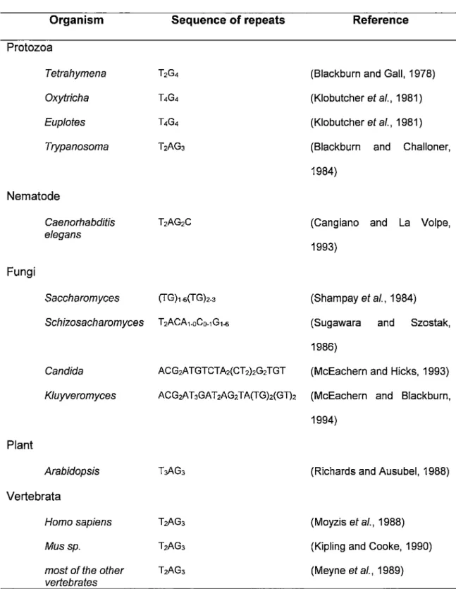
Table 1 Figure 1 Figure 2 Figure 3 Figure 4 Figure 5 Figure 6 Figure 7 Figure 8 Figure 9 Figure 10 Figure 11 Figure 12 Figure 13 Figure 14 Figure 15 Figure 16 Figure 17 Figure 18The sequence of telomeric repeats of the strand running 5'-3' towards the end of the chromosomes.
Organization of telomeric DNA in different eukaryotes. Telomere spatial structures in human and yeast. Telomere replication model.
Telomerase components in yeast. The telomerase reaction cycle.
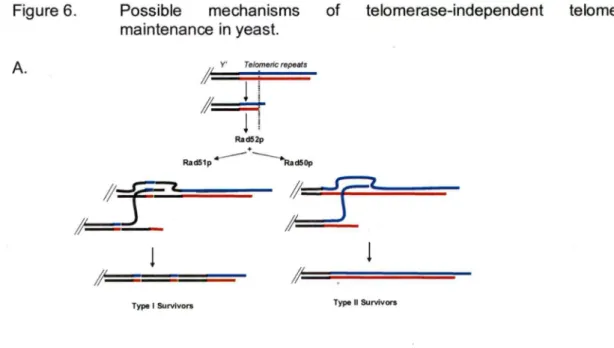
Possible mechanisms of telomerase-independent telomere maintenance in yeast.
The DNA damage checkpoint in Saccharomyces cerevisiae.
A spectrum of telomeric states.
T/01 and PTC2 deletion using a KanMX4 replacement cassette.
Pop-in I pop-out method.
Rationale for the SGA experiment.
Comparison of growth of double mutants (cdc13-115 yfg!J.) after 48 and 96 heurs of incubation.
An example of the comparative analysis of growth properties at 33°C after 96 heurs of incubation.
Growth properties of a primary candidate double-mutant at both permissive and restrictive conditions.
Verification of SGA results. Tetrad analysis. Replica plating. Example.
Verification of SGA results. Tetrad analysis. Streaking. Example.
T/01 and PTC2 ORF deletion verification.
Figure 19 Figure 20 Figure 21 Figure 22 Figure 23 Figure 24 Figure 25 Figure 26 Figure 27
Examples of generation of telomerase independent survivors. Mutant strains survivor type.
Requirement for gene products involved in adaptation to DSB in the formation of cap-independent survivors.
The rate of successful adaptation to telomere uncapping in survivors harbouring mutations in genes required for adaptation to a single DSB.
Schematic presentation of the procedure used to generate S2 survivors and second generation A2 cap-independent survivors.
Cell cycle progression in S1 survivors, S2 survivors and A1 li 13 strains after telomere uncapping.
Beth type 1 and type Il S2 survivors are able to form well developed viable colonies with much higher rate as compared to S 1 survivors.
The rate of successful adaptation to telomere uncapping in S2 survivors.
ABBREVIATIONS
50G 50 generations
200G 200 generations
oc
degrees CelciusLl delta, identifies deletion
µg microgram
µI micro litre
µM micromolar
A adenosine
ALT alternative telomere lengthening
ARS autonomously replicated sequence
BIR break-induced replication
bp base pares
c
cytosine Ci Curie cm centimeter dCTP deoxycytidine triphosphate ds double-stranded DSB double-strand breakdsDNA double-stranded deoxyribonucleic acid EDTA ethylenediamin disodium tetraacetate
FOA 5-fluoroorotic acid
FWD forward
g G2/M HU Li/Ac M min mg ml mM mm oie MMS MRX NHEJ nm nM nt OD ORF PCR PEG pH
REV
RT sec SGAgravitational rotation unit G2 cell cycle phase/mitosis hydroxyurea lithium acetate molar minute milligram millilitre millimolar mi Ili mole methylmethanesulfonate
complex of proteins Mre11 p/Rad52p/Xrs2p non-homologous end-joining
nanometre nanomolar nucleotide optical density open reading frame polymerase chain reaction poluethylene glycol
measure of the acidity or alkalinity of a solution reverse
room temperature second, a unit of time synthetic genetic array
SS ssDNA T Taq TE TPE Tris TRF ts TRF U or ura V
wt
w/vYC
YEPD yfg YPD single-strandedsingle-stranded deoxyribonucleic acid thymidine
DNA polymerase isolated from bacterium Thermus aquaticus buffer solution Tris-EDTA
telomere positioning effect
tris (hydroxymethyl) aminomethane telomere restriction fragment temperature sensitive
telomere restriction fragment uracil
volt wild-type weighUvolume
yeast complete (synthetic medium) yeast extract peptone dextrose "your favourite gene"
ABSTRACT
AN SGA APPROACH TC DISCOVER cdc13-f5 SUPRESSORS.
Telomeres, the DNA-protein complexes at the end of eukaryotic chromosomes, are essential for chromosomal stability. ln yeast, the telomeric single-strand binding protein Cdc13p has multiple important roles related to telomere maintenance:
telomeric "capping" - protection of telomeres by forming complexes with yKu?0/80 and with Stn1p/Ten1p;
• positive regulation of telomere replication via interaction with Est1 p, which is a part of telomerase;
negative regulation of telomerase by the recruitment of telomere elongation suppressors Stn1 p and Ten1 p.
ln an attempt to identify genes that are involved in the deleterious outcome of an absence of Cdc13p, we screened the yeast gene knock-out library for genes that could suppress the growth defect of cdc13-1 cells at 33°C. For this purpose, we performed an SGA array experiment. We scored for the ability of double mutant haploids to grow at 33°C. Eventually, we hoped to find the elusive genes involved in telomere 5'-end processing (exonucleases).
Based on the comparative analysis of growth properties of the strains (23°C vs 33°C), the initial screen identified up to 111 genes that displayed an apparent growth at 33°C. ln order to verify these results, diploids were regenerated, sporulated, microdissected, and haploid double mutants cdc13-1 yfgLJ. were isolated from 38 potential cdc13-1 suppressors. Unfortunately, this verification failed to reproduce a suppression of the growth defect by any of the selected genes at any temperature. While disappointing, the results reemphasize that careful re-examination of large
scale SGA approaches are indispensable before going on to more involved experimentation.
SIMILARITIES AND DIFFERENCES BETWEEN ADAPTATION TO DNA DOUBLE-STRAND BREAK AND TO TELOMERE UNCAPPING IN YEAST Saccharomyces cerevisiae.
lt was previously shown that a certain proportion of telomerase negative survivor cells (both type 1 and type Il cells) is able to survive in the absence of the telomere capping protein Cdc13p. These strains (named b.13s) were characterized in great detail and one of their discovered features was a striking ability to continuously inactivate DNA-damage checkpoints. Based on structural similarities between DNA double strand breaks (DSB) and unprotected telomeres, we attempted to verify if the molecular mechanisms regulating adaptation to a single irreparable DSB aise regulate adaptation to a loss of Cdc13p. For this purpose we created three tlc1 fJ. cdc13fJ. strains aise harboring DSB adaptation related mutations tid1/J., ptc2/J. and rfa1-t11. After deprotection of their telomeres, mutant survivor cells showed similar cell cycle progression patterns as compared to the cells where a single irreparable DSB was introduced. Adaptation defective mutants tid1/J. and ptc2/J. demonstrated an inability to adapt to telomere uncapping and to resume cell cycle. lnterestingly, cells harboring the rfa 1-t11 allele, which was reported to suppress adaptation defects of other mutations, did net show any distinguishable phenotype in terms of initial adaptation to telomere deprotection; i.e. rfa1-t11 mutant survivors do escape the G2/M arrest and re-enter the cell cycle. However, ail three mutant survivor strains failed to produce viable b.13 capping independent cells, which is consistent with the
hypothesis that adaptation to loss of Cdc13p depends on the same pathway as the previously reported adaptation phenomenon.
Finally, we report the surprising finding that if cells had once experienced an adapted b.13 state, they will re-produce capping negative survivors much more readily. Thus, while a culture of type Il survivor cells generates b.13s at a rate of about 1 x10⁻⁵
events per division, cells that had been b.13s and re-transformed with a Cdc13p carrying plasmid will produce capping independent cells at about 1x10⁻² events per
division. We are currently examining why these cells re-generate b.13 cell lines more readily and suspect structural differences in telomere terminal sequence arrangements.
RÉSUMÉ
UNE APPROCHE SGA POUR DÉCOUVRIR DES SUPPRESSEURS DE cdc13-1
Les télomères, complexes nucléoprotéiques situés aux extrémités des chromosomes eucaryotes, sont essentiels à la stabilité chromosomique. Chez la levure, la protéine Cdc13p liant la partie simple-brin des télomères joue plusieurs rôles importants dans la maintenance des télomères:
capuchon télomérique - protection des télomères par la formation de complexes avec yKu?0/80 et avec Stn1 pfîen1 p;
régulation positive de la réplication télomérique par l'interaction avec Est1 p, l'un des constituants de la télomérase;
régulation négative de la télomérase par le recrutement des suppresseurs de l'élongation des télomères Est1 p et Ten1 p.
Dans le but d'identifier des gènes impliqués dans les effets néfastes due à l'absence de Cdc13p, j'ai criblé la banque de souches de levure «knock-out» à la recherche de gènes qui pourraient supprimer le défaut de croissance de cellules cdc13-1 à 33°C. En effet, j'ai analysé la capacité des doubles mutants haploïdes à pousser à 33°C. J'espérais ainsi trouver les gènes élusifs impliques dans la dégradation de l'extrémité 5' des télomères (exonucléases).
En se basant sur l'analyse comparative des propriétés de croissance des souches (23°C vs 33°C), le criblage initial a identifié 111 gènes dont l'absence permettait une croissance apparente à la température restrictive de 33°C. Parmi ces 111 candidats j'ai retenu les 38 gènes codant pour des protéines dont la présence dans le noyau a été démontrée. Dans le but de vérifier les résultats du criblage initial j'ai regénéré les diploïdes par croisement. Les souches diploïdes ont été sporulées, microdisséquées
et les souches haploïdes doubles mutants cdc13-1 yfgfJ. ont été isolées pour les 38 suppresseurs potentiels. J'ai encore une fois évalué les propriétés de croissance des doubles mutants cdc13-1 yfgfJ. en les comparant avec celles du mutant simple
cdc13-1. Malheureusement, la vérification n'a pas reproduit les résultats du criblage initial.
Le défaut de croissance de cdc13-1 n'a été supprimé par l'absence d'aucun gène et ceci quelle que soit la condition de température.
Malgré leur issue négative, les résultats soutiennent la nécessité d'un réexamen détaillé du criblage SGA à grande échelle avant de s'en aller vers des
expérimentations plus approfondies.
SIMILITUDES ET DIFFÉRENCES DE L'ADAPTATION À UNE CASSURE DOUBLE BRIN DE L' ADN ET À LA PERTE DU CAPUCHON TÉLOMÉRIQUE CHEZ LA LEVURE Saccharomyces cerevisiae.
Il a été précédemment démontré qu'une certaine proportion des cellules survivantes télomérase-négatives (de type 1 et type Il) sont capables de survivre dans les
conditions d'absence de la composante principale du capuchon télomérique, la protéine Cdc13p. Les souches déficientes en capuchon télomérique (appelées b.13) ont été caractérisées en détail et une des propriétés découvertes a été leur frappante capacité à inactiver leur point de contrôle de dommage à l'ADN d'une façon continue. En me basant sur les similarités structurales entre la coupure double brin de l'ADN et les télomères non protégés, j'ai essayé de vérifier si les mécanismes moléculaires régulant l'adaptation à une coupure double brin unique et irréparable de l'ADN régulent aussi l'adaptation à la perte de Cdc13p. Dans ce but, j'ai construit trois
souches tlc1 ~ cdc13~ en délétant dans chacune un des gènes responsables de la régulation de l'adaptation à une cassure double brin unique: tid1~, ptc2~ et rfa1-t11. Après la déprotection de leurs télomères les cellules survivantes mutantes
montraient un patron de progression de leur cycle cellulaire similaire à celui de cellules mutantes dans lesquelles une cassure double brin unique de l'ADN a été introduite. Les mutants déficients en adaptation tid1~ et ptc2~ se sont révélés incapables de s'adapter à la perte du capuchon télomérique et de rétablir la
progression du cycle cellulaire. Les cellules portant rfa 1-t11, l'allèle dont la capacité à
supprimer les défauts d'adaptation d'autres mutants a été publiée, n'a démontré aucun phénotype détectable en terme d'adaptation initiale après la perte de protection télomérique; i.e. les survivants mutants rfa1-t11 s'échappent de l'arrêt G2/M et rétablissent leur cycle cellulaire.
Par contre, aucune des trois souches mutantes survivantes n'a été capable de générer des cellules b.13 capuchon indépendantes viables. Ce résultat corrèle avec l'hypothèse selon laquelle l'adaptation à la perte du capuchon télomérique dépend de la même voie métabolique que le phénomène d'adaptation à une cassure double brin unique de l'ADN décrite précédemment.
Je décris finalement une découverte surprenante: les cellules ayant été exposées une fois à l'état de perte du capuchon télomérique (l'état b.13) reproduiront des survivants capuchon-indépendants beaucoup plus facilement. Ainsi, alors qu'une culture de cellules de survivant de type Il génère des cellules b.13 au taux d'environ 1 x 10⁻⁵ événement par division cellulaire, les cellules b.13 qui ont été retransformées
avec un plasmide exprimant cdc13-1 reproduisent des cellules capuchon indépendantes au taux d'environ 1 x 10⁻² événement par division cellulaire.
Je soupçonne que ce phénomène existe grâce à des réarrangements de séquences terminales télomériques dans les cellules 6.13. Cette hypothèse est actuellement étudiée dans notre laboratoire.
INTRODUCTION
1. Historical Preamble
The term telomere originates from two words: TÉÀOÇ (te/os) and µtpoç (meros), which in Greek mean respectively "an end" and "a part". This term was first proposed by the geneticist Hermann J. Müller in the 1930'5 of the last century (Muller, 1938). Working with chromosomes of the fruit fly (Orosophi/a melanogaster) at the Edinburgh Animal Genetics lnstitute, he had just observed that the ends of the irradiated chromosomes, in contrast ta ends elsewhere in the genome, did not present alterations such as deletions or inversions, thanks ta the presence of a protective cap that he called «terminal gene» and afterwards «telomere». Just a few years later, Barbara McClintock, an investigator from the University of Missouri, who was dedicated ta the study of corn genetics (Zea mays), described how rupture of chromosomes resulted in adhesion and fusion of their ends, with the consequent formation of dicentric chromosomes (McClintock, 1941 ). She demonstrated that regardless of this damage, the ends could be restored thanks ta the acquisition of new telomeres. According ta her conclusions, telomeres play a crucial raie in the integrity of the chromosomes, since they prevent the appearance of «break-fusion-bridge» cycles which are catastrophic for cellular survival.
These studies founded the basis for the idea that the chromosome terminal fragments - telomeres - ought ta have features distinguishing them from DNA extremities resulting from DNA double-strand breaks. Later studies confirmed this idea and led ta discovery of specialized protective DNA-protein complexes located at the ends of eukaryotic linear chromosomes.
2. Telomere Structure
2.1 . Telomeric DNA
ln most eukaryotes, chromosome terminal regions consist of three distinct regions (Figure 1 ):
subtelomeric regions (DNA sequence immediately adjacent to telomeric repeats and stretching towards the centromere)
telomeric regions perse, where one can distinguish between double stranded DNA (dsDNA) and
single stranded DNA area (ssDNA).
Figure 1.
To
S.cer~wl.Slo96
Organization of telomeric DNA in different eukaryotes. Adopted from (Chakhparonian and Wellinger, 2003)
om
sutiite;om le r ion m li mrcrs
>-4----c s·
1 d 1 5-15 10100 1 > OO 3' 25Scheme is not to scale. "C" corresponds to cytosine-rich strand; "G" corresponds to guanine-rich strand. Subtelomeric region varies from one organism to the other and can contain repetitive elements.
ln this study 1 have used Saccharomyces cerevisiae as a model organism. This budding yeast is a very convenient organism for molecular biology and genetic studies. lt can be readily manipulated genetically and biochemically and is easy ta grow in the laboratory. Additionally, telomere structure and function are highly conserved amongst eukaryotes (see below) and therefore, any results obtained with yeast may be applicable ta other eukaryotes, including humans. Therefore, 1 will focus my discussions on molecular aspects of telomere protection using budding yeast Saccharomyces cerevisiae as a model.
2.1.1. Subtelomeric Regions
ln the yeast Saccharomyces cerevisiae, subtelomeric DNA comprises distinct elements called Y' and X separated by irregular telomere-like repeats TG1-3'CA1_3.
(Chan and Tye, 1983a; Chan and Tye, 1983b; Louis and Haber, 1992; Walmsley et al., 1984). Y' element(s) comprise the most proximal part of subtelomeric regions. Two types of Y' elements can be found on S. cerevisiae chromosomes. They differ in length - 6, 7 kb and 5,2 kb - but are very conserved in sequence. Not all of S.
cerevisiae chromosome ends possess Y' elements: while about 30% of yeast telomeres lack Y' elements completely, on the rest of them, 1 ta 4 Y' elements can be found arranged in tandem (Chan and Tye, 1983b). The exact raies of Y' elements in yeast cell life are net yet known. Elevated levels of homologous recombination was documented for this region, and such events play an important raie in existing yeast subtelomeric region variability (Louis and Haber, 1990a; Louis and Haber, 1990b; Louis and Haber, 1992).
A high level of homologous recombination in subtelomeric areas aise has great importance for telomere maintenance in the absence of telomerase (Lundblad and Blackburn, 1993) and this phenomenon will be discussed in detail below. Y' elements aise contain an ARS (origin of replication) consensus site (Chan and Tye, 1983a; Louis, 1995). These ARSs can fire in the S phase of the cell cycle but do net seem to be essential as chromosomes having no Y' elements or artificial chromosomes lacking terminal ARSs are capable of replicating successfully (Wellinger and Zakian, 1989).
X elements are present in just one single copy per chromosome extremity and located on telomeres immediately after Y' element towards the centromere (Louis, 1995; Louis et al., 1994). X elements are rather variable, both in size (from 0,3 to 3.75 kb) and in sequence (Biessmann and Mason, 1992; Chan and Tye, 1983a; Zakian and Blanton, 1988). There is just one small 475 bp conserved sequence element which is characteristic of all X elements on ail chromosomes (Louis, 1995). This sequence is referred to as the 'core' X element. Just as for Y'-elements, 'core' X contains an ARS consensus (Brand et al., 1987) as well as binding sites for two essential proteins - Tbf1 p (unknown function) and Abf1 p (involved in transcriptional regulation and replication) (Liu and Tye, 1991 ). Besides being involved in allowing telomere maintenance in the absence of telomerase, yeast subtelomeric regions are believed to play some role in nuclear architecture (Gotta et al., 1996; Hediger et al.,
2002; Taddei and Gasser, 2006; Taddei et al., 2004).
2.1.2. Double-stranded Telomeric DNA
The first telomeric DNA sequence determined was that of a ciliated protozoan called
telomeric DNA of T. thermophila consists of 50 to 70 repetitions of the sequence TTGGGG/CCCCAA. Further, it was shown that the strand running 5' to 3' towards the chromosome end consists of tandem repeats TTGGGGG, and the CCCCAA repeats constitute the complementary strand oriented 3' to 5'. Nowadays, the sequences and structures of telomeric DNA are known for many organisms (Table 1 ). As one can see from the Table 1, in the majority of organisms, telomeric DNA is comprised of short repetitive sequences organized in tandem. The strand running 5' to 3' usually is rich in guanines and therefore is referred to as G-strand, as opposed to the complementary 3' to 5' strand, rich in cytosine and referred to as C-strand (reviewed in Chakhparonian and Wellinger, 2003)). Due to the semi-conservative nature of DNA replication, the G-rich telomeric strand is always a product of leading strand synthesis, while the C-rich is the result of lagging strand synthesis. From Table 1 we can also derive that in some taxa the telomeric repeats are perfectly regular (e.g. Protozoa, Nematoda, most of Vertebrata), while in others, they comprise different degrees of irregularity (e.g. Fungi).
ln the budding yeast Saccharomyces cerevisiae, the telomeric repeat sequence is irregular (TG)1_6 TG2_3'C2_;A(CA)1_6 (Shampay et al., 1984; Wang and Zakian, 1990).
The number of telomeric repeats varies greatly between organisms. The range is from as little as 14 bp in Ciliata and reaching 100 kpb (kilo bp) in mouse (Kipling and Cooke, 1990; Klobutcher et al., 1981). The length of the repeat arrays can also vary between chromosomes of the same organism. For instance, in S. cerevisiae the irregular telomeric repeat sequence form telomeres of variable lengths of about 300 ± 75 bp (Shampay et al., 1984).
Table 1. The sequence of telomeric repeats of the strand running 5'-3' towards the end of the chromosomes.
Organism Sequence of repeats Reference
Protozoa
Tetrahymena T2G4 (Blackburn and Gall, 1978)
Oxytricha î4G4 (Klobutcher et al., 1981)
Euplotes î4G4 (Klobutcher et al., 1981)
Trypanosoma T2AG3 (Blackburn and Challoner, 1984)
Nematode
Caenorhabditis T2AG2C (Cangiano and La Volpe,
elegans
1993) Fungi
Saccharomyces (TG)1-6(TG)2_3 (Shampay et al., 1984)
Schizosacharomyces T2ACA1-0Co-1G1-6 (Sugawara and Szostak, 1986)
Candida ACG2ATGTCT A2(CT 2)2G2 TGT (McEachern and Hicks, 1993)
Kluyveromyces ACG2AT 3GAT 2AG2 T A(TG)2(GT)2 (McEachern and Blackburn, 1994)
Plant
Arabidopsis T~G3 (Richards and Ausubel, 1988)
Vertebrata
Homo sapiens T2AGa (Moyzis et al., 1988)
Mussp. T2AG3 (Kipling and Cooke, 1990)
most of the other T2AG3 (Meyne et al., 1989)
2.1.3. The Single Stranded 3' Extremity
An extension of the strand making up the 3'end (G-rich strand) has been detected in all organisms studied so far. Therefore, a G-rich single-stranded (ss) overhang has been proposed to be a conserved chromosome-terminal structure (Makarov et al., 1997; McElligott and Wellinger, 1997). However, the length of the overhang differs significantly from one taxon to the other (see Fig.1). ln the ciliates Tetrahymena, Oxytrichia and Euplotes, the ss 3' extension is very short and varies between 14 and 16 nt (Henderson and Blackburn, 1989; Klobutcher et al., 1981; Pluta et al., 1982). Sorne recent studies showed that in Tetrahymena, the majority of telomeres have ss overhangs of 14-15 or 20-21 nt in length, and the length of these extremities varies very little throughout the cell cycle (Jacob et al., 2001 ). Similarly, in the yeast S. serevisiae, the 3' strand extensions are about 12 to 15 nt, but can measure up to 25-30 nt at the end of S phase (Larrivée et al., 2004; Wellinger et al., 1996; Wellinger et al., 1993b). ln humans, it was shown that telomeres retain G-strand overhangs throughout the cell cycle and their length varies between 150 and 350 nt depending on cell type (Makarov et al., 1997; McElligott and Wellinger, 1997; Wright et al., 1997). Lately, Cimio-Reale et al., using a method based on the ligation of telomeric oligonucleotides hybridized to non-denatured DNA under stringent conditions showed that lengths ranging from 108 to 270 nt represented only 37% of the whole molecule population, while 56-62% were <90 nt. At the same time they have detected G-rich regions >400 nt in length (Cimino-Reale et al., 2001). However there were conflicting data on the distribution of G-rich ss tails on the telomeres. Makarov and colleagues, using a "primer extension/nick translation" technique, suggested that 80% of human chromosome extremities possess G-rich overhangs (Makarov et al., 1997). On the other side, Wright and colleagues obtained evidence for an asymmetry of human
chromosome ends. According to their data, one terminus of the same chromosome has a long G-rich extension, but the opposite one has a shorter G-tail (<12 nt) (Wright et al., 1997). Later, the Wright and Shay group reported data invalidating the above conclusion and showed that 3' overhangs on both leading (G-rich) and lagging (C-rich) strand ends were established very quickly after replication (Wright et al., 1999). The authors aise suggested that leading-telomere overhangs were shorter than lagging-telomere overhangs (Wright et al., 1999), a conclusion supported by later observations (Chai et al., 2006). Such a view is aise supported by studies in budding yeast showing differential processing of leading- and lagging-strand ends in strains lacking Rad27p endonuclease (Parenteau and Wellinger, 2002). This conclusion was supplemented with the idea that the overhangs at the leading and lagging-strand daughter telomeres are generated differently in human cells, and telomerase may preferentially affect overhangs generated at the telomeres produced by leading-strand synthesis (Chai et al., 2006).
The same group aise discovered that the vast majority of telomeric C strands end with the sequence CCAATC-5' (Sfeir et al., 2005), which strongly suggests that in human cells, C strand processing is very strictly regulated (Sfeir et al., 2005).
2.2.
Telomeric Heterochromatin
ln budding yeast and in humans, subtelomeric regions are organized into nucleosomes in a similar way as the rest of the chromosome. However, the most distal parts of the chromosomes are organized in a specific non-nucleosomal chromatin referred to as the telosome (Lejnine et al., 1995; Tommerup et al., 1994; Wright et al., 1992).
dsDNA binding proteins;
proteins associated with telomeres via interaction with other proteins; ssDNA binding proteins and the telomeric cap.
2.2.1. dsDNA Binding Proteins
Rap1 p is a protein bound to dsDNA in S. cerevisiae and it plays a major role in telomere homeostasis (Conrad et al., 1990). lts orthologs have been characterized for different organisms, for example TRF1 and TRF2 for humans (Chong et al., 1995) or Taz1p for S. pombe (Cooper et al., 1997). They ail bind the species specific repeats of telomeric dsDNA. RAP1 is an essential gene and Rap1 p, a protein of 827 aa, is required for repression and activation of several genes (Shore, 1994), as well as for telomere length regulation (Shore, 1997). At the telomeres, Rap1 p binds specifically the duplex sequence TG1_./C1_~ (Conrad et al., 1990; Gilson et al., 1993; Konig et al., 1996; Wright and Zakian, 1995) using its highly conserved Myb demains located in the centre of the molecule. Rap1 p plays a negative role in telomere regulation via recruitment of Rif proteins and provides Telomere Positioning Effect (TEP) via recruitment of Sir proteins {Tham and Zakian, 2002)) (discussed below). Recent studies elucidated the positive role of Rpa1 p protein in hiding the DSBs adjacent to the telomere from checkpoint detectors and exonucleolytic activity (Negrini et al., 2007).
Another protein binding to the dsDNA portion of telomeres is the yKu protein complex (Gravel et al., 1998). yKu is a heterodimer composed of two proteins: yKu70p and yKu80p (reviewed in Dynan and Yoo, 1998) and (reviewed in Featherstone and Jackson, 1999). The yKu heterodimer binds with high affinity to ds extremities, independently of their sequence or structure (Blier et al., 1993; Feldmann and
Winnacker, 1993; Paillard and Strauss, 1991 ). lt plays a crucial role in double strand break (DSB) repair vial non-homologous end joining (NHEJ) in both Mammalia and yeast (Jones et al., 2001) and protects their telomeres against uncontrolled homologous recombination events (Boulton and Jackson, 1996; Celli et al., 2006; Featherstone and Jackson, 1999).
The demain organization of Ku proteins is very well conserved in both lower an higher eukaryotes (Downs and Jackson, 2004), which could suggest homology between yeast yKu and mammalian Ku heterodimers (Downs and Jackson, 2004). ln S. cerevisiae the yKu heterodimer is associated with telomeres in vivo (Gravel et al., 1998). Cells lacking yKu show various telomere related phenotypes: shorter telomeres (Porter et al., 1996) abnormally elongated ss G-tails (Gravel et al., 1998) altered expression of genes located in proximity to telomeres (Boulton and Jackson, 1996; Gravel et al., 1998; Laroche et al., 1998), an elevated level of telomere-telomere recombination, as well as temperature sensitivity (Polotnianka et al., 1998). ln yeast, the NHEJ function of yKu is mediated by a conserved a-helix on the yKu?O surface, while its role in telomere maintenance is provided by a distinct surface on the yKu80 subunit (Ribes-Zamora et al., 2007). Besides interacting with telomeres, yKu aise interacts with telomerase via the telomerase RNA component TLC1 (Peterson et al., 2001; Stellwagen et al., 2003).
ln mammalian cells Ku plays an important role in NHEJ (Fukushima et al., 2001; Pierce et al., 2001), and there is evidence that Ku associates with telomeres in mammalian cells as well (d'Adda di Fagagna et al., 2001; Hsu et al., 2000). However, in humans, the heterodimer Ku70/Ku86 does not bind telomeric DNA directly but rather interacts with TRF1 (Hsu et al., 2000). Although there are differences in the phenotypes observed when comparing mammalian and yeast cells lacking Ku, it does play a role in protection and maintenance of telomeres in both higher and lower
eukaryotes. While the primary function of yeast Ku is to counteract exonucleases and inhibit telomere-telomere recombination (Maringele and Lydall, 2002; Nugent et al., 1998; Polotnianka et al., 1998), loss of Ku in human cells leads to deregulation in telomere length and genomic instability (Myung et al., 2004). ln human cells, the Ku complex was shown to mediate telomere end fusions at both unprotected telomeres (Smogorzewska et al., 2002) and critically short telomeres (Espejel et al., 2002). More recent data however reveal a critical role for mammalian Ku in suppression of homologous recombination at dysfunctional telomeres in addition to its known function in promoting telomere fusions (Celli et al., 2006).
2.2.2. Proteins Associated With Telomeres Via Protein-Protein
Interactions
As mentioned above, Rap1 p can interact, via its C-terminus, with Rif (Rap1 lnteracting Factor) proteins - Rif1 p and Rif2p (Hardy et al., 1992; Wotton and Shore, 1997). The complex Rap1 p, Rif1 p and Rif2p plays an important role in negative regulation of telomere length (Levy and Blackburn, 2004). Rif1 p and Rif2p are functionally very similar. Mutation of either RIF gene results in moderate telomere elongation. However, deletion of both RIF1 and RIF2 in the same cell results in a dramatic increase in telomere length (Hardy et al., 1992; Wotton and Shore, 1997). Additionally, overexpression of either RIF1 or RIF2 leads to significant telomere length decrease (Wotton and Shore, 1997).
The C-terminal demain of Rap1 p can also interact with Sir proteins (Moretti et al., 1994). Members of this family, in particular Sir2p, Sir3p and Sir4p, are responsible for TPE (Telomere Position Effect) (Aparicio et al., 1991). ln yeast, the essence of TPE is the repression of the expression of genes located in proximity of telomeres
(Gottschling et al., 1990; Tham and Zakian, 2002). lt was shown that in the cells where SIR2, SIR3, SIR4 as well as YKU70 and YKUBO are deleted, TPE is almost completely abolished (Aparicio et al., 1991; Mishra and Shore, 1999).
2.2.3. ssDNA Binding Proteins And The Telomeric Cap.
ln yeast, Cdc13p specifically binds the ss 3' extremities TG1_3 (Lin and Zakian, 1996; Nugent et al., 1996). lt binds ta this substrate with very high affinity and it is found bound at all the telomeres in vivo (Boums et al., 1998; Tsukamoto et al., 2001 ). Seing sequence specific, Cdc13p aise shows a certain level of affinity ta ss human telomeric repeats in vivo, but this affinity is estimated ta be 10 times weaker as compared ta yeast TG1_3 repeats (Alexander and Zakian, 2003; Lin and Zakian, 1996;
Nugent et al., 1996). This essential protein of 925 aa has two identified demains: a Recruitment Domain (RD) and a DNA Binding Domain (DBD) (Anderson et al., 2002; Mitton-Fry et al., 2002). Cdc13p binds ta telomeric ssDNA via the DBD and requires a minimal sequence of 11 nt: dGTGTGGGTGTG (Hughes et al., 2000b). The Recruitment Domain of Cdc13p serves for telomerase recruitment, providing an important raie of Cdc13p in telomere replication (Pennock et al., 2001 ).
Stn1 and Ten1 are proteins interacting with Cdc13p and are very important components of the telomere cap (Gao et al., 2007). lt is thought that these three proteins together (Cdc13p, Stn1p and Ten1p) create the first and the most important line of defense against exonucleolytic degradation of chromosomes termini (Gao et al., 2007; Grandin et al., 2000; Grandin et al., 2001 b; Grandin et al., 1997; Martin et al., 2007; Pennock et al., 2001).
2.3.
Telomere Structure ln Yeast and Humans
Human proteins TRF1 and TRF2 are factors binding to ds telomeric repeats T2AG3
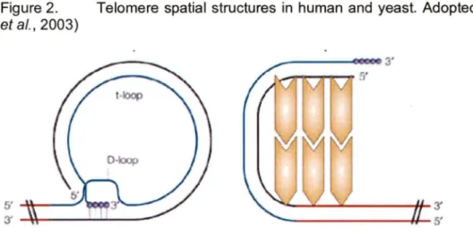
(Chong et al., 1995). They play a central role for human telomeric chromatin elaboration as they interact with various other proteins forming complexes (reviewed in Smogorzewska and de Lange, 2004)). Unlike its yeast ortholog Rap1p, human hRap1 interacts with telomeric DNA indirectly via TRF2 (Li et al., 2000). A major structural difference between human telomeres as compared to those of yeast was discovered via electron microscopy. Human telomeres appear to form loops, referred to as t-loops (telomeric-loop) (Griffith et al., 1999). The formation of t-loops is possible due to an invasion of the 3' ss G-rich extremity into ds telomeric repeats, where it finds complementary sequence, displacing the T2AG3 strand (Figure 2).
Figure 2. Telomere spatial structures in human and yeast. Adopted from (Vega et al., 2003)
Schematic representation of telomere organization in humans (a) and yeast (b). The blue line represents G-strand; the black line represents C-strand. Non-telomeric DNA is shown as a red line. More details are explained in the text. Telomere associated proteins are depicted as yellow polygons.
Similar telomeric structures were found in Mus sp., in chicken Gallus gal/us, in pea Pisum sativum, in the ciliate Oxytricha, and in the protozoan Trypanosoma (Cesare and Griffith, 2004; Munoz-Jordan et al., 2001; Murti and Prescott, 1999; Nikitina and Woodcock, 2004). Such structural similarity in a broad spectrum of taxa, suggests an evolutionary conservation of the t-loop structure. Surprisingly, t-loops have net yet been detected in yeast (reviewed in de Lange, 2004), but this could be due to their short ss 3'-overhangs or the short telomeres which would net loop as easily. Yet, there is evidence to suggest, that yeast telomeres form at least a sort of loop-back structure (de Bruin et al., 2001; Strahl-Bolsinger et al., 1997) (Figure 2).
3. Telomere Functions
Telomeres are essential for genome stability in all organisms with linear chromosomes. The specific roles include:
Chromosome end protection
Distinguishing chromosome ends from damaged DNA (DSB) Chromosome end replication
Architectural support in the nucleus Regulation of replication initiation timing Regulation of transcription
1 shall discuss the first three of the above mentioned functions as they are the most pertinent for understanding my experimental work.
3.1. Telomeres As Guards Of Chromosomes
3.1.1. The Telomeric Cap Protects Against Exonucleases
Telomeres protect eukaryotic chromosomes from exonucleolytic degradation with the aid of the telomeric cap. ln the budding yeast S. cerevisiae, the main component of the telomeric cap is the essential protein Cdc13p (see above). Garvik et al. discovered the protective raie of Cdc13p using a strain containing a temperature sensitive allele of CDC13, called cdc13-1 (Garvik et al., 1995). Exposed to a restrictive temperature above 28°C, cdc13-1 mutant cells suffer severe degradation of the 5' C-rich strand reaching the subtelomeric region and resulting in a rapid accumulation of 3' G-rich ssDNA. Accumulated ssDNA is detected by the Rad9p dependent checkpoint machinery (see "DNA DSB and DNA Damage Checkpoint"), the G2/M checkpoint is activated and the cell cycle arrested (Weinert and Hartwell, 1993). ln these conditions, yeast cells suffer progressive chromosome instability and subsequent cell death (Garvik et al., 1995). lt was suggested that Cdc13p bound to ss G-rich extremity protects the C-rich strand from degradation by one or several exonucleases (Nugent et al., 1996). Later, the interaction of two other essential proteins, namely Stn1 p and Ten1 p, with Cdc13p was shown to be absolutely required for efficient telomere protection (Grandin et al., 2000; Grandin et al., 2001 a). lndeed, the loss of function of either of these proteins leads to the same phenotype as
cdc13-115 mutant at restrictive temperature - C-strand degradation and Rad9p dependant
G2/M arrest (Grandin et al., 2001 a). The necessity of direct interaction between Cdc13p and Stn1p was shown in experiments with recombinant proteins in which the DNA binding demain of Cdc13p was fused to Stn1 p. Introduction of such recombinant proteins into a strain where CDC13 was deleted allowed the cells to recover viability (Pennock et al., 2001 ).
lnterestingly, C-strand degradation appears to be cell cycle dependent and regulated. (Vodenicharov and Wellinger, 2006; Vodenicharov and Wellinger, 2007). Abolishing the protective function of the essential capping proteins Cdc13p or Stn1 p causes telomere degradation in G2/M, but not in G1 of the cell cycle. (Vodenicharov and Wellinger, 2006). Completion of S phase and the activity of the S-Cdk1 kinase are required for telomere degradation. (Frank et al., 2006; Vodenicharov and Wellinger, 2006; Vodenicharov and Wellinger, 2007). Cdk1 regulated nucleolytic activity is required for the generation of 3' single-strand overhangs at both native and de nove telomeres in normally functioning cells (Frank et al., 2006; Vodenicharov and Wellinger, 2006; Vodenicharov and Wellinger, 2007).
Proteins functionally similar yeast Cdc13p are present in a wide variety of organisms (reviewed in Lange, 2001; Smogorzewska and de Lange, 2004). For example, a protein binding telomeric ssDNA in Schizosaccharomyces pombe is called spPot1 (Protection Of Telomere) and this protein is aise essential for telomere stability (Baumann and Cech, 2001). The deletion of the spPot1 gene leads to rapid telomere degradation, loss of telomeric DNA and to chromosome circularization (Baumann and Cech, 2001 ). Ali the Cdc13p-like proteins (TEBPs in Ciliata (Gottschling and Cech, 1984; Gottschling and Zakian, 1986), AtPot1 and AtPot2 in Arabidopsis (Shakirov et al., 2005) , cPot1 in chicken (Wei and Price, 2004), two distinct Pot1 proteins in mouse (Hockemeyer et al., 2006) and hPot1 in human (Baumann et al., 2002)), share very limited sequence homology, but are very conserved at the structural level of their DNA binding demain (Mitton-Fry et al., 2002; Smogorzewska and de Lange, 2004). Ali these proteins recognize G-rich ss overhangs and protect the telomeres against C-strand degradation thereby preserving chromosome integrity and genome stability (Smogorzewska and de Lange, 2004).
3.1.2.
Hiding
Chromosome
Ends
From
Homologous
Recombination
Yeast cells primarily use homologous recombination to repair damaged DNA, particularly DNA ds breaks (DSBs). ln order to protect chromosome termini against homologous recombination events, telomeres should not be recognized as damaged DNA (DSB in particular). Therefore, it is thought that telomeric chromatin is structured in a way such that chromosome ends are hidden from proteins whose raie is to detect DSBs, initiate checkpoint activation and subsequent cell cycle arrest (reviewed in Longhese, 2008; Lydall, 2003)). ln fact the very first observations on telomeres made by Müller and McClintock were the results of this feature of telomeres. Studies in S. cerevisiae showed that telomeres are essential for hiding chromosome ends from RAD9 dependent cell cycle arrest (Sandell and Zakian, 1993). These experiments were performed with a haploid strain harbouring two copies of chromosome VII (a so called disome) and in one of the chromosome VII homologues, a telomere could be removed in a controllable fashion. Such an elimination of one telomere led to cell cycle arrest (Sandell and Zakian, 1993). This experiment proved that telomeres are essential to allow the cells to distinguish between intact chromosome ends and a DSB.
3.2. Chromosome End-Replication
3.2.1. End Replication Problems
The transition from a circular genome of Procariota to linear chromosomes of Eukaryota created a problem, often referred to as "end replication problem". This
problem was first formally formulated in the beginning of 1970'5 of the last century (Olovnikov, 1973; Watson, 1972). The origin of the problem lays in the bidirectorial nature of DNA duplex molecule and the uniderectorial action of conventional DNA polymerases. They are able to polymerise new strands only in the 5' to 3' direction (Watson, 1972). ln addition, without a primer conventional DNA polymerases are unable to start polymerization.
Figure 3 .. Telomere replication model. Adopted from (Kelleher et al., 2002).
a. b. c. Parnnlal strands Sem!OO servativo DNA roplica!ion
'La991ng slrand tclomoro
+ 'Loadmg s1mnd' tolomoro Î: ~~,, ~~ ~~-~~l':._
3
• Loss of 3' overhang Resectioo of lh 5' end? 3'a. Parental stands before chromosome replication by conventional DNA polymerases. The G-rich 3'ss overhang is present.
b. After the passage of replication fork, two different 3' extremities are created : lagging strand 3' extremity (light green) and leading strand 3' extremity (red). RNA primers of lagging strand are represented by dark green arrows. Leading strand telomeres lose the 3' overhang since a blunt end is created.
c. The 5' end of the leading strand telomeres is processed in order for a regeneration of the 3' overhang.
During the replication of the lagging strand, the synthesis of Okazaki fragments is primed by short (8-12 nt) RNA primers, which are removed at a later stage. Removal of the most terminal RNA primer leaves the gap of at least 8-12 nt, which can net be filled (Lingner et al., 1995; Zakian, 1995). Additionally, the mode! of semi-conservative replication predicts that telomeres of leading strands will be in the form of a blunt end (Figure 3). lt was aise shown that 3' overhangs are present at bath ends of a chromosome, due ta C-rich 5'-end degradation by exonucleases (Makarov et al., 1997; Wellinger et al., 1996). A mode! depicting the above is presented in the Figure 3.
Only a few rounds of replication without restoration of telomeric repeats could shorten the telomere critically, leading ta degradation of coding DNA, genetic instability and eventually ta cell death (Biessmann and Masan, 1988; Lundblad and Szostak, 1989; Singer and Gottschling, 1994).
3.2.2. Telomerase
The solution ta the end replication problem was found in 1985. The telomerase enzyme was discovered in Tetrahymena (Greider and Blackburn, 1985). The telomerase holoenzyme consists of an RNA-moiety and a reverse transcriptase as its main elements, plus some other associated proteins. As in other organisms, the telomerase RNA TLC1 in S. cerevisiae (Figure 4) contains a sequence that serves as a template for the extension of the 3' G-rich strand of the telomeres (Feng et al., 1995; Shippen-Lentz and Blackburn, 1990; Singer and Gottschling, 1994).
ln budding yeast, telomerase consists of the TLC1 RNA subunit (Singer and Gottschling, 1994), Est2p, the reverse transcriptase catalytic su bu nit (Counter et al., 1997), proteins Est1 p and Est3p (Hughes et al., 2000a; Lendvay et al., 1996; Lin and
Zakian, 1995; Steiner et al., 1996), as well as the Ku heterodimer (yKu70p/yKu80p) (Peterson et al., 2001) and Sm7 protein complex (Seto et al., 1999).
Figure 4. Telomerase components in yeast. Adopted from (Kelleher et al., 2002).
Saccharomyces cerevisiae telomerase. The protein subunits are discussed in the text.
Concomitant interaction of the Ku heterodimer with telomeres and with TLC1 assures the presence of telomerase at the yeast telomere throughout the œll cycle (Fisher et al., 2004). Telomerase is recruited to the 3' end of the telomere via a direct interaction between the ss DNA binding protein Cdc13p and Est1 p in late S phase (Evans and Lundblad, 1999; Evans and Lundblad, 2002; Taggart et al., 2002). For yeast, the RNA component (TLC1) and reverse transcriptase (Est2p) are sufficient for telomerase activity in vitro (Cohn and Blackburn, 1995; Counter et al., 1997).
However, the presence of all of the above proteins is required in vivo (Cohn and Blackburn, 1995; Lingnereta/., 1997; Singer and Gottschling, 1994)
3.2.3. How Telomerase Elongates Telomeric Repeats.
Telomerase is responsible for proper telomere replication elongating the 3' strand via a reverse transcription type reaction (Greider and Blackburn, 1985; Lingner et al. , 1995; Nakamura and Cech, 1998). Telomerase uses its RNA component (see above) as a template for telomeric DNA synthesis (Singer and Gottschling, 1994; Yu et al., 1990).
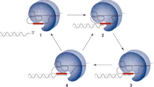
Figure 5. The telomerase reaction cycle. Adopted from (Kelleher et al., 2002)
/"\ ' /'\ F\ /\ ~ 3' \. V · 'A.../ V V 1
3
Different states of the reaction are represented. Telomerase is positioned at 3' extremity of a telomere (state 1 ). Telomere binding involves base-pairing with the RNA template (state 2). ln every reverse transcription cycle, one telomeric repeat is added (state 3). The 5' boundary of the template is the position where the telomeric substrate is most likely to either dissociate (state 4 to 1) or translocate (state 4 to 2) to the other end of the template.
The 3' extension-elongation takes place in late S phase of the cell cycle (Wellinger et al., 1993a; Wellinger et al., 1993b), at least in yeast. The reaction cycle is done in a multistep way, represented in Figure 5. Three stages of the elongation reaction can be distinguished (Kelleher et al., 2002):
Annealing the telomerase RNA component with the 3' G-rich strand; polymerization;
translocation (in mammals) or dissociation (in yeast) from telomere
ln Mammalia, telomere repeats addition is carried out in a similar way as in S. cerevisiae. The enzyme responsible for telomere elongation is also called telomerase and contains, at least in part, similar components as the yeast enzyme, such as hTR the RNA component. lt provides the template for reverse transcriptase and the hTERT protein catalyzes nucleotide addition (Counter et al., 1997; Feng et al., 1995; Harrington et al., 1997a; Harrington et al., 1997b; Meyerson et al., 1997; Nakamura
et al., 1997).
Human telomerase is active in germline cells and stem cells, but its activity is suppressed in somatic cells (Blackburn, 1992; Wright et al., 1996). lnterestingly, telomerase activity is reactivated in the majority of cancerous cells and in immortalized cell lines (Counter et al., 1992; Kim et al., 1994; Shay and Bacchetti, 1997; Shay and Wright, 1996).
3.3.
Telomere Length Regulation
ln all organisms with a constitutively active telomerase the length of telomeres remains stable. Therefore, we know that telomere length is tightly controlled, presumably by a number of mechanisms that remain ta be determined. ln yeast, not
all telomeres are elongated at the same time; telomerase acts on only a small portion of telomeres during one cell cycle (Teixeira et al., 2004). ln addition, there is accumulating evidence that telomerase shows a preference for elongating shorter telomeres (Arneric and Lingner, 2007; Hector et al., 2007; Teixeira et al., 2004; Viscardi et al., 2007).
As mentioned above, the members of the telomeric cap, Cdc13p, Stn1 p and Ten1 p play a very important role in the regulation of G-rich strand elongation by telomerase (Grandin et al., 2001 a; Grandin et al., 1997). The interaction between Cdc13p and Stn1 p has been shown to play an inhibitory role for G-rich strand elongation by telomerase (Ch and ra et al., 2001; Grand in et al., 1997). Furthermore, a S. cerevisiae strain harbouring a cdc13-5 allele shows very long telomeres and ss 3' extensions. The same phenomena were observed in a strain mutant for STN1 (Grandin et al., 1997). ln the case of overexpression of Stn1p in cdc13-5 cells, the long telomeres phenotype was suppressed, indicating that the inhibitory effect Cdc13-Stn1 p-Ten1 p complex on telomere elongation is due toits Stn1p subunit (Chancira et al., 2001). lt is aise believed that Cdc13p participates in telomerase recruitment onto telomeres during S phase via a direct interaction with the essential telomerase component Est1 p (Lustig, 2001; Pennock et al., 2001; Qi and Zakian, 2000). After the G-rich strand is elongated by telomerase and the complementary strand is synthesized, the ds telomeric region has increased in length, providing for new binding sites for telomere specific proteins such as Rap1 p. Marcand et al. showed that after creation of sufficient binding sites for Rap1 p (from 1 O to 20 molecules per one telomere) further telomere elongation in inhibited (Marcand et al., 1997). According to this model, once the optimal number of Rap1 p molecules are bound to a particular telomere, telomerase activity is inhibited, possibly via the telomere adopting some structure preventing telomerase access to its substrate. Conversely, should telomere
shortening result in less Rap1 p molecules bound, this inhibitory structure is disassembled and telomerase access is permitted (Marcand et al., 1997).
This model is supported by data showing extreme heterogeneity of telomere length in cells where Rap1 p is lacking its C-terminal demain required for protein-protein interactions at telomeres (Kyrion et al., 1992). Rap1 p binding proteins Rif1 p and Rif2p are aise involved in negative regulation of telomere length. Deletion of either of above the Rif proteins results in length increases, while deletion of both Rif proteins results in an synergistic effect and telomere length increases dramatically (Kyrion et al., 1992; Sussel et al., 1995). ln such cases of abnormal telomere extension, another mechanism of telomere homeostasis regulation can be revealed. This mechanism is referred to as Telomere Rapid Deletion (TRD). Using TRD, cells very quickly are able to shorten overelongated telomeres to their normal length. This mechanism is based on a single step telomere deletion, followed by 3' G-rich strand elongation and is conserved from yeast to mammals (Lustig, 2003).
4. Life After Loss Of Telomerase
Although telomerase is a major way to maintain the telomeres in Eukaryota, it is net the only way. A well described example of telomerase-independent telomeric DNA maintenance is the fly Drosophila melanogaster. ln Drosophila, telomeres are maintained via transposition of telomere specific retrotransposons (Biessmann and Mason, 1997). ln other insects such as the mosquito Anopheles and dipteran Chironomus, telomeres are maintained via telomere-telomere recombination pathways (Lopez et al., 1996; Roth et al., 1997). ln the alga Chlore/la, both telomerase-dependent and telomerase-independent pathways of chromosome end maintenance are present (Higashiyama et al., 1997).
The above examples show that telomerase is prevalent, but not the sole solution to the telomere maintenance problem. Therefore, it would be logical to expect the discovery of secondary, auxiliary pathways of telomere maintenance. The biological value of such pathways would be as a back-up means of telomere maintenance, should telomerase fail to function properly in organisms that normally rely on telomerase. lndeed, such pathways for normally telomerase-dependant eukaryotes were discovered some twenty years ago (Bernards et al., 1983; Dunn et al., 1984; Walmsley et al., 1984).
If yeast cells such as S. cerevisiae, S. pombe, or K. lactis, lose telomerase in one way or another, virtually all cells eventually die. However, the cell population does have a grace period, since telomerase loss first causes only a loss of about 5 bp per generation per telomere (Lundblad and Blackburn, 1993; Lundblad and Szostak, 1989; McEachern and Blackburn, 1996; Nakamura et al., 1997). This means that it takes approximately 60 to 1 OO generations before the telomeres lose functionality and due to the ensuing genomic instability, the cells stop growing (Hackett et al., 2001 ). However, invariably, there are cells that overcome this growth limitation and they are referred to as telomerase-independent survivors (Teng and Zakian, 1999). These cells maintain their telomeres via mechanisms relying on recombination (Chen et al., 2001; Huang et al., 2001; Le et al., 1999; Lendvay et al., 1996; Lundblad and Blackburn, 1993; Teng and Zakian, 1999; Teng et al., 2000). These mechanisms are virtually all dependent on the Rad52p protein (Chen et al., 2001; Huang et al., 2001; Le et al., 1999; Lendvay et al., 1996; Lundblad and Blackburn, 1993; Teng and Zakian, 1999; Teng et al., 2000).
Two types of survivors can be distinguished, depending on the kind of telomere rearrangements the cells have undergone: survivors type 1 and survivors type Il (Le
Type 1 survivors are characterized by heavily amplified subtelomeric Y' elements. Both, short and long Y' elements are amplified. The numbers of Y' elements in type 1 survivors can be more than 1 OO times higher compared to telomeres in wt cells (Le et al., 1999; Lundblad and Blackburn, 1993; Teng and Zakian, 1999). Even telomeres that normally do not possess Y' elements acquire Y' elements, despite the fact that in wt cells, interchromosomal Y' element exchange is a rare event (Horowitz and Haber, 1985). Telomeric repeats per se are very short in type 1 survivors (Le et al., 1999; Lundblad and Blackburn, 1993; Teng and Zakian, 1999), rarely reaching more than 1 OO bp. Nevertheless, they show high stability and do not shorten throughout the course of multiple generations. Supposedly, they originate from C1_:AfTG1_3 repeats separating the Y' elements.
lt is very important to mention that beside Rad52p, type 1 survivors require Rad51 p, Rad54p and Rad 57p proteins for growth (Chen et al., 2001; Le et al., 1999). Cells of type 1 survivors have a stable population of extrachromosomal circular DNA, containing one or two repetitions of Y' elements (Larrivée and Wellinger, 2006). These circles seem to be mostly double stranded structures. lt is however probable that some of them could be partially single stranded structures, as they are susceptible to degradation by mung bean nuclease (Larrivée and Wellinger, 2006). Mung bean nuclease is a single-strand-specific enzyme known ta be able to nick and degrade ss circular DNA, but not dsDNA (Kowalski et al., 1976; Sambrooke, 1989; Sung and Laskowski, 1962).
Type Il survivors do not amplify their Y' elements, but have very long and heterogeneous telomeres, consisting only of canonical C1_:AfTG1_3 repeat sequences generated via homologous recombination. The telomeric repeat tracts of type Il
survivors can be as long as 12 Kb (Teng and Zakian, 1999). Still being dependent on Rad52p, type Il survivors do not require Rad51 p, but do depend on the MRX complex members: Rad50p, Mre11 p, Xrs2p, as well as the recombination repair protein Rad59p and the DNA helicase Sgs1 p (Chen et al., 2001; Cohen and Sinclair, 2001; Huang et al., 2001; Le et al., 1999). Type Il survivors are stable over time, but their telomeres display dynamic changes in length, i.e. they continuously shorten and lengthen (Teng and Zakian, 1999). An analysis of the structures of the telomeric termini in type Il survivors revealed that they possess relatively normal 3' extensions (Larrivée and Wellinger, 2006). ln addition, it was shown that cells of type Il survivors have extrachromosomal circular DNA, consisting of telomeric repeats C1_:AfîG1-3 only. These circular DNA fragments proved to be at least partially single stranded circles (Larrivée and Wellinger, 2006).
Survivor types differ in terms of growth rates on plates, as well as in liquid cultures. lt takes just slightly longer for type 11 survivors to form colonies on agar plates comparing to wt cell (Teng and Zakian, 1999). ln contrast, type 1 survivors take significantly longer to form colonies (Teng and Zakian, 1999). lt was also observed that liquid cultures of type 1 survivors can end up as type Il survivors (Teng and Zakian, 1999). lt is thought that type 1 survivors might convert into type Il survivors, and as the latter proliferate significantly faster, the culture ends up with predominantly type Il survivor cells (Teng and Zakian, 1999). The reintroduction of functional telomerase into survivors suppresses recombination activity in telomeric areas and cells gradually restore telomeres to their initial wt length (Teng and Zakian, 1999). Type 1 survivors restore their telomeres to the wt state much faster than type Il survivors. (Lundblad and Blackburn, 1993; Teng and Zakian, 1999).