HAL Id: inserm-01466096
https://www.hal.inserm.fr/inserm-01466096
Submitted on 13 Feb 2017HAL is a multi-disciplinary open access
archive for the deposit and dissemination of sci-entific research documents, whether they are pub-lished or not. The documents may come from teaching and research institutions in France or abroad, or from public or private research centers.
L’archive ouverte pluridisciplinaire HAL, est destinée au dépôt et à la diffusion de documents scientifiques de niveau recherche, publiés ou non, émanant des établissements d’enseignement et de recherche français ou étrangers, des laboratoires publics ou privés.
Drugs in early clinical development for the treatment of
osteosarcoma
Marie-Françoise Heymann, Hannah Brown, Dominique Heymann
To cite this version:
Marie-Françoise Heymann, Hannah Brown, Dominique Heymann. Drugs in early clinical development for the treatment of osteosarcoma . Expert Opinion on Investigational Drugs, Taylor & Francis, 2017, 25 (11), pp.1265-1280. �10.1080/13543784.2016.1237503�. �inserm-01466096�
Drugs in early clinical development for the treatment of osteosarcoma
HEYMANN, Marie-Françoise1,2,3,4, BROWN, Hannah1,4 and HEYMANN, Dominique1,2,3,4
1. Department of Oncology and Metabolism, University of Sheffield, Medical School, Beech Hill Road, S10 2RX, Sheffield, UK
2. INSERM, UMR 957, Pathophysiology of Bone Resorption and Therapy of Primary Bone Tumours, Equipe Ligue 2012, University of Nantes, Faculty of Medicine, 44035 Nantes, France
3. Nantes University Hospital, Nantes 44035, France
4. European Associated Laboratory, INSERM-University of Sheffield, Sarcoma Research Unit, Medical School, S10 2RX, Sheffield, UK
Corresponding author:
Prof. Dominique Heymann
Department of Oncology and Metabolism Medical School
Beech Hill Road, S10 2RX, Sheffield, UK Tel.: +44 (0) 112 226 8464
Abstract
Introduction: Osteosarcomas are the main malignant primary bone tumours found in children
and young adults. Conventional treatment is based on diagnosis and resection surgery, combined with polychemotherapy. This is a protocol that was established in the 1970s. Unfortunately, this therapeutic approach has reached a plateau of efficacy and the patient survival rate has not improved in the last four decades. New therapeutic approaches are thus required to improve the prognosis for osteosarcoma patients.
Areas covered: From the databases available and published scientific literature, the present
review gives an overview of the drugs currently in early clinical development for the treatment of osteosarcoma. For each drug, a short description is given of the relevant scientific data supporting its development.
Expert opinion: Multidrug targeted approaches are set to emerge, given the heterogeneity of
osteosarcoma subtypes and the multitude of therapeutic responses. The key role played by the microenvironment in the disease increases the number of therapeutic targets (such as macrophages or osteoclasts), as well as the master proteins that control cell proliferation or cell death. Ongoing phase I/II trials are important steps, not only for identifying new therapies with greater safety and efficacy, but also for better defining the role played by the microenvironment in the pathogenesis of osteosarcoma.
Key words: Clinical Trials, Immunotherapy, Macrophages, Microenvironment,
Article highlights
• Tumour microenvironment is a key modulator in osteosarcoma development and is the source of new therapeutic targets
• Immunomodulators are promising drugs for controlling refractory and recurrent osteosarcoma (e.g. anti-GD2 therapy)
• Bone cells and bone matrix are two potential new targets for osteosarcoma (e.g. the anti-RANKL antibody, radium-223)
• Nanomedicine has led to the development of a new generation of compounds from “old” drugs (e.g. Nab-paclitaxel)
• Large biological cohorts with relevant clinical annotations are essential for rare tumours and will be an important source of new therapeutic targets
1. General features of osteosarcoma
Osteosarcoma accounts for 50% of all bone sarcomas, and is the most frequent primary malignant tumour found in children and young adults. With a peak of incidence at around 18 years, the male/female sex ratio is 1:4. A second peak of incidence is described in the elderly following radiotherapy, or in conjunction with Paget disease. The metaphyses of the long bones are their preferred development site. The proximal end of the tibia or humerus, as well as the distal end of the femur, is frequently affected. Sixty per cent of all cases of osteosarcoma are detected in the knee [1,2].
Osteosarcoma is part of a large family of heterogeneous histological tumour entities of mesenchymal origin. It expresses osteoblastic markers such as the runx2 master gene, alkaline phosphatase, osteocalcin or bone sialoprotein [10,11]. As a result, it has now been recognised that conventional osteoblastic osteosarcoma cells originate from a committed osteoblast in which an initial oncogenic event occurs, followed by secondary genetic alterations [12]. Osteosarcoma is thus a genetically complex disease. A recent study analysing a series of 44 osteosarcoma patients perfectly illustrates their high level of heterogeneity and complexity [13]. As expected, these authors demonstrated recurrent TP53 and RB1 somatic alterations and identified 84 point mutations and 4 deletions related to 82 different genes [13]. Similarly, Kovacs et al. studied the genetic alterations of 31 osteosarcomas and demonstrated that more than 80% of the cases could be explained by the fact that they exhibited a specific combination of single-base substitutions, a loss of heterozygosity, or large-scale genome instability. They identified alterations in 14 driver genes (TP53, RB1, BRCA2, BAP1, RET,
MUTYH, ATM, PTEN, WRN, RECQL4, ATRX, FANCA, NUMA1 and MDC1) with signatures
characteristic of BRCA1/2-deficient tumours [14]. They also proposed a new model for osteosarcoma development in which a TP53 and/or RB1 mutant cell initiated a monoclonal disease. This cell population exhibited higher chromosomic instability, leading to both the
emergence of new cell clones and polyclonal disease associated with these secondary genetic events [14]. The combination of multiple genetic events and a favourable microenvironment facilitate tumour growth [15-20]. It has been hypothesised that this microenvironment may be a sanctuary that sustains cell dormancy and contributes to drug resistance [20-22].
As osteosarcomas are bone-forming tumours, one of their signatures is the presence of osteoid tissue in close contact with spindle tumour cells. The morphology and organisation of tumour cells (such as extracellular matrix components) make it possible to identify various tumour subtypes, including osteoblastic, fibroblastic, chondroblastic, and highly vascularised telangiectactic forms, as well as giant cell enriched tumours [3-11]. Osteosarcomas are particularly prone to inducing lung metastases, which occur within 36 months of diagnosis and which have a strong impact on patient survival rate. Bone metastases can also occur in osteosarcoma, and they are associated with a worse survival rate than lung metastases [23]. The survival rate is estimated at around 50-70% after 5 years for non-metastatic patients and decreases dramatically to 30% when lung metastases are detected at the time of diagnosis (around 20% of patients) [24,25]. Unfortunately, these values have not changed in the last four decades [24]. The aim of the present review is to provide an overview of the main therapeutic approaches currently in development in the treatment of osteosarcoma.
2. Conventional therapeutic approaches to osteosarcoma
The therapeutic protocol currently in use for osteosarcoma was established by Rosen et al. at the end of the 1970s. It is a multimodal approach that combines surgery and polychemotherapy [26]. The advantages of chemotherapy were established by Link et al. in a randomised clinical trial that compared surgery with postoperative chemotherapy, and surgery alone [27]. Chemotherapy can be administered before (pre-operative, or neo-adjuvant, chemotherapy) and/or after surgery (post-operative, or adjuvant, chemotherapy). Overall, the
duration of the chemotherapy is around 6 to 12 months and combines doxorubicin, cisplatin, methotrexate and ifosfamide which are among the most efficient chemotherapeutic agents that have been identified for osteosarcoma [28]. The European Osteosarcoma Intergroup carried out a retrospective study of several clinical trials analysing various drug combinations and demonstrated the advantages of combining at least three drugs (reference combination: doxorubicin + methotrexate + cisplatin), and concluded that the doxorubucin/cisplatin association should no longer be considered as the standard chemotherapy combination for patients aged under 40 years with localised resectable osteosarcoma [29]. In addition, they demonstrated that chemotherapy-induced toxicity was a prognosis for overall survival, with the presence of greater toxicity generally associated with better survival [30]. However, although the advantages of neo-adjuvant chemotherapy have not been demonstrated [31], it is beneficial in several ways in treatment: i) it makes possible better delineation of tumours due to the formation of avascular collagenous pseudocaspules and then facilitates the definition of the surgical margin, ii) it reduces local tumour recurrence rates, iii) it makes it possible to evaluate the therapeutic response by means of histology, iv) it facilitates the preparation of definitive surgery for limb-salvage procedures by gaining time [32]. The Huvos grading system defines the therapeutic response and is established for the resected tumour, scoring the percentage of residual viable tumour cells (grade I >50%; grade II from 11 to 50 %; grade III from 1 to 10%; grade IV: no viable cells detected) [33]. Patients graded III and IV are considered to be good responders, and those graded I and II to be poor responders. As with a poor histological response, inadequate surgical margins are also an additional risk factor for local recurrence. The quality of the tumour resections, as evaluated by the quality of the surgical margins, is correlated with a high risk of local recurrence [34,35]. Unfortunately, at present, there is no consensus for staging and comparing these margins between all surgical/pathological teams. Although this histological assessment is a key parameter in
patient follow-up, the key challenge has been to determine whether the modification to post-operative treatment according to the therapeutic response analysed after the neo-adjuvant chemotherapy can improve the patients’ therapeutic response [36]. The European and American Osteosarcoma Study Group (EURAMOS), composed of the Children’s Oncology Group (COG), the Cooperative Osteosarcoma Study Group (COSS), the European Osteosarcoma Intergroup (EOI), and Scandinavian Sarcoma Group (SSG), analysed the impact of the nature of post-chemotherapy on 2,260 registered patients (good and bad responders) [37]. In a large clinical trial called EURAMOS-1, they compared the therapeutic advantages of MAP (Methotrexate/Doxorubicin/Cisplatin) and MAPIE (MAP/Ifosfamide/Etoposide) in bad responders, and MAP and MAPinf (MAP/Interferon-α). In bad responders, MAP vs MAPIE therapy did not show any difference in event-free survival [38]. Similarly, in good responders, MAPInf was not statistically different from MAP alone [39]. Overall, these results do not support adaptation of post-operative chemotherapy based on histological response. Osteosarcoma tumours are notoriously radioresistant [39]. However radiotherapy is used when adequate surgery is impossible, such as when the tumour is located in a high risk area (e.g. spine, pelvis, head and neck) [41,42]. Radiotherapy can thus help sterilise microscopic margins, and then contribute to local control of osteosarcoma growth in patients in whom surgical resection cannot lead to negative margins [43]. In addition, radiotherapy is a useful palliative tool for paediatric patients, especially when it comes to controlling bone pain [44].
3. Multi-target drugs and osteosarcoma
The poor results obtained with conventional therapeutic approaches led to the exploration of new, more effective treatments with less toxicity [45-47] (Figure 1). In this context, numerous clinical trials have been proposed, directly targeting cancer cells and/or their
microenvironment. Insulin-like growth factor-1 (IGF-1) and its receptor (IGF1-R) are expressed by osteosarcoma cells [48]. IGF-1 expression has been associated with the aggressiveness of the disease [48], However, IGF-1R status had no effect on median progression-free survival [50]. Based on this observation and an abundant literature exploring the advantages of blocking IGF-1 signalling in preclinical models, clinical trials targeting IGF-1 signalling using anti-IGF-1 or anti-IGF1R were set up [45]. Anti-IGF1-R antibodies were well-tolerated, although an extremely limited number of tumour responses were reported when it was used as a single or combined therapy [51]. These results can be explained by the existence of alternative signalling pathways that control cell proliferation [52], and/or by therapeutic escape through activation of phospho-AKT [53]. However, sirolimus, an mTOR inhibitor, has been identified as being a potentially interesting compound in osteosarcoma [54]. A phase I clinical trial [NCT02517918, “Metronomic chemotherapy in patients with advanced solid tumours with bone metastasis and advanced pretreated osteosarcoma (METZOLIMOS)”, 2015-2017, patients >13 years old] has been started. This study will include patients with unresectable locally advanced or metastatic osteosarcoma. The maximum tolerated dose is the primary outcome when sirolimus is administered in combination with cyclophosphamide, methotrexate and zoledronic acid.
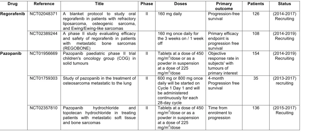
Numerous cytokines and growth factors act through activation of receptor tyrosine kinases (RTKs) and control cell proliferation, survival and migration [55]. Therefore, most of the RTK inhibitors (e.g. imatinib mesylate, dasatinib, sunitinib) considered to be multi-target therapies were assessed, although unfortunately their efficacy was low [55-65]. Pazotinib, which targets VEGFR, PDGFR and c-KIT [61,62], and sorafenib, which targets RET and VEGFR, show benefits in paediatric bone sarcomas by affecting angiogenesis [63,64]. Sofwat
et al. reported significant clinical responses in three metastatic osteosarcoma patients treated
patients are in progress to confirm the initial data obtained (Table 1). Grignani et al. studied the therapeutic effects of sorafenib in relapsed and unresectable high-grade osteosarcoma (clinical trial ref. NCT00889057, 35 patients) [64]. Thirty-five young and adult patients were enrolled and treated with 400 mg of sorafenib twice daily until progression or unacceptable toxicity. Sorafenib demonstrated activity as a second- or third-line treatment in terms of progression-free survival at 4 months, however the main limitation of this study was the lack of a control group. Associating sorafenib with everolimus did not produce any significant additional benefit compared to sorafenib alone [64]. Similarly, regorafenib is an oral multikinase inhibitor of angiogenic (VEGFR1-3, TIE2), stromal (PDGFR-β, FGFR), and oncogenic kinases (KIT, RET, and RAF). A phase I clinical trial revealed preliminary evidence of antitumor activity in patients with solid tumours including osteosarcoma [65]. A phase II trial started in 2014 is currently in the recruitment phase (Table 1).
c-MET (Mesenchymal Epithelial Transition) and its ligand hepatocyte growth factor are involved in many pathophysiological processes, especially in oncology. c-MET is a tyrosine kinase receptor encoded by the MET proto-oncogene and induces signalling pathways involving PI3K/Akt, MAPK and NFκB. Its transforming activity was initially identified in osteosarcoma cells and named MNNG HOS transforming gene [66]. Both proteins are expressed by musculoskeletal tumours [67], and osteosarcomas exhibit aberrant expression of the receptor [68-70]. In a preclinical model, c-Met inhibition reduced osteosarcoma growth, dysregulated bone remodelling [71], and sensitised cancer cells to chemotherapy [72]. These observations were the justification for setting up a phase II clinical trial using cabozantinib, a c-MET inhibitor (NCT02243605, “Cabozantinib-s-malate in treating patients with relapsed osteosarcoma or Ewing sarcoma”). Enrolment of 90 patients (> 12 years old) treated for relapsed osteosarcoma started in December 2014. The final data will be collected in June 2016 for the primary outcome measure. Dose use in sarcomas
corresponds to 60 mg tablets taken orally once a day in a 28-day cycle, repeated every 28 days in the absence of disease progression or toxicity. The primary outcome will be the antitumour activity of cabozantinib, in terms of 6-month objective response (complete response, partial response) and 6-month non-progression.
4. Targeting the bone microenvironment
Osteosarcoma cells are able to dysregulate the bone microenvironment by activating osteoclast differentiation and resorption, which in turn stimulate tumour growth by releasing proliferative factors stored in the extracellular matrix [17]. A vicious cycle is thus established between osteosarcoma and bone cells that identify osteoclasts as a potentially interesting target in bone sarcoma [73,74]. Preclinical investigations demonstrated that nitrogen-containing bisphosphonates decreased the proliferation of osteosarcoma cell lines in vitro and induced cell death [75,76]. In murine models, zoledronic acid decreased the volume of the primary tumour [77,78] and also the number of lung metastases induced [79,80]. In addition, combining it with chemotherapy revealed its value with regard to improving tissue repair and preventing tumour recurrence [81]. The mechanisms of action of zoledronic acid can be explained by its pleiotropic effects on osteosarcoma, especially modulating angiogenesis, and the bone and immune environment [82]. However, in 2010, Endo-Munoz et al. brought into question the therapeutic advantages of zoledronic acid, showing that a blockade of osteoclastogenesis played a part in the development of osteosarcoma lung metastases [83]. A phase III clinical trial called OS2006 (NCT00470223, “Combined chemotherapy with or without zoledronic acid for patients with osteosarcoma”) enrolled 318 patients (children and adults). This clinical trial was stopped prematurely due to the absence of any significant difference between the groups with or without zoledronic acid [84]. Various hypotheses can be advanced, including: i) the development of a resistance mechanism associated with
farnesyl diphosphate synthase in long-term treatment with zoledronic acid [85]; ii) the development of drug resistance due to the emergence of stemness properties in treated cancer cells [86]. A phase I clinical trial is in progress associating sirolimus with cyclophosphamide, methotrexate and zoledronic acid (NCT02517918, see paragraph 3). In addition to monocyte lineage, γ9δ2 T cells are key targets for zoledronic acid [87,88]. By inducing the release of phosphor-antigens, zoledronic acid induces the proliferation of these T lymphocytes. Interestingly, osteosarcoma cells are sensitised to zoledronic acid [89]. Using it to amplify ex
vivo γ9δ2 T cells and sensitise osteosarcoma to the immune response may be a future
treatment possibility. Based on an immonuregulatory effect, a phase I clinical trial is due to study the safety of transplantation with a haploidentical donor’s peripheral blood stem cell graft depleted of TCRαβ+ cells and CD19+ cells, in conjunction with zoledronic acid (NCT02508038, 21 patients, 2016-220, recruiting).
RANKL (Receptor Activator of Nuclear Factor Factor kappeB) and its receptor RANK clearly control osteoclast differentiation/activation, and consequently bone remodelling [90]. RANK is not only expressed by monocyte lineage (e.g. macrophages, dendritic cells, osteoclasts) and by endothelial cells, it is also expressed by osteosarcoma cells, as revealed by RT-qPCR and immunostaining. Depending on the series published, 18 to 69% of osteosarcoma cells express RANK [91-93]. A reverse correlation between RANK expression and the overall survival of patients with osteosarcoma has been demonstrated, but not with the response to chemotherapy [92]. Similarly, Bago-Horvath et al. reported that RANK expression is a negative prognostic factor for disease-free survival [93]. RANKL is also expressed by osteosarcoma cells [94,95]. One recent report has ignited controversy regarding the role of RANK/RANKL in the pathogenesis of osteosarcoma [95]. These authors did not detect the presence of RANK in osteosarcoma samples, and concluded that autocrine RANKL/RANK signalling in human osteosarcomas may not be operative, and anti-RANKL
therapy may not directly affect the tumour [95]. This discrepancy may be explained by the decalcification methods used and also by the source of the antibodies. Preclinical investigations demonstrated that RANKL blockade by osteoprotegerin, or soluble RANK delivery, has a strong impact on tumour development [96-98]. In other cancer cell types, tumour-infiltrating regulatory T cells appear to be the main source of RANKL, and may be a strong regulator of local immunity [99]. Denosumab is a fully humanised antibody that blocks RANKL binding to RANK and its functional activities [100]. In 2015, in a RANKL/RANK positive tumour, Cathomas et al. reported complete metabolic remission for over 18 months after treatment with combined sorafenib and denosumab, in a patient with progressive osteosarcoma after two lines of chemotherapy and radiotherapy [101]. A phase II clinical trial was thus initiated in 2015 led by the Children’s Oncology Group (NCT02470091, “Denosumab in Treating Patients With Recurrent or Refractory Osteosarcoma”). Ninety patients (age range: 11 to 50 years) who have relapsed or become refractory to conventional therapy with a regimen including some combination of high dose methotrexate, doxorubicin, cisplatin, ifosfamide and etoposide, will be included. Two cohorts will be formed: cohort I, patients with measurable disease according to RECIST, and cohort II, patients with complete resection of all sites of metastatic disease within 30 days prior to enrolment. Each patient will receive denosumab s.c. on day 1 (days 1, 8, and 15 in the first course of treatment). The treatment will be repeated every 4 weeks (28 days) for up to 24 months or 26 courses, whichever occurs first, in the absence of disease progression or unacceptable toxicity. At the end of the course of treatment, patients will be followed up for 3 years. The primary outcomes will be: i) the disease control rate at 4 months (cohort I), compared to historical Children’s Oncology Group experience with an objective response rate greater than 5%; ii) the disease control rate at 12 months, compared to historical Children’s Oncology Group experience (cohort II); iii) and the RECIST response at 4 months, compared to historical
Children’s Oncology Group experience with an objective response rate greater than 5%. The final data collection date for the primary outcome measure is April 2019. Secondary objectives include: i) investigation of pharmacokinetics and pharmacodynamics; ii) description of the tolerability of denosumab; iii) a review of the disease control rate and objective response rate for patients with recurrent osteosarcoma restricted to bone; iv) investigation of the biological markers associated with the therapeutic response to denosumab.
5. Immunomodulating drugs and osteosarcoma
Several reports have underlined the therapeutic value of using immunotherapies or immunomodulatory-based therapies for osteosarcoma (see reviews [102-105]). In this context, the number of new drugs activating the immune system has exploded in the last 10 years and numerous phase I and II clinical trials are in progress in osteosarcoma.
5.1. Mifamurtide (L-MTP-PE)
Mifamurtide is a synthetic analogue of a bacterial cell wall component that is a potent activator of the immune response, especially macrophages, in addition to standard chemotherapy [106,107]. This immunomodulator improved overall survival from 70 to 78% (p=0.03) in combination with chemotherapy, and resulted in a one-third reduction in the risk of death from osteosarcoma [108,109]. Mifamurtide was denied approval by the U.S. Food and Drug Administration (FDA) in 2007 and authorised by the European Medicines Agency (EMA) in 2009. The therapeutic efficacy of mifamurtide still remains highly controversial [110,111].
L-MTP-PE stimulates both the macrophages’ cytotoxic function and the secretion of high numbers of soluble mediators, including TNF, IL-1, IL-6 or IL-8 which stimulate the
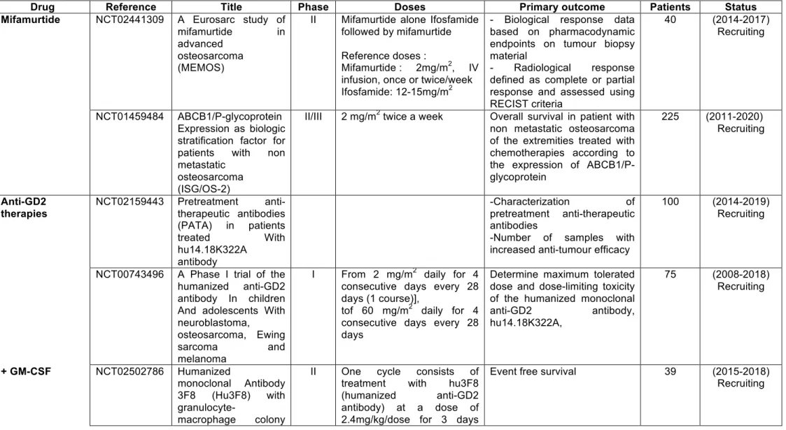
angiogenesis and development of metastases [112]. The density of tumour-associated macrophages is thus linked to poor prognosis. In osteosarcoma, Buddingh et al. showed that macrophages exhibit M1 and M2 phenotypes and demonstrated a link between M2 macrophages and angiogenesis [113]. Similarly, in preclinical models of osteosarcoma, the recruitment of the M2 subtype is correlated with tumour angiogenesis and lung metastasis [114]. Overall, these studies confirm the key role played by macrophages in the pathogenesis of osteosarcoma. The clinical investigations into the clinical benefits of mifamurtide continue, with an ongoing clinical trial combining mifamurtide and ifosfamide (Table 2). In osteosarcoma, around 50% of patients are poor responders to intensive conventional chemotherapy and these poor/no responses are frequently related to the over-expression of Multi-Drug Resistance protein-1 (MDR1 or P-gp for P-Glycoprotein or ABCB1). ABCB1 is also involved in the drug resistance mechanism associated with numerous compounds, including certain protein kinase inhibitors which increase its expression [115]. Patient stratification of high-grade osteosarcoma patients was suggested in 2006 by Serra et al. [116]. The effect of mifamurtide combined with chemotherapy will be re-evaluated in relation to ABCB1 expression. More than 200 non-metastatic patients will be included (ongoing recruitment, 2011-2020) in NCT014559484 trials in which overall survival will be the primary outcome (Table 2). Recently, Pahl et al. observed that the induction of macrophage anti-tumour activity (M1 subtype) by mifamurtide required IFN-γ [117]. This approach may be highly relevant for optimising mifamurtide therapy in osteosarcoma patients, and may open up new opportunities for this drug even if the combination of interferon and chemotherapy has not revealed any significant difference compared to conventional chemotherapy alone [55].
5.2. Disialoganglioside (GD2)
In 1987, Heiner et al. described the preferential accumulation of an anti-GD2 monoclonal antibody (3F8, a murine IgG3) at the tumour site in a preclinical model of osteosarcoma similar to previous observations made in neuroblastoma [118]. Ten years later, a phase I clinical trial revealed that a human-mouse chimeric monoclonal antibody (mAb) ch 14.18 directed against disialoganglioside (GD2) appeared to be clinically safe and effective with no specific toxicity after repeated administration [119]. An immunohistochemical study demonstrated that all the osteosarcoma tumours analysed were positive for GD2 in a series composed of 44 patients [120], and persisted upon recurrence [121]. In vitro, GD2 was suspected of enhancing the aggressiveness of the osteosarcoma [122]. Based on these observations, several clinical trials have been activated very recently (Table 2). Of them, one phase II trial (NCT02502786, sponsor: Memorial Sloan Kettering Cancer Center) will investigate the therapeutic advantages of the corresponding humanised form of the 3F8 antibody at a dose of 2.4mg/kg/dose for 3 days (days 1, 3, and 5) in the presence of GM-CSF. Patients (age range: 13 months-40 years) with recurrent high-grade osteosarcoma will be enrolled and the primary outcome will be event-free survival (time frame: 12 months) (Table 2). Another phase II protocol referenced NCT02484443 (sponsor: National Cancer Institute; Children’s Oncology Group) is in progress and is studying the effects of a human-mouse anti-GD2 monoclonal antibody ch14.18 in combination with sargramostim (GM-CSF) in patients with recurrent osteosarcoma (Table 2). Patients up to the age of 29 years will receive sargramostim s.c. on days 1-14 and dinutuximab i.v. over 20 hours on days 4 and 5 (the dinutuximab infusion can be extended for an additional 2 days for anticipated toxicities). The treatment will repeat every 28 days for up to 5 courses in the absence of disease progression or unacceptable toxicity. The primary outcome will be disease control after 12 months.
with the OKT3/3F8 bispecific antibody and will be administered in combination with IL-2 and GM-CSF (NCT02173093). The first objective is to determine the maximum tolerated dose and to analyse its efficacy and side effects (Table 2). Interestingly, endothelin A receptor, which has been implicated in osteosarcoma progression and the metastatic process, potentiates the inhibitory effects of the anti-GD2 antibody on invasiveness and tumour cell viability, opening up new potential clinical investigations [123].
5.3. Nivolumab
Nivolumab is an immunomodulator which acts by blocking the activation of programmed cell death-1 (1), induced by its ligand on subset activated T and pro-B lymphocytes [124]. PD-1 is part of the immunoglobulin superfamily that interacts with programmed cell death ligand 1 (PDL1), which is a cell-surface protein expressed in numerous cancer cells including osteosarcoma [125]. By interacting with PD-1, PDL-1 induces inhibitory signalling and suppresses cytotoxic T-cell-mediated tumour responses [126,127]. PD-1 has a dual effect, promoting apoptosis in antigen-specific T lymphocytes located in lymph nodes, and decreasing apoptosis in regulatory T cells. Consequently, PD-1 can be considered to be an immune checkpoint, down-regulating the immune system by preventing T lymphocyte activation. The inflammatory process in the tumour microenvironment is the source of many soluble factors such as IFN-γ, which may increase PDL-1 expression in cancer cells and suppress local immune responses [128]. Numerous preclinical investigations have demonstrated that inhibition of the interaction between PD-1 and PD-L1 enhances the T-cell response, resulting in increased antitumour activity. A phase I/II trial will be concluded in 2016 on refractory solid tumours and sarcomas, including osteosarcoma. 242 patients will be enrolled and treated with nivolumab IV over 60 minutes on days 1 and 15. Courses repeat every 28 days in the absence of disease progression or unacceptable toxicity (Table 2). PDL-1
expression in the tumour microenvironment is a key aspect in terms of therapeutic strategy (i.e. patient selection, predictive markers, follow-up biomarkers) and the initial investigations suggest that positive PDL-1 tumour expression is linked to a better therapeutic response. However, benefits were described in patients whose cancer cells were PDL-1 negative, which raises new questions regarding the mechanism of action of this molecule [129].
5.4. Immunity and dendritic cell vaccine
Dendritic cells have the specific ability to initiate and modulate adaptive immune responses [130]. This specificity, associated with their role in antigen presentation, has led to their use in vaccine approaches to cancer. Matured autologous dendritic cells loaded with tumour lysates derived from tumour tissue were used as the vaccine product. In a pre-clinical model of osteosarcoma, it has been demonstrated that killer dendritic cells were able to induce an adaptive antitumour immune response with a decrease in tumour development after cross-presentation of the tumour cell-derived antigen [131]. A phase I clinical trial demonstrated the feasibility and good tolerance of dendritic cells pulsed with MAGE-A1, MAGE-A3 and NY-ESO-1 full length peptides in combination with decitabine. Antitumor activity was observed in some patients [132]. In 2012, 12 osteosarcoma patients were vaccinated with tumour lysate pulsed dendritic cells, but evidence of a clinical benefit was observed in only 2 of these patients [133]. These authors concluded that osteosarcoma patients may be relatively insensitive to DC-based vaccine treatments. A new clinical trial was initiated, enrolling 56 patients (>1 year) with confirmed sarcoma, either relapsed or without known curative therapies, and treated with autologous dendritic cells pulsed with tumour lysate (Table 2). NCT02409576 is a pilot trial (“Pilot Study of Expanded, Activated Haploidentical Natural Killer Cell Infusions for Sarcomas (NKEXPSARC)”) analysing the effect of donor NK cells on clinical response determined by imaging. Twenty patients (aged 6 months to 80 years) will
be included between 2015 and 2016. The patients will receive lymphodepleting chemotherapy with cyclophosphamide (1 day) followed by fludarabine (5 days) and each patient will receive IL-2 1 day before infusion of the NK cell (total 6 doses).
6. Targeted alpha radiotherapy: Radium-223
The principle of alpha radiotherapy is to induce double strand breaks in DNA [134]. Radium-223 (223Ra) is a bone-seeking alpha-emitter which has been studied extensively in preclinical models [135]. Its half-life is 11·4 days. Its biodistribution in mice revealed that bone matrix is its preferred location of retention. Radium-233 is well tolerated, with doses of 50–250 kBq/kg, and has antitumour effects in preclinical murine models [136]. A first phase I clinical trial confirmed its potential clinical interest in skeletal metastases [137]. A recent phase III (NCT00699751) clinical trial in 921 patients with symptomatic castration-resistant prostate cancer with two or more bone metastases demonstrated the clinical benefit of radium-233 therapy [138]. In light of the marked retention of radium-233 in the bone matrix, a phase I trial has been set up for osteosarcoma to determine the maximum tolerated dose (NCT018335201, “Phase I Dose Escalation of Monthly Intravenous Ra-223 Dichloride in Osteosarcoma”, 2013-2017, ongoing but not recruiting) in 15 patients (> 15 years). The phase I starting dose was 50 kBq/kg Ra-223 dichloride i.v. over several minutes on day 1 of each 4-week cycle.
7. Alternative compounds for the treatment of osteosarcoma
Numerous targeted therapies are due to be assessed in clinical trials (Table 3). Of these drugs, those using the signalling pathways or enzymes involved in the cell cycle appear particularly interesting.
7.1. CC-115: a dual mTOR-DNA protein-dependent protein kinase inhibitor
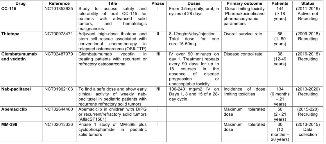
Optimisation of a series of triazoles led to the discovery of CC-115, which is able to both bind to mTOR and the DNA-protein dependent protein kinases involved in DNA repair mechanisms, and inhibit both of them [55,139]. CC-1115 inhibits both raptor-mTOR (TOR complex 1 or TORC1) and rictor-mTOR (TOR complex 2 or TORC2), and decreases the proliferation of cancer cells. DNA-PK is a serine/threonine kinase and from the PI3K-related kinase family of protein kinases. DNA-PK is activated following DNA damage and is involved in repairing breaks in double-stranded DNA via the DNA nonhomologous end joining (NHEJ) pathway [140]. By inhibiting DNA-PK, CC-115 impacts the DNA-repair mechanisms of tumour cells, inhibits the proliferation of numerous cancer cell lines, and increases cell apoptosis [141]. CC-115 has an anti-tumour effect in vivo as demonstrated by the inhibition of solid tumour growth in pre-clinical models of prostate cancer [139]. Interestingly, targeting DNA-PK increased the sensitivity of osteosarcoma cells to chemotherapeutic agents [142]. Treating cancer cells with CC-115 increases sensitivity to both chemo- and radiotherapy. A phase I trial has been set up (NCT01353625) in which 144 patients will receive increasing doses of oral CC-115 (starting with 0.5mg daily, in cycles of 28 days) (Table 3).
7.2. Abmaciclib: a CDK4 and CDK6 inhibitor
Cell cycle progression is controlled by cyclin-dependent kinases (CDK), which are dysregulated in numerous cancer cells, leading to uncontrolled cell proliferation. Of the various kinases identified, CDK4 and related CDK6 play a part in the progression of cells into the DNA synthetic phase of the cell-division cycle. CDK4 and CDK6 act more specifically in the first gap phase (G1) of the cell cycle and they assemble with D-type cyclins (D1-D3) in
response to various extracellular signals (i.e. mitigen activities and cytokine-induced signalling) to constitute enzymatically-active holoenzyme complexes [143]. Abmaciclib (LY2835219) is a CDK4 and CDK6 inhibitor capable of blocking the growth of cancer cells. Abemaciclib specifically inhibits CDK4/6 and related associated phosphorylation cascades such as Rb phosphorylation in early G1. Inhibition of Rb phosphorylation prevents CDK-mediated G1-S phase transition, blocking the cell cycle in the G1 phase, suppressing DNA synthesis and reducing cancer cell proliferation. This drug is currently being assessed in a phase I trial in children with recurrent or refractory solid tumours (NCT02644460) (Table 3).
7.3. Glembatumumab vedotin: an anti-gpNMB therapy
Glycoprotein non-metastatic melanoma protein B(gpNMB)/osteoactivin is a transmembrane glycoprotein that is highly expressed in various types of cancer. gpNMB is known to promote the invasion, migration and metastatic progression of cancer cells by modulating matrix metalloproteinase expression, but also by inhibiting the activation of tumour-reactive T lymphocytes via its binding to syndecan-4. gpNMB is also expressed by immune cells, including antigen-presenting cells, and may promote their adhesion to endothelial cells in an integrin-dependent manner. Furthermore, gpNMB decreases cell apoptosis and increases vascular density [144]. Recently, Roth et al. demonstrated that osteosarcoma gpNMB and its targeting by the antibody-drug conjugate glembatumumab vedotin resulted in cytotoxic activity [145]. A phase I/II trial has been initiated (NCT02487979) in 38 recurrent or refractory patients (Table 3).
7.4. Nanomedicine: Nab-paclitaxel and MM-398
Nanoparticles offer the possibility of encapsulating poorly soluble drugs and improving their half-life, bioavailability and efficacy [146]. Nab-paclitaxel is a new formulation of
conventional paclitaxel. It is solvent free, and comes in a nanoparticle albumin-bound (Nab) form. Nab-paclitaxel was designed to reduce the side effects of paclitaxel and docetaxel. Its activity is similar to paclitaxel, and it blocks the cell cycle in G2/M by stabilising the microtubules and consequently blocking chromosome duplication. Nab-paclitaxel has demonstrated its therapeutic advantages over paclitaxel in preclinical models, and combining it with gemcitabine in osteosarcoma may be of great interest [147]. A phase I/II trial was initiated in 2013 in paediatric patients with recurrent/refractory solid tumours, including osteosarcoma (NCT01962103, Table 3).
Based on similar technology, MM-398 is a stable nanoliposomal irinotecan with higher cytotoxicity than the original drug. The drug was assessed successfully in a preclinical model of Ewing sarcoma [148] and the results provoked the initiation of a phase I trial (NCT02013336) in paediatric solid tumours (Table 3).
8. Radiotherapy, miscellaneous trials and preparation for future investigations
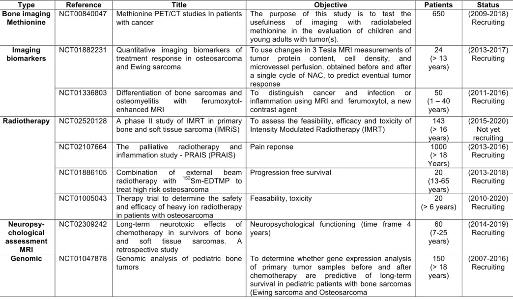
Although osteosarcoma is considered to be a radioresistant form of cancer, radiotherapy is used in the treatment of osteosarcoma in high-risk locations (such as the spine) to control local and recurrent development of tumours, and reduce pain, especially in a palliative context [44,149]. Several clinical trials are currently in progress to evaluate its efficacy in controlling bone pain and/or its therapeutic impact (Table 4). Recently, carbon ion radiotherapy was shown to be of interest in the management of unresectable osteosarcomas by providing good local control of the tumour without unacceptable morbidity [150,151]. Complementary investigations are required to validate carbon ion radiotherapy as a curative option in these patients.
Establishing biological cohorts for rare tumours takes a very long time. Such cohorts are nevertheless one of the key points for studying the pathogenesis of a specific disease,
especially heterogeneous pathologies when they are associated with clinical annotations. Several trials have been initiated to collect biological samples from osteosarcoma patients (e.g. tissue, blood) and will be open until 2100, enrolling 1000 patients (trials NCT02132182, NCT00580385, NCT00954473, NCT00899275, Table 4). These biological cohorts are and will be useful for helping define various differential diagnoses (trial NCT01336803, Table 4).
9. Conclusion
The key role played by the microenvironment in the pathogenesis of osteosarcoma increases the number of therapeutic targets (e.g. blood vessels, T cells, macrophages, and bone cells) in addition to the master proteins that control cell proliferation or cell death. Targeting the tumour microenvironment is the main objectives of the current phase II clinical trials in osteosarcoma and will provide very useful information on its clinical relevance in the near future. However, the key to success probably lies in better characterization of the disease, as this leads to better patient stratification and, consequently, to personalised medicine. Better understanding of how to control cancer-initiating cells, characterising their genotype, and identifying their functional links with their close environment are the scientific/medical challenges of the next few years. Biological cohorts will play a part in this challenge. Ongoing phase I/II trials are important steps, not only for identifying new therapies with greater safety and efficacy, but also for better defining the role of the microenvironment in the pathogenesis of osteosarcoma.
10. Expert opinion
Osteosarcoma is the most common malignant bone tumour. Like other bone sarcomas, osteosarcomas are largely insensitive to conventional therapies, have a tendency to form distant metastases (principally in the lungs), and show other common biological features, such
as dysregulation of bone remodelling, and the presence of disseminated cells considered to be cancer stem cells. However, despite progress in multidrug chemotherapy protocols and conservative limb salvage surgery, osteosarcoma survival rates have not improved for more than 30 years. Transcriptomic and phosphoproteomic assessments have identified key intracellular signalling pathways that are activated by cytokines/growth factors and sustain cancer cell proliferation. These data led to the development of a large panel and several generations of tyrosine-kinase inhibitors, which were initially promising multi-target drugs. Unfortunately, most of the drugs considered had low efficacy in osteosarcoma patients due to the development of resistance mechanisms [55-65]. However, many clinical trials failed to clearly evaluate their therapeutic value in the context of osteosarcoma with very high levels of heterogeneity. It is necessary to revisit their efficacy in view of the full expression profile of the tyrosine kinases of each patient. Sorafenib showed interesting clinical advantages, although unfortunately they remain difficult to analyse in the absence of an adequate control group. Complementary clinical trials are thus required [64]. Pazopanib [61,62], regorafenib [66] and cabozantinib (NCT02243605) may also be interesting therapeutic options.
Using the tumour microenvironment as a potential therapeutic target indicates the start of a new era for osteosarcoma patients. Immune modulators are some of the promising drugs in development in osteosarcoma (see section 5). A recently set up clinical trial is studying whether or not to associate ipilimumab, a fully human monoclonal antibody that binds CTLA-4 and blocks its interaction with CD80 and CD86 [152]. However, it is too early to conclude on any therapeutic advantages to this approach (Table 2). Mifamurtide is the frontrunner in the immunoregulator family, and it has been authorised after much debate in the Europe, but not in USA. This controversial drug was nevertheless the first to produce a significant improvement in survival rates in osteosarcoma. Although the effect was modest, this observation nevertheless identifies the concept of macrophage modulation as therapeutic
option. In the last decade, several authors demonstrated the key role played by macrophages in the pathogenesis of osteosarcoma, and, more specifically, the key point seems to be the balance in the M2/M1 macrophage subtype [112,114]. Since the development of mifamurtide [106-111, 115-117], the anti-GD2 antibody [118-123], and genetically-modified T cells, vaccines have been proposed and are currently undergoing clinical trials. The main idea here is not only to decrease or slow down tumour development, but also to control the disease. This is a significant modification to the philosophical approach used in oncology: associating curative aspects and control of a disease via the immune system. Radium-233 is also a promising new therapeutic agent that is retained preferentially in the bone matrix (tumour environment) close to the cancer cells [134-138]. The clinical benefits shown in the bone metastases of prostatic cancers heighten its clinical value. Clinical trials in progress will soon provide us with the answer.
Identifying and characterising early tumour recurrence and metastasis dissemination remains necessary if we are to propose better adapted therapeutic strategies. These early events can in fact be considered as biomarkers and include all the biological parameters that reflect the recurrent disease. More specifically, they reflect all the specific signatures at the transcriptional and/or protein level, as well as the isolated circulating tumour cells characterised by a specific phenotype. Metastatic spread to specific target sites (the lungs and/or bones) is a clinically intractable feature of osteosarcoma’s state of dormancy (quiescence), evading detection whilst remaining primed to colonise the target metastatic organ upon induction of the right environmental cues [153-155].
Circulating tumour cells have also been isolated from osteosarcoma patients [156,157] and new technologies (e.g. microfluidic) provide an opportunity to both isolate tumour cells and “cancer initiating cells” from fixed paraffin embedded samples at the single cell level, and better define tumour heterogeneity [158,159]. Based on the heterogeneity of
osteosarcoma subtypes and therapeutic response, new patient stratification may be proposed and new multidrug targeted approaches adapted to each patient (personalised medicine) will emerge. The biological cohorts established will be one of the key factors in these developments. The gap between the new generation of drugs and conventional chemotherapy will be filled by new formulations of “old” drugs (such as Nab-paclitaxel) thanks to nanomedicine, thus improving their bioavailability, efficacy, and safety, and reducing their side effects [146,147].
REFERENCES
1.Mirabello L, Troisi RJ, Savage SA. International osteosarcoma incidence patterns in
children and adolescents, middle ages and elderly persons. Int J Cancer 2009;125:229–234.
2. Hauben EI, Hogendoorn PCW. Epidemiology of primary bone tumors and economial
aspects of bone metastases.In Bone Cancer (Ed D. Heymann, Academic Press, San Diego), 2015; 5-9.
3.Inwards C, Suire J. Low-grade central osteosarcoma. In Who Classification of tumours of
soft tissue and bone (IARC ed, Lyon, France) 2013;281-282.
4. Rosenberg AE, Cleton-Jansen AM, de Pinieux G, et al. In Who Classification of tumours of
soft tissue and bone (IARC ed, Lyon, France) 2013;282-288.
5. Oliveira AM, Okada K, Suire J Telangiectatic osteosarcoma. In Who Classification of
tumours of soft tissue and bone (IARC ed, Lyon, France) 2013;289-290.
6. Kalil RK, Suire J. Small cell osteosarcoma. In Who Classification of tumours of soft tissue
and bone (IARC ed, Lyon, France) 2013;291.
7. Lazar A, Mertens F. Parosteal osteosarcoma. In Who Classification of tumours of soft
tissue and bone (IARC ed, Lyon, France) 2013;92-293.
8. Montag AG, Suire J. Periosteal osteosarcoma. In Who Classification of tumours of soft
tissue and bone (IARC ed, Lyon, France) 2013;294.
9. Wold LE, McCarthy EF, Squire J. High-grade surface osteosarcoma. In Who Classification
of tumours of soft tissue and bone (IARC ed, Lyon, France) 2013;295-296.
10. Heymann D, Redini F. Bone sarcomas: pathogenesis and new therapeutic approaches.
IBMS BoneKEy 2011;8:402-414.
11.Mohseny AB, Hogendoorn PC. Concise review: mesenchymal tumors: when stem cells go
mad. Stem Cells 2011;29:397-403.
12. Mutsaers AJ, Walkley CR. Cells of origin in osteosarcoma: mesenchymal stem cells or
osteoblast committed cells? Bone 2014;62:56-63.
••An important review summarizing the origin of osteosarcoma
13. Bousquet M, Noirot C, Accadbled F, et al. Whole-exome sequencing in osteosarcoma
reveals important heterogeneity of genetic alterations. Ann Oncol. 2016;27:738-44.
14. Kovac M, Blattmann C, Ribi S, et al. Exome sequencing of osteosarcoma reveals
mutation signatures reminiscent of BRCA deficiency. Nat Commun 2015; 6:8940.
15. Walkley CR, Qudsi R, Sankaran VG, et al. Conditional mouse osteosarcoma, dependent on p53 loss and potentiated by loss of Rb, mimics the human disease. Genes Dev 2008;22:1662–1676.
16. Lin PP, Pandey MK, Jin F, et al. Targeted mutation of p53 and Rb in mesenchymal cells
of the limb bud produces sarcomas in mice. Carcinogenesis 2009;30:1789–1795.
17. Berman SD, Calo E, Landman AS, et al. Metastatic osteosarcoma induced by inactivation
of Rb and p53 in the osteoblast lineage.Proc Natl Acad Sci U S A 2008;105:11851–11856.
18. Mutsaers AJ, Ng AJ, Baker EK, et al. Modeling distinct osteosarcoma subtypes in vivo
using cre:Lox and lineage-restricted transgenic shRNA. Bone 2013;55:166–178.
19. Wittrant Y, Théoleyre S, Chipoy C, et al. RANKL/RANK/OPG: new therapeutic targets
in bone tumours and associated osteolysis. Biochim Biophys Acta 2004;1704:49-57.
20. Borovski T, De Sousa E Melo F, et al. Cancer stem cell niche: the place to be. Cancer Res 2011;71:634-639.
• A interesting review describing the role of local niches in the cancer development 21. Perrot P, Rousseau J, Bouffaut AL, et al. Safety concern between autologous fat graft,
mesenchymal stem cell and osteosarcoma recurrence. PLoS One 2010;5:e10999.
22. Avril P, Duteille F, Ridel P, et al. Opposite Effects of Soluble Factors Secreted by
Adipose Tissue on Proliferating and Quiescent Osteosarcoma Cells. Plast Reconstr Surg 2016;137:865-75.
23. Clark JC, Dass CR, Choong PF. A review of clinical and molecular prognostic factors in
osteosarcoma. J Cancer Res Clin Oncol 2008;134:281-297.
24. Allison DC, Caarney SC, Ahlmann ER, et al. A meta-analysis of osteosarcoma outcomes
in the modern medical era. Sarcoma 2012;2012:704872.
25. Mirabello L, Troisi RJ, Savage SA. Osteosarcoma incidence and survival rates from 1973
to 2004: data from the surveillance, epidemiology, and end results program. Cancer 2009;115:1531-1543.
26. Rosen G, Tan C, Sanmaneechai A, et al. The rationale for multiple drug chemotherapy in
the treatment of osteogenic sarcoma.Cancer 1975;35:936-45.
••Key reference describing the conventional therapeutic protocol currently used in all clinical centres
27. Link MP, Goorin AM, Miser AW, et al. The effect of adjuvant chemotherapy on
relapse-free survival in patients with osteosarcoma of the extremity. N Engl J Med 1986;314:1600– 1606.
28. Bacci G, Longhi A, Fagioli F, et al. Adjuvant and neoadjuvant chemotherapy for
osteosarcoma of the extremities: 27 year experience at Rizzoli Institute, Italy.Eur J Cancer 2005;41:2836–2845.
29. Whelan JS, Jinks RC, McTiernan A et al. Survival from high-grade localised extremity
osteosarcoma: combined results and prognostic factors from three European Osteosarcoma Intergroup randomised controlled trials. Ann Oncol 2012;23:1607-1616.
30. McTiernan A, Jinks RC, Sydes MR, et al. Presence of chemotherapy-induced toxicity
predicts improved survival in patients with localised extremity osteosarcoma treated with doxorubicin and cisplatin: a report from the European Osteosarcoma Intergroup. Eur J Cancer.2012;48:703-712.
31. Goorin AM, Schwartzentruber DJ, et al. Presurgical chemotherapy compared with
immediate surgery and adjuvant chemotherapy for nonmetastatic osteosarcoma: Pediatric Oncology Group Study POG-8651. J Clin Oncol 2003;21:1574-1580.
32. Carrle D, Bielack SS. Current strategies of chemotherapy in osteosarcoma. Int
Orthop;30:445-451.
33. Huvos A. Osteogenic osteosarcoma. In Huvos A (ed): Bone Tumors: Diagnosis.
Treatment and Prognosis, 2nd ed. Philadelphia: WB Saunders 1991 ; 85-156.
34. Gouin F, Heymann MF. Margins and bone tumors- what are we talking about ?In Bone
Cancer (Ed D. Heymann, Academic Press, San Diego), 2015;287-291.
35. Ennecking WF. In Enncking WF, Editor.Musculoskeletal tumor surgery.Churchill
Livingstone New York, Edinburgh, London and Melbourne, 1983; 89-99.
36. Bielack S, Kempf-Bielack B, Von Kalle T, et al. Controversies in childhood
osteosarcoma. Minerva Pediatr 2013;65:125-148.
37. Whelan JS, Bielack SS, Marina N, et al. EURAMOS-1, an international randomised study
for osteosarcoma: results from pre-randomisation treatment. Ann Oncol 2015;26:407-414.
38. Marina N, Smeland S, Bielack, et al MAPIE vs MAP as postoperative chemotherapy in
patients with a poor response to preoperative chemotherapy for newly-diagnosed osteosarcoma: results from EURAMOS-1. Connective Tissue Oncology Society (CTOS) 2014,paper 032.
39. Bielack SS, Smeland S, Whelan JS, et al. Methotrexate, Doxorubicin, and Cisplatin
(MAP) Plus Maintenance Pegylated Interferon Alfa-2b Versus MAP Alone in Patients With Resectable High-Grade Osteosarcoma and Good Histologic Response to Preoperative MAP: First Results of the EURAMOS-1 Good Response Randomized Controlled Trial. J Clin Oncol. 2015;33:2279-2287.
40. Schwarz R, Bruland O, Cassoni A, Schomberg P, Bielack S. The role of radiotherapy in
oseosarcoma. Cancer Treat Res 2009;152:147-164.
41. Ozaki T, Flege S, Kevric M, et al. Osteosarcoma of the pelvis: experience of the
Cooperative Osteosarcoma Study Group. J Clin Oncol 2003;21:334-341.
42. DeLaney TF, Park L, Goldberg SI, et al. Radiotherapy for local control of osteosarcoma.
Int J Radiat Oncol Biol Phys 2005;61:492-498.
43. Errani C, Longhi A, Rossi G, et al. Palliative therapy for osteosarcoma. Expert Rev
44. Rahn DA 3rd, Mundt AJ, Murphy JD, et al. Clinical outcomes of palliative radiation
therapy for children. Pract Radiat Oncol 2015;5:183-187.
45. Heymann D, Redini F. Targeted therapies for bone sarcomas. BoneKey Rep 2013 ;2 :378. 46. Ando K, Heymann MF, Stresing V, et al. Current therapeutic strategies and novel
approaches in osteosarcoma. Cancers 2013;5:591-616.
47. Hattinger CM, Fanelli M, Tavani E, et al. Advances in emerging drugs for osteosarcoma.
Expert Opin Emerging Drugs 2015;20:495-514.
48. Burrow S, Andrulis IL, Pollak M, et al. Expression of insulin-like growth factor receptor,
IGF-1, and IGF-2 in primary and metastatic osteosarcoma. J Surg Oncol 1998;69: 21-27.
49. Jentzsch T, Robl B, Husmann M, et al. Worse prognosis of osteosarcoma patients expressing IGF-1 on a tissue microarray. Anticancer Res 2014;34:3881-3889.
50. Schwartz GK, Tap WD, Qin LX, et al. Cixutumumab and temsirolimus for patients with bone and soft-tissue sarcoma: a multicentre, open-label, phase 2 trial. Lancet Oncol 2013;14: 371–382.
51. Bagatell R, Herzog CE, Trippett TM, et al. Pharmacokinetically guided phase 1 trial of the IGF-1 receptor antagonist RG1507 in children with recurrent or refractory solid tumors. Clin Cancer Res 2011;17:611-619.
52. Avnet S, Sciacca L, Salerno M, et al. Insulin receptor isoform A and insulin-like growth factor II as additional treatment targets in human osteosarcoma. Cancer Res 2009;69:2443-2452.
53. Wan X, Harkavy B, Shen N, et al. Rapamycin induces feedback activation of Akt
signaling through an IGF-1R-dependent mechanism. Oncogene 2006;26:1932–1940.
54. Penel-Page M, Ray-Coquard I, Larcade J, et al. Off-label use of targeted therapies in osteosarcomas: data from the French registry OUTC'S (Observatoire de l'Utilisation des Thérapies Ciblées dans les Sarcomes). BMC Cancer 2015;15:854.
55. Ségaliny AI, Tellez-Gabriel M, Heymann MF, et al. Receptor tyrosine kinases:
Characterisation, mechanism of action and therapeutic interests for bone cancers. J Bone Oncol 2015;4:1-12.
56. Chugh R, Wathen JK, Maki RG, et al. Phase II multicenter trial of imatinib in 10
histologic subtypes of sarcoma using a bayesian hierarchical statistical model. J Clin Oncol 2009;27:3148-3153.
57. Bond M, Bernstein ML, Pappo A, et al. A phase II study of imatinib mesylate in children
with refractory or relapsed solid tumours: a Children’s Oncology Group study. Pediatr Blood Cancer 2008;50:254-258.
of dasatinib: a report from the children’s oncology group phase I consortium. J Clin Oncol 2011;29:839-844.
59. Maris JM, Courtright J, Houghton PJ, et al. Initial testing (stage 1) of sunitinib by the
pediatric preclinical testing program. Pediatr Blood Cancer 2008;51:42-48.
60. Dubois SG, Shusterman S, Ingle AM, et al. Phase I and pharmacokinetic study of
sunitinib in pediatric patients with refractory solid tumours: a children’s oncology group study. Clin Cancer Res 2011;17:5113-5122.
61. Keir ST, Morton CL, Wu J, et al. Initial testing of the multitargeted kinase inhibitor
pazopanib by the pediatric preclinical testing program. Pediatr Blood Cancer 2012;59:586-588.
62. Safwat A, Boysen A, Lücke A, et al. Pazopanib in metastatic osteosarcoma: significant
clinical response in three consecutive patients. Acta Oncol 2014;53:1451-1454.
63. Navid F, Baker SD, McCarville MB, et al. Phase I and Clinical Pharmacology Study of
Bevacizumab, Sorafenib, and Low-dose Cyclophosphamide in Children and Young Adults with Refractory/Recurrent Solid Tumors. Clin Cancer Res 2013;19:236-246.
64. Grignani G, Palmerini E, Dileo P, et al. A phase II trial of sorafenib in relapsed and
unresectable high-grade osteosarcoma after failure of standard multimodal therapy: an Italian Sarcoma Group study. Ann Oncol 2012;23:508-516.
• Publication describing the clinical interest of sorafenib in osteosarcoma
65. Grignani G, Palmerini E, Ferraresi V, et al. Sorafenib and everolimus for patients with unresectable high-grade osteosarcoma progressing after standard treatment: a non-randomised phase 2 clinical trial. Lancet Oncol 2015;16:98-107.
66. Mross K, Frost A, Steinbild S, Hedbom S, et al. A phase I dose-escalation study of regorafenib (BAY 73-4506), an inhibitor of oncogenic, angiogenic, and stromal kinases, in patients with advanced solid tumors. Clin Cancer Res 2012;8:2658-2667.
• Publication describing the clinical interest of regorafenib in osteosarcoma
67. Cooper CS, Park M, Blair DG, et al. Molecular cloning of a new transforming gene from a chemically transformed human cell line. Nature 1984;311:29-33.
• Orginial publication describing the role of cMET in osteosarcoma cells
68. Scotlandi K, Baldini N, Oliviero M, et al . Expression of Met/hepatocyte growth factor receptor gene and malignant behavior of musculoskeletal tumors. Am J Pathol 1996;149:1209-1219.
69. Ferracini R, Angelini P, Cagliero E, et al. MET oncogene aberrant expression in canine
osteosarcoma. J Orthop Res 2000;18:253-256.
70. Oda Y, Naka T, Takeshita M, et al. Comparison of histological changes and changes in nm23 and c-MET expression between primary and metastatic sites in osteosarcoma: a clinicopathologic and immunohistochemical study. Hum Pathol 2000;31:709-716.
71. Sampson ER, Martin BA, Morris AE, et al. The orally bioavailable met inhibitor PF-2341066 inhibits osteosarcoma growth and osteolysis/matrix production in a xenograft model. J Bone Miner Res 2011;26:1283-1294.
• Preclinical investigation demonstrating the therapeutic interest of cMET inhibition in osteosarcoma
72. Wang K, Zhuang Y, Liu C, et al. Inhibition of c-Met activation sensitizes osteosarcoma
cells to cisplatin via suppression of the PI3K-Akt signaling. Arch Biochem Biophys 2012;526:38-43.
73. Heymann D, Ory B, Gouin F, et al. Bisphosphonates : new therapeutic agents for the
treatment of bone tumors. Trends Mol Med 2004;10:337-343.
74. Moriceau G, Ory B, Gobin B, et al. Therapeutic approach of primary bone tumours by
bisphosphonates. Curr Pharm Des 2010;16:2981-2987.
75. Ory B, Blanchard F, Battaglia S, et al. Zoledronic acid activates the DNA S-phase
checkpoint and induces osteosarcoma cell death characterized by apoptosis-inducing factor and endonuclease-G translocation independently of p53 and retinoblastoma status. Mol Pharmacol 2007;71:333-343
76. Muraro M, Mereuta OM, Carraro F, et al. Osteosarcoma cell line growth inhibition by
zoledronate-stimulated effector cells. Cell Immunol 2007;249:63-72.
77. Heymann D, Ory B, Blanchard F, et al. Enhanced tumor regression and tissue repair when
zoledronic acid is combined with ifosfamide in rat osteosarcoma. Bone 2005;37:74-86.
78. Dass CR, Choong PF. Zoledronic acid inhibits osteosarcoma growth in an orthotopic
model. Mol Cancer Ther 2007;6:3263-3270.
79 Ory B, Heymann MF, Kamijo A, et al. Zoledronic acid suppresses lung metastases and
prolongs overall survival of osteosarcoma-bearing mice. Cancer 2005;104:2522-2529.
80. Koto K, Horie N, Kimura S, et al. Clinically relevant dose of zoledronic acid inhibits
spontaneous lung metastasis in a murine osteosarcoma model. Cancer Lett 2009;274:271-278.
81. Benassi MS, Chiechi A, Ponticelli F, et al. Growth inhibition and sensitization to cisplatin by zoledronic acid in osteosarcoma cells. Cancer Lett. 2007;250:194-205.
82. Ohba T, Cates JM, Cole HA, et al. Bone 2014;63:110-120.
83. Endo-Munoz L, Cumming A, Rickwood D, et al. Loss of osteoclasts contributes to
development of osteosarcoma pulmonary metastases. Cancer Res 2010;70:7063-7072.
84. Piperno-Neumann S, Le Deley M, Rédini F, et al. Zoledronate does not reduce the risk of
treatment failure in osteosarcoma: results of the French multicentre OS2006 randomised trial. Annals of Oncology 2014;25 (suppl_4): iv494-iv510.
86. Yoshiyama A, Morii T, Ohtsuka K, et al. Development of Stemness in Cancer Cell Lines Resistant to the Anticancer Effects of Zoledronic Acid. Anticancer Res 2016;36:625-631.
87. Dieli F, Gebbia N, Poccia F, et al. Induction of gammadelta T-lymphocyte effector
functions by bisphosphonate zoledronic acid in cancer patients in vivo. Blood 2003;102(6):2310-2311.
88. Muraro M, Mereuta OM, Carraro F, et al. Osteosarcoma cell line growth inhibition by zoledronate-stimulated effector cells. Cell Immunol 2007;249:63-72.
89. Li Z, Peng H, Xu Q, Ye Z. Sensitization of human osteosarcoma cells to Vγ9Vδ2
T-cell-mediated cytotoxicity by zoledronate. J Orthop Res 2012;30(5):824-830.
90. Theoleyre S, Wittrant Y, Kwan Tat SK, et al. The molecular triad OPG/RANK/RANKL:
involvement in the orchestration of pathophysiological bone remodeling. Cytokine Growth Factor Rev 2004;15:457-475.
91. Mori K, Le Goff B, Berreur M, et al. Human osteosarcoma cells express functional
receptor activator of nuclear facto-kappa B. J Pathol 2007;211:555-562.
92. Trieb K, Windhager R. Receptor activator of nuclear factor κB expression is a prognostic
factor in human osteosarcoma.Oncol Lett 2015;10:1813-1815.
93. Bago-Horvath Z, Schmid K, Rössler F, et al. Impact of RANK signalling on survival and
chemotherapy response in osteosarcoma.Pathology 2014;46:411-415.
94. Lee JA, Jung JS, Kim DH, et al. RANKL expression is related to treatment outcome of patients with localized, high-grade osteosarcoma. Pediatr Blood Cancer 2011;56:738-743.
95. Branstetter D, Rohrbach K, Huang LY, et al. RANK and RANK ligand expression in
primary human osteosarcoma. J Bone Oncol 2014;4:59-68.
96. Lamoureux F, Picarda G, Rousseau J, et al. Therapeutic efficacy of soluble receptor
activator of nuclear factor-kappa B-Fc delivered by nonviral gene transfer in a mouse model of osteolytic osteosarcoma. Mol Cancer Ther 2008;7:3389-3398.
97. Qiao B, Shui W, Cai L, et al. Human mesenchymal stem cells as delivery of
osteoprotegerin gene: homing and therapeutic effect for osteosarcoma. Drug Des Devel Ther 2015;9:969-976.
98. Chen Y, Di Grappa MA, Molyneux SD, et al. RANKL blockade prevents and treats
aggressive osteosarcomas. Sci Transl Med 2015;7:317ra197.
99. Tan W, Zhang W, Strasner A, et al. Tumour-infiltrating regulatory T cells stimulate
mammary cancer metastasis through RANKL-RANK signalling. Nature 2011;470:548-553.
•• This work revealed T cells as the main source of RANKL in the tumour microenvironment