HAL Id: tel-02861904
https://tel.archives-ouvertes.fr/tel-02861904
Submitted on 9 Jun 2020HAL is a multi-disciplinary open access archive for the deposit and dissemination of sci-entific research documents, whether they are pub-lished or not. The documents may come from teaching and research institutions in France or abroad, or from public or private research centers.
L’archive ouverte pluridisciplinaire HAL, est destinée au dépôt et à la diffusion de documents scientifiques de niveau recherche, publiés ou non, émanant des établissements d’enseignement et de recherche français ou étrangers, des laboratoires publics ou privés.
Development of conducting polymer devices for the
monitoring of in vitro barrier tissue models
Magali Ferro
To cite this version:
Magali Ferro. Development of conducting polymer devices for the monitoring of in vitro barrier tissue models. Other. Université de Lyon, 2018. English. �NNT : 2018LYSEM017�. �tel-02861904�
!
N°d’ordre NNT : 2018LYSEM017
THESE de DOCTORAT DE L’UNIVERSITE DE LYON
opérée au sein del’Ecole des Mines de Saint-Etienne
Ecole Doctorale
N° 488Sciences, Ingénierie, Santé
Spécialité de doctorat
: MicroélectroniqueDiscipline
: BioélectroniqueSoutenue publiquement le 03/10/2018, par :
(Magali Patricia Marie Ferro)
Development of conducting polymer
devices for the monitoring of 3D barrier
tissue models
Devant le jury composé de :Mamessier, Emilie CR1 Aix-Marseille Univeristy Présidente
Mamessier, Emilie, Chargée de recherche Université Aix-Marseille Rapporteure André, Frederic, Maitre de conférences Université Aix-Marseille Rapporteur Heilshorn, Sarah, Assistant Professeur Stanford Univeristy Examinatrice Van der Meer, Andries, Assistant Professeur Twente University Examinateur
Owens, Roisin Assistant Professeur Cambridge University Directrice de thèse
O’Connor, Rodney grade/qualité EMSE Co-directeur de
thèse
ABSI Nabil MR Génie industriel CMP
AUGUSTO Vincent CR Image, Vision, Signal CIS
AVRIL Stéphane PR2 Mécanique et ingénierie CIS
BADEL Pierre MA(MDC) Mécanique et ingénierie CIS
BALBO Flavien PR2 Informatique FAYOL
BASSEREAU Jean-François PR Sciences et génie des matériaux SMS
BATTON-HUBERT Mireille PR2 Sciences et génie de l'environnement FAYOL
BEIGBEDER Michel MA(MDC) Informatique FAYOL
BLAYAC Sylvain MA(MDC) Microélectronique CMP
BOISSIER Olivier PR1 Informatique FAYOL
BONNEFOY Olivier MA(MDC) Génie des Procédés SPIN
BORBELY Andras MR(DR2) Sciences et génie des matériaux SMS
BOUCHER Xavier PR2 Génie Industriel FAYOL
BRODHAG Christian DR Sciences et génie de l'environnement FAYOL
BRUCHON Julien MA(MDC) Mécanique et ingénierie SMS
CAMEIRAO Ana MA(MDC) Génie des Procédés SPIN
CHRISTIEN Frédéric PR Science et génie des matériaux SMS
DAUZERE-PERES Stéphane PR1 Génie Industriel CMP
DEBAYLE Johan MR Sciences des Images et des Formes SPIN
DEGEORGE Jean-Michel MA(MDC) Génie industriel Fayol
DELAFOSSE David PR0 Sciences et génie des matériaux SMS
DELORME Xavier MA(MDC) Génie industriel FAYOL
DESRAYAUD Christophe PR1 Mécanique et ingénierie SMS
DJENIZIAN Thierry PR Science et génie des matériaux CMP
DOUCE Sandrine PR2 Sciences de gestion FAYOL
DRAPIER Sylvain PR1 Mécanique et ingénierie SMS
FAUCHEU Jenny MA(MDC) Sciences et génie des matériaux SMS
FAVERGEON Loïc CR Génie des Procédés SPIN
FEILLET Dominique PR1 Génie Industriel CMP
FOREST Valérie MA(MDC) Génie des Procédés CIS
FRACZKIEWICZ Anna DR Sciences et génie des matériaux SMS
GARCIA Daniel MR(DR2) Sciences de la Terre SPIN
GAVET Yann MA(MDC) Sciences des Images et des Formes SPIN
GERINGER Jean MA(MDC) Sciences et génie des matériaux CIS
GOEURIOT Dominique DR Sciences et génie des matériaux SMS
GONDRAN Natacha MA(MDC) Sciences et génie de l'environnement FAYOL
GONZALEZ FELIU Jesus MA(MDC) Sciences économiques FAYOL
GRAILLOT Didier DR Sciences et génie de l'environnement SPIN
GROSSEAU Philippe DR Génie des Procédés SPIN
GRUY Frédéric PR1 Génie des Procédés SPIN
GUY Bernard DR Sciences de la Terre SPIN
HAN Woo-Suck MR Mécanique et ingénierie SMS
HERRI Jean Michel PR1 Génie des Procédés SPIN
KERMOUCHE Guillaume PR2 Mécanique et Ingénierie SMS
KLOCKER Helmut DR Sciences et génie des matériaux SMS
LAFOREST Valérie MR(DR2) Sciences et génie de l'environnement FAYOL
LERICHE Rodolphe CR Mécanique et ingénierie FAYOL
MALLIARAS Georges PR1 Microélectronique CMP
MOLIMARD Jérôme PR2 Mécanique et ingénierie CIS
MOUTTE Jacques CR Génie des Procédés SPIN
NEUBERT Gilles FAYOL
NIKOLOVSKI Jean-Pierre Ingénieur de recherche Mécanique et ingénierie CMP
NORTIER Patrice PR1 Génie des Procédés SPIN
O CONNOR Rodney Philip MA(MDC) Microélectronique CMP
OWENS Rosin MA(MDC) Microélectronique CMP
PERES Véronique MR Génie des Procédés SPIN
PICARD Gauthier MA(MDC) Informatique FAYOL
PIJOLAT Christophe PR0 Génie des Procédés SPIN
PINOLI Jean Charles PR0 Sciences des Images et des Formes SPIN
POURCHEZ Jérémy MR Génie des Procédés CIS
ROUSSY Agnès MA(MDC) Microélectronique CMP
ROUSTANT Olivier MA(MDC) Mathématiques appliquées FAYOL
SANAUR Sébastien MA(MDC) Microélectronique CMP
STOLARZ Jacques CR Sciences et génie des matériaux SMS
TRIA Assia Ingénieur de recherche Microélectronique CMP
VALDIVIESO François PR2 Sciences et génie des matériaux SMS
VIRICELLE Jean Paul DR Génie des Procédés SPIN
WOLSKI Krzystof DR Sciences et génie des matériaux SMS
XIE Xiaolan PR0 Génie industriel CIS
YUGMA Gallian CR Génie industriel CMP
EMSE : Enseignants-chercheurs et chercheurs autorisés à diriger des thèses de doctorat (titulaires d’un doctorat d’État ou d’une HDR) Spécialités doctorales Responsables :
SCIENCES ET GENIE DES MATERIAUX K. Wolski Directeur de recherche MECANIQUE ET INGENIERIE S. Drapier, professeur GENIE DES PROCEDES F. Gruy, Maître de recherche SCIENCES DE LA TERRE B. Guy, Directeur de recherche SCIENCES ET GENIE DE L’ENVIRONNEMENT D. Graillot, Directeur de recherche
Spécialités doctorales Responsables
MATHEMATIQUES APPLIQUEES O. Roustant, Maître-assistant INFORMATIQUE O. Boissier, Professeur SCIENCES DES IMAGES ET DES FORMES JC. Pinoli, Professeur GENIE INDUSTRIEL N. Absi, Maitre de recherche MICROELECTRONIQUE Ph. Lalevée, Professeur
! "#$ %&% '( )* %+% %,-.,/. /,01
iii
École Nationale Supérieure des Mines
de Saint-Étienne
NNT : Communiqué le jour de la soutenance
Magali FERRO
Development of conducting polymer devices for the monitoring of
3D barrier tissue models
Speciality:
BioelectronicsKeywords:
Barrier tissue, ionic selectivity, tight junctions, 3D in vitro cell models, organic electrochemical transistorAbstract
In vitro cell models are widely accepted platforms for toxicological studies and drug screening. However starting from the conventional 2D models, improvements are needed to reproduce the native physiological environment of the targeted tissue. As they usually represent the first line to cross for the adsorption of substances, barrier tissues are extremely interesting for toxicology and drug delivery. Major advances in the field of tissue engineering have given rise to barrier tissue models that accurately recreate cell-cell and cell-matrix 3-dimentional interactions. However, the development of characterization platforms to quantify the permeability of barrier tissue models hasn’t followed the rapid pace of complexification of the biological models. In this work I explore the possibilities to design conductive polymer-based devices adapted for the characterization of 3D barrier tissue models.
The regulation of passive ion diffusion across barrier tissues mostly happens through the paracellular route. Electrical measurements are traditionally used to quantify ion flow across barrier tissues and evaluate integrity and tightness of these tissues. Conventional electrical tools are made of metal electrodes placed on each side of the barrier tissue. They measure the current flow that crosses the cell layer. This technology suits most of the conventional 2D cell culture configurations. However, it presents limitations when it comes to
iv
analyzing customized 3D tissue models due to size and stiffness issues in traditional electrical instruments.
As an alternative option to metal electrodes, organic electronic materials have shown great promise to interface with biological tissues. In particular the Organic ElectroChemical Transistor (OECT) using poly(3,4- ethylenedioxythiophene) doped with poly (styrene sulfonate) (PEDOT:PSS) has already shown great efficiency to quantify electrical properties of barrier tissues in 2D. Thanks to microfabrication techniques they can be miniaturized and tuned to form mechanically compliant interface with a range of biological tissues. PEDOT:PSS related volumetric capacitance improves OECT ionic conductance sensitivity.
In this thesis, OECT devices have been adapted to a range of 3D cell culture configurations for in vitro modeling of barrier tissues. Compatibility with models such as tracheal cell culture at the air-liquid interface, spheroid models and microvessel-on-a-chip system has been tested. The achievements described in this work present significant progress in the field of in vitro platforms of barrier tissue modeling for toxicology and drug discovery testing.
v
École Nationale Supérieure des Mines
de Saint-Étienne
NNT : Communiqué le jour de la soutenance
Magali FERRO
Développement de dispositifs à base de polymères conducteurs
pour le suivi de modèles 3D de barrières tissulaires
Spécialité:
BioélectroniqueKeywords:
Barrière tissulaire, sélectivité ionique, jonctions serrées, modèle cellulaire in vitro 3D, transistor organique électrochimiqueAbstract
La réglementation européenne des 3Rs (Remplacer, Réduire, Raffiner) impose de diminuer le nombre d’animaux utilisés à des fins de recherches scientifiques. Elle répond à des exigences éthiques en soutenant le développement de méthodes alternatives telles que les modèles cellulaires in vitro. Les récents progrès en micro-fabrication et techniques d’ingénierie tissulaire ont permis une avancée remarquable dans la complexification des plateformes de cultures cellulaires .Ces dernières favorisent l’agencement tridimensionnel (3D) des cellules et les interactions biophysiques et biochimiques avec leur microenvironnement. Elles ont favorisé le développement de modèles de tissus et organes en laboratoire capables de mimer les principales fonctions physiologiques du corps humain. Bien qu’elles soient primordiales, les plateformes de caractérisations cellulaires et tissulaires n’ont pas su suivre l’évolution rapide des modèles in vitro. Au cours de cette thèse nous évaluerons le potentiel des dispositifs électroniques à base de polymères conducteurs pour caractériser la sélectivité ionique des nouveaux modèles de barrières tissulaires.
Les protéines qui forment les jonctions serrées permettent la régulation de la diffusion passive des ions au travers des barrières tissulaires. Ces protéines se comportent comme des canaux sélectifs qui peuvent être dans un état ouvert ou fermé. Les outils conventionnellement utilisés pour quantifier le niveau de
vi
sélectivité des barrières tissulaires sont composés d'électrodes en métal. Elles sont placées de chaque côté de la barrière et mesurent la capacité du tissu à conduire le courant électrique .Ces outils sont particulièrement bien adaptés pour les modèles 2D comme les Transwells®. En revanche, il est difficile d'intégrer ces électrodes au sein des nouveaux modèles 3D à cause de leurs tailles et de leurs rigidités.
Une des alternatives possibles à ces limitations repose sur l’utilisation des transistors organiques électrochimiques (OECTs) à base de PEDOT:PSS (poly(3,4- ethylenedioxythiophene ) dopés par des anions de polystyrène sulfonate). Il s’agit d’un matériel organique conducteur qui a déjà montré son efficacité pour la caractérisation de barrières tissulaires de configuration plane. Contrairement aux électrodes en métal, les dispositifs à base de PEDOT:PSS présentent une capacitance volumétrique qui leur permet de détecter les changements de concentrations ioniques avec une sensibilité et une résolution spatiale améliorée Les OECTs offrent également une grande flexibilité de configuration. Ils pourraient ainsi être amenés à concurrencer les outils conventionnels pour la mesure de conductance ionique de barrières tissulaires intégrées au sein de nouvelles plateformes 3D.
Les travaux menés dans cette thèse ont permis de développer des plateformes électriques à base d’OECTs pour caractériser des sphéroïdes, des cultures cellulaires à l’interface air-liquide ou encore les systèmes ‘organ-on-a-chip’. Ces plateformes représentent une avancée majeure dans le domaine des modèles in vitro pour les essais toxicologiques.
vii
Acknowledgments
ix
Table of Contents
Chapter 1: Introduction ... 1
1.1 Thesis overview ... 1
1.2 Collaborators ... 3
1.2.1 Collaborators for Chapter 2 ... 3
1.2.2 Collaborators for Chapter 3 ... 4
1.2.3 Collaborators for Chapter 4 ... 4
1.2.4 Collaborators for Chapter 5 ... 5
1.2.5 Collaborators for Chapter 6 ... 5
1.3 References ... 6
Chapter 2: In vitro platforms to assess barrier tissue integrity ... 8
2.1 Abstract ... 8
2.2 Introduction ... 8
2.2.1 Barrier tissues: structure and role ... 8
2.2.2 Barrier tissue disruption-related pathology ... 10
2.2.3 Toward a complexification of in vitro barrier tissue models 11 2.3 Barrier tissue in vitro characterization assay ... 12
2.3.1 Tight junctions regulate the paracellular diffusion ... 12
2.3.2 Assessing size selective pathway ... 13
2.3.3 Assessing charge selective pathway ... 15!
2.4 Conclusion and Opportunities ... 18
2.4.1 A need for TEER measurement standardization ... 18
2.4.2 Using bioelectronic devices to characterize in vitro barrier tissues ... 19
2.5 References ... 22
Chapter 3: Effect of E cigarette emissions on tracheal cells monitored at the air-liquid interface using an organic electrochemical transistor ... 28
3.1 Abstract ... 28
3.2 Introduction ... 28
3.3 Results... 31
x
3.5 Material & Method ... 42
3.6 Acknowledgments ... 46
3.7 References ... 46
Chapter 4: A planar impedance sensor for 3D spheroids ... 52
4.1 Abstract ... 52
4.2 Introduction ... 52
4.3 Results... 54
4.3.1 Microtrap platform design ... 54
4.3.2 Microtrap platform performance ... 60
4.4 Discussion ... 66
4.5 Conclusion ... 68
4.6 Material & Method ... 69
4.7 Acknowledgments ... 72
4.8 References ... 72
Chapter 5: Dynamic blood brain barrier 3D model with integrated real-time electrical cell barrier monitoring ... 79
5.1 Abstract ... 79
5.2 Introduction ... 79
5.3 Results... 81
5.3.1 Engineering 3D fluidic brain microvascular network ... 81
5.3.2 OECTs integration with organ-on-a-chip device ... 82
5.3.3 Blood brain barrier formation in 2D model vs 3D model .... 84
5.4 Discussion and Conclusion ... 85
5.5 Material & Method ... 86
5.6 Acknowledgments ... 88
5.7 References ... 88
Chapter 6: New materials for in vitro blood brain barrier models ... 92
6.1 Abstract ... 92
6.2 Introduction ... 92
6.3 The demand for in vitro models of the blood brain barrier ... 93
6.4 Structure and molecular composition of the blood brain barrier matrix ... 95
xi
6.5.1 Porous, non-degradable material for 2D blood brain barrier
models ... 98
6.5.2 Matrix modeling as an important feature for vascular function ... 102
6.6 Degradable biomaterials as a matrix design element for 2.5D blood brain barrier models ... 104
6.6.1 Decellularized in vitro-secreted extracellular matrix ... 104
6.6.2 Electropsun membranes ... 105
6.6.3 Hydrogels ... 107
6.7 3D microvascular blood brain barrier models ... 111
6.7.1 General information about endothelial cell sprouting ... 112
6.7.2 Biomaterials for blood brain barrier vasculogenesis ... 113
6.7.3 3D pinting bioinks... 115
6.8 Conclusion and future opportunities ... 116
6.9 References ... 119
xiii
List of Tables
Number Page
Table S3.2 Comparison of vaping parameters used in vitro versus human behavior ... 35
Table S3.4 E-cigarette aerosols composition. ... 36
Table 6.1 Overview of the blood brain barrier basal membrane composition ... 97
Table 6.2 Porous non degradable substrates for 2D BBB cell culture ... 101
Table 6.3 Biomaterials used to replace porous membrane for 2.5D model ... 109
Table 6.4 Summary of new materials for BBB modeling and mechanical stimuli associated with investigate/improve cell-matrix interactions. ... 117
xv
List of Figures
Number Page
Figure 2.1 Diversity of barrier tissues in the body ... 9
Figure 2.2 Cell barrier junctional complexes ... 10
Figure 2.3 Mechanism of diffusion regulated by tight junctions ... 13
Figure 2.4 Permeability assays to assess size selective diffusion ... 15
Figure 2.5 Electrical platforms to assess charge selective diffusion ... 17
Figure 2.6 Typical organic electrochemical transistor structure ... 20
Figure 2.7 OECT devices integrated with 2D barrier tissues ... 22
Figure 3.1 Electrical Resistance Monitoring Platform for ALI Airway Epithelium Model ... 31
Figure 3.2 Fg-OECT device for continuous airway epithelium resistance monitoring ... 33
Figure S3.1 Fg-OECT-induced ciliary beating frequency variance ... 35
Figure S3.3 Kinetics of e-cigarette aerosol deposition. ... 36
Figure 3.3 E-cigarette aerosol-induced cytotoxicity on MucilAirTM epithelium . 40 Figure 4.1 Microtrap platform design. ... 57
Figure 4.2 Spheroid impedance sensing ... 60
Figure 4.3 Size-selective microtrap platform performance. ... 62
Figure 4.4 Barrier-selective microtrap platform performance ... 64
Figure 4.5 Dynamic performance of the microtrap platform ... 66
Figure 5.1 Integration of OECTs within microvessel-on-a-chip device ... 83
Figure 5.2 Electrical characterization of blood brain barrier ionic permeability 84 Figure 6.1 Schematic representation of the neurovascular unit ... 96
Figure 6.2 Selected History of in vitro BBB Models ... 98
Figure 6.3 Porous non degradable substrate for 2D BBB models ... 100
Figure 6.4 Representative image of ZO-1 tight junction proteins ... 102
Figure 6.5 Decellularized cell secreted-ECM in vitro ... 105
Figure 6.6 Electropsun membrane for BBB 2.5D model ... 107
Figure 6.7 Strategies to move from 2D to 2.5D cell culture systems in a Transwell configuration ... 108
xvi
Figure 6.8 BBB-on-chips strategies ... 111 Figure 6.9 Hydrogel composition and casting conditions impact collagen
!" "
Chapter 1: Introduction
1.1. Thesis overview
Nowadays, more than $2 billion and about 10 years are required to develop one new drug on average1. In vitro cell models represent a popular tool for drug testing as well as a convenient alternative to animal models. However, conventional flat cell culture fail to accurately predict what will happen in humans when they are treated with a drug. This may be due in part to a mismatch between how cells work in our body and the 2D in vitro models we use. This is mainly due to the interactions with the surrounding microenvironment. For example, the complexity of the blood brain barrier (BBB) is usually reduced to cell culture on a flat, stiff surface and diffusion of soluble growth factors, which doesn’t take into account the matrix mechanical cues and cell-cell interactions. To address this global healthcare challenge, microphysiological systems that aim to mimic the native cell microenvironment through the 3D cell culture are on the rise.
In this thesis work, I present new approaches to characterize barrier tissues within a range of 3D cell culture platforms. I reasoned that preserving the integrity of both cell-cell and cell-matrix interactions will allow more predictive data in the field of drug testing and toxicological assays. To do so, I worked to interfaces and integrate bioelectronics with in vitro barrier tissues. Organic electrochemical transistor (OECT) represents an innovative device to interface with living tissue by recording ionic signals. OECTs are able to detect micromolar changes in ionic concentration with a millisecond temporal resolution2. They have been successfully used to characterize ionic barrier tissue selectivity in 2D configurations. We took advantage of the OECT flexibility to design compact devices that match with 3D barrier tissue models.
To begin with, in chapter 2, I bring a broad overview of barrier tissue structure and physiological functions. It details the molecular organizations of tight junction proteins that not only bridge endothelial or epithelial cells but also regulate ions and solutes passive diffusion across barrier tissue. I review the main features of 2D and 3D barrier tissue in vitro models and present available
#" "
characterization platforms to assess ionic and solute barrier selectivity. Firstly designed for 2D models, most of these platforms present technical limitations. Because of this, they end up giving contradictory results when integrated within 3D cell culture systems. Thus OECTs are introduced as a promising way to overcome these challenges. Made of organic materials, they benefits from miniaturization thanks to their fabrication via photolithography and can easily be integrated within barrier tissues in a seamless and non-invasive way. They have been designed to monitor transepithelial resistance (TER) in a Transwell® model3 as well as TER and cell coverage in a planar configuration4.
In chapter 3, I present an electrical platform able to record for the first time ionic selectivity of tracheal cells under physiological conditions. Tracheal cells are part of the airway epithelium which makes a physical separation between the external and the internal environment. As a result, tracheal cells are usually cultured at the air-liquid interface (ALI), which improves tissue functions (ie mucus secretion and clearance). However, there is a mismatch between in vitro models and electrical platforms: current electrical monitoring systems are usually made of rigid metal electrodes and require the tissue to be fully-immersed in an electrolyte to operate. I propose to pattern organic material on a flexible electrode so ion exchanges happen through the cell-secreted mucus. I highlight specific cellular response to e-cigarette aerosols by performing for the first time continuous monitoring using an adapted flexible electrode OECT.
In chapter 4, I coupled an OECT with a microfluidic trapping device to assess spheroid impedance sensing. The production of spheroids falls within the rapid expansion of 3D cell models. The method consists in culturing adhesive cells on a non-attachable substrate and forcing them to agglomerate and secrete their own extracellular matrix. In that configuration, cell-cell and cell-matrix interactions are better preserved. However, there are only a few in-line monitoring platforms to study spheroids. In this chapter I show how I modified the conventional OECT fabrication protocol to integrate it with a polydimethylsiloxane (PDMS) microdevice to accurately measure the impedance of spheroids.
In chapter 5, compact planar OECTs are integrated into a BBB-on-a-chip system. The organs-on-chip technology offers the possibility to culture cells in
$" "
microfluidic tubes under highly controlled conditions. Mechanical forces can be applied to the PDMS chip to mimic physiological events such as breathing and blood pressure. The integration of rigid metal electrodes within micron-size chambers of organ-on-a-chip device remains challenging. In this section I detail a method to integrate multiple OECTs along microvessel-networks designed into a collagen matrix. I introduce how OECTs enable to accurately compare in vitro BBB functions in 2D configuration with 3D configuration.
In chapter 6, I review the new materials used to improve in vitro BBB models. The BBB is constituted by microvascular endothelial cells that form a highly selective ionic barrier. They are surrounded by several cell types (including astrocytes and pericytes) and also by a defined matrix called the basal membrane (BM). BBB endothelial cells require several biochemical and physical cues coming from the surrounding environment, which render the tissue difficult to reproduce on the bench. I highlight materials that have been used to mimic native tissue microstructure. I partly focus on matrix mechanical features to improve BBB ionic selectivity. BBB is also surrounded by neurons that generate electrical signals. There is therefore an interest in understanding how BBB react after electric stimulation. The use of organic conducting materials is envisioned as promising because it electrically conducts substrate to culture BBB endothelial cells.
The thesis ends with conclusions and suggested areas of continued scientific investigation (see Chapter 7 Conclusions).
Each chapter starts with a list of Collaborators who contributed to the work presented in that chapter and ends with a comprehensive list of references. Detailed contributions are provided for each chapter in the following section (section 1.2). At the beginning of each chapter, it is specified if the work presented has been published or submitted to a peer-reviewed journal at the time of submission of this thesis. For chapters presenting experimental results, a brief Materials & Methods section is also included within.
1.2. Collaborators
%" "
Information contained within this chapter was compiled and written independently. There are no collaborators for this chapter.
1.2.2. Collaborators for Chapter 3
Collaborators: Dr Lara Leclerc1, Dr Mohamad Sleiman2, Bastien Marchiori3, Dr Jérémie Pourchez1, Dr Roisin M. Owens4,5* and Dr Marc Ramuz3*
1
Mines Saint-Étienne Department of Biomaterials and Inhaled Particles
2
Sigma Clermont Department of Photochemical
3
Mines Saint-Étienne Department of Flexible Electronics
4
Mines Saint-Étienne Department of Bioelectronics
5
Cambridge University Department of Chemical Engineering and Biotechnology
Contributions: Various collaborators contribute to this work. The project was first initiated by both Dr Ramuz and Dr Owens. Dr Ramuz set parameters for pulse-time response recording after introduction of tracheal barrier tissue and adapted the Matlab code for the extraction of the time constant. B. Marchiori prepared OECTs using glass substrates and polyurethane materials. Dr Leclerc and Dr Purchez helped to define e-cigarette aerosol treatment conditions and made analysis of vapor deposition cinetic. Dr Sleiman leaded the analysis of e-cigarette vapor composition. M. P. Ferro defined the experimental setup and made cellZscope measurements. Data collection and analysis were made by both Dr Ramuz and M. P. Ferro.
1.2.3. Collaborators for Chapter 4
Collaborators: Dr Vincenzo F. Curto1, Federica Mariani2, Dr Erica Scavetta2, Dr Roisin M. Owens1,3
1
Mines Saint-Étienne Department of Bioelectronics
2
Universita di Bologna, Departimento di Chimica Industriale
3
Cambridge University Department of Chemical Engineering and Biotechnology
Contributions: The project was initiated by both Dr Curto and Dr Owens. Dr Curto designed the microfluidic chip and the OECT platform as well as the experimental setup. F. Mariani developed the chemistry formula to improve adhesion of PEDOT:PSS film on top of the gold. M. P. Ferro optimized the
&" "
protocol for spheroid mono- and co-culture and made immunostaining and imaging of spheroids. Dr. Curto and M. P. Ferro worked together to make OECT measurements and M. P. Ferro made data analysis. All collaborators were involved in the editing of this chapter.
1.2.4. Collaborators for Chapter 5
Collaborators: Dr Vincenzo F. Curto1, Dr Rayan Nagao2, Dr Ying Zheng2, Dr Roisin M. Owens1,3
1
Mines Saint-Étienne Department of Bioelectronics
2
University of Washington Department of Bioengineering
3
Cambridge University Department of Chemical Engineering and Biotechnology
Contributions: Dr Curto first initiated the project and learned the technique of microvessel design into the collagen matrix from Dr Nagao and Dr Zheng (previously published paper). Dr Curto also adapted this technique and the design of OECTs to match both of them in a single sensing platform. M.P.Ferro made microvessel construction, assembly on the OECTs and cell culture into the device for each samples presented in that section. She collected and analyzed the data as well as the immunostaining images. Figure construction and writing were completed by M. P. Ferro.
1.2.5. Collaborators for Chapter 6
Collaborators: Dr Sarah Heilshorn1, Dr Roisin M. Owens2
1
Stanford University Department of Material Science & Engineering
2
Cambridge University Department of Chemical Engineering and Biotechnology
Contributions: Dr Heilshorn proposed the subject of this review. Dr Owens and Dr Heilshorn provided consultation on the selection of relevant topics and specific case studies for inclusion within this chapter. They also provided relevant suggestions on the design of the figures. The review and compilation of chosen topics, final selection of case studies, figure reconstruction, and writing were completed by M. P. Ferro.
'" "
1.3. References
1. Schuhmacher, A., Gassmann, O. & Hinder, M. Changing R&D models in
research-based pharmaceutical companies. J. Transl. Med. 14, (2016).
2. Rivnay, J. et al. Using white noise to gate organic transistors for dynamic
monitoring of cultured cell layers. Sci. Rep. 5, 11613 (2015).
3. Jimison, L. H. et al. Measurement of Barrier Tissue Integrity with an
Organic Electrochemical Transistor. Adv. Mater. 24, 5919–5923 (2012).
4. Ramuz, M., Hama, A., Rivnay, J., Leleux, P. & Owens, R. M. Monitoring
of cell layer coverage and differentiation with the organic electrochemical
(" "
!" "
Chapter 2:
In vitro platforms to assess barrier tissue integrity
Publication Status: This chapter has not been published or submitted for publication at the time
of submission of this thesis.
Collaborators: Information contained within this chapter was compiled and written
independently. There are no collaborators for this chapter.
2.1 Abstract
Tight junction proteins regulate the diffusion of ions and solutes across a barrier tissue. The selectivity of diffusion mainly happens through the charge and the size of the solute or ion. We review main techniques to assess barrier tissue selectivity, including permeability assays and trans-epithelial -endothelial electrical resistance. These techniques are challenging to integrate within recent 3D cell culture platforms. I highlight organic electrochemical transistors (OECTs) to characterize in vitro barrier tissues. The easy processing and chemical tunability of organic material makes OECTs particularly suitable for integration with 3D barrier tissue models.
2.2 Introduction
2.2.1 Barrier tissues: structure and role
Most tissues and organs require a well-defined microenvironment to accomplish their function. Barrier tissues are made of juxtaposed endothelial or epithelial cells. They guarantee tissue homeostasis by regulating exchanges of solutes between two compartments. Epithelium/ endothelium cellular organization depends on physiological function of the tissue in question. Thus, barrier tissues that make the separation between the internal and the external environment are usually thick and stratified to protect the body from physical or chemical damages. However, a simple monolayer of cell line internal organs, can also allow rapid gas and nutrient exchanges between the external environment and the underlined blood vessels (Figure 2.1). Vascular and lymphatic systems are made of endothelial cell monolayers. Mural cells, including pericytes and smooth
#" "
muscular cells, wrap around the endothelial cells to increase blood pressure mechanical strain resistance. The morphology of endothelial cells that forms blood or lymphatic capillaries is function-dependent. Fenestrated endothelium is found in tissues that are involved in filtration and secretion, while continuous endothelium is associated with highly selective barrier1.
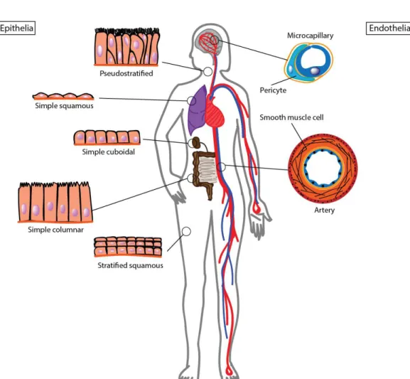
Figure 2.1: Diversity of barrier tissues in the body. a) Illustration of barrier
tissue diversity along the organism. On the left, epithelia showing cellular organization and shape diversity depending on the tissue function. On the right, endothelial cells that form blood vessels.
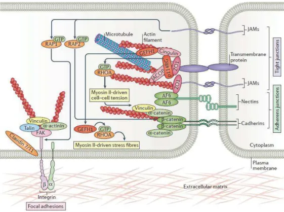
The primary physiological role of barrier tissue is to regulate solute and ion exchanges between two compartments. The regulation of passive ion and solute diffusion mainly happens through lateral junction proteins that bridge the cells. Among these proteins, adherens junctions (AJs), which cadherins are core components, act as cell-cell adhesion complexes2. Tight junction (TJ) proteins are mainly composed of claudins and occludins that present an extracellular loop able
$%" "
to interact with the loop from the neighboring cell to seal the intercellular gap3. The intracellular loop links with cell actomyosin cytoskeleton (actin filaments and microtubules) via adaptor proteins including zona occludens (ZO) (Figure 2.2). TJs and AJs are usually closely associated and reside at the apical end of the lateral membrane and are referred to as the ‘apical junctional complex’.
Figure 2.2: Cell barrier junctional complexes. Representation of junctional complexes that bridge the cell with neighboring cell and surrounding ECM. It includes tight junction proteins that interact with the internal cellular cytoskeleton via cytosolic plaques 3.
2.2.2 Barrier tissue disruption-related pathology
Barrier tissues guarantee organ and tissue homeostasis. Hence, disruption of barrier tissues might be an integral part of many disease pathophysiologies. Loss of barrier properties might be the consequence of tissue destruction as well as TJ network disruption. The latter may be induced by pathogenic agents able to modify junctional protein synthesis, interaction, distribution and phosphorylation status.
A couple of examples illustrate the relation between barrier tissue disruption and diseases pathology. E-cadherin expression was decreased in bronchial
$$" "
epithelial cells from subjects with asthma crisis4. Abnormal TJ structure causes an increase in intestinal epithelium permeability and has been correlated to celiac disease, an immune-mediated disorder of the small intestine triggered by gluten intolerance5,6. This junctional complex was also found of major importance in both inflammatory bowel disease and irritable bowel syndrome7. Blood brain barrier disruption has been involved in several neurodegenerative disorders including Alzheimer’s disease, Parkinson’s disease, and multiple sclerosis.8
2.2.3 Toward a complexification of in vitro barrier tissue models
The first successful in vitro cell culture was led by Katherine Sanford & co-workers in 1948. Cells were cultured in a petri-dish within an arbitrary formulated cell culture media. Since then, the influence of surrounding biochemical and physical signaling cues on cell and tissue functions has been largely demonstrated. Hence, the design of cell culture systems, which provide cell with a defined microenvironment increased drastically9.
Transwell® inserts (referred to as ‘transwells’ in the rest of the chapter) are usually made of track etched polycarbonate (PC) or polyethylene terephthalate (PET) suspended membranes. They offer the possibility to culture cells on either side of the membrane while leaving free access to the manipulator in both compartments. Transwells have been thus a widely used system to model in vitro barrier tissues, in particular for toxicology assays and drug screening. Although this system offers many technical advantages, cells grow on a flat and stiff surface, which strongly limit cell-cell and cell-matrix interactions. Since 2010, the convergence of microfabrication techniques with tissue engineering paved the way for a new class of microphysiological systems10. They offered to synthesize minimal functional units that recapitulate key tissue functions within a device about the size of a computer memory stick. They are referred to as organ-on-a-chip systems and are composed by two polydimethylsiloxane (PDMS) layers that define microfluidic channels. These micron-size chambers are continuously perfused and cells are seeded either on a horizontal porous membrane or along the wall of the tube11. From a technical point of view, organ-on-a-chip device offers an advantage to decrease required cell numbers and biological reagent quantity.
$&" "
2.3 Barrier tissue in vitro characterization assay
There are two main ways for a solute or an ion to cross a barrier tissue: across the cell (via transcellular route) or in between the cells (via paracellular route). The passive diffusion of solute and ions along the paracellular route is mainly regulated by TJ proteins that apply a selective diffusion based on molecule size and charge.
2.3.1 Tight junctions regulate the paracellular diffusion
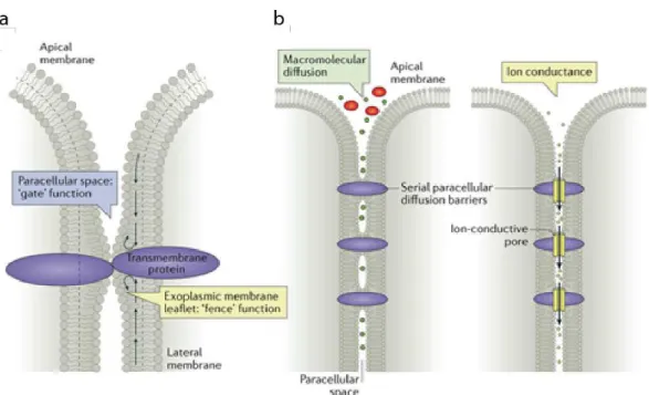
Freeze fracture electron microscopy of epithelial tissues done in 1973 revealed junction organization in fibril meshwork. At the interface between two or three adjacent cells, tight junction proteins allow the close proximity of neighboring cell membrane by an interlacing of protein extracellular loops. It results in an apparent hemifusion of lipid bilayers shown as ‘kissing’ points (Figure 2.3a). These points prevent the lateral diffusion of transmembrane proteins and participate to the establishment of an apico-basal polarization. Tetraspan proteins of the claudin family and MARVEL domain proteins including occludins represent main components of TJs. TJs provide physical tissue stability and participate to the selective property of barrier tissues.
Nowadays, we describe two junctional paracellular pathway routes depending on selection parameter: (1) size selective permeation pathway regulates the diffusion of larger solutes and macromolecules of 30-60 Å. (2) charge selective permeation pathway regulates the diffusion of ions and uncharged small molecules through pores of about 4-8 Å (Figure 2.3b). Evidence has led to the conclusion that pores are gated, however the underlying mechanism is still unknown. Claudins are an important determinant of the charge selective pathway. Depending on their isotypes, they support cation selectivity (claudin-2), anion selectivity (claudin-17) or sealing property (claudin-1).
$'" "
Figure 2.3: Mechanism of diffusion regulated by tight junctions. a) Illustration of gate and fence functions of tight junctions. It regulates the lateral diffusion of lipids and proteins along the cell membrane. b) Illustration of macromolecular and ionic passive diffusion regulation by tight junctions. From 3
Several assays have been developed in order to visualize TJ proteins and estimate paracellular selectivity. Western blotting and confocal microscopy are two powerful complementary techniques that can allow the visualization and quantification of TJ proteins, in particular claudins and occludin. In the case of confocal microscopy, these proteins are coupled to fluorescent labels, which emission intensity provides a quantitative measure of the junctional network development. However, due to the highly-time consuming of this method, it is not scalable to high throughput
2.3.2 Assessing size selective pathway
The size selective pathways can be visualized by mixing chomophore, fluorophore, or radioisotope labeled tracers on one side of the barrier tissue to create a gradient of concentration. By measuring the time required for the tracer to diffuse through the barrier and reach the other compartment, we can evaluate their passive diffusion rate. Lucifer yellow or Fluorescein isothiocyanate (FITC) coupled dextran or albumin are among the most used macromolecule markers of paracellular permeability and offer a range of molecular size from 3 to 70 kDa.
$(" "
Permeability assays are quite easy to operate in transwell cell culture configuration as the water-soluble tracer diffuse homogeneously in each compartment. The manipulator, who has easy access to both sides of the cell barrier, collects samples at determined time points and analyse the intensity of fluorescence overtime (Figure 2.4a, left). However, it necessities another strategy leading permeability assay on 3D barrier tissues when surrounded by viscoelastic hydrogel. Indeed, gel viscosity slow down the diffusion of the dye. Recently, a method based on confocal microscopy analysis has been developed to evaluate permeability of vascular organ-on-chip system. Then, region of interests (ROIs) have been defined around the tubular structure lined by endothelial cells and a conversion from green fluorescence to color map using image analysis software for determining the variation of intensity of FITC-dextran’s fluorescence in the collagen gel surrounding the capillary (Figure 2.4a, right). 12 This technique provides a rapid and cost-effective means of assessing size selective diffusion through barrier tissues over a wide range of molecular size without the need of additional equipment. However, this technique gives an integral permeability measurement and do not allow for new molecules to determine if the diffusion happens through the paracellular or the transcellular route. Progress in lithographic techniques on silicon substrates overcomes these limitations by designing micro-cuvette-like structure on the top of a silicon chip on which cells can be cultivated. The density of a micro-cuvette is high enough to underline most of the cellular membrane and intercellular space. After the dye crosses the cell barrier it fills the pores underneath the cell. This technique improves spatial resolution of macromolecular selective diffusion assay (Figure 2.4b).13 As another alternative, FITC-conjugated avidin dispersed in the culture media interact with biotin bound to viscoelastic hydrogels through a high-affinity interaction. The technique allows improvement in assay spatial resolution while offering the possibility to use cell culture substrates with improved mechanical properties such as collagen or gelatin (Figure 2.4c).14
$)" "
Figure 2.4: Permeability assays to assess size selective diffusion. a) Using
Transwell inserts, the tracer is added on one side of the cell barrier and samples are collected from the opposite side at defined time points to investigate tracer diffusion rate (on the left). Similarly, for organ-on-chip systems, the tracer is added into the tubular structure and let to diffuse through the barrier into the surrounding material such as collagen gel. Diffusion rate is determined using image analysis and color map transformation (on the right). b) Improvement of the spatial resolution in permeability assay can be reach through the design of micro-cuvette onto silicon substrate or c) biotin-avidin interactions.
Permeability assays are time-consuming and the lack of automatization makes this technique irrelevant for high-throughput screening. Moreover, the use of tracers can interfere with the transport process under study but also barrier integrity, making tested cells unusable for further experiments.
2.3.3 Assessing charge selective pathway
Contrary to permeability assays, techniques to assess charge selective diffusion are usually rapid, label free, and largely easy to automate. These techniques require two metal electrodes that are placed on either sides of the barrier tissue. A direct current (DC) or an alternative current (AC) voltage signal is applied to the electrodes, which forces the ionic current to flow across the cell barrier. The corresponding inter-electrode resistance value is calculated as the ratio between the voltage and the resulting current. Due to the high resistance of
$*" "
cell membranes, and the small conductance of ions through the cell membrane, the current predominantly flows through a paracellular route, making the resistance of the tissue approximately equal to the resistance of the TJs. The trans-epithelial/endothelial electrical resistance (TEER) is thus defined by multiplying the inter-electrode resistance by the barrier tissue area.
Electric cell-substrate impedance spectrum (ECIS) has been first used in 1984 to monitor fibroblast shape and behavior. In that system, cells are cultured directly on top of metal electrodes and an AC voltage signal is applied. The presence of cells induces a capacitive effect between the conductive cell media and the electrode. It allows a quantifiable cell spread and number.15 The AC voltage is usually applied over a range frequency going from 1Hz to 1MHz. 16 It allows the measurement of both cell capacitance and resistance. The design of an electrical equivalent circuit shows the ion flow pathways across a barrier tissue (Figure 2.5a). At low frequency range (kHz) most of the ion flows through the paracellular route. The resistance of ion flow along the basal membrane (Rseal) may be very high due to the presence of cell-matrix anchor proteins.17 ECIS has been rapidly adapted for the quantification of cell barrier paracellular ionic transport exploring the resistive impact of TJ selective permeation pathway.18 Several commercial systems based on multi-electrode array have been developed including ECIS (Applied BioPhysics, Troy NY) and xCELLigence (RTCA Systems-ACEA Biosciences Inc.) modules. They are widely used during high throughput screening assays.
The possibility to carry out both permeability assays and TEER measurement in parallel might give valuable insights about barrier tissue functions. Recently, the design of cell culture substrate that contains microgrooves have been integrated with a blood-retinal barrier organ-on-a-chip system.19 The microgroove surface was coated with gold to perform ECIS measurements while permeability assays were performed following the same protocol as previously described (Figure 2.4c).
Spheroids are valuable models for high throughput screening assays. However, spheroid impedance sensing is very challenging as the cells do not adhere to the electrodes anymore. Several platforms have been designed including microfluidic channels 20 and microcavity trap21 in order to maintain the spheroid
$+" "
in between gold electrodes (Figure 2.5b). In the latter microelectrodes are designed on the four sides of the cavity and monitor two independent bipolar EIS measurements using a frequency range of 133 kHz. However, spheroid placement between the electrodes may induce resistance variation that are difficult to extract.21 To overcome this issue, a new platform composed by a central channel confined by retaining pillars has been designed. Coplanar EIS electrodes are placed in the center of the channel and the full device is placed on a tilting stage. The spheroid is placed within the channel and is allowed to roll by gravity back and forth on top of the electrode. Impedance measurements are thus recorded in a dynamic way.22
Figure 2.5: Electrical platforms to assess charge selective diffusion. ECIS
platform for barrier tissue impedance sensing. Cells are cultured on top of planar metal electrodes (a) or in a spheroid configuration (b). c) TEER platforms developed for transwell insert (left and middle) and organ-on-a-chip system (right). Illustrations show the ion flow (black arrow) and the related equivalent circuit.
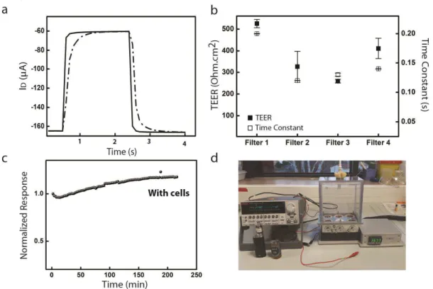
Cell impedance sensing is also widely performed on transwells. In this configuration, metal electrodes are placed in the upper and lower compartments (Figure 2.5c). Commercial modules to perform TEER measurements in transwells include the Epithelial Voltohmmeter (EVOM2, World Precision Instruments, Sarasota, FL) and the cellZscope (nanoAnalytics GmbH, Germany). The EVOM2, also referred as ‘chopstick’, applies a low AC voltage squared signal (~10 µA) with a signal frequency of 12.5 Hz. The TEER is calculated following the Ohm’s law. The assumption of non-conducting cells is valid, because at low frequency
$!" "
AC, capacitive impedance offered by the lipid bilayer is significantly higher than the junctional resistance, channeling the electric current through the ‘path of least resistance’23. The cellZscope module provides an impedance spectroscopy measurement to give a more accurate representation of TEER. Amplitude and phase of the resulting current is analyzed over a range of frequency from 1Hz to 1MHz and the cell layer impedance is calculated as Z = V(t)/I(t). The impedance spectrum shows three distinct frequency regions that are dominated by specific equivalent circuit elements.24
The micrometer-size of organ-on-a-chip chambers rends difficult barrier tissues access. Thus integrating rigid metal electrodes into the system turns out to be challenging. Electrode wires are slipped into the tubular chamber, however there are risks of cell damage and device contamination. To overcome this limitation, micropatterned electrodes may be embedded into PDMS layers (Figure 5c, right).
2.4 Conclusion and opportunities
2.4.1 A need for TEER measurement standardization
Brain microcapillary ionic resistance was measured in vivo for the first time in 1982 and serves today as a strong reference in the field of blood brain barrier. Two set of electrodes were placed within the capillary. The ion permeability was supposed to be constant along the structure and the loss of potential was expressed as a function of capillary length, thus TEER was reported in Ω.cm2. To compare in vitro TEER value with in vivo recordings, commercial systems were used to divide the calculated resistance by the cell layer surface, although this method is only valid if the current flow is uniform across the barrier. Non-uniform current density is one of the main reasons for erratic TEER values in customized microfluidic systems. The tissue segment closes to the electrodes may participate more to the ion flow than the rest of the structure, which results in an overestimation of the effective area and thus of the TEER value.
Additional factors including media conductivity, electrode size and distance from the cell layer induce artifactual differences in measured barrier tissue resistance. If the cell barrier can easily be moved in and out from the electrodes
$#" "
the contribution of these variables is reduced by subtracting a blank resistance. However, this method is prone to error for organ-on-a-chip systems as electrode placement and media conductivity may vary between the blank system and the tissue-containing system.25 Four electrodes were used (two electrodes placed on each side of the cell barrier) in a microfluidic device to generate six resistance measurements and calculate a TEER value independent of channel properties. 26,27 Mathematical models are able to describe customized systems using the finite element analysis method and separate raw resistance coming from the system itself and from a biological event.28,29
Nowadays, there is still a missing practical and standardized approach to integrate electronic devices into the newly developed microphysiological systems.19,30,31 One way to overcome this issue has been to match the shape of the in vitro system with conventional transwell membrane and use commercial systems to measure TEER value.32 Although this method makes easier the comparison with published data, it obviously limits the design of the microsystem.
2.4.2 Using bioelectronic devices to characterize in vitro barrier tissues
Bioelectronic materials couple signals from electronic devices and biological systems. They might transduce biological event into electronic recordings and generate biological response through electronic stimulation. The field of bioelectronics date from the work of Luigi Galvani (18th century) who made a detached frog legs twitch by applying a small voltage to the muscle. Today, bioelectronic devices principally extend to the medical field. Biosensors have been developed to improve healthcare, including glucose sensor for diabetics and pacemaker to support heart failure. Ultrathin implantable electrodes have been developed for brain electrical recording in accordance with the EU Flagship Initiative “Human Brain Project” and the US equivalent “Brain Activity Map”. Both projects started in 2013 with the aim to increase comprehension in brain activity and neurological diseases.33
The organic electrochemical transistor (OECT) is widely used to record ionic-based biological events. Developed by Wrighton and co-workers in the
mid-&%" "
1980s, the OECT is composed by an organic semiconductor film that makes a channel between two metal electrodes called the source and the drain. A third electrode, the gate, is connected to the channel through an electrolyte (Figure 2.6). The working principle of an OECT relies on an oriented flow of holes or electrons through the channel and the injection of ions into the channel to change the conductivity of the organic film. A typical material for the OECT is the conducting polymer poly(3,4̻ethylenedioxythiophene) doped with poly(styrene sulfonate) (PEDOT:PSS). PEDOT is a p-type doped polymer, which lead to the creation of mobile holes able to hop from one PEDOT chain to another. When a negative drain voltage is applied, these holes are compensated by sulfonate anions of PSS and a drain current flow through the channel. It defines the ON state of the transistor. The OECT works in depletion mode, which means the application of a positive gate voltage lead to the injection of positive ions from the electrolyte into the conductive polymer bulk. These cations compensate PSS anions, which decreases the number of holes available in the channel and induces a drop in the drain current. It leads to the OFF state of the transistor. Contrary to metal electrodes, change in channel conductivity happens through the all volume of the film and not only at the interface with the electrolyte. Thanks to this volumetric response, the OECT transduces small gate voltage signals into large drain current variations. OECT transduction efficiency is illustrated by the transconductance (gm=∂ID/∂VG), which is very high for the OECTs and an important figure of merit
for transistors.34
Figure 2.6: Typical organic electrochemical transistor structure. The
illustration shows the typical structure of the organic electrochemical transistor (OECT). It shows the source (S), drain (D) and gate (G) electrodes immersed into an electrolyte. d corresponds to the channel thickness. A drain voltage (VD) and
&$" "
gate voltage (VG) are applied to the system and the resulting drain current (ID) is
measured. From 34.
Bernards and Malliaras first investigated the use of OECT technology to characterize lipid bilayer. The bilayer lipid membrane (BLM) was placed in between the gate and the transistor channel. As lipid membrane act as ionic capacitors, the presence of an intact BLM fully suppress the gating of the OECT while the presence of ion channels partially restore it.35 This experiment paved the way for the use of OECTs to characterize in vitro barrier tissues.
OECT has thus been integrated into transwell models for cell impedance sensing. In that configuration, the transistor channel is patterned at the bottom of the lower compartment, while the gate electrode is immersed into the upper compartment (Figure 2.7a). 36–38 Thus barrier tissue ionic selectivity impact OECT gating efficiency as fewer ions are allowed to penetrate the channel. Toxicology assay were successfully performed as barrier disruption restore the progressively the gating. In contrast to the transwell model, a planar model may also be envisaged. Cell culture on top of the channel transistor allows to differentiate simple cell coverage from cell barrier formation.39,40 OECTs are easily represented by an RC circuit, which describe the electrolyte resistance and capacitance of both gate/electrolyte interface and PEDOT:PSS channel. The integration of an OECT with a barrier tissue, leads to an additional parallel RC circuit in series with the previous one, which corresponds to the resistance and capacitance of the cell layer (Figure 2.7b). Contrary to metal electrodes, PEDOT:PSS films are optically transparent, which allow cell layer visualization during impedance sensing. This optical property allowed to correlate electrical signals with visual information during a wound healing assay41.
&&" "
Figure 2.7: OECT devices integrated with 2D barrier tissues. a) Illustration of an
OECT integrated with transwell model (left) or within cell culture substrate (right). b) Electrical equivalent circuit showing resistance and capacitance of the cell layer (RCell
and CCell) grown on top of the transistor channel. RMedia corresponds to the resistance
of the media and COECT to the OECT capacitance. Cleft resistance (RCleft) describes the
presence of cell adhesion molecules that regulate ion flow in parallel of the channel.
2.5 References
1. Potente, M. & Mäkinen, T. Vascular heterogeneity and specialization in
development and disease. Nat. Rev. Mol. Cell Biol. 18, 477–494 (2017).
2. Harris, T. J. C. & Tepass, U. Adherens junctions: from molecules to
morphogenesis. Nat. Rev. Mol. Cell Biol. 11, 502–514 (2010).
3. Zihni, C., Mills, C., Matter, K. & Balda, M. S. Tight junctions: from simple
barriers to multifunctional molecular gates. Nat. Rev. Mol. Cell Biol. 17, 564–580
&'" "
4. Lambrecht, B. N. & Hammad, H. The airway epithelium in asthma. Nat. Med.
18, 684–692 (2012).
5. König, J. et al. Human Intestinal Barrier Function in Health and Disease. Clin.
Transl. Gastroenterol. 7, e196–e196 (2016).
6. Schumann, M., Siegmund, B., Schulzke, J. D. & Fromm, M. Celiac Disease:
Role of the Epithelial Barrier. Cell. Mol. Gastroenterol. Hepatol. 3, 150–162 (2017).
7. Landy, J. et al. Tight junctions in inflammatory bowel diseases and
inflammatory bowel disease associated colorectal cancer. World J. Gastroenterol. 22,
3117 (2016).
8. Sweeney, M. D., Sagare, A. P. & Zlokovic, B. V. Blood–brain barrier
breakdown in Alzheimer disease and other neurodegenerative disorders. Nat. Rev.
Neurol. 14, 133–150 (2018).
9. Sakolish, C. M., Esch, M. B., Hickman, J. J., Shuler, M. L. & Mahler, G. J.
Modeling Barrier Tissues In Vitro: Methods, Achievements, and Challenges.
EBioMedicine 5, 30–39 (2016).
10. Zhang, B., Korolj, A., Lai, B. F. L. & Radisic, M. Advances in organ-on-a-chip
engineering. Nat. Rev. Mater. 3, 257–278 (2018).
11. Bhatia, S. N. & Ingber, D. E. Microfluidic organs-on-chips. Nat. Biotechnol.
32, 760–772 (2014).
12. Pauty, J. et al. A Vascular Permeability Assay Using an In Vitro Human
Microvessel Model Mimicking the Inflammatory Condition. Nanotheranostics 1, 103–
113 (2017).
13. Michaelis, S. et al. Macroporous silicon chips for laterally resolved,
&(" "
14. Dubrovskyi, O., Birukova, A. A. & Birukov, K. G. Measurement of local
permeability at subcellular level in cell models of agonist- and ventilator-induced lung
injury. Lab. Invest. 93, 254–263 (2013).
15. Giaever, I. & Keese, C. R. Monitoring fibroblast behavior in tissue culture with
an applied electric field. Proc. Natl. Acad. Sci. 81, 3761–3764 (1984).
16. Xu, Y. et al. A review of impedance measurements of whole cells. Biosens.
Bioelectron. 77, 824–836 (2016).
17. Buitenweg, J. R., Rutten, W. L. C., Willems, W. P. A. & van Nieuwkasteele, J.
W. Measurement of sealing resistance of cell-electrode interfaces in neuronal cultures
using impedance spectroscopy. Med. Biol. Eng. Comput. 36, 630–637 (1998).
18. Tiruppathi, C., Malik, A. B., Del Vecchio, P. J., Keese, C. R. & Giaever, I.
Electrical method for detection of endothelial cell shape change in real time:
assessment of endothelial barrier function. Proc. Natl. Acad. Sci. 89, 7919–7923
(1992).
19. Yeste, J. et al. A compartmentalized microfluidic chip with crisscross
microgrooves and electrophysiological electrodes for modeling the blood–retinal
barrier. Lab. Chip 18, 95–105 (2018).
20. Hildebrandt, C., Büth, H., Cho, S., Impidjati & Thielecke, H. Detection of the
osteogenic differentiation of mesenchymal stem cells in 2D and 3D cultures by
electrochemical impedance spectroscopy. J. Biotechnol. 148, 83–90 (2010).
21. Kloß, D., Fischer, M., Rothermel, A., Simon, J. C. & Robitzki, A. A. Drug
testing on 3D in vitro tissues trapped on a microcavity chip. Lab. Chip 8, 879 (2008).
22. Bürgel, S. C., Diener, L., Frey, O., Kim, J.-Y. & Hierlemann, A. Automated,
Multiplexed Electrical Impedance Spectroscopy Platform for Continuous Monitoring
&)" "
23. Sun, T. et al. On-chip epithelial barrier function assays using electrical
impedance spectroscopy. Lab. Chip 10, 1611 (2010).
24. Srinivasan, B. et al. TEER measurement techniques for in vitro barrier model
systems. J. Lab. Autom. 20, 107–126 (2015).
25. Elbrecht, D. H., Long, C. J. & Hickman, J. J. Transepithelial/endothelial
Electrical Resistance (TEER) theory and ap-plications for microfluidic body-on-a-chip
devices. tc 1, 1 (2016).
26. van der Helm, M. W. et al. Direct quantification of transendothelial electrical
resistance in organs-on-chips. Biosens. Bioelectron. 85, 924–929 (2016).
27. van der Helm, M. W. et al. Fabrication and Validation of an Organ-on-chip
System with Integrated Electrodes to Directly Quantify Transendothelial Electrical
Resistance. J. Vis. Exp. (2017). doi:10.3791/56334
28. Khire, T. S. et al. Finite element modeling to analyze TEER values across
silicon nanomembranes. Biomed. Microdevices 20, (2018).
29. Odijk, M. et al. Measuring direct current trans-epithelial electrical resistance in
organ-on-a-chip microsystems. Lab Chip 15, 745–752 (2015).
30. Henry, O. Y. F. et al. Organs-on-chips with integrated electrodes for
trans-epithelial electrical resistance (TEER) measurements of human trans-epithelial barrier
function. Lab Chip 17, 2264–2271 (2017).
31. Arık, Y. B. et al. Barriers-on-chips: Measurement of barrier function of tissues
in organs-on-chips. Biomicrofluidics 12, 042218 (2018).
32. Wang, Y. I., Abaci, H. E. & Shuler, M. L. Microfluidic blood-brain barrier
model provides in vivo-like barrier properties for drug permeability screening:
Microfluidic BBB Model Mimics In Vivo Properties. Biotechnol. Bioeng. 114, 184–