Université de Montréal
Immuno-oncology of human prostate cancer :
phenotypical characterization and study of the tumor-derived, androgen-regulated immunosuppressive microenvironment
Par
Philippe O. Gannon
Programme de biologie moléculaire Faculté de médecine
Thèse présentée à la Faculté de médecine en vue de l’obtention du grade de Ph.D.
en biologie moléculaire
Mars 2010
Université de Montréal
Cette thèse intitulée :
Immuno-oncology of human prostate cancer :
phenotypical characterization and study of the tumor-derived, androgen-regulated immunosuppressive microenvironment
présentée par : Philippe O. Gannon
a été évaluée par un jury composé des personnes suivantes : Dr. Jean-Claude Labbé
Président rapporteur
Dre. Anne-Marie Mes-Masson Directeure de recherche Dr. Fred Saad Co-directeur de recherche Dr. Jean-François Cailhier Membre du jury Dr. Alain Lamarre Examinateur externe
Dre. Sylvie Mader
RÉSUMÉ
Le cancer de la prostate est le cancer le plus fréquemment diagnostiqué chez les hommes canadiens et la troisième cause de décès relié au cancer. Lorsque diagnostiqué à un stade précoce de la maladie, le cancer de la prostate est traité de manière curative par chirurgie et radiothérapie. Par contre, les thérapies actuelles ne peuvent éradiquer la maladie lorsqu’elle progresse à des stades avancés. Ces thérapies, comme la chimiothérapie et l’hormonothérapie, demeurent donc palliatives. Il est primordial d’optimiser de nouvelles thérapies visant l’élimination des cellules cancéreuses chez les patients atteints des stades avancés de la maladie. Une de ces nouvelles options thérapeutiques est l’immunothérapie.
L’immunothérapie du cancer a fait des progrès considérables durant les dernières années. Cependant, les avancements encourageants obtenus lors d’essais précliniques ne se sont pas encore traduits en des résultats cliniques significatifs. En ce qui concerne le cancer de la prostate, les résultats négligeables suivants des interventions immunothérapeutiques peuvent être causés par le fait que la plupart des études sur le microenvironnement immunologique furent effectuées chez des modèles animaux. De plus la majorité des études sur l’immunologie tumorale humaine furent effectuées chez des patients atteints d’autres cancers, tels que le mélanome, et non chez les patients atteints du cancer de la prostate. Donc, le but central de cette thèse de doctorat est d’étudier le microenvironnement immunologique chez les patients atteints du cancer de la prostate afin de mieux définir les impacts de la tumeur sur le développement de la réponse immunitaire antitumorale. Pour réaliser ce projet, nous avons établi deux principaux objectifs de travail : (i) la caractérisation précise des populations des cellules immunitaires infiltrant la tumeur primaire et les ganglions métastatiques chez les patients atteints du cancer de la prostate; (ii) l’identification et l’étude des mécanismes immunosuppressifs exprimés par les cellules cancéreuses de la prostate. Les résultats présentés dans cette thèse démontrent que la progression du cancer de la prostate est associée au développement d’un microenvironnement immunosuppressif qui, en partie, est régulé par la présence des androgènes.
ii
L’étude initiale avait comme but la caractérisation du microenvironnement immunologique des ganglions drainant la tumeur chez des patients du cancer de la prostate. Les résultats présentés dans le chapitre III nous a permis de démontrer que les ganglions métastatiques comportent des signes cellulaires et histopathologiques associés à une faible réactivité immunologique. Cette immunosuppression ganglionnaire semble dépendre de la présence des cellules métastatiques puisque des différences immunologiques notables existent entre les ganglions non-métastatiques et métastatiques chez un même patient. La progression du cancer de la prostate semble donc associée au développement d’une immunosuppression affectant les ganglions drainant la tumeur primaire.
Par la suite, nous nous sommes intéressés à l’impact de la thérapie par déplétion des androgènes (TDA) sur le microenvironnement immunologique de la tumeur primaire. La TDA est associée à une augmentation marquée de l’inflammation prostatique. De plus, les protocoles d’immunothérapies pour le cancer de la prostate actuellement évalués en phase clinique sont dirigés aux patients hormonoréfractaires ayant subi et échoué la thérapie. Cependant, peu d’information existe sur la nature de l’infiltrat de cellules immunes chez les patients castrés. Il est donc essentiel de connaître la nature de cet infiltrat afin de savoir si celui-ci peut répondre de manière favorable à une intervention immunothérapeutique. Dans le chapitre IV, je présente les résultats sur l’abondance des cellules immunes infiltrant la tumeur primaire suivant la TDA. Chez les patients castrés, les densités de lymphocytes T CD3+ et CD8+ ainsi
que des macrophages CD68+ sont plus importantes que chez les patients contrôles.
Nous avons également observé une corrélation entre la densité de cellules NK et une diminution du risque de progression de la maladie (rechute biochimique). Inversement, une forte infiltration de macrophages est associée à un plus haut risque de progression. Conjointement, durant cette étude, nous avons développé une nouvelle approche informatisée permettant la standardisation de la quantification de l’infiltrat de cellules immunes dans les échantillons pathologiques. Cette approche facilitera la comparaison d’études indépendantes sur la densité de l’infiltrat immun. Ces résultats nous ont donc permis de confirmer que les effets pro-inflammatoires de la TDA chez les patients du cancer de la prostate ciblaient spécifiquement les lymphocytes T et les macrophages.
iii
L’hypothèse intéressante découlant de cette étude est que les androgènes pourraient réguler l’expression de mécanismes immunosuppressifs dans la tumeur primaire.
Dans le chapitre V, nous avons donc étudié l’expression de mécanismes immunosuppressifs par les cellules cancéreuses du cancer de la prostate ainsi que leur régulation par les androgènes. Notre analyse démontre que les androgènes augmentent l’expression de molécules à propriétés immunosuppressives telles que l’arginase I et l’arginase II. Cette surexpression dépend de l’activité du récepteur aux androgènes. Chez les patients castrés, l’expression de l’arginase II était diminuée suggérant une régulation androgénique in vivo. Nous avons observé que l’arginase I et l’arginase II participent à la prolifération des cellules du cancer de la prostate ainsi qu’à leur potentiel immunosuppressif. Finalement, nous avons découvert que l’expression de l’interleukin-8 était aussi régulée par les androgènes. De plus, l’interleukin-8, indépendamment des androgènes, augmente l’expression de l’arginase II. Ces résultats confirment que les androgènes participent au développement d’une microenvironnement immunosuppressif dans le cancer de la prostate en régulant l’expression de l’arginase I, l’arginase II et l’interleukin-8.
En conclusion, les résultats présentés dans cette thèse témoignent du caractère unique du microenvironnement immunologique chez les patients atteints du cancer de la prostate. Nos travaux ont également permis d’établir de nouvelles techniques basées sur des logiciels d’analyse d’image afin de mieux comprendre le dialogue entre la tumeur et le système immunitaire chez les patients. Approfondir les connaissances sur les mécanismes de régulation du microenvironnement immunologique chez les patients atteint du cancer de la prostate permettra d’optimiser des immunothérapies mieux adaptées à éradiquer cette maladie.
MOTS CLÉS
Cancer de la prostate; Immunologie; Immunosuppression; Androgène; Arginase; Immunohistochimie
iv
SUMMARY
Prostate cancer is the most frequently diagnosed cancer in Canadian men and the third cause of cancer related death. When diagnosed at an early stage, prostate cancer can be effectively cured by surgery and radiotherapy. However, current therapies do not eradicate the advanced stages of the disease. Treatment of prostate cancer via chemotherapy or hormonotherapy remains palliative. It is thus essential to optimize novel therapies whose goal is to eliminate tumor cells in patients with advanced prostate cancer. One such approach is immunotherapy.
Cancer immunotherapy has made important strides in recent years. The encouraging progress observed in pre-clinical trials has nonetheless not translated to significant results in the clinical setting. Concerning prostate cancer, the limited clinical efficacy of current immunotherapeutic protocols could be explained by the lack of studies directly evaluating the immunological microenvironment in prostate cancer patients and not in animal models or in patients afflicted by other malignancies, such as melanoma. Thus, the fundamental goal of this Ph.D. thesis is to study the immunological microenvironment in prostate cancer patients in order to better understand the impact of the tumor on the development of the anti-tumoral immune response. To realize this project, we established two main working objectives: (i) to precisely characterize the immune cell populations in tumor draining lymph nodes (LNs) and in the primary tumor of prostate cancer patients; (ii) to identify and to study the immunosuppressive pathways expressed by prostate cancer cells. The results detailed in this thesis demonstrate that prostate cancer progression is associated with the development of an immunosuppressive microenvironment, which is regulated, in part, by the presence of androgens.
The initial study was based on the characterization of the immunological microenvironment of tumor draining LNs of prostate cancer patients. The results presented in chapter III allowed us to demonstrate that metastatic lymph nodes displayed cellular and histopathological evidence associated with a reduced immunological reactivity. This LN immunosuppression seemed to be dependant on
v
the presence of metastatic cells as we noted significant immunological differences between non-metastatic and metastatic lymph nodes of the same patient. Prostate cancer progression was thus associated with the development of an immunosuppressive state, which affected tumor-draining lymph nodes.
Next, we studied the impact of androgen deprivation therapy (ADT) on the immunological microenvironment of the primary tumor. Following ADT, there is a marked augmentation in intra-prostatic inflammation. Immunotherapeutic protocols currently evaluated in clinical trials are targeted at hormone refractory patients, which have received and failed ADT. However, very little information is available regarding the nature of the post-ADT immune infiltrate. Thus, it becomes essential to understand whether this post-ADT infiltrate could positively react to immunotherapy. In chapter IV, I present the results of the quantification of the immune cell abundance within the primary tumor. In patients who have received ADT prior to surgery, there was an elevated density of CD3+ and CD8+ T lymphocytes as well as CD68+
macrophages compared to control patients. We also observed an inverse correlation between the NK cell density and the risk of disease progression (biochemical recurrence). Conversely, an elevated macrophage infiltration was associated with a higher risk of progression. Furthermore, for this study, we developed a novel computerized approach allowing for the standardization of the quantification of immune cell infiltrate. This approach could facilitate the interpretation of results from independent studies on the density of immune cells within pathological specimens. This study confirmed that the pro-inflammatory impact of androgen deprivation therapy in prostate cancer patients target specifically the T lymphocyte and macrophage populations. The interesting hypothesis arising from this study was that androgens could positively regulate the expression of immunosuppressive pathways within the primary tumor.
In chapter V, we evaluated the immunosuppressive mechanisms expressed by prostate cancer cells and regulated by androgens. Our analysis demonstrate that androgens increase the expression of molecules with immunosuppressive properties, such as arginase I and arginase II in an androgen receptor dependent manner. This androgen regulated expression of arginase II was also observed in prostate cancer
vi
patients treated by ATD. We observed that arginase I and arginase II participate in prostate cancer cell proliferation as well as in their immunosuppressive potential. Finally, we discovered that interleukin-8 expression was also regulated by androgens. Moreover, interleukin-8, independently of androgens, increased the expression of arginase II. Altogether, these results confirmed that androgens participate in the development of an immunosuppressive microenvironment in prostate cancer by regulating the expression of arginase I, arginase II and interlukin-8.
In conclusion, the results presented in this thesis attest to the unique character of the immunological microenvironment in prostate cancer patients. Our work has also allowed to establish novel software-based analysis methods in order to better understand the dialogue between the tumor and the immune system. Further understanding of the regulatory pathways involved in the immunological microenvironment will allow for the optimization of immunotherapies better suited to eradicate prostate cancer.
KEY WORDS
Prostate cancer; Immunology; Immunosuppression; Androgen; Arginase; Immunohistochemistry
vii TABLE OF CONTENTS Résumé ... i Mots Clés ...iii Summary... iv Key Words... vi
Table of Contents ... vii
List of Tables ... xiv
List of Figures... xv
List of Abbreviations ... xvii
Remerciements / Acknowledgments... xxii
CHAPTER I ...1
INTRODUCTION ...1
1.1 PROSTATE CANCER... 1
1.1.1 Cancer Statistics ... 1
1.1.2 The Prostate Gland ... 2
1.1.3 Pathologies of the Prostate... 4
1.1.3.1 Prostatitis ... 4
1.1.3.2 Focal Atrophy ... 4
1.1.3.3 Prostatic Intraepithelial Neoplasia ... 4
1.1.3.4 Benign Prostatic Hyperplasia... 5
1.1.4 Prostate Cancer ... 5
1.1.5 Risk Factors for Prostate Cancer... 6
1.1.5.1 Age... 6 1.1.5.2 Environmental Causes... 6 1.1.5.3 Hereditary Causes... 6 1.1.5.4 Race ... 7 1.1.5.5 Androgens ... 7 1.1.5.6 Inflammation... 8
viii
1.1.6 Prostatic Inflammation ... 8
1.1.6.1 Inflammation in the Normal Prostate... 9
1.1.6.2 Causes of Prostatic Inflammation... 9
1.1.6.3 Association between Inflammation and Prostate Cancer... 12
1.1.7 Therapies for Prostate Cancer ... 14
1.1.7.1 Active Surveillance and Radical Prostatectomy ... 14
1.1.7.2 Radiotherapy ... 14
1.1.7.3 Androgen Deprivation Therapy... 15
1.1.7.4 Chemotherapy... 15
1.1.8 Cancer Immunotherapy... 16
1.1.8.1 Prostate Tumor-Associated Antigens... 17
1.1.8.2 Clinical Trials in Immunotherapy of Prostate Cancer ... 17
1.1.8.2.1 Sipuleucel-T (Provenge) ... 17
1.1.8.2.2 GVAX... 18
1.1.8.2.3 Prostvac-Vf ... 18
1.1.8.2.4 Anti-CTLA-4 Therapy... 19
1.1.8.2.5 Combination of Immunotherapy With Conventional Therapy ... 19
1.1.9 Summary ... 20
1.1.9.1 Studying the Prostate’s Immunological Environment in order to Develop Prostate Specific Immunotherapies. ... 20
1.1.9.2 Understanding the Prostate’s Immunological Environment in Human... 20
1.1.9.3 Immunosuppression in Human Prostate Cancer ... 21
1.2 BASIC CONCEPTS OF AN IMMUNE RESPONSE...21
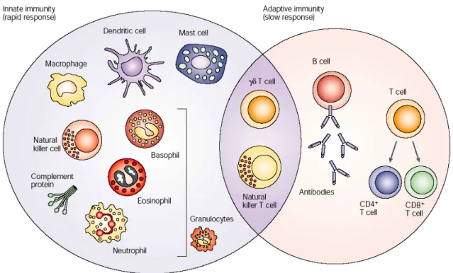
1.2.1 The Innate and the Adaptive Arm of the Immune System... 21
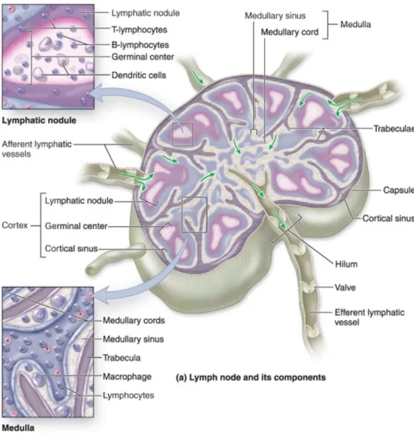
1.2.2 Secondary Lymphoid Organ: the Lymph Node... 24
1.2.3 Important Immunological Concepts ... 26
1.2.3.1 Pathogen and Malignant Cell Recognition ... 26
1.2.3.2 Antigen Presentation... 27
ix
1.2.3.4 Termination of the Immune Response... 29
1.2.3.5 Inflammation-Induced Carcinogenesis ... 29
1.2.4 Cells of the Innate Immune System... 34
1.2.4.1 Granulocytes ... 34
1.2.4.2 Natural Killer Cells... 35
1.2.4.3 Natural Killer T Cells... 35
1.2.4.4 Monocytes and Macrophages ... 36
1.2.4.5 Dendritic Cells ... 40
1.2.4.6 Mast Cells... 41
1.2.4.7 Myeloid-Derived Suppressor Cells... 42
1.2.5 Cells of the Adaptive Immune System ... 42
1.2.5.1 T Lymphocytes ... 42 1.2.5.2 CD4+ T Lymphocytes... 43 1.2.5.3 Regulatory T Cells ... 43 1.2.5.4 CD8+ T Lymphocytes... 45 1.2.5.5 B Lymphocytes ... 45 1.2.6 Summary ... 46
1.3 THE ANTI-TUMORAL IMMUNE RESPONSE ...46
1.3.1 Immune Elimination... 47
1.3.1.1 Evidence of Immune Elimination in Mice... 49
1.3.1.2 Evidence of Immune Elimination in Humans... 49
1.3.2 Immune Equilibrium... 50
1.3.2.1 Evidence of Immune Equilibrium in Mice... 51
1.3.2.2 Evidence of Immune Equilibrium in Humans ... 51
1.3.3 Immune Escape ... 52
1.3.4 Immunosuppression in Prostate Cancer... 55
1.3.4.1 Defects in Antigen Presentation ... 55
1.3.4.2 Production of Immunosuppressive Cytokines... 55
1.3.4.3 Immunosuppression Through Amino Acid Depletion ... 56
1.3.4.4 COX-2 and Prostaglandin E2... 57
x
1.3.5 Summary ... 58
1.4 L-ARGININE AND ARGINASE...58
1.4.1 L-Arginine Homeostasis... 58
1.4.1.1 Polyamines and Tumor Cell Proliferation ... 59
1.4.1.2 L-Arginine Intracellular Transport... 62
1.4.1.3 Pathological Disorders Associated with L-Arginine Deficiency... 62
1.4.1.4 Regulation of L-Arginine Metabolism ... 63
1.4.1.4.1 NO Production in Cancer... 63
1.4.2 Arginase I and Arginase II... 64
1.4.2.1 Regulation of Arginase Expression in Animal Models... 64
1.4.2.1.1 Androgenic Regulation of Arginase Expression in Animal Models ... 65
1.4.2.2 Regulation of Arginase Expression in Humans... 65
1.4.2.3 Immunosuppressive Effects of L-Arginine Depletion... 66
1.4.3 L-Arginine Supplementation ... 69
1.4.3.1 L-Arginine Supplementation in Cancer... 69
1.4.3.2 Polyamine Inhibition to Minimize Tumor Growth ... 70
1.4.3.3 Arginase Inhibitors... 70
1.4.4 Summary ... 70
1.5 REGULATION OF IMMUNE RESPONSES BY SEXUAL HORMONES ...71
1.5.1 Higher Incidence of Autoimmune Diseases in Women... 72
1.5.2 Sexual Hormones in Thymic Development ... 72
1.5.3 Estrogens Promote a TH2 Skewing of the Immune Response ... 73
1.5.4 Androgens Act as Non-Specific Immunosuppressant... 74
1.5.5 Expression of Estrogen and Androgen Receptor by Immune Cells ... 75
1.5.5.1 Estrogen Receptor... 75
1.5.5.2 Androgen Receptor... 76
1.5.5.2.1 iAR Genomic Signaling... 76
xi
1.5.6 Immunoregulatory Properties of Medical Castration... 77
1.5.6.1 Castration in Animal Models ... 78
1.5.6.2 ADT and Prostatic Inflammation... 79
1.5.7 Summary ... 79
CHAPTER II...80
DOCTORAL THESIS OBJECTIVES...80
CHAPTER III ...83
Presence of Prostate Cancer Metastasis Correlates with Lower Lymph Node Reactivity ...83
Abstract... 84
Introduction ... 85
Materials and Methods ... 88
Results ... 91 Discussion ... 96 Conclusions... 100 Acknowledgements ... 101 References ... 102 Figure 1 ... 108 Figure 2 ... 110 Figure 3 ... 112 Table 1... 114 Table 2... 115 CHAPTER IV... 116
Characterization of the Intra-Prostatic Immune Cell Infiltration in Androgen-Deprived Prostate Cancer Patients ... 116
Abstract... 117
Introduction ... 118
Materials and Methods ... 120
Results ... 123
Discussion ... 127
xii References ... 131 Figure 1 ... 135 Figure 2 ... 137 Figure 3 ... 139 Table 1... 141 Table 2... 142 Table 3... 143 Supplementary Table 1... 144 Supplementary Table 2... 145 CHAPTER V ... 146
Androgen-Regulated Expression of Arginase 1, Arginase 2 and Interleukin-8 in Human Prostate Cancer... 146
Abstract... 147
Introduction ... 148
Materials and Methods ... 150
Results ... 154 Discussion ... 159 Conclusion... 162 Acknowledgements ... 163 References ... 164 Figure 1 ... 169 Figure 2 ... 171 Figure 3 ... 173 Figure 4 ... 175 Figure 5 ... 177 Supplementary Table 1... 179 Supplementary Table 2... 180 Supplementary Figure 1... 181 Supplementary Figure 2... 183 CHAPTER VI... 185 DISCUSSION... 185
xiii
6.1 Immunosuppression in Tumor Draining Lymph Nodes of
Prostate Cancer Patients... 185
6.2 Androgen Deprivation Therapy Promotes the Infiltration of T Lymphocytes and Macrophages Within the Primary Tumor... 189
6.3 Androgen Regulated Immunosuppression Through Arginase Expression... 194
CONCLUSION...202
REFERENCES ...204
APPENDIX I:... i
SUPPLEMENTARY RESULTS... i
Index of Supplementary Figures: ... i
Supplementary Figure 1... ii
Supplementary Figure 2... iv
Supplementary Figure 3... v
Supplementary Figure 4... vi
APPENDIX II: ... vii
xiv
LIST OF TABLES CHAPTER I
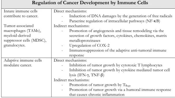
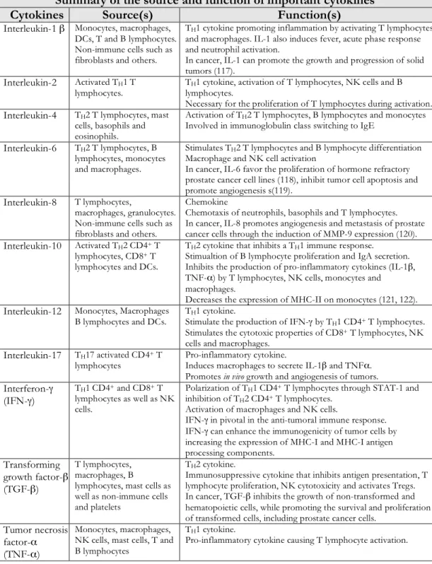
Table 1. Source of Intra-Prostatic Inflammation...11 Table 2. Regulation of Cancer Development by Immune Cells...31 Table 3. Source and Functions of Important Cytokines. ...32
CHAPTER III
Table 1. Clinical Characteristics of Prostate Cancer Patients ...114 Table 2. Pathological Analysis of LNs in Prostate Cancer Patients...115
CHAPTER IV
Table 1. Correlations Between Immune Cell Populations ...141 Table 2. Correlations Between Immune Cell Populations and Clinical Markers...142 Table 3. Univariate Cox Regression Analyses of Biochemical Recurrence ...143 Supplementary Table 1. Clinico-Pathological Characteristics of
Prostate Cancer Patients...144 Supplementary Table 2. Univariate and Multi-Variate Cox Regression Analyses of
Biochemical Recurrence ...145
CHAPTER V
Supplementary Table 1. Correlations between ARG2 Expression and Clinico-
Pathological Parameters...179 Supplementary Table 2. Correlations between ARG2 Expression and Immune
xv
LIST OF FIGURES CHAPTER I
Figure 1. Zonal Predisposition of Prostate Disease...3
Figure 2. Causes of Prostatic Inflammation...10
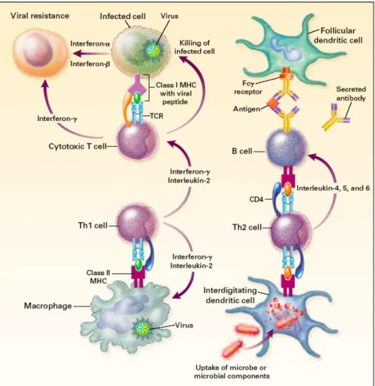
Figure 3. Immune and Adaptive Immune Cells. ...23
Figure 4. Histology of a Lymph Node...25
Figure 5. Lymphocyte Immune Response Activation. ...33
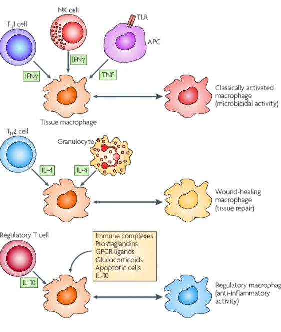
Figure 6. Human Macrophage Phenotypes...38
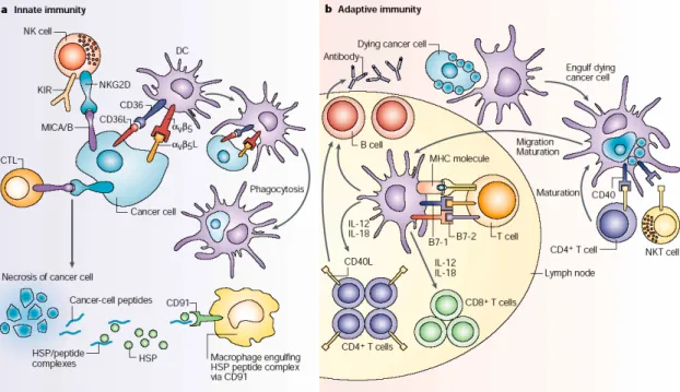
Figure 7. Direct And Indirect Pathways of Tumor Cell Recognition by Innate and Adaptive Immune Cells...48
Figure 8. Immunosuppressive Pathways in Cancer...54
Figure 9. L-Arginine Metabolism. ...61
Figure 10. Potential Inhibitory Pathways of L-Arginine Depletion. ...68
CHAPTER III Figure 1. Representative Images of Immunohistochemical Staining ...108
Figure 2. Percentage of Area Covered by Positively Stained Cells ...110
Figure 3. Histopathological Features Observed in Hematoxylin and Eosin (H&E) Stained LN Sections. ...112
CHAPTER IV Figure 1. Image Analysis with Image Scope and the Pixel-Count Algorithm...135
Figure 2. Immunohistochemical Staining of Immune Cells in Paraffin-Embedded Prostate Primary Tumors. ...137
Figure 3. Increased Abundance of Immune Cells in ADT Patients...139
CHAPTER V Figure 1. In vitro and in vivo Expression of ARG1 and ARG2 in PCa ...169
xvi
Figure 3. Reduced ARG2 Expression Following ADT...173 Figure 4. ARG1 and ARG2 are Metabolically Active...175 Figure 5. Androgens Induced Interleukin-8, which in turn Promotes
ARG1 and ARG2 Expression ...177 Supplementary Figure 1. Androgen Stimulation of PCa Cells...181 Supplementary Figure 2: ARG1 and ARG2 Induction Following IL-8 Stimulation. ...183
ANNEXE I
Supplementary Figure 1. Impact of R1881 on the Proliferation and Activation of
Human PBMCs...ii Supplementary Figure 2. Macrophages Differentiated in the Presence of R1881
Have an Immunosuppressive Phenotype in a
Mixed-Lymphocyte Reaction...iv Supplementary Figure 3. Expression of iAR by Human Monocyte
Derived Macrophages. ...v Supplementary Figure 4. Analysis of COX-2, MPEGS-1, MPGES-2 And CPGES
xvii
LIST OF ABBREVIATIONS
ACN Acetonitrile
ADC Arginine Decarboxylase
ADCC Antibody-Dependent Cell Cytotoxicity ADT Androgen Deprivation Therapy AGAT Arginine:Glycine Amidinotransferase APC Antigen-Presenting Cell
apc adenomatosis polyposis coli
AR Androgen Receptor
ARE Androgen Receptor Response Element
ARG1 Arginase I
ARG2 Arginase II
ASL Argininosuccinate Lyase ASS Argininosuccinate Synthase
BCR B Cell Receptor
BPH Benign Prostatic Hyperplasia
BRCA2 Breast Cancer Type 2 Susceptibility Protein
BrdU Bromodeoxyuridine
cAMP Cyclic Adenosine Monophosphate CAPB Cancer Prostate and Brain
CAT Cationic Amino Acid Transporters
CD40L CD40 ligand
CDK4 Cyclin-Dependent Kinase 4 cGMP Cyclic Guanosine Monophosphate
CMV Cytomegalovirus
COX-2 Cyclooxygenase-2
cPGES cytosolic Prostaglandin E2 Synthase
CTL Cytotoxic T Lymphocyte
xviii
DAB 3,3’-Diaminobenzidine
DC Dendritic Cell
DHT Dihydrotestosterone
EAE Experimental Autoimmune Encephalomyelitis eIF2 Eukaryotic Initiation Factor 2
ELAC2 Elac Homolog 2 (Escherichia Coli) ELISA Enzyme-Linked Immunosorbent Assay eNOS Endothelial Nitric Oxide Synthase
ER Estrogen Receptor
ERE Estrogen Receptor Responsive Elements
FasL Fas Ligand
FFPE Formalin-Fixed Paraffin-Embedded
Foxp3 Forkhead Box P3
GCN2 General Control Non-Derepressible-2
GITR Glucocorticoid-Induced Tumor Necrosis Factor Receptor Family Related Protein
GM-CSF Granulocyte-Macrophage Colony-Stimulating Factor GnRH Gonadotropin-Releasing Hormone
GPCR G-Protein-Coupled Receptor
HA Hemagglutinin
H&E Hematoxylin and Eosin H2O2 Hydrogen Peroxide
HHV8 Human Herpes Virus Type 8
HPC-1 Hereditary Prostate Cancer Locus On Chromosome-1 HPC-2 Hereditary Prostate Cancer Locus On Chromosome-2 HPC-X Hereditary Prostate Cancer Locus On Chromosome-X
HPV Human Papillomavirus
HRP Horseradish Peroxidase
HRPC Hormone Refractory Prostate Cancer HSV2 Human Herpes Simplex Virus Type 2
xix
iAR Intracytoplasmic Androgen Receptor IDO Indoleamine 2,3-Dioxygenase
IFN-γ Interferon-γ
IL Interleukin
IMPACT trial Immunotherapy For Prostate Adenocarcinoma Treatment trial iNOS Inducible Nitric Oxide Synthase
IRF3 Interferon Regulatory Factor 3 ITIM Intracytoplasmic Inhibitory Motif KAR Killer-Activating Receptor KIR Killer-Inhibitory Receptor
LHRH Luteinizing Hormone Releasing Hormone
LN Lymph Node
L-NMMA NG-Monomethyl-L-Arginine
LPS Lipopolysaccharide
MAPK Mitogen-Activated Protein Kinases
mAR Membrane Androgen Receptor
MCA 20-Methylchol-Anthrene
MCP-1 Monocyte Chemotactic Protein-1 mDC Myeloid Dendritic Cell
MDSC Myeloid-Derived Suppressor Cell mER Membrane Estrogen Receptor
MHC-I Major Histocompatibility Complex Class I MHC-II Major Histocompatibility Complex Class II MIC1 Macrophage Inhibitory Cytokine 1
MICA/B MHC Class I-Related Chain Molecules A/B Molecule mPGES-1 membrane PGE2 Synthase-1
MS Multiple Sclerosis
MSR1 Macrophage Scavenger Receptor 1
MyD88 Myeloid Differentiation Primary-Response Gene 88 NDA Naphthalene-2,3-Dicarboxaldehyde
xx
NK cell Natural Killer cell NKT cell Natural Killer T cell
nNOS Neuronal Nitric Oxide Syntase
NO Nitric Oxide
NOHA N-Hydroxy-Nor-L-Arginine
NOS Nitric Oxide Syntase
NSAIDs Non-Steroidal Anti-Inflammatory Drugs NSCLC Non-Small Cell Lung Cancer
OAT Ornithine Aminotransferase ODC Ornithine Decarboxylase
PABPN1 Poly(A) Binding Nuclear Protein 1 PAP Prostatic Acid Phosphatase
PBMC Peripheral Blood Mononuclear Cell
PCa Prostate Cancer
PCAP Predisposing for Cancer of the Prostate pDC Plasmacytoid Dendritic Cell
PGG2 Prostaglandin G2
PHA Phytohemagglutinin
PhIP 2-Amino-1-Methyl-6-Phenylimidazol[4,5-B]Pyridine PI3K Phosphatidyl-Inositol 3-Kinase
PIN Prostatic Intraepithelial Neoplasia
PKA Protein Kinase A
PKC Protein Kinase C
PLC Phospholipase-C
PSA Prostate-Specific Antigen PSCA Prostate Stem Cell Antigen
PSMA Prostate Specific Membrane Antigen qPCR Quantitative Polymerase Chain Reaction
RA Rheumatoid Arthritis
Rag-2 Recombinase Activating Gene-2
xxi
rf-PSA Recombinant Fowlpox Virus Expressing PSA RNASEL Ribonuclease L
ROS Reactive Oxygen Species
RRM RNA-Recognition Motif
rv-PSA Recombinant Vaccinia Virus Expressing PSA SCID Severe Combined Immunodeficiency
SHBG Sex Hormone Binding Globulin SLE Systemic Lupus Erythematosus
STAT1 Signal Transducer and Activator of Transcription-1 TAA Tumor-Associated Antigen
TAMs Tumor-Associated Macrophages
TCR T Cell Receptor
TEAA Triethylammonium Acetate TGF-β Transforming Growth Factor-β
TH T Helper lymphocyte
TIL Tumor-Infiltrating Lymphocyte
TLR Toll-Like Receptor
TMA Tissue Microarray
TNF-α Tumour Necrosis Factor-α
TRAIL TNF-Related Apoptosis-Inducing Ligand TRAMP Transgenic Adenocarcinoma Mouse Prostate TREG Regulatory T Cell
TURP Transurethral Resection of the Prostate VEGF Vascular Endothelial Growth Factor
xxii
REMERCIEMENTS / ACKNOWLEDGMENTS
To my two directors, Anne-Marie and Fred, you have given me intellectual freedom with the necessary amount of motivational and technical support. You have allowed me to be independent, to travel to various conferences and to gain confidence in my research. I am grateful for all the writing that we did and the sense of professional responsibility that I have gain in the last six years, which I owe to your mentorship. I am sincerely thankful for having spent my formative scientific years under your supervision. Réjean, merci pour toutes ces discussions enrichissantes que nous avons eu dans ton bureau. Tes connaissances et l’étroite collaboration avec ton laboratoire furent instrumentales à l’aboutissement de mon Ph.D.. Nathalie, les efforts que tu accomplis avec tous les étudiants avec qui tu travailles sont inestimables et ne passent pas inaperçus. Merci! Je veux aussi remercier le « groupe prostate » avec qui j’ai travaillé, voyagé et rigolé durant les six dernières années. À Jean-Simon Diallo, Hervé Koumakpayi, Laurent Lessard, Benjamin Péant, Cécile Le Page, Mona Alam Fahmy, Blandine Betton et Ingrid Labouba, je vous remercie.
J’aimerais également remercier tous mes collègues, présents et passés, de l’Institut du cancer de Montréal. La grande famille que forme l’ICM m’a apporté un support technique, mais de manière bien plus importante, un environnement chaleureux et un support moral sans lequel mes études auraient été bien misérables. Je retiendrais toujours des souvenirs heureux de mon passage à l’ICM. Particulièrement, j’aimerais remercier Nicolas Parent, Jessica Godin-Éthier, Julie Lafontaine, Marie-Andrée Forget, Alexandre Reuben, Isabelle Cousineau, Catherine Chabot, et Jason Madore. Je désire également souligner le support des membres des laboratoires des Drs Jean-François Cailhier, Nathalie Arbour et Alexandre Prat ainsi que des divers laboratoires du centre de recherche du CHUM et de l’Université McGill avec lesquels j’ai collaboré de près et de loin. Je veux aussi reconnaître la contribution des stagiaires d’été, Audrey Djoukhadjian, Meghan Aversa et Alexis Poisson. Je tiens à remercier de façon particulière Louise Champoux, Manon de Ladurantaye, Sylvie Dagenais, Louise
xxiii
Portelance, Marlène Siewers, Vivianne Jodoin, Nathalie Tapp et Maral Tersakian pour leur aide administrative et leurs efforts à facilité le cheminement des étudiants.
Finalement, je dois remercier mes parents, ma famille et mes amis qui ont toujours su comment m’encourager dans les moments difficiles et célébrer les moments heureux.
CHAPTER I INTRODUCTION
The introductory chapter of this Ph.D. thesis contains five sections. Following a summary of important aspects of prostate cancer, I will describe basic concepts of the immune system and of the current theories regarding the anti-tumoral immune response. Finally, I will review the literature on arginase and on the immunoregulatory properties of sexual hormones.
1.1 PROSTATE CANCER 1.1.1 CANCER STATISTICS
Cancer affected 166,400 Canadians and claimed the lives of 73,800 patients in 2008 (1). According to the current cancer incidence rate, 40% of Canadian women and 45% of Canadian men will develop cancer in their lifetime. In Canada, cancer is the second most common cause of mortality (30.2% of deaths) after circulatory diseases and fourth cause of hospitalization (7.4%) (2).
As for prostate cancer, in 2008, 24,700 Canadian men were diagnosed with the disease and 4,300 patients died from prostate cancer related complications making it the most diagnosed cancer (28.4% of newly diagnosed cancer in men) and third cause of cancer related deaths (11.1% of all male cancer related deaths) (1). Improvements in early detection protocols through the widespread use of prostate-specific antigen (PSA) screening accounts for the continuing increasing numbers of patients diagnosed with prostate cancer, which are now being diagnosed at a younger age and with less aggressive tumors (3). Over 95 % of prostate cancer patients have a relative survival rate exceeding five years and prostate cancer mortality has decreased by 2.9% annually between 1995 and 2004. This decreased in prostate cancer associated mortality is attributed to PSA screening, surgery, higher doses of radiotherapy and earlier onset of androgen deprivation therapy (ADT) (1).
2
1.1.2 THE PROSTATE GLAND
The prostate is an exocrine gland part of the male reproductive system. The prostate surrounds the urethra below the bladder. Its organogenesis begins at the onset of puberty and is under the control of androgens. In healthy men, the prostate is roughly the size of a walnut. Through the contraction of prostatic muscles during ejaculation, the prostate secretes a milky alkaline fluid that constitutes 25-30% of the semen along with spermatozoa and seminal vesicle fluid. This fluid is primarily composed of sugars and electrolytes (citrate, zinc) (4) with less than 1% being proteins such as proteolytic enzymes, prostatic acid phosphatase (PAP) and PSA. The prostatic fluid protects the genomic material of spermatozoa and promotes their motility and survival by providing the necessary nutrients as well as by regulating the pH of the environment (5).
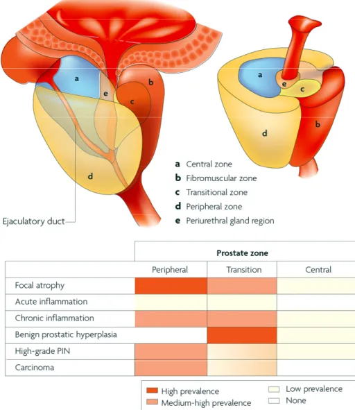
The prostate contains five distinct glandular zones: central zone, fibromuscular zone, transitional zone, peripheral zone and periurethral zone. There is a distinctive prevalence of prostatic pathologies between the different zones (Figure 1 on page 3). Benign prostatic hyperplasia (BPH) is mostly present within the transition zone. Adenocarcinomas are most often located in the peripheral zone, with few in the transition zone and with rare occurrence in the central zone. The peripheral zone is also more susceptible to inflammation, high-grade prostatic intraepithelial neoplasia (PIN) and more aggressive adenocarcinomas. Adenocarcinomas within the transition zone are generally less aggressive (6).
3
Figure 1. Zonal predisposition of prostate disease.
Most cancers develop in the peripheral zone, with few in the transition zone and with rare occurrence in the central zone. Focal atrophy, chronic inflammation and high-grade PIN are also more prevalent in the peripheral zone. BPH lesions develop preferentially in the transition zone without affecting the peripheral zone.
4
1.1.3 PATHOLOGIES OF THE PROSTATE 1.1.3.1 Prostatitis
Prostatitis is the inflammation of the prostate gland. Symptomatic prostatitis affects 3 % to 16 % of men (7). The American National Institute of Health (NIH) recognizes four categories of prostatitis (8). Acute (class I) and chronic (class II) symptomatic bacterial prostatitis are caused by Escherichia coli or other Gram-negative bacteria. However, 90% of patients with prostatitis have symptomatic chronic non-bacterial prostatitis of unknown etiology (class III) (9). The prevalence of asymptomatic prostatitis (class IV) is unknown. More details on prostatic inflammation and its implication in prostate cancer can be found in section 1.1.6 on page 8.
1.1.3.2 Focal Atrophy
Histologically, areas of focal epithelial atrophy are frequently associated with acute or chronic immune cell infiltration. These atrophic lesions contain elevated numbers of proliferative epithelial cells, which fail to differentiate as columnar secretory cells. This pathology is defined as proliferative inflammatory atrophy (10). Proliferative inflammatory atrophy lesions occur in the peripheral zone and are associated with the development of high-grade PIN and prostate cancer (6).
1.1.3.3 Prostatic Intraepithelial Neoplasia
PIN is the benign proliferation of prostate epithelium cells within the glandular lumen in the absence of basal membrane invasion. PIN can remain unchanged or regress with time. It is classified as either low-grade or high-grade PIN. High-grade PIN is associated with an increased risk of developing prostate cancer.
5
1.1.3.4 Benign Prostatic Hyperplasia
BPH refers to the non-malignant proliferation of the prostate’s stromal and epithelial cells frequently diagnosed in older men. Over 70% of 60 year old men and > 90% of 70 year old men have histological evidence of BPH (11). BPH is also caused by the proliferation and increased muscle tone of the prostate’s stromal smooth muscle cells, which lead to the formation of nodules and the enlargement of the transition zone. The enlarged prostate constricts the prostatic urethra thereby causing urinary difficulties, a common symptom of BPH and prostate cancer. BPH can be treated by medication such as α-adrenoreceptor blockers (tamsulosine, alfuzosin), by 5-α reductase inhibitors (finasteride, dutasteride) or by surgery. Surgery for BPH is usually through transurethral resection of the prostate (TURP).
1.1.4 PROSTATE CANCER
Prostate cancer is a slow progression cancer, which can remain asymptomatic for a relatively long period. Approximately 70% of men in their 60s have asymptomatic prostate cancer (12). Prostate cancer is an adenocarcinoma caused by the uncontrolled proliferation of prostate epithelial cells. Prostate cancer statistics are detailed in section 1.1.1 on page 1.
Prostate cancer diagnosis is based on digital rectal examination and the pathological evaluation of prostate biopsies. Serum PSA level is used in diagnosis and in disease monitoring. Nomograms are used for risk assessment and prostate cancer prognosis. Current nomograms are composed of clinico-pathological features such as Gleason score, pTNM stage, pre-operative serum PSA levels and seminal vesicle invasion (13). The Gleason scoring system categorizes the degree of tissue differentiation, with “1” representing well-differentiated and “5” undifferentiated tissues. The sum of the two most prevalent histologies is used as the Gleason score. Current nomograms however lack the desired precision for identification of patients at higher risk of prostate cancer progression. An important domain of prostate cancer research focuses on the optimization of nomograms using molecular and/or cellular markers. Such markers could be evaluated on biopsy samples prior to surgery and
6
complement current clinico-pathological markers in the early prognosis of prostate cancer patients.
1.1.5 RISK FACTORS FOR PROSTATE CANCER 1.1.5.1 Age
Age is the primary risk factor for prostate cancer, with an average age at diagnosis of 70. Older age is associated with an increased risk of prostate injury and infection resulting in chronic prostate inflammation, as well as with hormonal changes and decreasing immunological functions. All of these factors participate in the development of prostate cancer.
1.1.5.2 Environmental causes
Similar to other cancers, prostate cancer has a multi-factorial etiology. Several environmental factors increase the risk of developing the disease. The importance of environmental factors in the development of prostate cancer is apparent in the population of Southeast Asian men who immigrate to a westernized country. These men, who naturally have a low incidence of prostate cancer, develop an increased rate of prostate cancer often within one generation following immigration. This rise is attributed to diet, pattern of sexual behavior, alcohol consumption, exposure to ultraviolet radiation and occupational exposure. A diet rich in red meat and animal fat escalate the risk of prostate cancer. Conversely, an Asian diet rich in soy as well as a Nordic diet with high content of rye lowers the risk of developing prostate cancer. Epidemiological data suggests that consumption of dietary anti-oxidants and micronutrients, such as lycopene, selenium, vitamin D and vitamin E, may also be protective (14), but remains unproven in prospective studies to date.
1.1.5.3 Hereditary causes
Studies comparing the occurrence of prostate cancer in monozygotic and dizygotic twins reveal that prostate cancer has the strongest hereditary component of
7
any cancer (15). Prostate cancer is hereditary in 5-10% of cases and 10-20% of prostate cancer patients have a family history of the disease (16). While canonical cancer mutations have been identified in other cancers (k-ras in pancreatic cancer and
adenomatosis polyposis coli (apc) in colon cancer), few specific genetic risk factors have
been identified in prostate cancer. Linkage analyses have however identified several gene locus associated with an increased risk of developing the disease. These locus include the hereditary prostate cancer locus on chromosome-1 (HPC1), HPC-2, HPC-X, predisposing for cancer of the prostate (PCAP) locus, cancer prostate and brain (CAPB) locus, which together contain a hereditary mutations in genes such as
elaC homolog 2 (Escherichia coli) (elac2), ribonuclease l (rnasel) and macrophage scavenger receptor 1 (msr1) [reviewed in (17)]. Mutations in breast cancer type 2 susceptibility protein (brca2)
also augment the risk of prostate cancer and could be attributed to 5% of cases in patients younger than 55 years (13).
1.1.5.4 Race
Southeast and East Asian men have the lowest incidence of prostate cancer (18). Conversely, compared to Caucasian men, African American men have a 34% higher incidence of prostate cancer, less favorable stages at diagnosis and a two-fold higher risk of prostate cancer associated mortality (19). PSA levels are also higher and more variable in African American men without prostate cancer (20) or with localized prostate cancer (21).
1.1.5.5 Androgens
Although there is no direct association between androgen serum levels and the development of prostate cancer (22), androgens do play an important role in the disease. The initial stages of prostate cancer are termed “hormone sensitive” and medical castration causes prostate atrophy and a temporary elimination of symptoms associated with prostate cancer metastasis (discussed in section 1.1.7.3 on page 15) Testosterone is the most abundant sex hormone in men and is converted to dihydrotestosterone (DHT) by 5α-reductase. Compared to testosterone, DHT has a
8
greater affinity (8-fold) for the androgen receptor (AR) (23). Finasteride, an inhibitor of 5α-reductase, is the only agent to date proven to reduce the risk of developing prostate cancer (24, 25). Testosterone can also be converted into estrogen by the enzymatic activity of a cytochrome p450 aromatase. Exposure to environmental or developmental estrogen is also associated with the development of prostate cancer (26, 27).
1.1.5.6 Inflammation
Prostatic inflammation is associated with an increased risk of prostate cancer (28) and will be discussed in details in the following section.
1.1.6 PROSTATIC INFLAMMATION
From an immunological standpoint, the prostate is a rather complex organ. The prostate was long thought to be an immunologically privileged organ similar to the eye (29, 30). Such is no longer the case. Immune cells secreting a wide-array of cytokines infiltrate the prostate and there is evidence of immune responses directed against prostate specific antigens.
The prostate may nonetheless contain a unique immunological microenvironment. For instance, during puberty the androgen-dependent organogenesis of the prostate causes the expression of novel prostate specific antigens. Remarkably, there is no immune response targeting these novel prostate antigens. The prostate also has a low density of lymphatic vessels. Furthermore, androgens, which are present at their highest tissue concentration within the prostate, have documented immunosuppressive functions (discussed in section 1.5.4 on page 74) and could regulate a state of immunological tolerance (31, 32). Such observations suggest that the prostate may possess a strong immunoregulatory potential that accompanies its organogenesis.
9
1.1.6.1 Inflammation in the normal prostate
B and T lymphocytes, macrophages and mast cells infiltrate the normal prostate. Within the normal prostate tissue, most T lymphocytes are CD8+, whereas
CD4+ T lymphocytes predominate in prostatitis lesions. Compared to non-activated
T lymphocytes in normal tissues, T lymphocytes in inflamed tissues express major histocompatibility complex of class II (MHC-II) and CD45RO suggesting that they are activated and antigen experienced (33).
1.1.6.2 Causes of prostatic inflammation
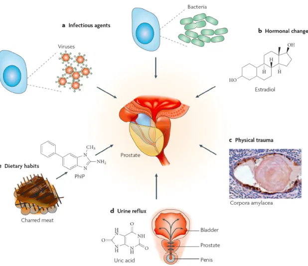
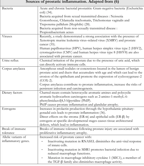
As discussed earlier, 90 % of men diagnosed with prostatic inflammation have prostatitis of unknown etiology. Various factors have been described to have pro-inflammatory effects within the prostate such as: infection, urine reflux, dietary factor, age, and hormonal imbalance [reviewed in (6)] (see Figure 2 on page 10 and Table 1 on page 11).
10
Figure 2. Causes of prostatic inflammation.
Details on the sources of prostatic inflammation are found in Table 1.
11
Sources of prostatic inflammation. Adapted from (6)
Bacteria Acute and chronic bacterial prostatitis: Gram-negative bacteria (Escherichia coli) (34).
Bacteria acquired from sexual transmitted diseases : Neisseria Gonorrhoeae, Chlamydia trachomatis, Trichomonas vaginalis and Treponema pallidum (Shyphilis) (28).
Bacteria acquired from non-sexually transmitted disease : Propionibacterium acnes
Viruses Recently, a study demonstrated a strong association with the presence of Xenotropic murine leukemia virus–related virus (XMRV) and prostate cancer (35).
Human papillomavirus (HPV), human herpes simplex virus type 2 (HSV2), cytomegalovirus (CMV) and human herpes virus type 8 (HHV8) are also associated with prostate cancer.
Urine reflux Chemical irritation of the prostate due to the presence of uric acid, which can directly activate immune cells.
Corpus amylacea Amorphous small nodules or concretions located in the lumen of benign prostate acini and ducts that accumulate with age and which can lead to the erosion of the epithelium and promote the expression of cyclooxygenase-2 (COX-2).
Corpus amylacea contribute to prostate inflammation, increase the risks of persistent infection and carcinogenesis.
Dietary factors Charred meats contain heterocyclic aromatic amines and polycyclic aromatic hydrocarbon carcinogens such as 2-amino-1-methyL-6-phenylimidazol[4,5-b]pyridine (PhIP).
PhIP causes prostate inflammation and glandular atrophy.
Estrogens Increases in prolactin production through the hypothalamic-pituitary-gonadal axis leads to prostate inflammation (36).
Direct effects on the stroma (ER-α) and epithelial cells (ER-β) by estrogens at specific developmental stages causes tissue architectural defects, which lead to inflammation.
Break of immune
tolerance Breaks of immune tolerance following prostate injury are associated with proliferative inflammatory atrophy. Allelic variants of
inflammatory genes. Increased risk of prostate cancer with: - Inactivating mutation in RNASEL diminishes the anti-viral response of innate cells.
- Inactivating mutation in MSR1 promotes bacterial infection due to reduced macrophage functions.
- Mutation in macrophage inhibitory cytokine 1 (MIC1), a member of the TGF-β family also diminishes macrophage activity.
12
1.1.6.3 Association between inflammation and prostate cancer
Chronic inflammation is linked to an increased risk of developing prostate cancer (37). Prostatitis increases the risk of prostate cancer and this preferentially in younger men (38). Several studies demonstrate that the infiltration of various immune cell populations correlates with disease progression or with various clinico-pathological parameters of prostate cancer patients. Increased immune cell infiltrate correlates with an increased rate of tumor recurrence (39), whereas elevated density of CD4+ T lymphocytes (40) and mast cells (41) are associated with poor survival and
higher Gleason score, respectively. Strong tumor-infiltrating lymphocytes (TILs) density also correlates with perineural and capsular invasion and a shortened time to PSA recurrence (42). Conversely, one study finds that a high TILs density was protective against disease progression (43). Results from these studies are however difficult to analysis due to different staining and analysis methods (discussed in Chapter IV on page 116)
Furthermore, it is only recently that the activation status through detailed phenotypical analysis has been documented. CD3+ and CD4+ T lymphocytes do
infiltrate the tumor tissue but do not express perforin and interferon-γ (IFN-γ) suggesting that they are functionally inactive (44). Several studies have also evaluated the presence of Foxp3+ regulatory T cells (T
REGs) in prostate cancer. TREGs are
important for the maintenance of immune tolerance and the inhibition of the anti-tumoral immune response (more details in section 1.2.5.3 on page 43). In a transgenic adenocarcinoma mouse prostate (TRAMP) mouse model expressing the influenza hemagglutinin (HA) antigen under a prostate specific promoter, adoptive transfer of HA-specific CD4+ T lymphocytes resulted in a skewing into a T
REG phenotype both
at the transcriptional and functional level attesting to the tolerogenic power of the prostate microenvironment (45). In prostate cancer patients, a recent study demonstrates an increased infiltration of TREGs, PD-1+ and B7-H1+ immune cells
within the prostate primary tumors (46). Foxp3+ T lymphocytes were also more
present in tumor tissue than in BPH tissue or normal prostate samples from healthy men (46). The TREG skewing was apparent even at the earlier stages of the disease and
13
elevated levels of TREGS with increased suppressive potential (47) are also present in the blood and primary tumors of prostate cancer patients (48). Although present at a higher density in malignant tissues compared to benign tissues, TREG density does not
correlate with disease progression (49). Another study illustrated that CD4+ T
lymphocyte population was skewed towards a TH17 and a TREG phenotypes (50). High
abundance of TH17 CD4+ T cells did correlate with lower Gleason scores (50). The
roles of TH17 cells in prostate cancer remain undefined. Altogether, as the primary
tumor is able to convert antigen-specific T lymphocytes into immunosuppressive immune cells thereby promoting immune evasion, these results argue that the simple assessment of immune cell numbers is inaccurate as it is their activation status that is relevant.
Finally, there are correlations between cytokines levels and the risk of prostate cancer progression. For example, elevated serum levels of IL-6, IL-8, transforming growth factor-β (TGF-β) and tumor necrosis factor-α (TNF-α) are associated with higher Gleason score, development of metastatic disease and poor prognosis (51, 52). The cytokines are produced by infiltrating immune cells and by prostate cancer cells through the activation of NF-κB signaling (7). The prolonged presence of infiltrating lymphocytes and the production of cytokines and other immunological mediators can inhibit the anti-tumoral immune response and provide pro-angiogenic and tumor growth factors.
Altogether, these data suggest that prostatic inflammation correlates with a more aggressive disease and a more rapid disease progression. Interestingly, TREG
removal in murine model of prostate cancer decreased prostatic inflammation and reduced the risk of developing prostate cancer (53). Nonetheless, from these studies, it is difficult to establish the roles of prostatic inflammation in actual prostate cancer development. Due to a lack of appropriate experimental model suited for the evaluation of prostatitis prior to prostate carcinogenesis, it remains unclear whether inflammation is a causative agent of prostate cancer or whether inflammation is induced following the development of prostate cancer.
14
1.1.7 THERAPIES FOR PROSTATE CANCER
Localized prostate cancer is highly curable through radical prostatectomy or radiotherapy. Treatment of advanced prostate cancer is however palliative (54). Advanced prostate cancer is characterized by: (i) high-risk locally advanced disease or metastatic disease, (ii) PSA recurrence after local therapy or (iii) increasing PSA level despite treatment with ADT, which is also termed hormone-refractory prostate cancer (HRPC) (55). The median survival of patients with HRPC ranges from 24 to 40 months for patients diagnosed with skeletal metastasis and averages 68 months for patients without skeletal metastasis (56).
1.1.7.1 Active Surveillance and Radical Prostatectomy
Prostate cancer is well-suited for active surveillance as the cancer grows relatively slowly in most patients. Approximately 70% of men in their 60s have asymptomatic prostate cancer (12). A study evaluating the influence of radical prostatectomy versus active surveillance demonstrates that the benefits of surgery on cancer-related mortality, risks of metastasis and local progression were mainly apparent at 10 years post-surgery (57). As such, it is recommended that men with a life expectancy of less than 10 years and who are diagnosed with early-stage prostate cancer be actively monitored without undergoing aggressive therapy in the absence of progression (13). Side effects also remain a major problem following radical prostatectomy, which include erectile dysfunction (frequent) and urinary incontinence (less frequent).
1.1.7.2 Radiotherapy
Radiotherapy, by brachytherapy with the insertion of radioactive sources within the prostate, or by external beam radiotherapy, is a curative therapy for early-stage prostate cancer. Patients with locally advanced prostate cancer (positive surgical margins, seminal vesicle invasion) can also be cured by radiotherapy through dose escalation and by combination with ADT.
15
1.1.7.3 Androgen Deprivation Therapy
ADT is the primary treatment option for patients with advanced prostate cancer. Huggins and Hodges first reported in 1941 that ADT causes prostate cancer regression and alleviation of pains associated with metastatic prostate cancer (58). Dr Charles B. Huggins, born in Halifax, Nova Scotia, won the Nobel Prize in Medicine in 1966 for his discovery. Their work demonstrated the androgen dependency of normal and neoplastic prostate cells. ADT blocks cellular proliferation and causes the involution of the prostate gland through the apoptosis of hormone sensitive epithelial cells (59). Successful in 70-80% of patients, ADT was the only treatment clinically proven to prolong patient survival, until 2005 when two docetaxel (Taxotere) regiments proved to have survival benefits (60-63). Unfortunately, ADT remains a palliative treatment option with a response window limited 18 to 24 months (64).
ADT targets circulating androgens and/or the AR. Testicular androgens are eliminated through surgical castration or via agonists and antagonists of the gonadotropin-releasing hormone (GnRH) receptors and luteinizing hormone releasing hormone (LHRH). AR activity is blocked with cyproterone (Androcur), cyproterone acetate (Androcur) and non-steroidal anti-androgens such as flutamide (Euflex), bicalutamide (Casodex) and nilutamide (Anandron).
1.1.7.4 Chemotherapy
Recent reports suggest that docetaxel increases the survival of metastatic HRPC patients by 2.9 months. Docetaxel is an anti-mitotic agent that promotes microtubule assembly and stability. Combination of docetaxel with prednisone (Deltasone) decreases PSA levels and increases survival of HRPC patients (65-67). Prednisone is a synthetic corticosteroid, which is converted to prednisolone in the liver, and acts mainly as an anti-inflammatory agent. Docetaxel-based therapy is now the standard of care for chemotherapy against HRPC as recommend by American and European guidelines.
16
1.1.8 CANCER IMMUNOTHERAPY
The goal of immunotherapy is the induction of a cytotoxic immune response targeting the tumor cell. The therapeutically induced anti-tumoral immune response must achieve three criteria: (i) in vivo generation of sufficient numbers of tumor-specific immune cells; (ii) trafficking and infiltration of these tumor-tumor-specific immune cells within the tumor: (iii) activation of the immune cell’s effector functions within the tumor (68). Although a humoral immune response could be beneficial, it is the cell-mediated immune response that is essential for tumor rejection. In mouse models, transfer of T lymphocytes, and not antibodies, was protective against tumor challenges. Elimination of CD8+ T lymphocytes also abrogated both the protective
and therapeutic actions of the anti-tumoral immune response. Finally, in the context of a potent immunotherapy promoting the induction of a cytotoxic CD8+ T
lymphocytes response, both dendritic cells (DCs) and CD4+ T lymphocytes need to
be involved.
Cancer immunotherapy is classified as either active or passive. Active immunotherapy involves the in vivo stimulation of the immune system either specifically or non-specifically through administration of cytokines and interleukins. Passive immunotherapy involves the ex vivo activation of immune cells, which are transferred back into the patient. The inherent specificity of immunotherapy should theoretically decrease the normal tissue toxicity observed with chemotherapy. However, results from clinical trials demonstrate that patients that do develop clinically manageable autoimmunity associated with the development of the anti-tumoral immune response, such as vitiligo in the case of immunotherapy against melanoma, have a more favorable clinical response to therapy.
In theory, prostate cancer is ideally suited for immunotherapy. Innate and adaptive immune cells infiltrate the prostate and the therapeutic window is relatively long (69). Moreover, for patients diagnosed at the earlier stages of the disease, immunotherapy could be administered during the period of active surveillance or prior to ADT (see section 1.1.8.2.5 on page 19). The prostate also expresses several specific antigens (see section 1.1.8.1 on page 17), which are recognized by T
17
lymphocytes. Finally, prostate cancer patients are generally diagnosed at an age when they no longer have children. As such, the prostate becomes a non-essential organ and there is no need to discriminate between normal and neoplastic prostatic epithelium as the entire prostate can be targeted by immunotherapy. In practice however, the prostate’s immunosuppressive mechanism may be the cause of the lack of success in the clinical setting.
1.1.8.1 Prostate Tumor-Associated Antigens
Several prostate tumor-associated antigens (TAAs) have been described as potential immunotherapeutic targets (70, 71). PSA is an active serine protease that participates in the liquefaction of the seminal fluid (72). PSA transcription is regulated by androgens and is specifically expressed by the prostate epithelium. Prostate cancer causes PSA levels to increase up to 10,000 fold. Serum PSA levels are also influenced by BPH, prostatitis, age, body-mass index and race (73). Increases in serum PSA levels are not caused by elevated PSA expression, which actually decreases during cancer, but by a higher release of PSA in blood caused by a disruption of prostate architecture (74). Circulating CD8+ T lymphocytes are present in patients with
prostatitis (75) or prostate cancer (76). Other prostate tumor-associated antigens include prostate specific membrane antigen (PSMA), a transmembrane glycoprotein overexpressed in primary tumors and metastases, prostate stem cell antigen (PSCA) (77) and PAP (78), a glycoprotein whose expression is more specific to the prostate than that of PSA or PSMA (79).
1.1.8.2 Clinical trials in immunotherapy of prostate cancer
Several immunotherapy strategies for prostate cancer using cell-based approaches, viral vectors or antibodies are currently in clinical trials. The following sections will describe the immunotherapies that have had the most promising results.
18
Sipuleucel-T (Provenge) is a DC-based vaccine. DCs are loaded with PAP peptides and granulocyte macrophage-colony stimulating factor (GM-CSF). PAP was chosen becase of its localization on the cytoplasmic membrane and its success in pre-clinical models, where it could elicit prostate-specific immune responses and autoimmune prostatitis (80). GM-CSF promotes DC differentiation into potent TH1
activator (81). Activated DCs promote the activation of cytotoxic CD8+ T
lymphocytes targeted against PAP epitopes. In two phase III trials, Sipuleucel-T offered a survival advantage of 4.5 months for HRPC patients (82, 83). Following recent positive results from the IMPACT trial (Immunotherapy for Prostate AdenoCarcinoma Treatment) the American Food and Drug Administration has approved Sipuleucel-T for treatment of HRPC. It is the first immunotherapy to be approved for cancer treatment.
1.1.8.2.2 GVAX
GVAX is tumor cell-based vaccine in which LNCaP and PC3 cell lines are transfected with GM-CSF, irradiated and injected in patients. The premise of the GVAX vaccine is that the prostate cancer cells will be phagocytosed by the patient’s DCs, which will then present several prostate TAAs to cytototoxic CD8+ T
lymphocytes. In a Phase II trial, GVAX increased median survival of HRPC patients by 8.2 months (84). A phase III clinical trial is currently underway in North America and Europe evaluating GVAX as a single agent (VITAL-1) in comparison to docetaxel plus prednisone in HRPC patients. A VITAL-2 phase III trial evaluating the combination of GVAX with docetaxel was terminated due to increased mortality in the GVAX docetaxel arm.
1.1.8.2.3 PROSTVAC-VF
PROSTVAC-VF is a recombinant vaccinia virus expressing PSA (rV-PSA). The premise is that the anti-viral response will promote the activation of PSA-specific CD8+ T lymphocytes. Initial trials showed that pre-existing immunity to the virus and
immunodominance of viral proteins limited the efficacy of the therapy (85). The protocol was modified to include a fowlpox virus (rF-PSA) as a boost to improve
19
effectiveness. A phase II trial showed that rV-PSA + rF-PSA increased time to progression from 9.2 months to 18.2 months compared to rV-PSA or rF-PSA individual injection and an increase survival of 8.5 months (86). A phase III trial (PARADIGM, Therion/NCI/ECOG) is currently underway.
1.1.8.2.4 Anti-CTLA-4 therapy
Cytotoxic T lymphocyte-associated antigen-4 (CTLA-4) signaling inhibits activated T lymphocytes. Blocking of CTLA-4 causes the activation of CD8+ T
lymphocytes. In two phase I trials with HRPC patients, anti-CTLA4 therapy (Ipilimumab) was shown to be safe and cause a decrease in PSA levels (87, 88).
1.1.8.2.5 Combination of immunotherapy with conventional therapy
Combination of immunotherapy with radiotherapy has synergistic effects in prostate cancer (89) and in other cancers (90). In squamous cell carcinoma, the beneficial effects of combining radiotherapy and chemotherapy are associated with the elimination of TREGS and an increase in homeostatic proliferation (91-93). In a TRAMP mouse model, cyclophosphamide temporarily decreases TREG numbers and
could potentially improve immunotherapy (94). In prostate cancer, chemotherapy with docetaxel however inhibits the anti-tumoral effect of immunotherapy (95).
The most beneficial combinatory effects have been obtained from co-therapy with ADT. ADT improves the survival benefits of immunotherapy (96) when vaccination occurs early in the treatment regiment (90) and prior to ADT (97, 98). The favorable effects of ADT are related to the improvements of DC maturation, costimulation marker expression and cytokine secretion. However, it is noteworthy that, in the current clinical settings for immunotherapy, patients undergoing immunotherapy have already failed other therapies including ADT. It is thus important to understand the immunological consequences of ADT, and other therapies, on the prostate’s immunological environment.