Université de Montréal
Anatomical Correlates of Working Memory Deficits in
Schizophrenia
par Virginie Petel Légaré
Département de Neurosciences Faculté de Médecine
Mémoire présenté
en vue de l’obtention du grade d’une maîtrise en Neurosciences
Juillet, 2017
Université de Montréal Faculté de Médecine
Ce mémoire intitulé
Anatomical Correlates of Working Memory Deficits in Schizophrenia
Présenté par Virginie Petel Légaré
À été évalué par un jury composé des personnes suivantes
Dr. Karim Jerbi Président-Rapporteur Dr. David Luck Directeur de Recherche Dr. Maxime Descoteaux Membre du Jury
Résumé
La mémoire de travail — c’est-à-dire la capacité limitée de retenir et de manipuler temporairement l’information — est un déficit cognitif central en schizophrénie. La perturbation de cette fonction possède un fort impact dans la vie quotidienne des patients. Des travaux récents de notre laboratoire ont pu mettre en évidence que ces troubles de mémoire de travail ne sont pas homogènes et que certains processus sont plus perturbés que d’autres. Par exemple, une méta-analyse du laboratoire a démontré que l’encodage volontaire d’information est une des fonctions spécifiquement affectée en schizophrénie (Grot, Potvin et al. 2014). Plus spécifiquement, l’association volontaire d’informations distinctes en un ensemble cohérent (par exemple, un objet et sa position spatiale) est déficitaire chez les patients. Ce déficit spécifique est notamment sous-tendu par une hypoactivation du cortex préfrontal et pariétal chez les patients (Grot, Légaré et al. 2017). Ces deux régions sont liées à l’attention, à la manipulation d’information, et aux stratégies d’encodage, ce qui confère l’habilité et la flexibilité nécessaire à la mémoire de travail (Kane and Engle 2002, Baddeley 2003). Il est intéressant de noter que de nombreuses études rapportent aussi une réduction de l’épaisseur corticale de ces régions chez les patients, ainsi qu’une altération des fibres blanches les interconnectant (Goldman, Pezawas et al. 2009). En ce sens, notre étude a montré qu’une modification anatomique du réseau préfrontal-pariétal pourrait expliquer le déficit spécifique de mémoire de travail en schizophrénie. Plus spécifiquement, la latéralisation gauche de ce réseau serait atténuée en schizophrénie, et engendrerait le déficit observé en mémoire de travail.
Mots-clés: Schizophrénie, Mémoire de Travail, Imagerie de Diffusion, Épaisseur Corticale
Abstract
Working memory, which is the limited capacity to temporarily maintain and manipulate information, is a core cognitive deficit in schizophrenia. This impairment has a strong impact on the daily lives of patients. A previous study of our laboratory observed that working memory deficits are not homogeneous and that some processes are more disturbed than others (Grot, Potvin et al., 2014). This was supported by a subsequent study, which showed that the voluntary association of distinct information into a coherent whole (i.e. an object and its spatial position) was specifically impaired in patients with schizophrenia (Grot, Légaré et al., 2017). This specific deficit, which is referred to as active binding, is underpinned by a hypoactivation of the left prefrontal and parietal cortex in patients (Grot, Légaré et al., 2017). These two regions are related to attentional processes, manipulation, and encoding strategies, which confer the skills and flexibility required for working memory (Kane and Engle 2002, Baddeley 2003). Interestingly, numerous studies report a cortical thickness reduction in these regions, as well as an alteration of the white fibres interconnecting them in patients with schizophrenia (Goldman, Pezawas et al., 2009). Accordingly, our study showed that anatomical modifications of this network could underpin the specific active binding deficit observed in schizophrenia patients. More specifically, a reduced leftward lateralization of the prefrontal-parietal network could contribute to this specific working memory deficit in patients.
Table of Content
Introduction ... 1
1. Schizophrenia ... 1
a) General ... 1
Prevalence, Impact on Society and Patients ... 1
Impact on Patients ... 2
Impact on Society ... 3
b) History ... 4
Kraeplin, Bleuler and Scheinder ... 4
c) Diagnosis of Schizophrenia ... 6
d) Evolution of Disease ... 8
2. Cognitive Symptoms of Schizophrenia ... 10
a) Attention ... 13
Cerebral Regions Associated With Attention ... 17
b) Verbal and Visual Learning and Memory ... 18
Cerebral Regions Associated With Verbal and Visual Memory ... 20
c) Executive Functions: Cognitive Control and Flexibility ... 21
Cerebral Regions Associated with Executive Functions ... 22
Anatomical Alterations in Schizophrenia ... 24
3. Working Memory ... 26
a) Models ... 27
b) Working memory Deficit in Schizophrenia ... 34
Encoding ... 36
Maintenance ... 37
c) Cerebral Regions Associated with Working Memory: ... 39
The Prefrontal-Parietal Network ... 39
The Prefrontal-Parietal Network in Schizophrenia ... 41
4. Binding ... 43
b) Binding in Schizophrenia ... 45
c) Cerebral Regions Associated with Binding ... 47
d) Regions Associated with Binding in Schizophrenia ... 50
5. Anatomical Alterations in Schizophrenia and Binding Deficits ... 52
a) The Prefrontal-Parietal Network Alterations in Schizophrenia ... 52
Gray Matter ... 52
White Matter ... 53
b) The Prefrontal-Medial Temporal Network Alterations in Schizophrenia ... 56
Gray Matter ... 56 White Matter ... 58 Objectives ... 62 Hypotheses ... 63 Methodology ... 65 1. Participants ... 65 a) Patient Recruitment ... 65 b) Clinical Evaluation ... 66 c) Intellectual Testing ... 67 2. Cognitive Task ... 69 3. Data Acquisition ... 72 a) Scanning Procedures ... 72 4. Anatomical Analysis ... 73
a) Diffusion-Weighted Imaging ... 73
Pre-processing ... 73
Deterministic Tractography ... 74
b) Cortical Thickness Analysis ... 83
c) Statistical analysis ... 84
Performance ... 84
Diffusion Tensor Imaging ... 85
Cortical Thickness Analysis ... 85
Results ... 87
1. Binding Performance ... 87
2. Diffusion-Weighted Imaging Results ... 88
a) Whole fibres: ... 88
b) SLF Subcomponents ... 91
3. Cortical Thickness ... 94
Discussion ... 97
1. Summary of Results ... 97
2. Specific Active Binding Deficit in Schizophrenia ... 98
a) Active Binding and Passive Binding in Schizophrenia ... 98
b) Encoding and Maintenance in Schizophrenia ... 100
3. Prefrontal-Parietal Network Leftward Lateralization Contributes to the Executive Aspect of the Active Binding Task ... 103
a) Prefrontal-Parietal Network ... 103
b) White Matter: Leftward Lateralization of the Prefrontal-Parietal Network ... 104
Encoding Strategies and Lateralization ... 105
Manipulation, Attentional Processes and Lateralization ... 106
c) Gray Matter: Prefrontal Lateralization ... 109
Limits ... 113
Conclusion ... 116
References ... 117
List of Tables
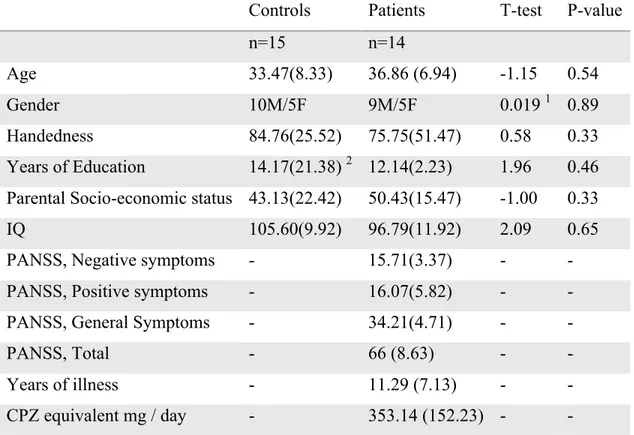
Table I: Socio-demographic and neuropsychological average values (standard deviation) of both groups ... 68 Table II: Pearson Correlations Between the Tasks and the FA Values of the UF and SLF Fibres... 89 Table III: Correlations Between the BaBp contrast and the FA Values of the UF and SLF Fibres ... 89 Table IV: Pearson Correlations Between the Tasks and the LI Values of the UF and SLF Fibres... 90 Table V: Pearson Correlations Between the BaBp contrast and the LI Values of the UF and SLF Fibres ... 90 Table VI: Pearson Correlations Between the Tasks and the FA Values of the SLF II and Arcuate Fibres ... 92 Table VII: Pearson Correlations Between the BaBp contrast and the FA Values of the SLF II and Arcuate Fibres ... 92 Table VIII: Pearson Correlations Between the Tasks and the LI Values of the SLF II and Arcuate Fibres, Control Group ... 93 Table IX: Pearson Correlations Between the Tasks and the LI Values of the SLF II and Arcuate Fibres, BaBp Contrast ... 93 Table X: Pearson Correlations Between Asymmetry Values of the MFG ROI and Task for both Groups. ... 96 Table XI: Pearson Correlations Between Asymmetry Values of the MFG ROI and BaBp Contrast for both Groups. ... 96 Supplementary Table I: Left and Right FA Average Values (standard deviation) of all Fibre for both Groups ... 141 Supplementary Table II: LI Average Values (standard deviation) of all Fibres for both Groups ... 141 Supplementary Table III: Pearson Correlations Between the Cortical Thickness MFG Lateralization Values and the LI Values of the SLF and SLF II ... 141 Supplementary Table IV: Partial Pearson Correlations Between the Tasks and the LI Values of the SLF and SLF II, Controlled for Handedness ... 142
Supplementary Table V: Partial Pearson Correlations Between the Tasks and the LI Values of the SLF, and SLF II, Controlled for Handedness ... 142 Supplementary Table VI: Pearson Correlations Anatomical Values and IQ for both Groups ... 142 Supplementary Table VI: Pearson Correlations Anatomical Values and Age for both Groups ... 143
List of Figures
Figure 1: Figure Adapted from Green (2000); Learning Potential is Related to Everyday
Quality of Life ... 12
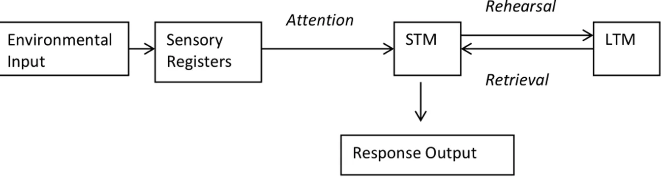
Figure 2: Figure Adapted From Atkinson and Shiffrin Model (1971) ... 27
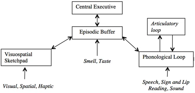
Figure 3: Figure Adapted from Revised Working Memory Model, Baddeley (2011) .... 32
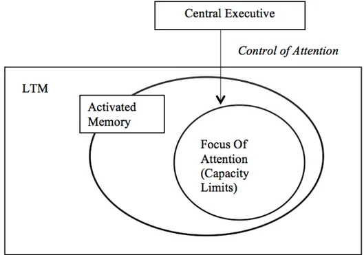
Figure 4: Figure Adapted from Cowan’s Working Memory Model (Cowan 2009) ... 34
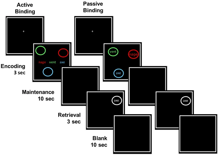
Figure 5: Representation of the Active and Passive Binding Tasks Performed by the Participants ... 71
Figure 6: ROIs Used to Reconstruct the Left Uncinate Fasciculus ... 76
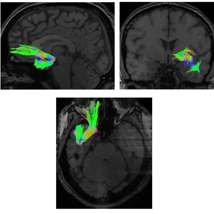
Figure 7: Left Uncinate Fasciculus Reconstruction ... 77
Figure 8: ROIs Used to Reconstruct the Left SLF ... 78
Figure 9: Left SLF Reconstruction ... 79
Figure 10: Left Arcuate Fasciculus Reconstruction ... 80
Figure 11: ROIs Used to Reconstruct the Left SLF II ... 81
Figure 12: Left SLF II Reconstruction ... 82
Figure 13: Performance (Pr value) by Group for the Active and Passive Binding Condition ... 87
Figure 14: Control>Patient T-map of Cortical Thickness ... 94
Figure 15: Cluster Representing the Area in which Controls have a Greater Leftward Cortical Thickness Asymmetry than Patients. ... 95
Supplementary Figure 1: Cortical Thickness Lateralization Group Differences, Corrected for Handedness ... 143
Abbreviations
ACC: Anterior Cingulate Cortex AF: Arcuate Fasciculus
ANOVA: Analysis of Variance BrA: Brodmann Area
Ba: Active Binding Bp: Passive Binding CSF: Cerebrospinal Fluid
CVLT: California Verbal Learning Test DLPFC: Dorsolateral Prefrontal Cortex DMN: Default Mode Network
DSM: Diagnostic and Statistical Manual of Mental Disorders DTI: Diffusion Tensor Imaging
DWI: Diffusion Weighted Imaging EP: Echo Planar
FA: Fractional Anisotropy FAL: False Alarm
FDR: False Discovery Rate
FMRI: Functional Magnetic Resonance Imaging GM: Gray Matter
IQ: Intelligence Quotient
IUSMM: Institut Universitaire en Santé Mentale de Montréal LI: Laterality Index
LTM: Long Term Memory MATRICS:
MGF: Middle Frontal Gyrus
MNI: Montreal Neurological Institute MRI: Magnetic Resonance Imaging NK: Newman-Keuls
PANSS: Positive and Negative Syndrome Scale PFC: Prefrontal Cortex
QC: Quality Control
RFT: Random Field Theory
ROFC: Rey–Osterrieth Complex Figure Test ROI: Region of Interest
Sec: Seconds
SCID: Structured Clinical Interview for DSM Disorder SLF: Superior Longitudinal Fasciculus
STM: Short Term Memory UF: Uncinate Fasciculus
UNF: Unité de Neuroimagerie Fonctionnelle UFOV: Useful Field of View
VLPFC: Ventrolateral Prefrontal Cortex WAIS: Weschler Adult Intelligence Scale WCST: Wisconsin Card Sorting Test WM: White Matter
Remerciements
J’aimerais tout d’abord remercier mon directeur de recherche, Dr. David Luck pour sa disponibilité, sa patience, et son encadrement tout au long de ma maitrise. Grâce à son support, j’ai pu développer de nombreuses connaissances et compétences qui, j’en suis sure, me seront utiles tout au long de mon futur cheminement.
De plus, j’aimerais remercier Stéphanie Grot pour son aide indispensable, son appui et ses conseils avisés. J’aimerais aussi remercier Marie-Ève Leclerc et Andras Tikasz pour leur écoute et leur soutient. Finalement, j’aimerais remercier Simon Beaulieu, Sacha Molderez, et Myrika Néron, que j’ai eu la chance de côtoyer au laboratoire.
J’aimerais aussi remercier la Faculté de Médecine de l’Université de Montréal de m’avoir accordé une bourse, ce qui m’a permis de me concentrer sur mes études, et de compléter ma maitrise.
Introduction
1. Schizophrenia
a) General
Schizophrenia is a chronic disease which alters the behaviour, thoughts, and general functioning of affected individuals (National Institute of Mental Health 2015). It is characterized by three main symptoms categories: positive, negative, and cognitive. Positive symptoms include psychotic manifestations such as hallucinations, delusions, and thought disorders (National Institute of Mental Health 2015). Negative symptoms refer to a diminution of normal functions, such as depression, reduced emotional expression, and lower general pleasure (National Institute of Mental Health 2015). Finally, cognitive symptoms include reduced attentional capacities, inability to understand and use information in order to attain a goal, and lower learning capacities (National Institute of Mental Health 2015). Those symptoms create a general lower quality of life, and the impacts on patients, but also on society are broad. The main characteristics and consequences of this devastating disease will be described below. Prevalence, Impact on Society and Patients
!
Schizophrenia affects about 1% of the Canadian population (Health Canada 2002), and about 21 million people worldwide (World Health Organization 2017). This percentage rises to 8-12% for individuals having a schizophrenic sibling or parent, and to 40-50% for identical twins (Tamminga and Holcomb 2004). Men and women are equally impacted, although men usually develop the symptoms approximately 5 to 7 years earlier (Health Canada 2002, Tandon, Nasrallah et al. 2009). Moreover, women usually present fewer cognitive impairments, fewer negative symptoms, better social functioning, typically respond better to treatment, and have a generally better global outcome (Grossman, Harrow et al. 2008, Luoma, Hakko et al. 2008, Tandon, Nasrallah et al. 2009, APA 2013).
Impact on Patients
The impact of this disease on a patient life is profound. Patients have a reduced ability to maintain a number of facets of their lives, such as social interactions, employment, self-care, as well as having a generally reduced quality of life (Health Canada 2002, Canadian Psychiatric Association 2005, Carpenter 2008). Schizophrenia arises in early adulthood, with an onset between 15 and 45 years (Tandon, Nasrallah et al. 2009). The age of onset is related to the severity of symptoms. Indeed, patients diagnosed before the age of 20 present more negative and cognitive symptoms, and usually have a worse prognosis (Tohka, Zijdenbos et al. 2004, Luoma, Hakko et al. 2008). Moreover, about 52% of the patients hospitalized are aged between 25 and 44 years old (Health Canada 2002). This early onset often refrains patients from forming families, creating social ties, maintaining employment, or completing their education (Health Canada 2002). In fact, 60 to 70% percent of patients do not marry, and many withdraw from social circles during the early years of the illness (Health Canada 2002).
Patients also have a reduced life expectancy (Canadian Psychiatric Association 2005). When untreated, their lifespan is shortened by roughly 15 to 20 years (Tandon, Nasrallah et al. 2009). This is due to a number of factors such as comorbid mental disorders including anxiety, depression, and intellectual disability (Ciapparelli, Paggini et al. 2007, Tandon, Nasrallah et al. 2009). Mental disorders also increase death by suicide. Indeed, suicide is higher in patients than in the general population, a phenomenon linked to comorbid depression, substance abuse, medication discontinuation, and increased impulsivity (Tandon, Nasrallah et al. 2009). This results in a 40 to 60% increase in suicide attempts, leading to a 10 to 15 times increased likelihood of death by suicide in schizophrenic patients compared to the average population (Elizabeth D. Radomsky, Gretchen L. Haas et al. 1999, Health Canada 2002). Furthermore, comorbid physical disorders also reduce lifespan, and include increased cardiovascular incidence, diabetes, metabolic syndrome, and pulmonary disease (APA 2013). Schizophrenic patients usually have an increased risk of physical disorders, due to increased obesity, cigarette smoking, sedentary life, as well as a reduction of access and quality of care (Tandon, Nasrallah et al. 2009, APA 2013). Lastly, prognosis is generally more favourable when a number of factors are present such as superior premorbid phase, greater neurocognitive capacity,
female gender, lack of substance use, and late onset (Flyckt 2006, Tandon, Nasrallah et al. 2009).
Impact on Society
Although representing a small proportion of the population, schizophrenia creates a high burden on the society, the health care system, as well as on the families. In 1996, the direct cost of schizophrenia in Canada was about 2.35 billion $(Health Canada 2002, Goeree, Farahati et al. 2005).This is due to hospitalization, assistance plans, incarceration costs, comorbidity, as well as reduced employment (Health Canada 2002). Indeed, the patient population is over-represented in incarceration and homelessness situations, with about only 1/5 of patients having a full-time job (Health Canada 2002, Robert Rosenheck, Douglas Leslie et al. 2006, Tandon, Nasrallah et al. 2009). A 2002 review of literature reported that on average, 11 % of homeless people suffer from schizophrenia (Folsom and Jeste 2002). As for incarceration, a longitudinal study showed that about 9% of patients will be incarcerated four years after the disease onset (Jonathan D. Prince , Ayse Akincigil et al. 2007). Finally, a proportion of patients as high as 80% will have episodes of substance abuse (Health Canada 2002). This increases suicidal thoughts, violence, incarceration rate, and reduces recovery (Munetz, Grande et al. 2001, Tandon, Nasrallah et al. 2009).
Importantly, about half of the patient population worldwide does not receive care, and if they do, it is mostly at the community and family level (World Health Organization 2017). Families of patients show higher subjective and objective burden than any other chronic disease (Magliano, Fiorillo et al. 2005, Tandon, Nasrallah et al. 2009). Moreover, they usually have lower social and professional support in comparison to families of patients with other chronic diseases (Magliano, Fiorillo et al. 2005, Tandon, Nasrallah et al. 2009). Finally, stigmatization leads to discrimination, fear, reduced self-esteem, which can precipitate social withdrawal (Read, Haslam et al. 2006).
All in all, schizophrenia is a debilitating disease, which has a great impact not only on patients, but also on their family, and society in general. The combination of symptoms, which have their onset very early in life, as well as the prejudice accompanying this disease, prevents a decent quality of life, and creates social isolation.
Patients are overrepresented in incarceration and in homelessness situations, and often have substance abuse problems, which create a number of other consequences such as violence, and suicide. Schizophrenia research is therefore crucial, in order to better understand its aetiology and pathophysiology, and support patients suffering from this devastating disease.
b) History
The progression of the inclusion criteria of schizophrenia not only affected diagnosis, but research as well, as its evolution led the way in the development of pharmacotherapies. It is critical to assess the progression of the inclusion criteria in order to understand the current knowledge of this disease. Schizophrenia is a highly heterogeneous disease, and the diagnostic weight put on individual symptoms created a bias towards psychotic symptoms. In fact, research has not only been delayed due to the complex nature of the disease, but also because of the lack of consensus in the identification, and core nature of the disease (Carpenter 2008).
Kraeplin, Bleuler and Scheinder
In the late nineteenth century, Emil Kraepelin described a large cluster of diseases sharing a common pattern, which he named “dementia praecox”, literally meaning “cognitive decline with onset in youth” (Hoenig 1983, Tandon, Nasrallah et al. 2009, Jablensky 2010). Manifestations of the disease all lead to cognitive and behavioural decline, and seemed to follow the same temporal course, such as similar type and time of onset (Carpenter 2008). What he called “dementia praecox” included diseases like paranoia, catatonia, and thought disorder.
Later, Bleuler (1911) coined the term “schizophrenia”, literally split mind, insisting on the fact that “the split of several psychic functions is one of its most important characteristic” (Bleuler 1951, Hoenig 1983). Split mind highlighted the hypothesis that patients had a mental rupture within thought, affect and behaviour (Carpenter 2008). It also permitted the use of the plural form “schizophrenias”, as Bleuler insisted that schizophrenia was not a disease by itself, but rather a group of diseases (Tandon, Nasrallah et al. 2009). Bleuler also divided the symptoms into two categories, basic and accessory (Jablensky 2010). The basic, fundamental symptoms, which he
believed were present in all cases, included thought disorder, discordant affect, loosening of associations, autism, and withdrawal from reality (Fonov, Evans et al. 2009, Jablensky 2010). On the other hand, the accessory symptoms resembled what is now called positive symptoms, such as hallucinations, and delusions (Jablensky 2010).
The symptom balance changed dramatically in the mid-twentieth century, due to the increasing need for a reliable, specific diagnosis. Psychotic symptoms being more easily characterized were thus put in the forefront of the diagnostic criteria. Schneider proposed that nine groups of psychotic symptoms be of crucial weight in the diagnosis; such as delusions, out of body experience, and voices (Schneider 1950, Jablensky 2010). Those “first rank symptoms” were included in the DSM-III with the hopes of increasing the diagnosis accuracy, thereby renouncing to the central cognitive deficit present in Kraeplin and Bleuler’s schizophrenia model, and establishing reality distortion as the major manifestation of this disease. From this point, most clinical trial measured antipsychotics therapeutic response through psychosis, while cognitive and negative symptoms were mostly left out (Carpenter 2008). In fact, the psychosis treatment efficacy was considered as efficacy to treat the whole schizophrenia spectrum (Carpenter Jr 2004). Still, a number of studies showed that those psychotic symptoms do not accurately predict the patient outcome (Michael Foster 1996, Waters 2009). In fact, psychotic symptoms were shown to be the worst predictors of functional outcomes of all three symptom categories (Michael Foster 1996). On the other hand, certain neurocognitive measures, such as the Wisconsin sorting card task (WSCT), were more predictive of functional outcome than psychotic symptoms (Michael Foster 1996).
Finally, in an attempt to characterize schizophrenia more accurately, Strauss (1974) introduced the tripartite model in 1974 (Carpenter Jr 2004). This concept was proposed to address the issue of the low association between various clinical symptoms. The separation of psychotic positive, negative, and cognitive symptoms permitted the etiologic study and treatment of each symptom category. Supporting this, Carpenter Jr (2004) found that while there was low within-subject correlation between the three symptom categories, each symptom category had a highly predictive power within itself for each individual. This therefore put back on the forefront the hypothesis that
schizophrenia is a group of diseases, emphasizing the importance of its study through various collection of symptoms (Carpenter Jr 2004).
All in all, there was an important evolution of the definition of schizophrenia, as this disease seems to differ between individual regarding the extent of the expression of each symptom category. Taking all those factors into account, the current diagnosis criteria of schizophrenia will be exposed below.
c) Diagnosis of Schizophrenia !
The diagnosis for schizophrenia is still broad, and mostly based on subjective measures (Jablensky 2010). The diagnosis criteria, as defined by the Diagnostic and
Statistical Manual of Mental Disorders V (DSM-V, 2013), are as follows1:
A.!Two (or more) of the following, each present for a significant portion of time during a 1-month period (or less if successfully treated). At least one of these must be (1), (2), or (3):
1. Delusions. 2. Hallucinations.
3. Disorganized speech (e.g., frequent derailment or incoherence). 4. Grossly disorganized or catatonic behaviour.
5. Negative symptoms (i.e., diminished emotional expression or avolition). B.!For a significant portion of the time since the onset of the disturbance, level of
functioning in one or more major areas, such as work, interpersonal relations, or self-care, is markedly below the level achieved prior to the onset (or when the
!!!!!!!!!!!!!!!!!!!!!!!!!!!!!!!!!!!!!!!!!!!!!!!!!!!!!!!!
onset is in childhood or adolescence, there is failure to achieve expected level of interpersonal, academic, or occupational functioning).
C.!Continuous signs of the disturbance persist for at least 6 months. This 6-month period must include at least 1 month of symptoms (or less if successfully treated) that meet Criterion A (i.e., active-phase symptoms) and may include periods of prodromal or residual symptoms. During these prodromal or residual periods, the signs of the disturbance may be manifested by only negative symptoms or by two or more symptoms listed in Criterion A present in an attenuated form (e.g., odd beliefs, unusual perceptual experiences).
D.!Schizoaffective disorder and depressive or bipolar disorder with psychotic features have been ruled out because either 1) no major depressive or manic episodes have occurred concurrently with the active-phase symptoms, or 2) if mood episodes have occurred during active-phase symptoms, they have been present for a minority of the total duration of the active and residual periods of the illness.
E.!The disturbance is not attributable to the physiological effects of a substance (e.g., a drug of abuse, a medication) or another medical condition.
F.!If there is a history of autism spectrum disorder or a communication disorder of childhood onset, the additional diagnosis of schizophrenia is made only if prominent delusions or hallucinations, in addition to the other required symptoms of schizophrenia, are also present for at least 1 month (or less if successfully treated).
Diagnosis can also be made with specifiers such as the number of episodes (first episode, or multiple episodes), the current period (acute, remission, partial remission, or continuous), the severity of symptoms, or if the illness is accompanied by catatonia (APA 2013)
Following this definition it is clear that the diagnosis is still highly skewed towards psychotic symptoms, as hallucinations, disorganized speech or delusion must be present for at least one month in order to be diagnosed. This represents the Schneiderian point of view that schizophrenia is mainly a psychotic illness. Negative symptoms are part of the diagnosis, however, not crucial. The chronic aspect of the disease is also present, as impairment in various functions has to be present for at least 6 months (Criterion C). Interestingly, cognitive symptoms are not part of the core diagnosis. The addition of those symptoms to the DSM-V was considered, but they were excluded in the end. Indeed, some argued that cognitive symptoms were not adequate for differential diagnosis, as they are present in a number of other psychotic disorders (Barch, Bustillo et al. 2013). For example, the neurocognitive profile of bipolar and schizophrenic patients is quite similar for verbal memory and speed processing (Barch, Bustillo et al. 2013).Yet, the progression of cognitive impairments can usually distinguish between those two disorders. Indeed, schizophrenia is characterized by cognitive impairments present before the onset, which will stay stable throughout the disease. On the other hand, bipolar individuals present increased cognitive dysfunction during active phases, such as mania or depression (Barch, Bustillo et al. 2013). Nonetheless, cognitive symptoms still have a strong influence on Criterion B, which is the impairment of functioning in one or more aspect of life (APA 2013). Moreover, the cognitive decline present before the onset of the disease could also be important for early diagnosis, as described below.
d) Evolution of Disease !
Schizophrenia develops over time, and can be divided into a number of periods. Yet, it is important to note that those phases are not always clearly demarked (Tandon, Nasrallah et al. 2009). During childhood, individuals who will later develop the disease have a number of subtle cognitive, motor and social impairments (Tandon, Nasrallah et al. 2009). This constitutes the premorbid phase, which includes attention deficits, poor social interactions, and late motor developments (Tandon, Nasrallah et al. 2009). The prodromal phase occurs before the first full-scale psychosis, and is characterized by cognitive and negative symptoms (Tandon, Nasrallah et al. 2009). This phase can last
from months to years, with 5 years as the mean (APA 2013). Attenuated positive symptoms, such as subthreshold hallucinations or delusions, also transpire up to one year before the first episode psychosis hospitalization (Tandon, Nasrallah et al. 2009). Individuals can show eccentric beliefs, perceptions, disorganized speech and behaviour, as well as social withdrawal (APA 2013). As stated, this phase is part of the diagnosis criteria, and must occur for at least 6 months before onset (Criterion C) (APA 2013). Illness onset is defined by the diagnostic criteria of the DSM-V, and therefore individuals must present their first “full-blown” psychotic episode (Tandon, Nasrallah et al. 2009, APA 2013). After the first psychotic episode, the course of the disease is usually made up of episodes of recovery intertwined with relapsing psychotic breaks, with greater functional deterioration during the first five years (Tandon, Nasrallah et al. 2009). The symptoms then stabilize, as the positive symptoms usually attenuate with years, a phenomenon thought be related to the decline in dopamine with age (Tandon, Nasrallah et al. 2009, APA 2013). On the other hand, negative symptoms usually increase (Tandon, Nasrallah et al. 2009, APA 2013). As for cognitive symptoms, they stay stable, or show low improvement over the course of the illness (Tandon, Nasrallah et al. 2009, APA 2013, Keefe 2014). Thus, cognition is not only impaired before psychosis, but those deficits are also stable throughout life, a pattern typical of schizophrenia (Barch, Bustillo et al. 2013) The absence of improvement of those functions is an important obstacle for recovery (APA 2013).
Taken together, the three principal symptoms of schizophrenia are regarded in a very different way, and their individual importance evolved throughout history. Positive symptoms have the greatest diagnosis weight, and are the principal target of pharmacotherapy (Carpenter Jr 2004, Tandon, Nasrallah et al. 2010). Yet, those symptoms usually become less prominent during illness progression. Negative symptoms are presently not treated by medication, and usually persist or worsen (Carpenter 2008). Finally, cognitive symptoms are present before onset, and have a relative stability throughout the disease. This could be not only important for early diagnosis, but for the study of cognitive symptoms as endophenotypes. In fact, cognitive symptoms are present in first-degree relatives, who generally show intermediate values, suggesting that there could be a genetic contribution, and that cognitive symptoms could be a cause, and not a
consequence of the disease (Tamminga and Holcomb 2004, Braff 2015). This could be advantageous, as the study of first-degree relatives could eliminate many variable such as chronic medication, institutionalization and psychosis (Cornblatt 1994). Moreover, given the high heterogeneity of the disease, it would be advantageous to study more quantifiable values, such as performance in certain cognitive tasks, than the subjective characteristics of positive and negative symptoms (Tamminga and Holcomb 2004). Cognitive symptoms are the greatest impediment to quality of life and recovery, social life and work, and predict a worst prognosis (Michael Foster 1996). Furthering our understanding of cognitive impairment in schizophrenia could not only increase the quality of life of the patients, but also improve the understanding of the basic underlying deficits of schizophrenia.
2. Cognitive Symptoms of Schizophrenia
!
Cognitive deficits are a core symptom in schizophrenia, and more than 80% of patients have a deficit in at least one measure (Toulopoulouand and Murray 2004, Jablensky 2010). In average, patients will be between half to one and a half standard deviation below the general population for a number of cognitive measures (Heinrichs and Zakzanis 1998, Cirillo and Seidman 2003). The remaining of the population is considered as “high functioning”, as they show similar performance as healthy controls on several cognitive tests. Yet, they are still considered below normal range (Hoff , Palmer, Heaton et al. 1997, Toulopoulouand and Murray 2004). For instance, it was suggested that the 20% of patients scoring in the normal range had higher than normal functioning before onset (Wilk, Gold et al. 2005). This was supported by a study which observed that when matched by Intelligence Quotient (IQ), patients performed below controls in a number for neuropsychological tests (Wilk, Gold et al. 2005). Following this logic, “high-functioning” patients would have a deficit compared to their pre-onset values, but would fall into the general population values after onset. Nonetheless, it is important to mention that most patients have a lower IQ than the average population (Bowie and Harvey 2006). This is observed during the premorbid phase, and it seems to decline with onset (Bowie and Harvey 2006). Moreover, as for high-functioning patients, patient with low IQ will still show cognitive deficits even when matched by IQ to healthy
controls (Kremen, Seidman et al. 2001, Bowie and Harvey 2006). Therefore, cognitive deficits are not a result of a lower general intelligence.
As pointed out in the previous section, cognitive deficits appear before onset, are not a consequence of the illness, and will persist in a relatively stable manner during the illness (Jablensky 2010). During the premorbid phase, cognitive deficits have an average effect size of 0.5 (Tandon, Nasrallah et al. 2009, Trotta 2015). Before or at the time of onset, cognitive impairment usually increases, after which there is generally a mild improvement or stability. Still, the cognitive performance of patients is usually lower than controls throughout the illness, and occasionally declines with the disorder’s duration (Kremen, Vinogradov et al. 2010). Moreover, antipsychotic medication as no to little effect on cognitive symptoms (Keefe, Bilder et al. 2007). Generally, second generation atypical antipsychotics are more effective than first generation medication, but the effects on cognitive symptoms are still low (Keefe, Silva et al. 1999). Supporting this, a meta-analysis including 15 studies using atypical antipsychotics showed that verbal fluency and executive function are some of the most improved functions, with attention responding to a lesser extent, and learning and memory being the least affected by medication (Keefe, Silva et al. 1999). Yet, a more recent meta-analysis suggested that first-generation antipsychotics are not totally ineffective (Mishara and Goldberg 2004). Indeed, it was found that they have modest to moderate effects sizes on a number of cognitive symptoms such as automatic and perceptual processing, attention, memory and language, and executive function (Mishara and Goldberg 2004). Nonetheless, most studies agree that pharmacological improvements of cognitive symptoms are still very low, even if some beneficial effects are observed.
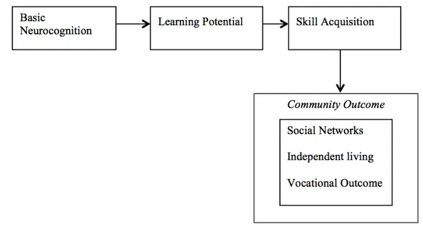
Cognitive symptoms are also present as an intermediate phenotype in relatives (Toulopoulou, Picchioni et al. 2007, Tandon, Nasrallah et al. 2009), and are greatly related to functional outcome (Michael Foster 1996, Green, Kern et al. 2004, Carpenter 2008). Indeed, Green’s influential reviews proposed that global outcome would be related to cognitive deficits (Green 1996, Green, Kern et al. 2004). Deficits such as episodic memory, working memory, vigilance, and executive functioning, have the greatest correlation to functional outcome (Green, Kern et al. 2004). Indeed, Green (1996) argues that learning potential is correlated with everyday quality of life, as it permits the
acquisition of new skills, which could contribute to a number of crucial aspects in life (Figure 1).
Figure 1: Figure Adapted from Green (2000); Learning Potential is Related to Everyday Quality of Life
The importance of cognitive symptoms on global outcome led to a tremendous increase in the cognitive deficit research in schizophrenia, which resulted in the Measurement and Treatment Research to Improve Cognition in Schizophrenia (MATRICS) initiative. This conference established seven categories of core cognitive deficits, such as speed of processing, attention/vigilance, working memory, verbal learning and memory, visual learning and memory, reasoning and problem solving, and social cognition (Nuechterlein, Barch et al. 2004, McClure, Romero et al. 2007). Yet, not all functions are affected the same way in every patient. A meta-analysis by Fioravanti et al. (2005) showed that although there was a global reduced cognitive function in patients with schizophrenia compared to healthy control, there was a high heterogeneity in the degree to which each process is affected. This finding was re-established seven years later, illustrating the stability of the finding (Fioravanti, Bianchi et al. 2012). As there is a high heterogeneity of symptoms, this review will concentrate on a few cognitive deficits,
which are considered to be the most consistently affected in patients (Cirillo and Seidman 2003).
a) Attention
Attention is crucial, as it provides a filter for the great amount of information perceived in everyday life. Inputs compete for priority, and attention can create a bias towards certain information (Braff 1993, Daniel H. Mathalon, Theda Heinks et al. 2004, Luck and Gold 2008). Attention is thus an essential process as it regulates perception, memory, and response selection, thereby influencing a number of other cognitive processes, such as working memory and executive control (Luck and Gold 2008). Impaired attention in schizophrenic patients has been highly documented, and is now considered a fundamental deficit (Fioravanti, Carlone et al. 2005, Fioravanti, Bianchi et al. 2012). The deficits in attention are present before onset, and are shared with first-degree relatives, suggesting a genetic contribution (Bowie and Harvey 2006). Although the definition of attention seems quite straightforward, this function is multifaceted, and modulated by a number of factors (Carter, Bizzell et al. 2010). The various types of attention will be described below.
Sustained attention. First, attention can be transient or sustained. Sustained attention is the ability to focus continuously, without being distracted, for the period of time necessary to complete a task. For example, it is required when reading a book, as individuals with impaired sustained attention would constantly be distracted and unable to finish a page. On the other hand, transient attention is the ability to increase attention at a moment when a task is more demanding. Therefore, while sustained attention is needed throughout a task, transient attention is required at specific moments that are important for the task’s performance (Bozikas, Andreou et al. 2005, Fioravanti, Carlone et al. 2005, Carter, Bizzell et al. 2010). When performing a task, patients with schizophrenia will show a greater decline in sustained attention with time compared to controls, suggesting that they are unable to maintain a constant attentional level (Hahn, Robinson et al. 2012). This decline increases with task difficultly and cognitive demand (Hahn, Robinson et al. 2012). However, even at the beginning of the task, attentional levels are lower in patients, suggesting that the sustained attention would not be responsible for the whole deficit (Hahn, Robinson et al. 2012). Carter, Bizzell et al. (2010) designed a mix
block/event-related task that allowed the separation transient and sustained attention. The overall performance in patients was reduced compared to controls, and altered activations were reported for both types of attention. Indeed, Carter et al. (2010) found that although the attentional network regions, which will be described in more details below, were more active during sustained attention, they presented a lower activation during the transient attention compared to controls (Carter, Bizzell et al. 2010). The authors hypothesized that this was due to an irrelevant control of attention. Indeed, while healthy controls increased their attention network during relevant stages, patients were unable to effectively control their attention, resulting in a hypoactivation during transient attention compared to controls. In short, patients might recruit the attentional network to a greater extent during less demanding tasks, until task demands would become too high, at a point which their activation would be unable to compensate (Karch, Leicht et al. 2009, Carter, Bizzell et al. 2010). This would therefore imply inefficient activation in patients (Karch, Leicht et al. 2009, Carter, Bizzell et al. 2010). Hence, patients are unable to maintain attentional levels during an extended period, representing impaired sustained attention. Furthermore, they are unable to increase their attention at critical moments, representing dysfunctional transient attention.
Selective attention. Selective attention is the capacity to choose and enhance the information processing of relevant information, while decreasing irrelevant information in order to attain a goal (Braff 1993, Cameron S. Carter, Mark Mintun et al. 1997). For example, in a big crowd, it would be necessary to focus on the speech of the person talking to us, while ignoring other conversations and sounds. Therefore, selective attention requires voluntary processing, and this function is impaired in patients (Fioravanti, Carlone et al. 2005). Indeed, patients have trouble focusing their attention on low valence cues (Braff 1993). On the other hand, involuntary attention is linked to the valence one attributes to a stimulus (Braff 1993). This type of attention is performed in a passive way, as the stimuli which are attributed a high valence will be processed in high priority. For instance, when the desired input has a high valence, its processing would require less control of attention. This type of attention seems to be spared in patients (Braff 1993). Supporting this, the importance of selective attention can be highlighted when the target used for goal directed behaviour is not the most salient (Braff 1993, Luck
and Gold 2008). This was illustrated in a study by Hahn, Robinson et al. (2010), which tested the effect of salience on selective attention in schizophrenia. During a visuospatial task, half of the target stimuli flickered, thereby increasing their salience, while the other half did not flicker. Increased target salience benefited both controls and schizophrenic patients. However, when the target had low salience (non-flickering target), controls preferentially remembered the non-flickering target, while patients preferably remembered the flickering stimuli. Thus, patients had trouble overcoming salient cues, supporting the hypothesis that patients have trouble in selective attention, and this especially when the target has low salience.
When discussing selective attention, an additional distinction was pointed out by Luck and Gold (2008). In fact, selective attention could be characterized by rule selection and input selection. Rule selection can be exemplified by the Stroop task, which usually consists of coloured words (e.g. the written word “Green” coloured in blue). In this task, participants are told to either read the words (here, “Green”), or state the colour of the word (here, “blue”). Rule selection is the process by which participants have to choose between competing rules, such as the “read the word” or “say the colour”. Therefore, rule selection is the ability to apply a rule to a task. On the other hand, input selection is the ability to choose an input, and is further characterized by the control of selection, and the implementation of selection. The control of selection is the process by which one determines which input needs to be selected, while the implementation of selection is the process by which one reduces irrelevant inputs and enhances relevant inputs. The authors argue that schizophrenic patients have trouble in the control of selection, but not in the implementation of selection. Indeed, patients usually are able to perform in spatial cueing paradigms, which requires individuals to enhance the processing of the cued location, and therefore suggests an intact implementation of selection in schizophrenia (Luck and Gold 2008). On the other hand, when patients are informed that the target will appear on the opposite side of the cued location, patients present a low performance. This involves controlling the selection, and overriding the prepotent rule of enhancing the cued location. Hence, patients have a deficit in the control of selection, directing their attention to the right target, especially when the input has low salience and requires top-down attention, or when the appropriate rule is not the default one. Top-down control is the
ability to control perception with cognition, while bottom-up control is the process by which perception controls cognition. For example, when the target stimulus is the most salient, bottom-up control will suffice, as that stimulus will be processed in priority. On the other hand, when the target stimulus is not the most salient, top-down control is crucial in order to select the appropriate information. Therefore, patients with schizophrenia are deficient in rule selection and input selection task, but only when the latter necessitates a high level of control of selection, and top-down control. The control of selection happens in the prefrontal and parietal cortex, while the implementation happens in the cortex of the input’s modality (Luck and Gold 2008). Therefore, patients would have a specific selective attention deficit, due to the fact that they are unable to accurately select the target or rule when they are in competition.
Divided attention. Divided attention is the ability to attend to multiple inputs at once, and thus “splitting” attentional resources to multiple targets. This can be illustrated by talking on the phone while writing. Indeed, in order to succeed, one needs to listen to the individual, as well as concentrate on writing the appropriate information. Patients have a deficit in broad attention, i.e. when one has to attend a high number of inputs (Hahn, Robinson et al. 2012). Indeed, Hahn and colleagues (2012) used a visuospatial attention task, in which either one, two or four possible target would be cued. The target then appeared in either one of the cued locations (valid trial), or in a non-cued location (invalid trial). They found that patients had a greater deficit when presented with four cues than in invalid trials. The authors conclude that patients have a deficit when a task requires broad monitoring (Hahn, Robinson et al. 2012). On the other hand, Gray, Hahn et al. (2014) performed a Useful Field of View (UFOV), in which it is possible measure the ability of individuals to divide their attention between peripheral and central locations. Participants had to identify the target that had been presented in both those locations, and a significant deficit in the divided attention was seen in patients compared to controls (Gray, Hahn et al. 2014) The authors concluded that this divided attention deficit could be due to either the inability to handle two stimuli at the same time, the inability monitor the environment in a broad way, or due to the fact that patients tend to give one location greater weight, resulting in hyperfocusing (Gray, Hahn et al. 2014) Following this, a recent study tested the hyperfocusing hypothesis, performing a test in
which individuals were asked to either focus on the central, or peripheral area (Kreither, Lopez-Calderon et al. 2017). Patients showed an increased processing of the central element, and increased inhibiting of peripheral information, but they failed to inhibit the central region when asked to focus on the peripheral region. This deficit was correlated to the UFOV performance of divided attention (Kreither, Lopez-Calderon et al. 2017). Therefore, patients show a general inability to spread their attention to a number of inputs, and tend to process central inputs that are irrelevant to a greater extent, resulting in the inability to divide their attention in an adequate manner.
Cerebral Regions Associated With Attention
A number of regions are inappropriately activated during attentional tasks in patients compared to controls. Those regions include the dorsolateral prefrontal cortex (DLPFC), the insula, the anterior cingulate cortex (ACC), the parietal cortex, the amygdala, and the hippocampus (Carter, Bizzell et al. 2010).
The ACC is associated with sustained attention (Tamminga and Holcomb 2004), and failure to activate this region will impair input selection (Cameron S. Carter, Mark Mintun et al. 1997). The ACC is believed to detect mismatch and conflict, and is connected to the DLPFC to signal when a response should be improved (Lesh, Niendam et al. 2011). In short, the ACC could attribute valence to certain inputs, and the DLPFC could select appropriate responses according to the goal suggesting that the ACC could be activated during control of selection (Bench, Frith et al. 1993, Cameron S. Carter, Mark Mintun et al. 1997, Orellana and Slachevsky 2013). This is supported by studies, which reported that controls and schizophrenic patients had different ACC activation during decision-making tasks, such as the Stroop task (Henry H. Holcomb, Adrienne C. Lahti et al. 2000, Tamminga and Holcomb 2004).
The parietal cortex is also important for attentional processes. It is thought that the parietal cortex maintains possible responses linked to a particular stimulus, while the prefrontal cortex is involved in the selection of those responses (Bunge, Hazeltine et al. 2002). The parietal and DLPFC are also activated during sustained attention (Lesh, Niendam et al. 2011). Moreover, the parietal region is also required for attention switching (Lesh, Niendam et al. 2011) Supporting this, the parietal cortex, the ACC and
the DLPFC were observed as hypoactivated during the Stroop task in unmediated patients (Weiss, Siedentopf et al. 2007).
b) Verbal and Visual Learning and Memory !
Verbal Memory and Learning. Verbal memory refers to the ability to remember words and phonological information. It is the ability to perceive information, store it, and retrieve it when necessary. For example, verbal memory is often used to remember events, ideas or facts. Impaired verbal learning and memory is a fundamental cognitive deficit in schizophrenia, and it has been extensively documented (Heinrichs and Zakzanis 1998, Toulopoulouand and Murray 2004, Bowie and Harvey 2006). Supporting this, Cirillo, Seidman, and colleagues (2003) reviewed 110 studies, and 101 of them showed that schizophrenic patients had a verbal memory deficit compared to controls, with negative studies mainly including other psychiatric disease, a small sample size, or patients diagnosed before the DSM-III. Verbal memory was found to be the greatest predictor of community outcome for schizophrenia patients (Michael Foster 1996, Green, Kern et al. 2004). This cognitive deficit is present during the premorbid phase, as well as in first-degree relatives and high-risk individuals (Cirillo and Seidman 2003). Verbal memory impairment is relatively stable throughout time, even though some studies show some small improvement, or decline (Toulopoulouand and Murray 2004).
The nature of the verbal deficits is still under debate. In their study, Cirillo, Seidman, and colleagues (2003) argued that the deficit in verbal memory was mainly due to an encoding deficit. Encoding is the first step of memory. It is necessary to create a representation of perceived information, which can then be stored. Cirillo, Seidman, and colleagues (2003) hypothesized that patients have trouble organizing inputs into semantic categories, but that the deficit would still be present even if the words could not be classified into groups. This ability to organize verbal inputs according to similar meaning is called semantic clustering. Semantic clustering is an encoding strategy in verbal memory, and is impaired in patients (Brebion, Amador et al. 1997, Gsottschneider, Keller et al. 2011). Supporting this hypothesis, a recent study showed that patients applied less semantic clustering in the California Verbal Learning Test (CVLT), resulting in a reduced performance in the patient group (Gsottschneider, Keller et al. 2011). Another
study showed that patients organizing words into semantic categories had better performance in immediate recall (Vaskinn, Sundet et al. 2008, Gsottschneider, Keller et al. 2011). Consequently, the encoding deficit seems to be related to inappropriate encoding strategies. Yet, patients could show less semantic clustering because of the requirement of higher cognitive demand during this process, or because they have trouble finding the similarities between the items (Brébion, David et al. 2004, Gsottschneider, Keller et al. 2011). Interestingly, Chan et al. (2000) found that providing an external cue about the categories in which words could be organised improved the performance of patients. This hints to the fact that patients could be unable to select an appropriate strategy by themselves. Moreover, this goes in line with a recent study by Ragland and colleagues (2015), which showed that when there were low cognitive demands, and that encoding strategies were given, patients did not show a deficit in a recognition task. Nevertheless, recall is more affected than recognition, which points to a deficit further than encoding. Moreover, when controlling for semantic clustering, Brébion et al. (2004) noted that the deficit was still present, suggesting that reduced encoding strategies did account for the whole verbal deficit, and that maintenance or retrieval might also be affected.
Visual Memory and Learning. Visual memory refers to the ability to remember visual information. This deficit was observed in schizophrenic patients, as well as first-degree relatives, although to a lesser extent than verbal memory and learning (Toulopoulou, Rabe-Hesketh et al. 2003, Skelley, Goldberg et al. 2008). The Rey– Osterrieth Complex Figure Test (ROFC) allows the assessment of visual memory and learning. This test consists of copying a complex figure, and then replicating it from memory, immediately or after variable delays (Kim, Namgoong et al. 2008). A deficit in immediate recall was observed in patients for this task, hinting to an encoding deficit (Seidman, Lanca et al. 2003). For instance, Seidman, Lanca et al. (2003) found that patients have a reduced ability to encode a picture as a whole, and tend to focus more on the details. This pointed to an organizational deficit during encoding, such as what is observed with the deficiency in creating semantic categories. The reduced organizational capacity finding was replicated (Kim, Namgoong et al. 2008), and related to the difficulty of the task (Glahn, Barrett et al. 2006). Furthermore, Seidman, Lanca et al. (2003) also
found that when controlling for the organizational deficit, the reduced recall persisted. In the same way, when testing immediate recall and controlling for organizational deficit, patients still present deficits which implies that the deficit is not only due to encoding dysfunction (Seidman, Lanca et al. 2003). Finally, it was suggested that patients give more importance to non-essential features, which reduces the attentional capacity to process significant part of the pictures (Seidman, Lanca et al. 2003, Kim, Namgoong et al. 2008). Therefore, as for verbal learning and memory, inappropriate strategies during encoding seem impaired, but other processes could also contribute to the deficit in visual learning and memory.
Cerebral Regions Associated With Verbal and Visual Memory
Verbal and visual memory deficits are attributed to a reduced communication and functional coupling between the prefrontal cortex and other regions such as the hippocampus, the thalamus and the cerebellum (Weiss and Heckers 2001, Toulopoulouand and Murray 2004). A reduction of the DLPFC gray matter was found in patients having verbal and visual memory deficits (Seidman et al., 1994). Accordingly, a meta-analysis by Ragland, Laird et al. (2009), showed that the most consistent finding was a reduction of lateral prefrontal activation during encoding and retrieval, suggesting its important role in memory deficits, and cognitive control (Ragland, Laird et al. 2009, Gsottschneider, Keller et al. 2011). Indeed, as stated, patients have trouble controlling and finding the right strategy to encode information, but the dysfunction will disappear if provided one. The lateral prefrontal cortex could thus be responsible for providing the appropriate strategy and control. Interestingly, the ventrolateral prefrontal (VLPLC) cortex and DLPFC cortex seem to be involved in different ways in verbal memory. Ragland, Laird et al. (2009) found that the VLPFC deficit disappeared when providing patients with encoding strategies, while the DLPFC deficit remained. The DLPFC is involved in organization, manipulation as well as monitoring the goal relevant information, and therefore crucial for task requiring flexibility and high cognitive processing. On the other hand, VLPFC is able to process semantic words that require less cognitive demand, or that are already in categories (Ragland, Ranganath et al. 2015). Therefore, an alteration in the DLPFC could impair encoding organization, and the VLPFC could compensate for the DLPFC during low demanding task. Some studies also
suggest that verbal deficits could also be attributed to hippocampal alterations, as this structure is involved in encoding and retrieval (Weiss and Heckers 2001). This hypothesis is noteworthy owing that the modification structure is one of the most consistent findings in patients (Seidman, Lanca et al. 2003). Finally, the reduced impairment of visual compared to verbal memory was attributed to a generally greater left hemisphere impairment in schizophrenia (Toulopoulouand and Murray 2004).! Indeed, it is hypothesized that visuospatial processes are lateralized to the right hemisphere, while verbal processes are lateralized to the left hemisphere (Toga and Thompson 2003) .
c) Executive Functions: Cognitive Control and Flexibility
Executive functions refer to the capacity of understanding and using information in order to attain a goal (National Institute of Mental Health 2015). They include a variety of functions, which are important for goal directed behaviour and voluntary control (Bowie and Harvey 2006, Orellana and Slachevsky 2013). Executive functions help individuals plan, make analogies, solve problems, adapt to a changing environment, as well as execute many tasks at the same time in order to achieve a goal (Orellana and Slachevsky 2013). In short, executive functions could be considered as higher cognitive processes.
Executive function impairments are observed in a consistent manner throughout schizophrenia (Orellana and Slachevsky 2013). Those impairments have been known for a long time, as Kraepelin (1913) described patients as having lower mental efficiency, and a distracted mind (Orellana and Slachevsky 2013). As for other cognitive symptoms, they are present before onset, in first-degree relatives, as well as high-risk individuals (Orellana and Slachevsky 2013). Patients have low adaptive capacity to a changing environment, as well as inflexible thinking (Pantelis, Barber et al. 1999, Orellana and Slachevsky 2013). Executive function deficit can be assessed with the Wisconsin Card Sorting Test (WCST). This test requires individuals to shift rules according to the changing information provided by the evaluator. During this test, the rules of associations shift, and individuals must adapt according to feedback. It is important to note that attention is required during this test, as patients need to find the correct dimension,
reflecting rule selection. As mentioned before, patients have trouble rule selection when they are in competition. Nevertheless, perseveration, which measures the ability to change rule to use the correct dimension, measures cognitive flexibility, while inefficient sorting might reflect attentional processes (Greve, Williams et al. 1996). Indeed, the latter represents the selection of the appropriate action for a goal-directed behaviour, and will be deficient especially when there is competition of various possible responses. On the other hand, perseveration errors represent the inability to take into account perceptual information (Chambon, Franck et al. 2008), and change their behaviour according to the presented stimuli (Koechlin, Ody et al. 2003). It is known that patients have trouble in the inhibition of previously learned behaviours, which will cause increased perseverative errors, representing a loss of cognitive flexibility (Orellana and Slachevsky 2013). Finally, schizophrenia patients also perform poorly on the Tower of Hanoi test, and will take much more time and move to solve the task (Bustini, Stratta et al. 1999, Orellana and Slachevsky 2013). The Tower of Hanoi test consists of discs that must be displaced according to a set of rules in order to create a tower, and deficits in this task represent failure to plan, but also impulsive responding (Bustini, Stratta et al. 1999)
Cerebral Regions Associated with Executive Functions
Deficits in executive functions are related to hypofrontality, and the connection between the prefrontal cortex and other structures such as the basal ganglia, the medial temporal lobe, the parietal lobe and the ACC (Orellana and Slachevsky 2013).
The prefrontal cortex is connected to various regions, and it is thought that this structure integrates information, which can then provide top-down control (Lesh, Niendam et al. 2011). Indeed, the PFC serves as online storage (Miller 2001), and can compare incoming information with internal goals, in order to select appropriate responses (Lesh, Niendam et al. 2011). To achieve this, the PFC will provide bias to other structures in order to “guide” them (Miller 2001). Simplistically, the PFC can “decide” which information is required to attain a specific goal, and will bias the information towards the appropriate one (Lesh, Niendam et al. 2011). This function is mostly achieved by the DLPFC, which thought to be the crucial structure for maintenance of rules, and for response selection (Lesh, Niendam et al. 2011). A good example is the Stroop task, in which schizophrenic patients are deficient (Bench, Frith et al. 1993). The
cognitive control provided by the DLPFC is necessary in the colour-naming situation, in order to overcome the default action of word reading. Therefore, the DLPFC would be responsible for cognitive control, assuming a top-down regulation, and thus a central player in executive function (Koechlin, Ody et al. 2003, Orellana and Slachevsky 2013).
Furthermore, all cognitive manifestations pointed above, including attention, verbal, and visual memory deficits are also related to a prefrontal dysfunction, or to a reduced connectivity of this region with other regions. Indeed, the prefrontal cortex is connected to a number of other regions, such as the ACC, parietal regions as well as the medial temporal lobe (Lesh, Niendam et al. 2011, Orellana and Slachevsky 2013). The reduced connectivity and coupling between keys region in cognitive processing represents Bleuler’s original thought about the disease. Schizophrenia means « split-mind», representative of the dysfunctional brain connections and integrative processes. Indeed, Bleuler thought that the decoupling of various functions was the main cause of the disease (van den Heuvel and Fornito 2014, Sakurai, Gamo et al. 2015). As mentioned above, attentional deficits are linked to prefrontal, ACC and parietal disconnection. On the other hand, visual and verbal learning and memory are linked to a prefrontal dysfunction, and reduced hippocampal connectivity. Therefore, the prefrontal cortex, and its connectivity seem to be primordial for the cognitive symptoms. This goes back to Kraepelin (1971) describing dementia praecox as “an orchestra without a conductor” (Lesh, Niendam et al. 2011). Interestingly, patients without medication show hypofrontality, which means it is inherent to the disease (Sakurai, Gamo et al. 2015). Moreover, even though cognitive symptoms seem sparse, the failure to control and self-regulate seems to be a shared characteristic.
Thus, numerous cerebral regions involved in cognitive deficits seem to display altered activation in schizophrenia. Interestingly, numerous anatomical modifications are reported in patients with schizophrenia. This could underpin a number of deficits, as functional activation has been linked to structural integrity (Guye, Parker et al. 2003, Greicius, Supekar et al. 2008, Bennett and Rypma 2013). A quick review of anatomical findings in schizophrenia will thus be presented below.