Université de Sherbrooke
Radiolabeled Bombesin Analogs to Improve Prostate Cancer
Diagnosis by PET Imaging
Par
Nematallah Mansour
Programme de sciences des radiations et imagerie biomédicale
Thèse présentée à la Faculté de médecine et des sciences de la santé
en vue de l’obtention du grade de philosophiae doctor (Ph.D.)
en sciences des radiations et imagerie biomédicale
Sherbrooke, Québec, Canada Avril, 2019
Jury
Prof. Martin Lepage Ph.D. Jury President
Department of Nuclear Medicine and Radiobiology, Université de Sherbrooke Prof. Roger Lecomte, Ph.D. Supervisor
Department of Nuclear Medicine and Radiobiology, Université de Sherbrooke Prof. Brigitte Guérin, Ph.D. Co-Supervisor
Department of Nuclear Medicine and Radiobiology, Université de Sherbrooke Prof. Robert Day, Ph.D. Internal Examiner
Department of Surgery, Urology Service, Université de Sherbrooke, Dr. Jean-Mathieu Beauregard, M.D. External Examiner
Department of Radiology, Université Laval
ii
To my beloved parents for their usual encouragements,
To my brothers and sisters
To my wife and daughter
To my special friends
iii
R
ÉSUMÉAnalogues radiomarqués de la bombésine pour améliorer le diagnostic du
cancer de la prostate par imagerie TEP
Par
Nematallah Mansour
Programme de sciences des radiations et imagerie biomédicale
Thèse présentée à la Faculté de médecine et des sciences de la santé en vue de l’obtention du diplôme de philosophiae doctor (Ph.D.) en science des radiations et imagerie biomédicale, Faculté de médecine et des sciences de la santé, Université de Sherbrooke, Sherbrooke, Québec,
Canada, J1H 5N4
Le récepteur du peptide de libération de la gastrine (GRPR) est un récepteur couplé aux protéines G. Ce récepteur est fortement exprimé dans le cancer de la prostate, il est ainsi un biomarqueur potentiel pour ce cancer. La bombésine est un peptide de 14 acides aminés qui se lie avec une forte affinité au GRPR. Les analogues radio-marqués de la bombésine ont été utilisés intensivement pour cibler ce récepteur. Le radio-marquage de la bombésine avec des isotopes métalliques, tel que le 64Cu, nécessite l'utilisation d’un chélateur bi-fonctionnel et d’un intermédiaire entre le chélateur et le peptide. Ces deux composants sont nécessaires pour maintenir l’activité de la bombésine.
Cette thèse a pour objectif le développement des sondes peptidiques radio-marquées, analogues de la bombésine. Ces sondes seront utilisées pour la TEP des cancers exprimant les GRPRs, notamment le cancer de la prostate. Dans cette thèse, nous avons utilisé un analogue de (l'antagoniste) la bombésine qui s’appelle RM26. Pour améliorer les propriétés physicochimiques et pharmacocinétiques de ce peptide, nous avons testé plusieurs chélateurs et intermédiaires. Cette thèse hybride est divisée en plusieurs parties. Nous avons consacré l’introduction à la présentation du contexte scientifique, des principaux biomarqueurs et traceurs TEP impliqués dans le cancer de la prostate et finalement nous avons récapitulé les développements que les radio-traceurs de la bombésine, les espaceurs et les nouveaux chélateurs ont reconnu au cours des dernières années. Le premier chapitre (article 1) se concentre sur l'étude des espaceurs (LK) portant des charges di-cationiques et di-anioniques ainsi que leur impact sur les paramètres pharmacocinétiques des analogues de type [64Cu]-NOTA-LK-RM26. Récemment, notre groupe a développé un chélateur bi-fonctionnel (DOTHA2) portant des bras acides
hydroxamates permettant une complexation plus rapide, plus efficace et plus forte au 64Cu. Dans le second chapitre (article 2), nous décrivons l'évaluation d'un nouvel antagoniste au GRPR, le
64Cu-DOTHA
2-PEG-RM26, pour visualiser les tumeurs prostatiques par imagerie TEP. Dans les
deux études précédentes, le [64Cu]-NOTA-PEG-RM26 a été utilisé comme composé de référence car le complexe 64Cu-NOTA possède une forte stabilité in vivo et une captation non spécifique globale très faible. Les résultats des études in vitro et in vivo, du complexe DOTHA2
-APCA-RM26 radio-marqué au 64Cu, sont présentés dans le troisième chapitre (en format traditionnel). Enfin, une discussion générale et les conclusions finales de la thèse sont présentées.
iv Mots clés : DOTHA2, cancer de la prostate, bombésine, chélateur, espaceur, RM26, tomographie
v
S
UMMARYRadiolabeled Bombesin Analogs to Improve Prostate Cancer Diagnosis by
PET Imaging
By
Nematallah Mansour
Radiation Science and Biomedical Imaging Program
Thesis presented at the Faculty of Medicine and Health Sciences to obtain a Doctor degree (Ph.D.) in Radiation Science and Biomedical Imaging, Faculty of Medicine and Health Sciences,
Université de Sherbrooke, Sherbrooke, Québec, Canada, J1H 5N4
The gastrin-releasing peptide receptor (GRPR) is a G-protein coupled receptor is highly expressed on prostate cancer and different other kind of cancer, hence its interest as a potential biomarker. Bombesin is a 14-amino acid peptide that binds with high affinity to the GRPR. Radiolabeled bombesin analogs have been used intensively to target GRPR. Bombesin radiolabeling with radiometal such as 64Cu requires the use of a bifunctional chelator and a linker. The aim of this thesis was to develop radiotracers, based on bombesin peptides, for PET imaging of GRPR expressing cancers, especially prostate cancer. In this thesis, we have used the bombesin antagonist RM26 for specific binding to the GRPR and we have modified the chelators and the linkers to improve the physicochemical and pharmacokinetic properties of radiolabeled peptides. The hybrid thesis is divided into several parts. In the introduction, we present the scientific context, the current state of biomarkers and PET tracers for prostate cancer and the trends in the development of bombesin radiotracers, linkers and new chelators over the last years. The first chapter of the thesis (Article 1) focuses on studying the impact of linker (LK) with dicationic and dianionic charges on the pharmacokinetics of the radiolabeled bombesin peptide analogues of the type [64Cu]-NOTA-LK-RM26. The three peptides showed similar affinity toward the GRPR expressed on PC3. The charged linkers (the dicationic and the dianionic linkers) are promising candidates for visualization of GRPR-expressing tumor using PET.
Recently, our group designed a bifunctional chelator DOTHA2 bearing hydroxamic acid arms for
a fast, efficient and strong 64Cu complexation. In the second chapter of the thesis (Article 2), we describe the evaluation of a novel 64Cu-DOTHA2-PEG-RM26, a GRPR antagonist to visualize
prostate tumors by PET imaging. In this study, the [64Cu]-NOTA-PEG-RM26 was considered as the reference compound because the 64Cu-NOTA complex was reported to have high in vivo stability and a very low overall nonspecific uptake. ). The [64Cu]-DOTHA2-PEG-RM26
conjugate was prepared with a labeling yield >95% and molar activity of 56 ± 3 GBq/µmol after a 5-minute room temperature labeling. The [64Cu]-DOTHA2-PEG-RM26 and [64
Cu]-NOTA-PEG-RM26 displayed similar tumor and normal tissue uptakes at early time point post injection. In conclusion, the DOTHA2 enables fast 64Cuchelation under mild condition, and as such could
be used advantageously for the development of other 64Cu-labeled peptide-derived PET tracers. The results of in vitro and in vivo studies are presented in the third chapter (traditional section) of the thesis for DOTHA2-APCA-RM26 radiolabeled with 64Cu. The [natCu]-DOTHA2
vi showed a promising biodistribution profile and high but totally unspecific accumulation on PC3 tumor. Finally, a general discussion and the final conclusions of the thesis are given.
Keywords: DOTHA2, prostate cancer, bombesin, chelator, linker, RM26, positron emission
vii
T
ABLE OFC
ONTENTSRésumé ... iii
Summary ... v
Table of Contents ... vii
List of Figures ...ix
List of Tables ...xi
List of Abbreviations ... xii
Introduction ... 1
1.1 Prostate Gland and Prostate Cancer ... 1
1.2 Prostate Cancer Biomarkers ... 1
1.2.1 Prostate Specific Antigen (PSA) ... 1
1.2.2 Prostate Specific Membrane Antigen (PSMA)... 2
1.2.3 G Protein Coupled Receptor (GPCR) and Gastrin releasing peptide receptor (GRPR)... 2
1.3 Positron Emission Tomography (PET) Imaging ... 3
1.3.1 Metabolic PET Tracers ... 4
1.3.2 Peptides and Radiolabeled Peptide Imaging ... 6
1.3.2.1 Bombesin Peptide ... 6
1.3.2.2 Linkers ... 8
1.3.2.3 Bifunctional Chelators for 64Cu ... 12
Chapter 1 ... 19
Chapter 2 ... 50
Chapter 3 ... 80
3.1 Introduction ... 81
3.2 Materials and Methods (traditional section) ... 81
3.3 Results ... 82
Competition binding study ... 82
Biodistribution study in female Balb/c mice ... 83
Biodistribution studies in PC3 tumor-bearing athymic nude mice ... 84
µPET imaging in PC3 tumor-bearing athymic nude mice ... 86
viii 3.5 Conclusion ... 89 Discussion... 90 Conclusion ... 93 Acknowledgment ... 94 List of References ... 96 Appendix ... 105
ix
L
IST OFF
IGURESINTRODUCTION
Figure 1 : Structure of [11C]-Choline ... 5
Figure 2 : Structure of [18F]-Fluorocholine (FCH) ... 5
Figure 3 : Structures of 4-Aba, 5-Ava, 6-Ahx, 8-Aoc and 12-Ado linkers ... 9
Figure 4 : Structures of tripeptide linkers ... 10
Figure 5 : Structures of unnatural amino acid linkers ... 11
Figure 6 : Structure of glycyl-aminobenzoic acid linker ... 11
Figure 7 : Structure of AMBA linkers ... 12
Figure 8 : Structure of 4-amino-1-carboxymethyl-piperidine linker ... 12
Figure 9 : Structures of DOTA, TETA, CB-TE2A and SarAr bifunctional chelators ... 14
Figure 10 : Structures of NOTA, NOTA-Bn-SCN, N-NE3TA and C-NE3TA, TRAP and NODAGA-NHA bifunctional chelators ... 15
Figure 11 : Strcuture of DOTHA2 and NOTHA2 bifunctional chelators ... 16
CHAPTER 1 Figure 1 : Structures of [64Cu]-NOTA-LK-RM26 ... 24
Figure 2 : Biodistribution data at 30 minutes p.i. of [64Cu]-NOTA-PEG-RM26 (PEG), [64Cu]-NOTA-APCA-RM26 (APCA) and [64Cu]-NOTA-AHDA-RM26 (AHDA) at 30 minutes p.i ... 27
Figure 3 : Representative PET images of [64Cu]-NOTA-PEG-RM26, (B) [64 Cu]-NOTA-APCA-RM26 and (C) [64Cu]-NOTA-AHDA-RM26 in PC3 tumor ... 30
Figure 4 : Positron emission tomography–derived uptake of [64Cu]-NOTA-PEG-RM26, [64Cu]-NOTA-APCA-RM26 and [64Cu]-NOTA-AHDA-RM26 for PC3 tumor ... 31
Figure 5 : Representative ex vivo autoradiograms of horizontal sections of human PC3 tumor ... 34
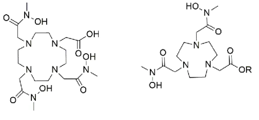
CHAPTER 2 Figure 1 : Structures of [64Cu]-NOTA-PEG-RM26 and [64Cu]-DOTHA2-PEG-RM26 ... 55
x
Figure 2 : Representation of cellular uptake studies of [64Cu]-DOTHA2-PEG-RM26 and
[64Cu]-NOTA-PEG-RM26 on PC3 human prostate cell line ... 62 Figure 3 : (A) Biodistribution study of [64Cu]-DOTHA2-PEG-RM26 and [64
Cu]-NOTA-PEG-RM26 in female balb/c mice ... 64 Figure 4 : Biodistribution study of [64Cu]-NOTA-PEG-RM26 and [64Cu]-DOTHA2
-PEG-RM26 in PC3 tumor-bearing athymic nude male mice ... 65 Figure 5 : Example of a 3D-MIP PET/CT of [64Cu]-DOTHA2-PEG-RM26 in PC3
tumor-bearing athymic nude male mice ... 66 Figure 6 : PET-derived time-activity curves from tumor-bearing mice injected with either
[64Cu]-DOTHA2-PEG-RM26 and [64Cu]-NOTA-PEG-RM26 ... 67
CHAPTER 3
Figure 1 : Structures of [64Cu]-NOTA-APCA-RM26 and [64Cu]-DOTHA2-APCA-RM26 . 81
Figure 2 : Biodistribution data of [64Cu]-DOTHA2-APCA-RM26 in female Balb/c mice ... 83
Figure 3 : Biodistribution data of [64Cu]-DOTHA2-APCA-RM26 in athymic male nude
mice... 85 Figure 4 : PET-derived time-activity curves from tumor-bearing mice injected with [64 Cu]-DOTHA2-APCA-RM26 ... 86
Figure 5 : Representative PET images of [64Cu]-DOTHA2-APCA-RM26 in PC3 human
xi
L
IST OFT
ABLESINTRODUCTION
Table 1 : Various radionuclides used in PET imaging ... 4 Table 2 : Amino Acid sequence of synthetic peptides ... 7
CHAPTER 1
Table 1 : Analytical data for NOTA‐LK‐RM26 peptides ... 25
Table 2 : Biodistribution data of [64Cu]‐NOTA‐PEG‐RM26 on female Balb/c mice ... 28
Table 3 : Biodistribution data of [64Cu]‐NOTA‐APCA‐RM26 and [64Cu]‐NOTA‐AHDA‐
RM26 on female Balb/c mice ... 29 Table 4 : Biodistribution data of [64Cu]‐NOTA‐PEG‐RM26 on PC3 tumor‐bearing athymic
nude mice ... 33 Table 5 : Tumor‐to‐tissue ratios of [64Cu]‐NOTA‐LK‐RM26 in PC3 tumor‐bearing athymic
nude male mice ... 34
CHAPTER 2
Table 1 : Affinities of DOTHA2-PEG-RM26 for GRPR on PC3 human prostate cell line. . 61
Table 2 : Ex vivo and PET-derived tumor-to-tissue ratios of [64Cu]-DOTHA2-PEG-RM26
and [64Cu]-NOTA-PEG-RM26 in PC3 tumor bearing athymic nude male mice ... 65 CHAPTER 3
Table 1 : Affinities for GRPR determined using [125I]-Tyr4-BBN on PC3 cells. ... 83 Table 2 : Biodistribution data of [64Cu]-DOTHA2-APCA-RM26 and [64
Cu]-NOTA-APCA-RM26 in Balb/c mice ... 84 Table 3 : Biodistribution data of [64Cu]-DOTHA2-APCA-RM26 in athymic nude male mice
xii
L
IST OFA
BBREVIATIONS %ID μM Aca AHDA APCA BBN BFC BSA CB-TE2A C-NE3TA DOTA DOTHA2 DRE DTPA EDTA FCH GPCR GRPR HEPE IC50 LK NE3TA N-NE3TA NOTA NOTHA2 PA PEG PET PSA PSMA RM26 SarAr TACN TETAPercentage of injected dose per gram Micro molar Aminocaproic acid Amino-hexanedioic-1-acid 2-Aminoethyl-piperazine-1-carboxylic acid Bombesin Bifunctional chelator Bovine serum albumin
1,4,8,11-Tetraazabicyclo[6.6.2]hexadecane-4,11-diacetic acid 4-carboxymethyl-7-[2-(carboxymethyl-amino)-3-(4-nitro-phenyl)-propyl]-[1,4,7]triazonan-1-yl-acetic acid
1,4,7,10-tetraazacyclododecane-1,4,7,10-tetraacetic acid
N-methylhydroxamates tetraazamacrocycles
Digital rectal exam
Diethylenetriamine pentaacetic acid Ethylenediamine tetracetic
Fluorothylcholine
G protein coupled receptor Gastrin releasing peptide receptor
(4-(2-hydroxyethyl)-1-piperazineethanesulfonic acid) Half-maximal inhibitory concentration
Linker 2,2’-(7-((carboxymethyl)amino)ethyl)-1,4,7-triazonanane-1,4-diyl)diacetic acid 4-carboxymethyl-7-[2-[carboxymethyl-(4-nitro-benzyl)-amino]-ethyl]-[1,4,7]triazonan-1-yl-acetic acid 1,4,7-triazacyclononane-1,4,7-triacetic acid N-methylhydroxamates triazamacrocycles Phosphoramidon Polyethylene glycol
Positron emission tomography Prostate specific antigen
Prostate specific membrane antigen [D-Phe6-Sta13-Leu14-NH2]bombesin(6-14)
1-N-(4-aminobenzyl)-3,6,10,13,16,19-hexaazabicyclo[6.6.6]-eicosane-1,8-diamine
1,4,7-triazacyclononane
1,4,8,11-tetraazacyclotetradecane-N, N’,N’’,N’’’-tetraacetic acid Triazacyclononane-phosphinate
1
I
NTRODUCTION1.1PROSTATE GLAND AND PROSTATE CANCER
The prostate is an exocrine gland in men. It is part of the reproductive system located below the bladder and in front of the rectum. It produces a fluid that helps to transport and nourish sperm as it passes from the testes, through the ejaculatory ducts and prostate, and out from the urethra (Denmeade et Isaacs, 2002). Prostate cancer can be defined as an abnormal and uncontrolled division of cells of the prostate gland. It is the second most common leading cause of cancer-related death in men in Canada. In USA, about 1.6 million new cancer cases are expected to be diagnosed in 2016 and 0.6 million cancer deaths will occur (Siegel et al., 2017). Prostate cancer is the most diagnosed cancer in men. The first prostate cancer case was discovered by J. Adams a surgeon at the London Hospital in 1853 (Denmeade et al., 2002). Prostate cancer is often a very slow growing cancer and it can be detected at late stage when it spreads (metastasis) to bones, lymph nodes, rectum and bladder (Mustafa et al., 2016).
1.2PROSTATE CANCER BIOMARKERS
There are different biomarkers expressed in prostate cancer such as Prostate Specific Antigen (PSA), Prostate Specific-Membrane Antigen (PSMA), Gastrin Protein Coupled Receptor (GPCR) and Prostate Specific G Protein (PSGP). Those biomarkers have been used for assessment and diagnosis of prostate cancer. The upregulation and downregulation of prostate cancer biomarkers are crucial for the clinical course for the disease and could be targeted for early detection and/or therapeutic applications in prostate cancer (Cao et al., 2015).
1.2.1PROSTATE SPECIFIC ANTIGEN (PSA)
Prostate specific antigen (PSA) is a sensitive prostate cancer biomarker. It is a 34 kD glycoprotein excreted by epithelial cells of the prostate gland. Elevation in PSA levels is specific to cancer presence. Also, the elevated PSA level could be due to the infection and inflammation of the prostate gland. PSA measurement is the most commonly used technique for prostate cancer screening, followed by digital rectal exam (DRE) (Crawford ED, Ventii K, 2014). The US Food and Drug Administration (FDA) approved PSA test in 1986 as a screening tool for early detection of prostate cancer (Jakobsen et al., 2017). PSA screening was conducted in Canada in the early 1990’s. A study performed in Quebec/Canada in early 1990’s has shown that increased
2 screening efforts with the PSA test were not correlated with the subsequent declining in mortality rate (Perron et al., 2002). PSA is useful for screening prostate cancer but the lack of specificity of PSA measurement as a tumor biomarker results in a high rate of unnecessary biopsies (Dickinson et al., 2016).
1.2.2PROSTATE SPECIFIC MEMBRANE ANTIGEN (PSMA)
Prostate specific membrane antigen (PSMA) is also known as folate hydrolase 1 or a carboxypeptidase II. It is a 100 kDa cell surface protein overexpressed in prostate cancer. PSMA consists of 750 amino acid residues (Silver et al., 1997). It plays a role in nutrient uptake in the prostate gland. The selective detection of PSMA overexpressed in prostate cancer offers the possibility to accurately diagnose and treat prostate cancer. It is very useful clinically as a diagnostic biomarker for prostate cancer due to the development of several selective PSMA-targeting moieties such as DUPA, PSMA-11 and Glu-Glu-urea (Banerjee et al., 2016), which can be radiolabeled and used for imaging.
1.2.3GPROTEIN COUPLED RECEPTOR (GPCR) AND GASTRIN RELEASING PEPTIDE RECEPTOR
(GRPR)
GPCR is one of the integral membrane protein that contains an extracellular terminal (NH2-)
region, and a seven-domain transmembrane alpha-helical structure (3 intracellular domains and 3 extracellular domains and a carboxylic terminal (COOH-). It responds to various molecules such as peptides, neurotransmitters and growth factors. Upon activation by ligands such as peptides, the GPCR undergoes a conformational change and then activates the G proteins by promoting the exchange of GDP/GTP associated with the Gα subunit (Xia et al., 2001;Weng et al., 2005). The Gα-GTP complex diffuses into the cytoplasm to regulate various intracellular activities. The activation of GPCR and its signaling pathways can contribute to normal cell functions of growth, survival and differentiation; however, they can enhance tumor growth and promote angiogenesis in cancer cells (Yeagle et Albert, 2007).
Gastrin releasing peptide receptor (GRPR) is a member of the GPCR family. This receptor is responsible for the signaling process of the neuropeptides in the nervous system. The GRPR family consists of four receptor subtypes: the neuromedin B receptor (NMB-R [BBr1, BRS-1]), the gastrin-releasing peptide receptors (GRP-R [BBr2, BRS-2]), the orphan receptor ([BBr3,
3 BRS-3]) and the amphibian receptor ([BBr4]). GRPRs are expressed in organs such as pancreas, breast and neuroendocrine cells of the lung, brain and prostate. They are overexpressed in different cancers including prostate, breast, lung and several other types of cancer cells. Presence of GRPR has been confirmed in breast cancer (71%), primary prostate cancer in proportions ranging from 63% to 100 %, and in its metastases (> 50%) (Markwalder et Reubi, 1999; Ohki-Hamazaki et al., 2005). The overexpression of GRPR receptors in various tumors has generated interest in the development of radiolabeled peptides for targeting these receptors.
Gleason Score is the grading system used to determine the aggressiveness of prostate cancer. The grading score of Gleason score was initiated in the 1960’s by a pathologist names Donald Gleason. The grading system is based on how prostate cancer looks under a microscope and how it is likely to spread. The score ranges from 2 to 10, the higher the score the more likely that the caner will grow and spread quickly. Gleason score (2 – 4) tend to be less aggressive, while cancers with higher Gleason scores (7 – 10) trend to be more aggressive. It is very useful for predicting the behavior of the prostate cancer (Gordetsky et al., 2016).
1.3POSITRON EMISSION TOMOGRAPHY (PET)IMAGING
PET technique shows a promising future in the imaging of prostate cancer (Hong et al., 2010). PET imaging requires radionuclides that emit positron (β+). Positron-emitting nuclides are generally produced using a cyclotron by bombarding target materials with protons or deuterons. The resulting nuclides are unstable and therefore, stabilize through decay by positron-emission. The positron is ejected from the nucleus and is then annihilated with a surrounding electron producing two 511 keV photons traveling in opposite directions. In the PET scanner, a ring of detectors detects those annihilation photons in coincidence. The sensitivity of PET is at least 2 orders of magnitude better than that of single photon imaging systems. Therefore, PET has the potential to play an important role in the detection of localized and metastatic prostate cancer. Short-lived organic radioisotopes such as 11C, 13N, 15O and 18F have limitations because they require fast manipulation, rapid chemical synthesis, which makes the clinical applications quite challenging and requires an on-site cyclotron for their production.
Recently, more efforts were directed toward producing positron-emitting radiometals such as Gallium-68 (68Ga), Copper-64 (64Cu) and Zirconium-89 (89Zr). Table 1 shows a list of several positron emitters ( Sarko et al., 2012; Price et Orvig, 2014). The increased use of copper
4 radiometal led to a noteworthy work for the synthesis of a multidentate ligand for its chelation (Jin et al., 2017). 64Cu has a few specific advantages compared to other radionuclides. Its relatively long physical half-life (T1/2 = 12.7 h) allows for quality control after production and
radiotracer preparation, and this long physical half-life can be useful when imaging at a relatively late time point is necessary. 64Cu is produced using a medical cyclotron by the reaction of 64Ni(p,n)64Cu. It can be produced in a cyclotron center and delivered to different local and remote hospitals.
64Cu decays by positron emission (18%, E
βmax = 656 keV, average = 288 keV), which can be
used for PET imaging to evaluate the kinetics of radiolabeled compounds. Its therapeutic potential comes from the beta emission (39%, Eβmax =573 keV) and electron capture (EC) with
corresponding gamma emission at 1346 keV, which represent 43.1% of the decay profile (Holland, Williamson, et al., 2016; Smith, 2004; Sun et Anderson, 2004). Via its complexation to a chelator, 64Cu can be conjugated to different delivery systems such as liposomes, microparticles, nanoparticles and dendrimers for diagnostic and therapeutic applications (Holland, Ferdani, et al., 2016).
Table 1 : Various radionuclides used in PET imaging
Radionuclide Half-life (min) Reaction Production method
11C 20.4 14N(p,α)11C Cyclotron 13N 9.96 16O(p,α)13N 13C(p,n)13N Cyclotron 15O 2.07 14N(d,n)15O 15N(p,n)15O Cyclotron 18F 109.7 18O(p,n)18F Cyclotron 64Cu 768 64Ni(p,n)64Cu Cyclotron 68Ga 68 68Ge/68Ga Generator 82Rb 1.25 Decay of 82Sr Cyclotron 89Zr 78.5 h 89Y(p,n)89Zr Cyclotron
1.3.1METABOLIC PETTRACERS
The most widely used radiotracer in PET imaging is [18F]-fluoro-2-deoxy-2-D-glucose (18 F-FDG), a glucose analogue. This tracer is used as an indicator of glycolytic activity in cells. Well-differentiated prostate cancer is well known for its low glucose metabolic rate due to the low
5 expression of glucose transport protein (GLUT1). [18F]-FDG fails in the early detection of the prostate cancer mainly because of the low metabolism of prostate cancer cells, but also because of the proximity to the bladder and urinary tract. The presence of high activity in the bladder during PET imaging could easily mask any prostate lesion (Jadvar, 2009).
Prostate cell membrane contains phospholipid and choline. Choline (trimethyl-2-hydroxyethylammonium) is a substrate for the synthesis of phospholipidcholine. It’s uptake is mediated by the upregulation of the choline kinase activity. The choline kinase is highly expressed in prostate cancer. Choline was radiolabeled with the short half-life positron emitter
11C (half-life = 20 min) (Figure 1). Its uptake is non-specific and observed in liver, bone marrow
and muscles. One of the major drawback of using 11C tracers is the requirement of an on-site cyclotron for its production and the short time window for imaging due to the short half-life of
11C (Awwad et al., 2012).
Figure 1 : Structure of [11C]-Choline
The advantage of using longer half-life 18F (half-life = 110 min) led to the synthesis of 18 F-Fluoromethylcholine (FCH) (Figure 2). FCH is a choline analogue. Imaging studies using FCH showed usefulness in detecting different tumors, including prostate cancer, but it showed non-specific binding which could lead to false-positive and misdiagnosis of cancer. 18F-FCH PET/CT could enhance prostate cancer detection in patients with elevated PSA level and improve patient management (DeGrado et al., 2001). However, the tracer failed to image the early recurrence of prostate cancer due to low sensitivity (<42%) to detect lesions in post-prostatectomy patients with low level of PSA.
6
1.3.2PEPTIDES AND RADIOLABELED PEPTIDE IMAGING
Peptides are defined as molecules that contain less than 50 amino acids chain and, as such, they have a low molecular weight. Amino acids in a chain are coupled by peptide bonds. They can be produced easily and inexpensively. Peptides bind with high affinity and specificity toward the overexpressed receptors on common cancers such as prostate, breast and colon cancers. Peptides showed favorable properties to be used as radiolabeled tracers. They can tolerate harsh chemical conditions during radiolabeling with different radionuclides (Okarvi, 2004; Craik et al., 2013). Radiolabeled peptide imaging to target prostate cancer is becoming a very interesting area of research (Fani et al., 2012). Researchers are focusing their efforts toward the development of radiolabeled peptides for molecular imaging and targeted therapy for prostate cancer (Boerman
et al., 2000; Weiner et Thakur, 2002). Several peptide receptors represents an important
breakthrough in the management of patients with neuroendocrine tumors (NETs) such as somatostatin receptor (SST) and cholecystokinin receptor (CCK) and gastrin releasing peptide receptor (GRPR) (Lee et al., 2010).
1.3.2.1BOMBESIN PEPTIDE
Bombesin (BBN) is a linear amphibian tetradecapeptide [pGlu-Gln-Arg-Leu-Gly-Asn-Gln-Trp-Ala-Val-Gly-His-Leu-Met-NH2], in which the natural carboxyl-terminal sequence (C-terminus)
BBN(8-14) is responsible for the binding. It is found naturally in the skin of the European fire-bellied toad bombina bombina (Anastasi et al., 1971). Bombesin was shown to resemble the Gastrin Releasing Peptide (GRP), a 27 amino acid peptide that both share the same seven COOH terminal amino acid (Table 2). Bombesin and GRP peptides play important roles in regulating smooth muscle contraction, stimulating action of gastric contraction and the regulating the growth of normal and malignant cells as it functions as an autocrine or paracrine growth stimulator. Both peptides bind with high affinity to the GRPR (Bologna et al., 1989; Sebesta et
7
Table 2: Amino Acid sequence of synthetic peptides
Peptide Amin Acid Sequence
GRP Val-Pro-Leu-Pro-Ala-Gly-Gly-Gly-Thr-Val-Leu-Thr-Lys-Met-Tyr-Pro-Arg-Gly-Asn-His-Trp-Ala-Val-Gly-His-Leu-Met- NH2 Bombesin pGlu-Gln-Arg-Leu-Gly-Asn-Gln-Trp-Ala-Val-Gly-His-Leu-Met- NH2 RM26 (Bombesin Antagonist) D-Phe-Gln-Trp-Ala-Val-Gly-His-Sta-Leu-NH2
The seven amino acids sequence (C-terminus) are responsible to determine the agonistic or antagonistic character of the bombesin peptide. Bombesin agonists bind on the surface of the tumor cells and further internalize into the cytoplasm and stimulate tumor growth and angiogenesis. Bombesin antagonists bind to the GRPR and remain on the cell surface and no further internalization occurs. Bombesin antagonists have shown reduced physiological activity and may prevent tumor proliferation.
Truncated bombesin (BBN(7–14)) and full-length bombesin derivatives were designed for efficient GRPR targeting. The truncated derivatives facilitate easy synthesis and modifications and provide longer biological half-life compared to full-length derivatives (Breeman et al., 1999; Tokita et al., 2001). Different groups reported the synthesis, development and characterization of several radiolabeled bombesin derivatives to target overexpressed receptors and identify tumor cells.
Yang et al. conjugated the DOTA chelator to the truncated BBN(7-14) via the Ɛ-aminocaproic acid (Aca) linker to produce the [64Cu]-DOTA-Aca-BBN(7-14) tracer and compared to the DOTA directly conjugated through the Lys3 side chain to full-length BBN to give the [64 Cu]-DOTA-[Lys3]BBN. Both conjugates were radiolabeled with 64Cu. The full-length derivative showed higher binding and higher tumor uptake on PC3 prostate cancer cells compared to the truncated derivative. (Yang et al., 2006). Lantry et al. used the truncated agonist DOTA-glycyl-4-aminobenzoic-Gln7Trp9Ala9Val10Gly11His12Leu13Met14-NH2 known as AMBA and
8 internalization rate and high PC3 tumor uptake (Lantry et al., 2006). Abd-Elgaliel et al. conjugated the DOTA chelator to the truncated antagonist [D-Phe6, Leu-NHCH2CH2CH313,desMet14]BBN(6-14) through the aminohexanoyl linker for 111In labeling. Due
to its antagonistic nature, the compound showed low internalization and high uptake (6.9 ± 1.1 %ID/g) on PC3 cells (Abd-Elgaliel et al., 2008).
Cescato et al. compared a potent full-length agonist
(Gln2Arg3Leu4Gly5Asn6Gln7Trp9Ala9Val10Gly11His12Leu13Met14-NH2) (Demobesin4) to the
truncated antagonist (Demobesin1) ([99mTc]-N4-Bzdig0,(D)Phe6,Leu-NHEt13,des-Met14
]BBN(6-14)). The two compounds were radiolabeled with 99mTc. However, the antagonist Demobesin1 had 4-fold higher tumor uptake than the agonist Demobesin4 on PC3 prostate cancer cell line (Cescato et al., 2008). Gourni et al. compared the truncated agonist derivative BBN(7-14) vs the full-length agonist bombesin. The two derivatives were radiolabeled with 99mTc and both showed fast clearance from the blood and elimination through the kidney and liver with slight advantage toward kidney elimination for the full-length derivative, which could be due to the high hydrophilicity of the truncated derivative (Gourni et al., 2009). Mansi et al. reported that the bombesin antagonist [111In]-RM1 showed superior uptake on PC-3 tumor compared to the agonist [111In]-AMBA at 4 h p.i. (Mansi et al., 2009). Varasteh et al. conjugated the NOTA chelator to the bombesin antagonist RM26 via the polyethylene glycol (PEG) linker for 68Ga and
111In radiolabeling. The tumor-to-background ratios were greater than 10 for both compounds
after 1 h (Varasteh et al., 2013). In summary, bombesin antagonist derivatives showed higher affinity and longer retention on tumor cells and low background in several tissues (Schumacher
et al., 2005; Lantry et al., 2006; Abd-Elgaliel et al., 2008).
1.3.2.2LINKERS
Linkers are chemical structures consisting of short amino acid sequences inserted between the bifunctional chelator and the peptide-based targeting molecules. They could be used to avoid steric hindrance that may be caused by the bifunctional chelator on the targeting molecules (Veronese et Pasut, 2005). Several attempts have been proposed to study the impact of modifying linkers on the pharmacokinetics of the radiolabeled peptide which could lead to reduction in the liver uptake and enhance kidney excretion pathway (Garcia et al., 2008). Various linkers were proposed for the preparation of the radiolabeled bombesin, among them,
9 aliphatic linear amino acids, amino acids, aromatic amino acids or charged linkers such as polyethylene glycol (PEG), cationic or anionic charged linkers. Aliphatic aminooctanoic acid (Aoc = 8 carbon) linker were introduced between the DOTA chelator and the truncated BBN[7-14] analog. The compound was radiolabeled with 64Cu. The resulting 64 Cu-DOTA-Aoc-BBN[7-14] conjugate showed specific localization on PC3 tumor-bearing mice. It was suggested to use more hydrophilic linkers with total neutral or negative charges to modulate the pharmacokinetics of BBN derivatives (Rogers et al., 2003). Parry et al. introduced a series of carbon spacer (Aba = 4-carbon, Ava = 5-carbon, Ahx = 6-carbon, Aoc = 8-carbon and Ado = 12-carbon) between the DOTA chelator and the BBN(7-14) to study the impact of linker length on tumor uptake. The bombesin analog with 8-carbon (Aoc linker) had the highest tumor uptake as determined by µPET imaging (Parry, et al., 2007) (Figure 3).
Figure 3 : Structures of 4-Aba, 5-Ava, 6-Ahx, 8-Aoc and 12-Ado linkers
In further studies to improve tumor uptake and renal clearance, Parry et al. have prepared a series of 64Cu-DOTA-linker-BBN(7-14) tracers using three amino acids (glycine (G), serine (S) and glutamic acid (E)) linkers between the DOTA chelator and the truncated agonist bombesin(7-14).
10
Figure 4 : Structures of tripeptide linkers
Negatively charged glutamic acid residues significantly reduced the affinity of bombesin analogues for GRPR as well as internalization into GRPR-expressing cells. The other analogues have shown specific tumor uptake and good imaging characteristics (Parry, et al., 2007) (Figure 4). De Visser et al. designed a series of truncated version of DTPA-bombesin (Cmp1 [DTPA-Pro1,Tyr4]BN). Non-natural amino acids were introduced between the DTPA chelator and the bombesin in order to increase the receptor affinity (Figure 5).
11
Figure 5 : Structures of unnatural amino acid linkers
The tracer containing the ACMPip non-natural amino acid showed the highest affinity and internalization rate, as well as the fastest clearance, which led to high tumor-to-blood ratio (De Visser et al., 2007). Lantry et al. proposed the glycyl-4-aminobenzoic aromatic linker between the DOTA bifunctional chelator and the truncated BBN(7-14)NH2 for 177Lu-labeling. The
conjugate had high affinity toward GRPR and high internalization, and the excretion was mainly through the kidneys (Lantry et al., 2006) (Figure 6).
Figure 6 : Structure of glycyl-aminobenzoic acid linker
Lane et al. introduced the NO2A chelator [64Cu-NO2A-(X)-BBN(7–14)NH2] and compared a
series of linkers, where (X) is AMBA, β-Ala, 5-Ava, 6 Ahx, 8-Aoc and 9-Anc. The conjugate containing the AMBA aromatic linker had high tumor uptake and retention and showed the lowest liver uptake as compared to the other linkers (Lane et al., 2010) (Figure 7).
12
Figure 7 : Structure of AMBA linkers
Mansi et al. introduced a positively charged linker, the 4-amino-1-carboxymethyl-piperidine to a bombesin antagonist to improve its pharmacological performance. The results showed that a positive charge at the N-terminal of bombesin-based peptides lead to improved bombesin receptor affinity (Mansi et al., 2011) (Figure 8).
Figure 8 : Structure of 4-amino-1-carboxymethyl-piperidine linker
The length of linker was found also to have an impact on tumor uptake and clearance (Nedrow et
al., 2014). In further study, Varastech et al. studied the effect of the mini-PEG on the
radiolabeled bombesin. The NOTA chelator was conjugated to the RM26 using a series of PEG linkers (n = 2, 3, 4 and 6). There was no significant difference in the in vitro binding affinity between the linkers and similar tumor uptake was observed on the two prostate cancer cell lines PC3 and BT-474 at 2 h p.i. (Varasteh et al., 2014). In summary, structural modifications on the length and the structure of the linkers aimed to reduce the high abdominal tissue uptake, optimize lower uptake in non-target organs such as liver and obtain high uptake in cancer cells.
1.3.2.3BIFUNCTIONAL CHELATORS FOR 64CU
The incorporation of a 64Cu to a biomolecule requires the use of a bifunctional chelator (BFC). BFC contains a metal-chelating group and a chemically reactive functional group to facilitate linking the 64Cu-chelator complex to variable biomolecules (Brechbiel, 2008). The design and synthesis of kinetically inert BFC for 64Cu radionuclide is an active area of research (Anderson et Ferdani, 2009). The reasons to develop new BFCs are to obtain easy 64Cu radiolabeling procedure, to form a stable complex and to prevent in vivo demetallation for early detection of tumor. As a number of 64Cu-labeled bombesin conjugates have been proposed as PET imaging
13 probes for prostate cancer, but there is still a need to develop a high in vivo stability bifunctional chelator (Hoffman et Smith, 2009; Cai et Anderson, 2014).
There are two main classes of bifunctional chelators: acyclic and macrocyclic. Acyclic ethylenediamine tetracetic acid (EDTA) and diethylenetriamine pentaacetic acid (DTPA) have poor kinetic inertness and their ability to chelate stably 64Cu is poor (Cai et Anderson, 2014). Macrocyclic chelators showed higher thermodynamic and kinetic stability compared to the linear DTPA chelator. The designs of many different macrocyclic chelators are based on the three following backbones: the 1,4,7-triazacyclononane (TACN), a nine-membered macrocycle; 1,4,7,10-tretraazacyclododecane (Cyclen), a twelve-membered macrocycle; and 1,4,8,11-tetraazacyclotetradecane (Cyclam), a fourteen-membered macrocycle (Sarko et al., 2012).
1,4,7,10-tetraazacyclododecane-1,4,7,10-tetraacetic acid (DOTA) is the most commonly used cyclen chelator for radiolabeling compounds using different trivalent and divalent radionuclides (Figure 9(a)). DOTA showed higher thermodynamic and kinetic stability compared with acyclic chelators. The DOTA chelator has been conjugated to bombesin and radiolabeled with different radionuclides: 64Cu and 68Ga for PET imaging, and 90Y and 177Lu for therapy (Schumacher et al., 2005; Lantry et al., 2006; De Visser et al., 2007). DOTA chelator have shown poor stability when chelated with 64Cu (Ait-Mohand, S, et al. 2011). A series of cyclam based chelators called 1,4,8,11-tetraazacyclotetradecane-N,N’,N’’,N’’’-tetraacetic acid (TETA and derivatives) were developed to enhance the in vivo stability for 64Cu radiolabeling for imaging and therapy (Bass et
al., 2000; Sprague et al., 2007) (Figure 9(b)). The cross bridged tetraazamacrocycle
1,4,8,11-tetraazabicyclo[6.6.2]hexadecane-4,11-diacetic acid (CB-TE2A) cyclam derivative was designed for 64Cu complexation (Figure 9(c)). Its corresponding chelates present high resistance to transmetalation reactions in vivo compared to TETA and DOTA (Garrison et al., 2007).
However, the CB-TE2A and its derivatives require harsh radiolabeling conditions, which can disable their bioconjugation to more fragile biomolecules. The 1-N-(4-aminobenzyl)-3,6,10,13,16,19-hexaazabicyclo[6.6.6]-eicosane-1,8-diamine (SarAr) BFC was developed (Figure 9(d)). It has the advantage of efficient radiolabeling with 64Cu at room temperature compared to CB-TE2A (at 70oC for CB-TE2A). It enhanced the labeling kinetics and in vivo stability (Lears et al., 2011). Overall, cross bridged TETA derivatives, the CB-TE2A and SarAr bifunctional chelators showed superior kinetic stability in vivo and higher tumor uptake compared to the TETA and DOTA chelators (Liu, 2008).
14
Figure 9 : Structures of DOTA, TETA, CB-TE2A and SarAr bifunctional chelators
Several studies showed that the 1,4,7-triazacyclononane-1,4,7-triacetic acid (NOTA) and its derivatives NO2A, NODAGA, NE3TA and TRAP have been proposed for 64Cu radiolabeling. An easy preparation of NOTA chelator on solid support has been presented by our group (Guérin et al., 2010). The 1,4,7-triazacyclononane-1,4-diacetic acid (NO2A), a NOTA derivative was developed, but it lacks a p-NCS-Bz arm and forms an amide linkage via the third carboxylic acid unit(Prasanphanich et al., 2009) (Figure 10(b)).
De Silva et al. have developed two NOTA chelators derived from the 2,2’-(7-((carboxymethyl)amino)ethyl)-1,4,7-triazonanane-1,diyl)diacetic acid (NE3TA) and called 4- carboxymethyl-7-[2-(carboxymethyl-amino)-3-(4-nitro-phenyl)-propyl]-[1,4,7]triazonan-1-yl-acetic acid) (C-NE3TA) and (4-carboxymethyl-7-[2-[carboxymethyl-(4-nitro-benzyl)- amino]-ethyl]-[1,4,7]triazonan-1-yl-acetic acid) (N-NE3TA) (Figure 10(d)). The two compounds showed their ability for 64Cu radiolabeling at room temperature (De Silva et al., 2012). The bifunctional chelator is expected to offer fast complexation rate and slow dissociation rate. The dissociation of the radiometal from the chelator could lead to poor image quality in diagnostic application. In a previous study, four chelators NOTA, NODAGA, DOTA and DOTAGA conjugated to the
(a) DOTA (b) TETA (c) CB-TE2A
15 antagonist RM26 via the PEG linker were radiolabeled with 68Ga and 111In (Figure 10(e)). The [68Ga]-NOTsA-PEG-RM26 and the [111In]-NODAGA-PEG-RM26 provided the highest tumor-to-organ ratios. The affinity of the RM26 antagonist was slightly improved when conjugated to the NOTA-PEG complex compared to the DOTA-PEG (Varasteh et al., 2013, 2015). The triazacyclononane-phosphinate (TRAP) chelator has been initially proposed for 68Ga radiolabeling, and later was shown to have improved radiolabeling properties with 64Cu. The three-phosphonate groups act as proton acceptors to chelate the radiometal. Radiolabeling using TRAP could be performed at pH < 1 and at room temperature (25oC) in very short time (5 minutes).
Figure 10 : Structures of NOTA, NOTA-Bn-SCN, N-NE3TA and C-NE3TA, TRAP and NODAGA-NHA bifunctional chelators
(a) NOTA (b) NOTA-Bn-SCN (c) TRAP
(e) NODAGA-NHS (d) N-NE3TA and C-NE3TA
16 The chelator can link three different biomolecules for different applications (Notni et al., 2011). Šimeček et al., conjugated the RGD peptide to the TRAP chelator and radiolabeled the compound with 64Cu (64Cu-TRAP(RGD)3) (Figure 10(c)). Conjugation of three RGD peptides
increased the kinetic stability of the structure (Šimeček et al., 2012). Jackson et al. developed a heterodimeric RGD-bombesin agonist conjugated to NO2A or NOTA for 64Cu labeling. This group clearly showed that the complexing agent/metal complex influences the biodistribution of the radiopharmaceutical (Jackson et al., 2012). On the other hand, the 1,4,7-triazacyclononane chelator,1-glutaric acid-4,7 acetic acid (NODAGA) showed high stability in vivo and the highest reported tumor/blood compared to several chelators such as DOTA and NOTA (Kim et al., 2015).
Two macrocyclic BFC bearing hydroxamic acid arms, called DOTHA2 and NOTHA2, were
designed by our group for 64Cu radiolabeling. Their structures are based on the tetraazamacrocycle for DOTHA2 and the triazamacrocycle for NOTHA2. The synthesis of these
chelators is straightforward and can be obtained in large quantities (Ait-Mohand et al., 2014). The novel hydroxamate BFCs offered very fast labeling kinetics at room temperature in a wide range of concentration and pH as compared to their acetic acid analogs DOTA and NOTA. Both
64Cu-DOTHA
2 and 64Cu-NOTHA2 showed high stability, low residual activity in various tissues
and fast clearance in healthy mice.These earlier findings highlight the potential of these BFC for PET imaging facilitating further exploration of 64Cu-peptide based tracers (Samia Ait-Mohand et
al., 2014). Figure 11 shows the chemical structure of two bifunctional chelators, DOTHA2 and NOTHA2.
17
HYPOTHESIS AND OBJECTIVES
Others and we have developed a series of promising 64Cu-peptide agonists and antagonists that specifically bind with high affinity to GRPR. While several potential peptide-tracer candidates were identified from our previous work, their stability and/or pharmacokinetic properties need to be improved and alternative tracer structural modifications are put forth in this thesis to supports the central HYPOTHESIS that radioligands for prostate cancer with a better biodistribution
profile can be used as tools for cancer diagnosis by PET. To this end, we propose modifications
of the linker and the chelator and we selected the BBN antagonist RM26 (D-Phe-Gln-Trp-Ala-Val-Gly-His-Sta-Leu-NH2) for specific binding to the GRPR. We hypothesized that the
introduction of charged linker could enhance the pharmacokinetics of the radiolabeled antagonist (Chapter 1) and the BFC DOTHA2 bearing hydroxamic arms could provide superior in vivo
stability compared to the current BFCs (Chapters 2 and 3).
The overall objective was to develop novel radiolabeled peptides as highly specific diagnostic tools for PET imaging of prostate cancer.
The first objective of this thesis was to study the influence of dicationic and dianionic linkers on the elimination kinetics of the radiolabeled RM26 by performing in vitro evaluation and conducting preclinical PET imaging studies in prostate cancer models. For this comparative study, three GRPR antagonists containing the RM26 sequence were synthesized and conjugated with NOTA via different linkers (LK): polyethylene glycol (PEG–neutral), APCA (dicationic) or AHDA (dianionic). The NOTA-LK-RM26 peptides were radiolabeled with 64Cu to assess their pharmacokinetic and PET imaging properties using PC3 tumor‐ bearing athymic nude mice. The second objective of this thesis was to investigate the influence of DOTHA2 on tumor uptake
and pharmacokinetics of 64Cu-radiolabeled GRPR peptide antagonists to assess their PET imaging properties for diagnosis of prostate cancer. DOTHA2-PEG-RM26 was conveniently and
efficiently assembled on solid support. The compound radiolabeled with 64Cu and its affinity, stability and cellular uptake on PC3 prostate cancer cells were evaluated. The in vitro and in vivo behavior of [64Cu]-DOTHA2-PEG-RM26 was examined by PET imaging using human PC3
prostate cancer xenografts and its behavior was compared to that of the analogous [64 Cu]-NOTA-PEG-RM26.
18 Our third objective was to evaluate 64Cu-DOTHA2-APCA-RM26, a novel BBN antagonist for
visualization of GRPR expressing tumor by PET. This study with 64Cu-DOTHA2-APCA-RM26
follows our previous work on the exploration of the impact of spacers and chelators used for the labeling of peptide tracers targeting GRPR for the diagnosis of prostate cancer.
19
C
HAPTER1
Impact of dianionic and dicationic linkers on tumor uptake and
biodistribution of [
64Cu]Cu/NOTA peptide-based gastrin releasing
peptide receptors antagonists (Article 1)
Nematallah Mansour, Véronique Dumulon-Perreault, Samia Ait-Mohand, Michel Paquette, Roger Lecomte, Brigitte Guérin
Article status: published in J Label Compd Radiopharm, 2017, 60(4):200-212
Contribution of the student: Nematallah Mansour has performed the in vitro, in vivo and ex vivo experiments and data analysis. He has contributed in the preparation of the first draft and
revised the article under the supervision of Roger Lecomte and Brigitte Guérin.
Résumé : Dans cette étude, nous avons étudié pour la première fois l'influence de l'acide
2-aminoéthylpipérazine-1-carboxylique (APCA) et de l'acide aminé-hexanedioïque-1 (AHDA) sur la captation tumorale et la cinétique d'élimination d’antagonistes peptidiques radiomarqués au
64Cu ciblant le récepteur à la relâche de gastrine (GRPR). Trois antagonistes du GRPR contenant
la séquence RM26 ont été synthétisés et conjugués au NOTA via différents espaceurs (LK): polyéthylène glycol (PEG-neutre), APCA (dicationique) et AHDA (dianionique). Les peptides NOTA-LK-RM26 ont été radiomarqués au 64Cu pour évaluer leurs propriétés pharmacocinétiques et de tomographie d’émission par positrons (TEP) en utilisant des souris immunodéficientes porteuses de tumeur PC3. Les constantes d'inhibition (Ki) des trois peptides natCu/NOTA-LK-RM26 portant des espaceurs PEG, dicationique et dianionique étaient
respectivement de 0,98 ± 0,48 nM, 0,95 ± 0,21 nM et 17,97 ± 2,79 nM. Les conjugués [64 Cu]-NOTA-LK-RM26 ont été préparés avec des rendements de marquage supérieurs à 95% et des activités spécifiques de 67 à 77 TBq/mmol. Les 3 radiopeptides étaient stables in vivo et présentaient une captation spécifique du GRPR dans le pancréas avec un efflux très rapide de ce tissu observé pour le peptide [64Cu]-NOTA-AHDA-RM26. Les résultats des études d'imagerie
20 ont montré une absorption spécifique à la tumeur PC3, ainsi que des éliminations rénales et hépatiques similaires et rapides pour les deux peptides [64Cu]-NOTA-APCA-RM26 et [64 Cu]-NOTA-AHDA-RM26. Compte tenu de leurs caractéristiques d'imagerie adéquates, les analogues [64Cu]-NOTA-LK-RM26 portant les espaceurs APCA et AHDA sont des candidats prometteurs ciblant le GRPR pour l’imagerie du cancer de la prostate par TEP.
21
Article 1
22
Impact of dianionic and dicationic linkers on tumor uptake and
biodistribution of [
64Cu]Cu/NOTA peptide-based gastrin-releasing peptide
receptors antagonists
Nematallah Mansour | Véronique Dumulon-Perreault | Samia Ait-Mohand | Michel Paquette | Roger Lecomte | Brigitte Guérin
Department of Nuclear Medicine and Radiobiology, Faculty of Medicine and Health Sciences, Université de Sherbrooke and Sherbrooke Molecular Imaging Centre, Centre de recherche du CHUS (CRCHUS), Sherbrooke, Canada
Abstract
In this study, we investigated for the first time the influence of 2-aminoethyl-piperazine-1-carboxylic acid (APCA) and amino-hexanedioic-1-acid (AHDA) on tumor uptake and elimination kinetics of [64Cu]-radiolabeled gastrin releasing peptide receptors (GRPR) antagonists. Three GRPR antagonists containing the RM26 sequence were synthesized and conjugated with NOTA via different linkers (LK): polyethylene glycol (PEG–neutral), APCA (dicationic) or AHDA (dianionic). The NOTA-LK-RM26 peptides were radiolabeled with 64Cu to assess their pharmacokinetic and positron emission tomography (PET) imaging properties using PC3 tumor-bearing athymic nude mice. The inhibition constants (Ki) of the 3
natCu/NOTA-LK-RM26 peptides bearing PEG, dicationic and dianionic linkers were 0.98 ±
0.48 nM, 0.95 ± 0.21 nM, and 17.97 ± 2.79 nM, respectively. The [64Cu]-NOTA-LK-RM26 conjugates were prepared with labeling yields superior to 95% and specific activities of 67 to 77 TBq/mmol. The 3 radiopeptides were stable in vivo and showed GRPR‐ specific uptake in pancreas with a very fast wash-out of this tissue observed for [64Cu]-NOTA-AHDA-RM26 peptide. Results from imaging studies displayed specific PC3 tumor uptake for both [64 Cu]-NOTA-APCA- and AHDA-RM26, similar kidney elimination and fast liver washout. Considering their adequate imaging characteristics, [64Cu]-NOTA-LK-RM26 bearing APCA- and AHDA-linkers are promising candidates for GRPR-targeted PET imaging prostate cancer.
KEYWORDS
64Cu-NOTA-RM26, prostate cancer, charged linker, Gastrin releasing peptide receptors, PC3
23
INTRODUCTION
Prostate cancer is the second most frequently diagnosed cancer worldwide in men, with an estimated 1.1 million new cases in 2012.1 Current pharmacological treatments are limited to antiandrogen strategies and the development of new diagnostic and therapeutic approaches remains a challenge. There has been a significant growth in the development of radiolabeled peptides for theranostic applications in oncology. Peptides have a number of distinct advantages for receptor imaging and tumor targeting including: small size, easy preparation, easy radiolabeling, ability to attach a chelator, rapid clearance from blood and nontarget tissues, high tumor-to-background ratio, low toxicity, and low immunogenicity.2,3 In recent years, gastrin-releasing peptide receptors (GRPRs) have been shown to be significantly overexpressed on a large proportion in prostate,4,5 breast,6–8 and other cancers.9–11 Reubi et al showed that metastases of receptor‐ positive primary carcinomas were all positive with usually a comparable range in GRPR density levels in primary tumors and in metastases.8 These receptors are prime candidates for receptor targeting with radiopharmaceutical for highly specific tumor imaging by positron emission tomography (PET).
Bombesin (BBN), a potent GRPR peptide agonist naturally found in the skin of the toad Bombina bombina,12 is often used for the design of GRPR radiotracers. Bombesin peptide radiolabeling has been reported using various radionuclides, including 177Lu, 99mTc, and 188Re derivatives.13–24 For PET imaging, Schuhmacher et al labeled DOTA-PEG2-[D-Tyr6
,β-Ala11,Thi13,Nle14]BBN(6-14) with 68Ga,25 while Chen et al used DOTA-Lys3-BBN with 64Cu.26 Smith et al successfully labeled modified BBN(7-14) analogs with 64Cu for potential use in diagnostic imaging using NOTA or NO2A as chelating agents and obtained stable compounds.27,28 More recently, Wieser et al showed that the preliminary patient data with 64 Cu-CB-PEG4-D-Phe-Gln-Trp-Ala-Val-Gly-His-Sta-LeuNH2], a GRPR antagonist, are consistent
with previous preclinical studies and suggest a favorable tumor uptake and image contrast for prostate cancer imaging.29 We also synthesized and labeled DOTA- and NOTA-BBN peptides with 68Ga and 64Cu.30–32 Although our group already designed BBN tracers functionalized with NOTA chelator displaying excellent affinities and very good receptor-mediated tumor uptake in our mouse xenograft model, their pharmacokinetics need to be improved by favoring urinary excretion over hepatobiliary clearance and optimizing the target-to-background ratio for imaging to render them eligible for clinical trials.30–32
24 Recently, it has been reported that radiolabeled GRPR antagonists give higher uptake and better imaging.11 Introduction of PEG, sugar moiety, amino acids, and oligoglycine linkers have been applied to improve the in vivo kinetics of radiopeptides33–35 and BBN radiotracers.36–40 Mansi et
al introduced a positively charged linker to a BBN antagonist to improve its pharmacological
performance.41 They have shown that a positive charge at the N-terminal of bombesin-based peptides lead to improved bombesin receptor affinities. Minor modifications in the design of these peptides may have an impact on their in vivo behavior.
The goal of this study was to investigate for the first time the influence of APCA and AHDA on tumor uptake and pharmacokinetics of [64Cu]-radiolabeled GRPR peptide antagonists to assess their PET imaging properties for diagnosis of prostate cancer. We have prepared 3 NOTA-LK-RM26 conjugates where NOTA-LK-RM26 is a synthetic truncated GRPR antagonist ((D-Phe-Gln-Trp-Ala-Val-Gly-His-Sta-Leu-NH2),41) and LK is either PEG, APCA or AHDA (Figure 1).
Figure 1 : Structures of [64Cu]-NOTA-LK-RM26 selected as leads to pursue the present study. AHDA, amino-hexanedioic-1-acid. APCA, 2-aminoethyl-piperazine-1-carboxylic
acid. PEG, polyethylene glycol
The APCA and AHDA linkers were chosen because of their different charges under physiological conditions; being respectively dicationic and dianionic species. Peptide bearing neutral PEG linker, previously labeled with 68Ga and 111In,42 was used for comparative studies.
In vitro GRPR‐ binding affinities were determined with competitive radioligand binding assays on PC3 human prostate cancer cells. In vivo stability and biodistribution of the radiolabeled
25 compounds were assessed in balb/c mice. In vivo receptor targeting potential and pharmacokinetic of 64Cu‐ radiopeptides were also evaluated in PC3 tumor‐ bearing athymic nude mice using PET imaging-derived time-dependent uptake.
RESULTS
Peptide synthesis
The NOTA-LK-RM26 conjugates were synthetized by automated solid-phase synthesis with Fmoc amino acids followed by manual synthesis of the NOTA chelating moiety as described previously. After purification, the peptides were assayed for purity (>97%) by analytical high performance liquid chromatography (HPLC) with UV detection at 223 nm and their identity confirmed by mass spectrometry (MS) (Table 1).
Table 1 : Analytical data for NOTA‐LK‐RM26 peptides
Peptide Mass Yield
(%) Purity (%)b Ki (nM)c Calcd Founda NOTA-PEG-RM26 (neutral) natCu/NOTA-PEG-RM26 1543 1544 48 99 0.39 ± 0.21 0.98 ± 0.48 NOTA-APCA-RM26 (dicationic) natCu/NOTA-APCA-RM26 1566 1567 42 98 0.33 ± 0.18 0.95 ± 0.21 NOTA-AHDA-RM26 (dianionic) natCu/NOTA-AHDA-RM26 1642 1643 43 97 15.29 ± 4.08 17.97 ± 2.79
aMasses were measured on API 3000 LC/MS/MS. bPurity was determined by HPLC analysis.
cAffinities for GRPR were determined using [125I]-Tyr4-BBN in PC3 cells.
Competition binding study
The results of the binding affinity of the 3 complexes for GRPR were fitted in sigmoid curves for the displacement of [125I]-Tyr4-BBN as a function of increasing concentration of bombesin analogs. As illustrated in Table 1, Ki values of the trois NOTA-LK-RM26 peptides bearing PEG
dicationic and dianionic linkers were 0.38 ± 0.21 nM, 0.33 ± 0.18 nM, and 15.29 ± 4.08 nM, respectively. The peptide was labeled with natural Cu(II) (natCu(II)) to evaluate the change in
26 affinity by addition of the metal in the chelator. The complexation of natCu(II) to NOTA did not alter the affinity of the 3 peptides for GRPR (Table 1). The peptides bearing dicationic and PEG linkers showed higher binding affinity to the GRPR receptors compared to the peptide with a dianionic linker.
Labeling with 64Cu and stability studies
NOTA-LK-RM26 conjugates were successfully radiolabeled with 64Cu at 100°C for 10 min with labeling yields, not decay corrected, superior to 95% and specific activities of 67 to 77 TBq/mmol. No demetallation occurred up to 1 hour in mouse circulation and 20 hours in plasma because no trace of free 64Cu was detected by radioTLC.
Biodistribution studies in Balb/c mice
Biodistribution studies of [64Cu]-NOTA-PEG-RM26, [64Cu]-NOTA-APCA-RM26, and [64 Cu]-NOTA-AHDA-RM26 were carried out in female balb/c mice; the results are summarized in Figure 2 and in Tables 2 to 3. The studies were performed at 3 time points: 15, 30, and 60 minutes post-injection (p.i.) (Table 2) for [64Cu]-NOTA-PEG-RM26. This radiopeptide had a rapid blood clearance from 13.67 ± 5.89 %ID/g at 15 minutes time point to 4.47 ± 1.91 %ID/g in circulation at 30 minutes followed by a further decrease 1.72 ± 0.60 %ID/g at 60 minutes. It also showed significant high uptake on pancreas, a GRPR rich tissue,43 with the highest value of 30.98 ± 9.73 %ID/g at 30 minutes p.i. and maintained good retention of 14.35 ± 1.94 %ID/g at 60 minutes. Hepatic and kidney uptakes decreased by 50% during the first hour p.i. moving from 11.10 ± 2.62 %ID/g at 15 minutes to 5.83 ± 0.08 %ID/g at 60 minutes and from 20.05 ± 9.07 %ID/g at 15 minutes to 11.39 ± 3.25 %ID/g at 60 minutes, respectively.
[64Cu]-NOTA-APCA-RM26 also showed significant high uptake in pancreas of 20.34 ± 4.15 %ID/g that can be significantly reduced to 5.37 ± 2.59 %ID/g at 30 minutes p. i. (P < .0005) in presence of blocking agent (Figure 2B, Table 3). There were high hepatic and kidney uptakes 30 minutes with values of 22.70 ± 15.55 and 22.19 ± 6.57 %ID/g, respectively. In addition, [64 Cu]-NOTA-APCA-RM26 showed high uptake in plasma (8.01 ± 1.68 %ID/g) adrenals (10.17 ± 4.82 %ID/g) and lungs (7.96 ± 4.07 %ID/g) at this time point. [64Cu]-NOTA-AHDA-RM26 also showed a rapid blood clearance from 6.95 ± 1.89 %ID/g at 15 minutes time point to 4.60 ± 1.22
27 %ID/g at 30 minutes. It showed a very low uptake on pancreas of 3.97 ± 1.18 %ID/g at 30 minutes.
Figure 2 : Biodistribution data (A) on female Balb/c mice at 30 minutes postinjection (p.i.) of [64Cu]-NOTA-PEG-RM26 (black, n=8), [64Cu]-NOTA-APCA-RM26 (dark grey, n=6), and [64Cu]-NOTA-AHDA-RM26 (light grey, n=8); (B) Pancreatic uptake on female Balb/c
mice at 30 minutes p.i. of [64Cu]-NOTA-PEG-RM26 (PEG), [64Cu]-NOTA-APCA-RM26 (APCA), [64Cu]-NOTA-AHDA-RM26 (AHDA), and as insert, pancreatic uptake on female Balb/c mice at 15 minutes p.i. of [64Cu]-NOTA-AHDA-RM26 without (dark grey) and with (light grey) co-injection of 0.5 μmol/kg of unlabeled peptide. Data presented as percentage
of the injected dose per gram (%ID/g SD). ***: P < .005;****: P < .0005. AHDA, amino-hexanedioic-1-acid. APCA, 2-aminoethyl-piperazine-1-carboxylic acid. PEG, polyethylene
28 Nevertheless, the pancreas uptake decreased by ~47% moving from 4.74 ± 0.62 %ID/g to 2.50 ± 0.29 %ID/g in the presence of an excess of unlabeled peptide (Figure 2B insert, Table 3). Hepatic and kidney uptake increased during the first 30 minutes p.i. moving from 7.26 ± 1.04 %ID/g at 15 minutes to 18.86 ± 6.31 %ID/g at 30 minutes and from 23.54 ± 9.32 %ID/g at 15 minutes to 32.39 ± 15.95 %ID/g at 30 minutes, respectively.
Table 2 : Biodistribution data on female Balb/c mice of [64Cu]-NOTA‐PEG‐RM26 at 15
minutes post-injection (n = 3), 30 minutes (n = 8), 60 minutes (n = 3), and co-injection of unlabeled peptide at 30 minutes (n = 4). Data presented as percentage of the injected dose
per gram (%ID/g SD)
Unblocked Unblocked Blocked Unblocked
Organs 15 min 30 min 30 min 60 min
Blood 13.67 ±5.89 4.47 ±1.91 13.20 ±3.53 1.72 ±0.60 Plasma 28.60± 19.61 7.81 ±3.26 19.31 ±8.28 3.11 ±1.32 Adrenals 6.01 ±1.12 3.69 ±2.26 3.40 ±3.45 0.66 ±0.30 Fat 2.56 ±1.42 1.73 ±1.73 2.20 ±1.94 1.30 ±0.94 Kidneys 20.05 ±9.07 13.73 ±7.28 14.07 ±12.87 11.39 ±3.25 Spleen 7.46 ±3.17 3.70 ±1.83 3.05 ±1.25 1.56 ±0.78 Pancreas 24.44 ±4.35 30.98 ±9.73 3.42 ±0.89 14.35 ±1.94 Liver 11.10 ±2.62 7.08 ±1.86 9.27 ±2.03 5.83 ±0.08 Heart 4.68 ±2.21 1.75 ±0.67 4.69 ±1.47 0.99 ±0.36 Lungs 10.70 ±5.01 4.01 ±2.68 7.11 ±2.88 1.94 ±0.41 Muscle 2.62 ±1.03 1.18 ±0.49 2.39 ±1.15 0.50 ±0.12 Bone 3.19 ±1.94 0.82 ±0.28 2.85 ±1.32 0.56 ±0.33 Brain 0.39 ±0.24 0.17 ±0.06 0.46 ±0.17 0.19 ±0.08 Tail 5.70 ±0.97 3.43 ±1.07 8.33 ±0.66 4.52 ±1.04
[64Cu]-NOTA-PEG-RM26 displayed the lowest kidney, liver, and lung uptakes (Figure 2A). To assess the GRPR-mediated accumulation of the tracers, biodistribution studies were also realized with co-injection of 0.5 μmol/kg of an unlabeled peptide. Blocking with unlabeled peptide
29 significantly decreased the tracer levels in pancreas uptake at 30 minutes for both [64 Cu]-NOTA-PEG-RM26 and [64Cu]-NOTA-APCA-RM26 (P<.0005) indicating GRPR-mediated uptake in this organ (Figure 2B). [64Cu]-NOTA-AHDA-RM26 showed similar pancreas uptake values without and with co-injection of unlabeled peptide at 30 minutes p.i. that is 5 to 6 times lower than those of [64Cu]-NOTA-APCA-RM26 and [64Cu]-NOTA-PEG-RM26 respectively (Figure 2B). However, significant receptor-dependent response was observed for [64 Cu]-NOTA-AHDA-RM26 at 15 minutes p.i. (Figure 2B insert, Table 3). All other measured organs presented low uptake and no significant difference between the 3 tracers (Tables 2 and 3).
Table 3 : Biodistribution data on female Balb/c mice of [64Cu]‐NOTA‐APCA‐RM26 and
[64Cu]‐NOTA‐AHDA‐RM26 at 15 and 30 minutes post-injection with and without co‐
injection of unlabeled peptide. Data presented as percentage of the injected dose per gram (%ID/g SD)
Unblocked Blocked Unblocked Blocked Unblocked Blocked
(n=6) (n=5) (n=8) (n=5) (n=4) (n=4)
Organs APCA-30 min AHDA-30 min AHDA-15 min Blood 4.53 ±1.07 17.22 ±6.75 4.60 ±1.22 8.72 ±5.59 6.95 ±1.89 9.69±7.59 Plasma 8.01 ±1.68 29.85 ±11.55 8.01 ±2.20 15.41 ±10.03 12.53 ±3.59 12.00 ±2.26 Adrenals 10.17 ±4.82 12.07 ±3.99 6.50 ±6.60 4.79 ±3.21 4.46 ±1.28 3.92 ±1.41 Fat 1.82 ±0.58 5.31 ±3.59 1.28 ±0.32 2.62 ±2.11 1.52 ±0.21 1.48 ±0.15 Kidneys 22.19 ±6.57 95.00 ±19.95 32.39 ±15.95 51.63 ±39.35 23.54 ±9.32 16.64 ±2.66 Spleen 3.70 ±1.87 4.28 ±1.42 6.26 ±4.26 4.88 ±2.26 2.03 ±0.37 1.76 ±0.21 Pancreas 20.34 ±4.15 5.37 ±2.59 3.97 ±1.18 3.68 ±2.64 4.74 ±0.62 2.50 ±0.29 Liver 22.70 ±15.55 10.82 ±4.49 18.86 ±6.31 10.43 ±4.43 7.26 ±1.04 6.11 ±0.73 Heart 3.33 ±1.71 5.41 ±2.21 3.25 ±0.83 3.90 ±1.72 3.53 ±0.91 3.65 ±0.73 Lungs 7.96 ±4.07 12.09 ±4.42 9.68 ±5.03 7.12 ±2.23 5.19 ±0.99 4.81 ±0.45 Muscle 2.43 ±1.65 2.77 ±1.43 1.61 ±0.89 2.16 ±1.33 2.03 ±0.89 1.62 ±0.34 Bone 1.50 ±0.47 2.19 ±0.92 1.18 ±0.52 1.88 ±0.95 1.19 ±0.36 0.95 ±0.13 Brain 0.51 ±0.30 0.69 ±0.31 0.33 ±0.19 0.25 ±0.10 0.21 ±0.04 0.17 ±0.04 Tail 4.84 ±1.15 7.14 ±0.91 3.75 ±1.25 5.74 ±3.25 5.03 ±1.64 5.35 ±1.32


![Figure 1 : Structures of [ 64 Cu]-NOTA-LK-RM26 selected as leads to pursue the present study](https://thumb-eu.123doks.com/thumbv2/123doknet/4998686.123893/37.918.131.789.515.765/figure-structures-nota-selected-leads-pursue-present-study.webp)

![Figure 2 : Biodistribution data (A) on female Balb/c mice at 30 minutes postinjection (p.i.) of [ 64 Cu]-NOTA-PEG-RM26 (black, n=8), [ 64 Cu]-NOTA-APCA-RM26 (dark grey, n=6), and [ 64 Cu]-NOTA-AHDA-RM26 (light grey, n=8); (B) Pancreatic uptake on female](https://thumb-eu.123doks.com/thumbv2/123doknet/4998686.123893/40.918.221.674.198.753/figure-biodistribution-female-minutes-postinjection-pancreatic-uptake-female.webp)
![Table 2 : Biodistribution data on female Balb/c mice of [ 64 Cu]-NOTA‐PEG‐RM26 at 15 minutes post-injection (n = 3), 30 minutes (n = 8), 60 minutes (n = 3), and co-injection of unlabeled peptide at 30 minutes (n = 4)](https://thumb-eu.123doks.com/thumbv2/123doknet/4998686.123893/41.918.129.797.421.945/biodistribution-injection-minutes-minutes-injection-unlabeled-peptide-minutes.webp)
![Table 3 : Biodistribution data on female Balb/c mice of [ 64 Cu]‐NOTA‐APCA‐RM26 and [ 64 Cu]‐NOTA‐AHDA‐RM26 at 15 and 30 minutes post-injection with and without co‐](https://thumb-eu.123doks.com/thumbv2/123doknet/4998686.123893/42.918.109.796.512.1072/table-biodistribution-female-apca-nota-ahda-minutes-injection.webp)
