R E S E A R C H A R T I C L E
Open Access
Actigraphy assessments of circadian sleep-wake
cycles in the Vegetative and Minimally
Conscious States
Damian Cruse
1*, Aurore Thibaut
2, Athena Demertzi
2, Julia C Nantes
1, Marie-Aurélie Bruno
2, Olivia Gosseries
2,
Audrey Vanhaudenhuyse
2, Tristan A Bekinschtein
3, Adrian M Owen
1and Steven Laureys
2See related commentary here http://www.biomedcentral.com/1741-7015/11/19
Abstract
Background: The Vegetative and Minimally Conscious States (VS; MCS) are characterized by absent or highly disordered signs of awareness alongside preserved sleep-wake cycles. According to international diagnostic guidelines, sleep-wake cycles are assessed by means of observations of variable periods of opening and eye-closure. However, there is little empirical evidence for true circadian sleep-wake cycling in these patients, and there have been no large-scale investigations of the validity of this diagnostic criterion.
Methods: We measured the circadian sleep-wake rhythms of 55 VS and MCS patients by means of wrist actigraphy, an indirect method that is highly correlated with polysomnographic estimates of sleeping/waking. Results: Contrary to the diagnostic guidelines, a significant proportion of patients did not exhibit statistically reliable sleep-wake cycles. The circadian rhythms of VS patients were significantly more impaired than those of MCS patients, as were the circadian rhythms of patients with non-traumatic injuries relative to those with traumatic injuries. The reliability of the circadian rhythms were significantly predicted by the patients’ levels of visual and motor functioning, consistent with the putative biological generators of these rhythms.
Conclusions: The high variability across diagnoses and etiologies highlights the need for improved guidelines for the assessment of sleep-wake cycles in VS and MCS, and advocates the use of actigraphy as an inexpensive and non-invasive alternative.
Keywords: Vegetative State, Minimally Conscious State, circadian rhythms, sleep, actigraphy
Background
The Vegetative State (VS) or Unresponsive Wakefulness Syndrome (UWS [1]) is thought to reflect the dissociation of the two primary components of consciousness -awareness and wakefulness [2,3]. A common tool for the assessment of awareness is the Coma Recovery Scale Revised [4] (CRS-R) which includes subscales designed to assess a range of functions, including auditory, visual, motor, verbal, communication and arousal. A brain-injured patient is considered to possess awareness if
they produce non-reflexive responses to stimulation, such as tracking an object that is moving in front of the eyes, or following a verbal command. Patients in the VS do not produce non-reflexive behavior and are, there-fore, considered to lack awareness [5,6]. Patients in the Minimally Conscious State (MCS) exhibit some repro-ducible but inconsistent signs of awareness, although communication remains absent [6,7].
Wakefulness, on the other hand, is thought to be pre-served in both VS and MCS patients. According to the standards for VS and MCS outlined by the Multi-Society Task Force for Permanent Vegetative State [5] and the Royal College of Physicians [6], ‘wakefulness’ refers to the presence of typically cycling periods of eye-closure
* Correspondence: dcruse@uwo.ca
1
Brain and Mind Institute, University of Western Ontario, 1151 Richmond Street, London, ON N6A 3K7, Canada
Full list of author information is available at the end of the article
© 2013 Cruse et al; licensee BioMed Central Ltd. This is an Open Access article distributed under the terms of the Creative Commons Attribution License (http://creativecommons.org/licenses/by/2.0), which permits unrestricted use, distribution, and reproduction in any medium, provided the original work is properly cited.
and eye-opening that give the appearance of sleep-wake cycles. While a great deal of behavioral and neuroima-ging research has focused on the assumption of una-wareness in these patients [4,8-10], very little is known regarding the assumption of preserved sleep-wake rhythms.
A typical sleep-wake cycle follows a circadian rhythm, with a period of between 19- and 28-hours [11]. Electro-encephalography (EEG), in combination with other phy-siological measures as part of polysomnography, is the gold-standard approach for the assessment of sleep-wake cycles [12]. However, the results of the limited EEG investigations of circadian sleep-wake cycling in VS and MCS patients are inconsistent with the assumption of preserved wakefulness. Landsnesset al. [13] observed sleep-wake-like changes in the EEG of six MCS patients across one day, while the EEG of five VS patients remained unchanged between periods of eye-opening and eye-closure. Isono et al. [14] also reported an absence of EEG sleep-wake changes in 4 out of 12 VS patients. High variability has also been observed in other physiological circadian rhythms in VS and MCS, includ-ing body temperature and hormone levels [15,16], blood pressure and heart rate [16,17], and sleep-related erec-tions [18]. Circadian-like variaerec-tions in arousal have also been reported in both VS and MCS patients, as indexed by fluctuating behavioral abilities across the day [19]. Bekinschteinet al. [20] observed well-formed circadian rhythms in the body temperatures of two VS patients with traumatic brain injuries (TBI), but absent rhythms in three VS patients who had sustained non-traumatic brain injuries (non-TBI), indicating the potential rela-tionship between etiology and circadian rhythms. It appears, therefore, that, contrary to the diagnostic guidelines describing these conditions, a great deal of variability exists both within and across VS and MCS patient groups with regard to the relative preservation of circadian rhythms.
An indirect and inexpensive approach to detecting cir-cadian sleep-wake cycles from large numbers of patients is wrist actigraphy, in which a wrist-mounted device is used to record the frequency and amplitude of motor activity [12]. This method is known to correlate well with polysomnographic measurements of sleep and wakefulness in healthy individuals, as well as non-ambu-latory patients, such as those with C5 to C7 tetraplegia [21-23]. A number of algorithms have been developed in order to produce minute-to-minute estimations of sleeping/waking from short-term variations in actigraphy data in healthy individuals. Broadly, these algorithms judge an individual to be awake or asleep at a given sample point by weighting the amount of movement in a number of preceding sample points by a set of prede-fined constants. Such approaches have reported between
88 and 97% concordance with polysomnography in healthy individuals (see [21] for a full review). However, none of these approaches have been validated with VS or MCS patients by means of concurrent polysomnogra-phy and actigrapolysomnogra-phy recordings. Nevertheless, a circadian sleep-wake rhythm - that is, more activity during waking hours and less activity during sleeping hours - can be readily identified from raw actigraphy recordings, and makes fewer assumptions than these un-validated algo-rithms (for example, [24,25]). In the only article to report actigraphy-based assessments of sleep-wake rhythms in VS, Bekinschtein et al. [26] described a greater deterioration in the circadian rhythmicity evident in the actigraphy of one VS patient relative to an MCS patient. De Weeret al. [27] also reported day-night var-iation in the amount of movement (as measured by acti-graphy) in two TBI MCS patients, but not in a non-TBI MCS patient. However, in neither of these studies was circadian rhythmicity examined statistically.
In order to investigate the relative preservation of cir-cadian sleep-wake rhythmicity in patients in the VS and MCS, we recorded wrist actigraphy from 55 patients (18 VS, 37 MCS) across four days, and subjected the data to cosinor rhythmometry analyses (see Methods), a stan-dard statistical approach for circadian rhythm identifica-tion. By definition, all of these patients are considered to possess circadian sleep-wake cycles [5-7]. In keeping with the studies described above, however, we expected to see variability in the extent to which circadian sleep-wake rhythms were preserved across patients as a func-tion of etiology (TBI vs. non-TBI) and diagnosis (VS vs. MCS). We also predicted significant relationships between the behavioral profiles of these patients - as indexed by their CRS-R subscales - and the relative pre-servation of their circadian sleep-wake rhythms.
Methods
Patients
Fifty-five patients were recruited from the University Hospital of Liège, Belgium. Actigraphy recordings were made for at least four days. All patients were VS or MCS. During their admission, all patients were manually turned in their beds four times per day. No patient had skin pressure sores that required more frequent manual turning. No patient required mechanical ventilation. All patients were admitted as part of the same research pro-tocol, and completed the same tasks across each day, for example, behavioral tests, positron emission tomography (PET), and magnetic resonance imaging (MRI). Across their admission, all patients were assessed multiple times with the CRS-R [4]. The highest CRS-R score and diagnosis across this period are shown in Table 1, along with other demographic information. In total, 18 VS patients (mean age 38.0, SD 14.8; 7 TBI) and 37 MCS
Table 1 Demographics and circadian rhythm fits for all patients.
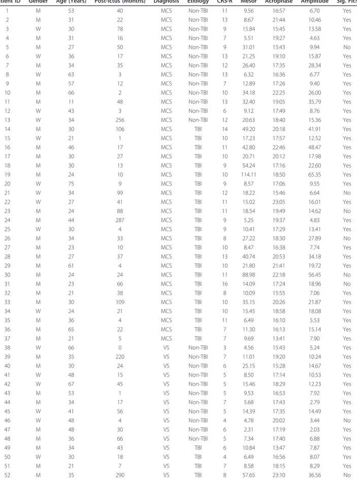
Patient ID Gender Age (Years) Post-Ictus (Months) Diagnosis Etiology CRS-R Mesor Acrophase Amplitude Sig. Fit? 1 M 53 40 MCS Non-TBI 11 9.56 16:57 6.70 Yes 2 M 31 22 MCS Non-TBI 13 8.67 21:44 10.46 Yes 3 W 30 78 MCS Non-TBI 9 15.84 15:45 13.58 Yes 4 M 31 16 MCS Non-TBI 7 5.51 19:27 4.63 Yes 5 M 27 50 MCS Non-TBI 9 31.01 15:43 9.94 No 6 W 36 17 MCS Non-TBI 13 21.25 19:10 15.87 Yes 7 M 34 35 MCS Non-TBI 12 26.40 17:35 28.34 Yes 8 W 63 3 MCS Non-TBI 13 6.32 16:36 6.77 Yes 9 M 57 12 MCS Non-TBI 7 12.89 17:26 9.40 Yes 10 M 66 2 MCS Non-TBI 10 34.18 22:25 26.00 Yes 11 M 11 48 MCS Non-TBI 13 32.40 19:05 35.79 Yes 12 W 43 3 MCS Non-TBI 6 9.12 17:49 8.76 Yes 13 W 34 256 MCS Non-TBI 12 20.63 18:40 15.36 Yes 14 M 30 106 MCS TBI 14 49.20 20:18 41.91 Yes 15 W 21 1 MCS TBI 10 17.23 17:57 12.52 Yes 16 M 46 17 MCS TBI 11 42.80 22:46 48.47 Yes 17 M 30 27 MCS TBI 10 20.71 20:12 17.98 Yes 18 M 30 13 MCS TBI 9 54.24 17:16 22.60 Yes 19 M 24 10 MCS TBI 10 114.11 18:50 65.35 Yes 20 W 75 9 MCS TBI 9 8.57 17:06 9.55 Yes 21 W 34 99 MCS TBI 12 18.22 15:46 6.64 No 22 W 27 41 MCS TBI 11 15.02 23:05 16.01 Yes 23 M 24 88 MCS TBI 11 18.54 19:49 14.62 No 24 M 44 287 MCS TBI 9 5.25 19:37 4.83 Yes 25 W 30 4 MCS TBI 9 10.41 17:29 13.41 Yes 26 M 34 33 MCS TBI 8 27.22 18:30 27.89 No 27 M 23 10 MCS TBI 10 8.47 16:38 7.74 Yes 28 M 27 37 MCS TBI 13 40.74 20:53 34.18 Yes 29 M 61 4 MCS TBI 10 21.80 21:41 19.72 Yes 30 M 24 24 MCS TBI 11 88.98 22:18 56.45 No 31 M 23 66 MCS TBI 16 14.09 17:24 18.96 No 32 M 21 38 MCS TBI 8 10.09 15:55 7.06 Yes 33 M 30 109 MCS TBI 10 35.15 20:26 21.87 Yes 34 W 24 21 MCS TBI 10 15.45 18:58 18.08 Yes 35 M 36 4 MCS TBI 11 6.49 16:10 5.53 Yes 36 M 65 22 MCS TBI 7 11.30 16:13 15.14 Yes 37 M 21 5 MCS TBI 7 9.69 13:41 7.90 Yes 38 W 66 0 VS Non-TBI 3 4.56 15:43 5.24 Yes 39 M 35 220 VS Non-TBI 7 11.01 19:20 10.24 Yes 40 M 30 24 VS Non-TBI 6 25.15 15:28 14.67 Yes 41 W 48 15 VS Non-TBI 5 8.50 17:14 10.53 Yes 42 W 67 45 VS Non-TBI 5 15.46 18:29 12.23 Yes 43 M 53 1 VS Non-TBI 5 9.53 16:53 7.92 Yes 44 M 34 17 VS Non-TBI 7 5.68 17:43 2.79 Yes 45 W 41 56 VS Non-TBI 5 14.39 17:35 14.49 Yes 46 W 48 4 VS Non-TBI 4 4.78 20:02 3.44 No 47 M 48 30 VS Non-TBI 6 2.31 17:19 2.03 Yes 48 M 36 66 VS Non-TBI 5 7.34 17:40 6.88 Yes 49 M 34 43 VS TBI 6 10.84 13:47 7.87 Yes 50 W 30 18 VS TBI 4 6.49 16:56 8.07 Yes 51 M 21 7 VS TBI 7 8.58 18:15 8.29 Yes 52 M 35 290 VS TBI 8 57.65 23:10 36.56 No
patients (mean age 35.7, SD 15.2; 24 TBI) contributed data to the study. There was no significant difference in the proportions of each etiology contributing to the VS and MCS groups. Two two-way ANOVAs with factors of diagnosis (VS, MCS) and etiology (TBI, non-TBI) conducted on age (in years) and months post-ictus revealed only a reliable main effect of etiology on age (F(1, 51) = 10.363,P <.01) reflecting the older average age of non-TBI patients. Informed consent was obtained from the patients’ surrogate decision makers. The Ethics Committee of the University and University Hospital of Liège provided ethical approval for the study.
Procedure
Actigraphy recordings were made with a Philips Acti-watch Spectrum (Philips Healthcare, Best, Brabant, The Netherlands) attached to the wrist with the highest range of movement (never the hemiplegic side) for a minimum of four days, sampled in one-minute epochs. In order to normalize across patients, only the first four days of actigraphy data were included in the analyses for those patients who were admitted for longer than four days. The first two hours of data were also excluded to avoid initial artifacts from attachment of the Actiwatch. Circadian rhythm analyses
Cosinor rhythmometry analyses [28] were performed on each patient’s dataset individually. This approach uses the least squares method to fit a sine wave with a period of 24 hours to the raw actigraphy data [11,12,28]. The rhyth-micity of the fit can be described by three parameters: the amplitude, the acrophase, and the mesor. The amplitude of the fit refers to half the distance between the peak and the trough of the fitted wave - in effect describing the amount of movement produced during periods of activity. The acrophase describes the point in the cycle at which activity is maximal. Finally, the mesor (an acronym for midline-estimating statistic of rhythm [28]) describes the rhythm-adjusted mean of the wave, or the value around which the fitted wave oscillates. For equidistant data sam-ples (as employed here), the mesor is equivalent to the arithmetic mean of the fitted wave, or the average amount of activity produced across the recording period. The goodness-of-fit of the wave - that is, the statistical reliabil-ity of the circadian rhythm - can also be determined by means of a zero-amplitude F-test [28].
In order to control for over-fitting of noise to the sine wave, this goodness-of-fitP-value was subsequently sub-jected to a permutation test. Specifically, a set of sine waves with periods ranging in 10-minute intervals from 6 hours to 48 hours were fit to the data (excluding rhythms between 19 and 28 hours since these are defined as circadian periods; see Introduction [11]). The P-values from these 200 zero-amplitude tests were then used to form a surrogate distribution to test the hypoth-esis that a 24-hour rhythm does not fit the data better than a non-circadian period. When the goodness-of-fit P-value associated with the 24-hour rhythm fell below the smallest 5% of surrogate P-values, the circadian rhythm was considered to be significant atP <.05.
Results
A total of 46 out of the whole group of 55 patients (84%) exhibited significant 24-hour rhythms in their actigraphy data after permutation testing. This proportion is signifi-cantly lower than the diagnostic expectation that all patients retain significant circadian rhythms (Fisher’s Exact Test,P <.01). When separated according to diagno-sis, 15/18 VS patients (83%) and 31/37 MCS patients (84%) returned circadian rhythms that passed this statisti-cal test. When separated according to etiology, 24/31 TBI patients (77%) and 22/24 non-TBI patients (92%) exhib-ited circadian rhythms. There was no significant effect of diagnosis or etiology on the proportions of patients exhi-biting circadian rhythms (Fisher’s Exact Tests, all P >.14). While age significantly differed across etiologies, it did not significantly correlate with any of the four rhythmi-city variables (mesor, amplitude, acrophase or goodness-of-fit, as indexed by the log-transformed zero-amplitude F-ratio).
VS versus MCS patients
Four one-way ANOVAs with diagnosis (VS, MCS) as the factor of interest revealed the main effects of mesor (F(1,54) = 4.441, P <.05), amplitude (F(1,54) = 6.819, P <.05), and goodness-of-fit (F(1,54) = 16.517, P <.001), but not acrophase. Together these reflect the greater average amount of movement across the four days (mesor), the greater amount of movement during peri-ods of activity (amplitude), and greater statistical relia-bility of the circadian rhythms (goodness-of-fit) of MCS patients relative to VS patients (see Figure 1).
Table 1 Demographics and circadian rhythm fits for all patients. (Continued)
53 M 21 8 VS TBI 6 9.95 19:22 6.45 Yes 54 M 13 1 VS TBI 6 5.78 21:24 3.12 No 55 M 25 15 VS TBI 5 10.02 16:19 9.05 Yes
The final column indicates whether the circadian rhythm fit was significant or not. MCS, Minimally Conscious State; TBI, Traumatic Brain Injury; VS, Vegetative State
Due to the high inter-correlations between these three significant rhythmicity variables (all absolute r > .33), all three variables were entered into a backward stepwise logistic regression in order to determine their relation-ships with diagnosis, over and above the effects of the other two variables. This regression retained only goodness-of-fit in the model as a significant predictor (Wald = 10.189, Beta (SE) = -2.043 (.640),P < .01), indi-cating significantly weaker circadian rhythms in VS patients relative to MCS patients, regardless of the amount of movement produced by these patients. Traumatic versus non-traumatic brain injury
Four one-way ANOVAs with etiology (TBI, non-TBI) as the factor of interest revealed the main effects of ampli-tude (F(1,54) = 4.299, P < .05) and goodness-of-fit (F(1,54) = 4.226), P < .05), but not mesor or acrophase. These effects reflect the greater amount of movement during periods of activity (amplitude) and the greater statistical reliability of the circadian rhythms (goodness-of-fit) of TBI patients relative to non-TBI patients.
As with the analyses across diagnosis, due to the high inter-correlations between the two significant rhythmi-city variables, both were entered into a backward step-wise logistic regression in order to determine their relationships with etiology, over and above the effect of the other variable. This regression retained neither vari-able as a significant predictor, likely due to the weak effects of etiology on these variables (contrast F-values above with those in the analyses across diagnosis). Relationship between rhythmicity and behavioral profile Four backward linear regressions were conducted on the four rhythmicity variables with the six subscales of the
CRS-R as predictors. Diagnosis was also included as a predictor since the higher scores on each subscale are also more likely to be associated with MCS and the lower scores with VS. The motor sub-scale was found to signifi-cantly predict mesor (F(1,54) = 7.792,P < .01, B(SE) = 6.174 (2.212),P < .01) and amplitude (F(2,54) = 6.178, P < .01, B(SE) = 3.462 (1.453),P < .05). The visual sub-scale was found to significantly predict acrophase (F(1,54) = 4.636,P < .05, B(SE) = -.108 (.050), P < .05), and both the visual and motor subscale together were found to predict goodness-of-fit (F(2,54) = 16.487,P < .001, B-visual(SE) = .208 (.055),P < .001, B-motor(SE) = .225 (.071), P < .005).
Discussion
On the basis of periodic eye-opening and eye-closure, patients in the VS and MCS are considered to have pre-served circadian sleep-wake rhythms [5,6]. However, by means of an indirect measure of sleep-wake rhythmicity - wrist actigraphy - we have shown that a significant proportion of these patients do not exhibit statistically reliable circadian sleep-wake rhythms. The observed variability across patients is consistent with previous smaller studies of circadian rhythmicity in VS and MCS (see Background), and is the first evidence from a large-scale study of sleep-wake cycling using the inexpensive and non-invasive method of wrist actigraphy.
While there was no significant difference in the propor-tion of patients exhibiting significant sleep-wake rhythms between VS and MCS patients, the goodness-of-fit of the circadian rhythms in the data of MCS patients were signif-icantly higher than those of the VS patients (see Figure 1). This result indicates that the circadian sleep-wake cycles of MCS patients were significantly more statistically reli-able than those of VS patients. Importantly, this remained
Figure 1 Actigraphy data from four representative patients. Each panel shows intensity of activity across each recording day. Red lines indicate the fit of the circadian rhythm. Note the periodic structure of the activity of the two patients with significant rhythms (left), compared with those without (right). Patients 19, 21, 52 and 45 are shown (clockwise from top-left). Log activity data smoothed across five minutes is plotted for clarity of visualization.
true when taking into account the morphology of the rhythm (that is, its mesor and amplitude), indicating that the effect of diagnosis on the statistical reliability of the circadian rhythms is not driven by simple differences in the amount that a patient moves, but rather reflects differ-ences in the circadian rhythmicity with which this move-ment occurs.
A master biological clock in the hypothalamic supra-chiasmatic nuclei (SCN) is considered to maintain the timing of circadian rhythms. The SCN in turn modu-lates the activity of the ascending reticular activating system (ARAS) - a circuit of subcortical nuclei responsi-ble for promoting wakefulness (see [29] for a review). One region of the ARAS - the central thalamus - is known to be crucial for the regulation of arousal and has been linked to the disorders of consciousness exhib-ited by VS and MCS patients [30]. Indeed, the extent of atrophy in this region of the thalamus has been asso-ciated with the degree of disability exhibited by these patients [31]. More broadly, greater thalamic atrophy has been observed in VS patients relative to MCS patients using in vivo diffusion tensor imaging [32] (DTI). The weaker circadian sleep-wake rhythms observed in the VS patients in the current study are, therefore, entirely consistent with these differential pat-terns of damage to the thalamus.
Etiology was also shown to have a small effect on the amount that patients moved during periods of activity (amplitude) and the statistical reliability of the circadian rhythm (goodness-of-fit). Similarly, Bekinschteinet al. [20] observed reliable circadian temperature rhythms in only TBI VS patients, but not in non-TBI patients, while De Weeret al. [27] detected sleep-wake activity changes in only TBI MCS patients. The primary neuropathology associated with TBI is diffuse axonal injury with relative preservation of the cortex, while non-TBI involves more widespread damage to the cortex and basal ganglia [33-39]. The greater impairment of circadian rhythms in non-TBI patients relative to TBI patients reported here is, therefore, consistent with the general patterns of neu-ropathology associated with the two etiologies. Indeed, mouse models of hypoxic brain injury have been shown to result in impaired sleep-wake cycling [40].
Significant relationships were also observed between the behavioral profiles of the patients - as indexed by their CRS-R sub-scales - and aspects of their circadian rhythmicities. A significant positive relationship was found between the motor subscale and the mesor and amplitude of the rhythm. The motor subscale of the CRS-R is scored from flaccid motor tone at its lowest, to object manipulation and automatic motor responses at its highest (before emergence from MCS). Since wrist movements were used to indirectly measure the circadian rhythms, it is unsurprising that greater amounts of
movement exhibited by patients across the recording per-iod (mesor, amplitude) are related to their overall abilities to produce motor output during behavioral assessments. This result suggests the need for caution in the use of actigraphy for assessing circadian sleep-wake rhythms since they rely on motor output for a rhythm to be detected. Nevertheless, our analyses have demonstrated that significant changes in the statistical reliability of the rhythms across diagnoses are not dependent on the amount of movement produced, suggesting that actigra-phy can be used to assess the statistical reliability of cir-cadian sleep-wake cycles, regardless of the degree of activity exhibited by the patients.
A combination of the visual and motor subscales signifi-cantly predicted the goodness-of-fit of the circadian rhythms. The visual subscale score describes behaviors from absent visual startle at its lowest, through fixation and pursuit, to object recognition at its highest. This rela-tionship is of particular interest since the master clock for circadian rhythms, the SCN, is itself timed by light inputs from the retina during the day, as well as melatonin from the pineal gland at night [29]. The more purposeful eye-movements of those scoring high on the visual subscale may allow for differing levels of light to reach the retina -perhaps through a greater ability to orient toward light or to maintain eye-opening for longer periods - and, conse-quently, result in a strengthening of the rhythm via the SCN. The predictive value of the visual subscale could, therefore, be considered to be consistent with our under-standing of the biological generators of sleep-wake rhyth-micity. This conclusion is necessarily speculative, however, since it is unclear whether high visual functioning is asso-ciated with a greater degree of orientation toward light or longer periods of eye-opening. Further investigation of this relationship will contribute to our understanding of the exogenous cues that drive circadian rhythms in VS/MCS patients.
A significant relationship was also found between the visual subscale of the CRS-R and the acrophase (time of maximal activity) of the rhythm, over and above the con-tribution of the other CRS-R subscales, or of the diagno-sis of the patient. The relationship with acrophase reflects the tendency for patients with higher visual func-tioning to be most active later in the afternoon than patients with lower visual functioning (Visual Score >= 1, Mean acrophase (SD) 18:20 (three hours); Visual Score = 0, Mean acrophase (SD) 17:20 (two hours)). Consistent with this observation, exposure to higher levels of light has been associated with later peaks of activity in institu-tionalized individuals [41,42]. However, the activity peaks of healthy individuals occur earlier in the day than those observed in the patients here, typically between approxi-mately 13:30p.m. and approxiapproxi-mately 16:00p.m. [24]. It has been observed that the levels of light experienced
by institutionalized patients are considerably lower than those of non-institutionalized individuals [42,43], and since the patients in the current study were residing on a hospital ward during the recording period, it is likely they were exposed to abnormally fluctuating levels of light compared with healthy individuals. Unfortunately, we were unable to record light levels alongside actigraphy; however, future studies investigating their contribution to the timing of activity of VS and MCS patients will be invaluable.
Since we inferred the circadian rhythms of patients from wrist actigraphy, it is likely that the recordings contain some levels of exogenous activity, perhaps from nurses moving the patient from bed to chair. Since these patients were all admitted to the same ward of the University Hospital of Liège as part of the same research protocol, they all received equivalent levels of care and were involved in the same assessments throughout the day - for example, behavioral tests, PET and MRI. As a result, the potential exogenous noise in the data would then be equally distributed across all patients. Our con-clusions regarding the effects of diagnosis, etiology and behavioral profile on sleep-wake cycles, therefore, would remain valid despite this potential confound. The use of simultaneous video-recordings would allow for the exclusion of activity that is generated exogenously and would further validate our findings.
Some prescribed medications may also have an effect on actigraphy-detected circadian rhythms. For example, treatment for spasticity (for example, with baclofen) is common in VS/MCS patients and may increase the amount of movement that will be detected with actigra-phy, while psychoactive medications (for example, amantadine) may also serve to exogenously modulate a patient’s level of arousal. Caution in this regard is not limited to actigraphy, however, since psychoactive medi-cations will also alter the resting EEG of a patient, thereby modulating the level of wakefulness that will be inferred from polysomnography. Due to differences in the wishes of families and physicians, a wide variety of medications are prescribed to VS and MCS patients (see Table 2 for details). As a result, it is not possible to sta-tistically control for each of these drugs individually, nor for their many interactions. Nevertheless, there is no reason to believe that prescribed medications would sys-tematically differ between VS and MCS groups due to the paucity of treatment recommendations for all patients with disorders of consciousness ([44]). Future controlled clinical trials are needed in order to provide insights into the effects of specific medications not only on circadian rhythmicity, but also on VS/MCS patient outcome in general.
A final caveat is that the apparent absence of reliable circadian rhythms in some of our patients may be a
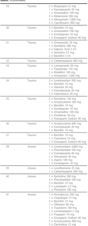
Table 2 Patient etiology and prescribed daily medications.
Patient ID Specific etiology Daily medication dosage 1 Anoxia 1 × Clonazepam 2 mg 1 × Phenytoin 100 mg 1 × Clopidogrel 75 mg 1 × Acetylcysteine 600 mg 3 × Baclofen 25 mg 3 × Levetiracetam 500 mg 2 Anoxia 1 × Amantadine 100 mg 2 × Clonidine 150 mg 1 × Bisopropol 2.5 mg 1 × Paroxetine 20 mg 1 × Tetrazepam 50 mg 1 × Acetylcysteine 600 mg 3 Anoxia 2 × Lamotrigine 50 mg 1 × Levetiracetam 1,000 mg 3 × Diazepam 5 mg 3 × Baclofen 25 mg 1 × Esomeprazole 20 mg 1 × Domperidome 10 mg 4 Anoxia 1 × Vancomycin 2,000 mg 4 × Piperacillin 4,000 mg 4 × Amikacin 1,000 mg 1 × Enoxaparin Sodium 50 mg 3 × Paracetamol 1,000 mg 1 × Diazepam 10 mg 3 × Baclofen 15 mg 1 × Acetylcysteine 600 mg 5 Anoxia 3 × Valproic Acid 40 ml
3 × Diazepam 10 mg 2 × Terbutaline 5 mg 3 × Baclofen 10 mg 3 × Dantrolene 25 mg 1 × Enoxaparin Sodium 40 mg 2 × Fluconazole 50 mg 1 × Aspirin 160 mg 6 Anoxia 1 × Amantadine 100 mg 1 × Zolpidem 10 mg 1 × Esomeprazole 20 mg 1 × Trihexyphenidyl 4 mg 7 Tumor/Hemorrhage 2 × Lamotrigine 100 mg 1 × Aspirin 80 mg 1 × Omeprazole 20 mg 1 × Escitalopram 10 mg 8 Anoxia 1 × Aspirin 100 mg 3 × Tizanidine 4 mg 1 × Enoxaparin Sodium 40 mg 1 × Omeprazole 20 mg 9 Anoxia 1 × Esomeprazole 20 mg 2 × Valproic Acid 500 mg 10 Anoxia 1 × Esomeprazole 20 mg 3 × Valproic Acid 2 ml 2 × Levetiracetam 7.5 ml 1 × Enoxaparin Sodium 40 mg 11 Anoxia 3 × Baclofen 10 mg 1 × Omeprazole 10 mg 4 × Domperidone 1 mg 2 × Clonazepam 1 mg 12 Aneurysm 1 × Enoxaparin Sodium 40 mg 13 Anoxia 2 × Carbamazepine 200 mg 14 Trauma 1 × Valproic Acid 500 mg
Table 2 Patient etiology and prescribed daily medica-tions. (Continued)
15 Trauma 1 × Phenobarbital 100 mg 3 × Baclofen 10 mg 16 Trauma 1 × Valproic Acid 500 mg
1 × Bisoprolol 5 mg 3 × Piracetam 1,200 mg 3 × Baclofen 25 mg 17 Trauma 1 × Omeprazole 20 mg 2 × Flecainide 100 mg 2 × Levetiracetam 10 ml 3 × Baclofen 10 mg 2 × Sodium Valproate 600 ml 1 × Clonazepam 2 mg 18 Trauma 1 × Escitalopram 10 mg 4 × Alprazolam 250 mg 1 × Trazodone 100 mg 1 × Prothipendyl 80 mg 19 Trauma 2 × Baclofen 10 mg 1 × Paracetamol 500 mg 1 × Esomeprazole 40 mg 2 × Levetiracetam 7.5 ml 20 Trauma 1 × Bisopropol 2.5 mg 1 × Pantoprazole 20 mg 2 × Tizanidine 2 mg 1 × Enoxaparin Sodium 40 mg 21 Trauma 2 × Ranitidine 150 mg 3 × Baclofen 25 mg 1 × Enoxaparin Sodium 40 mg 22 Trauma 3 × Baclofen 25 mg 2 × Tizanidine 4 mg 1 × Enoxaparin Sodium 20 mg 1 × Amantadine 50 mg 23 Trauma 3 × Baclofen 25 mg 3 × Domperidome 10 mg 2 × Clonazepam 2.5 mg 1 × Promethazine 16 mg 24 Trauma 1 × Amantadine 100 mg 2 × Baclofen 10 mg 1 × Esomeprazole 20 mg 1 × Tizanidine 4 mg 25 Trauma 2 × Valproic Acid 7.5 ml
1 × Lamotrigine 25 mg 3 × Baclofen 10 mg 2 × Esomeprazole 20 mg 1 × Enoxaparin Sodium 20 mg 26 Trauma 1 × Esomeprazole 40 mg 3 × Clonazepam 2 mg 3 × Paracetamol 1,000 mg 2 × Levetiracetam 500 mg 3 × Benserazide 250 mg 27 Trauma 1 × Acetylcysteine 600 mg 1 × Esomeprazole 20 mg 1 × Baclofen 25 mg 1 × Atenolol 50 mg 1 × Enoxaparin Sodium 40 mg 1 × Glycopyrrolate 10 mg 28 Trauma 1 × Amantadine 100 mg 1 × Paroxetine 20 mg 3 × Domperidone 10 mg 1 × Esomeprazole 20 mg
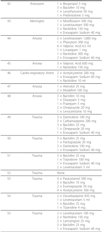
Table 2 Patient etiology and prescribed daily medica-tions. (Continued) 29 Trauma 2 × Bisopropol 2.5 mg 1 × Esomeprazole 20 mg 1 × Amantadine 100 mg 3 × Metamizole 500 mg 3 × Meropenem 1,000 mg 3 × Ciprofloxacin 400 mg 30 Trauma 3 × Baclofen 25 mg 1 × Amantadine 100 mg 1 × Escitalopram 10 mg 1 × Enoxaparin Sodium 40 mg 31 Trauma 1 × Omeprazole 20 mg 1 × Ranitidine 300 mg 3 × Valproic Acid 2 ml 1 × Sertraline 2.5 mg 2 × Baclofen 5 ml 32 Trauma 2 × Carbamazepine 400 mg 33 Trauma 1 × Lansoprazole 30 mg 1 × Topiramate 150 mg 2 × Modafinil 100 mg 1 × Aniracetam 1,500 mg 34 Trauma 3 × Levetiracetam 500 mg 3 × Baclofen 10 mg 1 × Atenolol 50 mg 1 × Esomeprazole 20 mg 3 × Indometacin 50 mg 35 Trauma 2 × Ranitidine 150 mg 1 × Acetylcysteine 200 mg 3 × Baclofen 10 mg 1 × Escitalopram 10 mg 1 × Amantadine 100 mg 2 × Diclofenac 50 mg 1 × Enoxaparin Sodium 40 mg 36 Trauma 1 × Acetylcysteine 600 mg 1 × Esomeprazole 20 mg 1 × Baclofen 10 mg 37 Trauma 1 × Baclofen 10 mg 1 × Trazodone 25 mg 1 × Enoxaparin Sodium 40 mg 38 Anoxia 1 × Levetiracetam 3,000 mg 1 × Phenobarbital 100 mg 1 × Esomeprazole 40 mg 1 × Simvastatin 40 mg 1 × Aspirin 100 mg 1 × Escitalopram 10 mg 39 Anoxia 1 × Levothyroxine 25 mg 1 × Carbamazepine 200 mg 40 Anoxia 1 × Ranitidine 300 mg 3 × Phenobarbital 100 mg 3 × Baclofen 25 mg 5 × Lorazepam 2.5 mg 1 × Phenytoin 100 mg 41 Anoxia 1 × Amiodarone 200 mg 1 × Clopidogrel 75 mg 3 × Baclofen 25 mg 2 × Diltiazem 60 mg 1 × Trazodone 100 mg 1 × Lormetazepam 2 mg 2 × Prazepam 10 mg 1 × Enoxaparin Sodium 40 mg 2 × Acetylcysteine 400 mg 3 × Dantrolene 25 mg
result of a lack of sensitivity of the actigraphy method, rather than the true absence of those rhythms. While wrist actigraphy has been validated for sleep assessment in patients with C5 to C7 tetraplegia [23], these patients are nevertheless capable of small but purposeful wrist
movements. Patients in the VS, however, are by defini-tion unable to produce purposeful movements, although spontaneous movements are common. Similarly, due to the heterogeneity of brain injuries of these patients, it is not clear whether the presence of actigraphy-detected waking is necessarily indicative of concurrent cerebral waking. Future validation of the relationship between polysomnography and actigraphy measures of sleeping and waking in VS and MCS patients is needed in order to fully characterize the nature of their circadian rhythms.
Conclusions
Our analyses indicate a greater impairment of circadian sleep-wake cycling in patients in the VS compared with those in the MCS, and in those with non-TBI compared with TBI. The significant differences observed between VS and MCS patients support the conclusion that these are diagnostically distinct entities. However, they also suggest that despite periods of closure and eye-opening, sleep-wake cycles are not necessarily present despite the clinical criteria for these conditions [5-7]. Wrist actigraphy is considerably less expensive and less invasive than other forms of sleep-wake monitoring and may, therefore, provide a reliable means of determining the extent to which these cycles are preserved in indivi-dual patients. These recordings could also allow clini-cians and researchers to identify the time of day in which a patient is most active, in order to schedule behavioral and/or neuroimaging assessments for a time that maximizes the likelihood of detecting an appropri-ate response (see [26]). Future validation of the relation-ship between actigraphy and polysomnography measures of sleeping/waking in VS and MCS patients will allow for a more complete understanding of the physiological nature of these circadian rhythms. Follow-up studies will also determine the prognostic utility of wrist acti-graphy for VS and MCS patients.
Abbreviations
ARAS: ascending reticular activating system; CRS-R: Coma Recovery Scale Revised; DTI: diffusion tensor imaging; EEG: electroencephalography; MCS: Minimally Conscious State; MRI: magnetic resonance imaging; TBI: non-traumatic brain injuries; PET: positron emission tomography; SCN: suprachiasmatic nuclei; TBI: traumatic brain injuries; UWS: Unresponsive Wakefulness Syndrome; VS: Vegetative State.
Authors’ contributions
DC designed and conducted the analyses and wrote the manuscript. AT, AD, MAB, OG and AV collected all data and contributed to the final manuscript. JCN contributed to the analyses and the final manuscript. TAB, AMO and SL provided conceptual input and contributed to the final manuscript. All authors approved the final version of the manuscript. Competing interests
The authors declare that they have no competing interests.
Table 2 Patient etiology and prescribed daily medica-tions. (Continued) 42 Aneurysm 1 × Bisopropol 5 mg 3 × Baclofen 10 mg 1 × Levothyroxine 50 mg 1 × Prednisolone 5 mg 43 Meningitis 1 × Moxifloxacin 500 mg 1 × Levetiracetam 500 mg 2 × Ranitidine 150 mg 1 × Enoxaparin Sodium 40 mg 44 Anoxia 2 × Levetiracetam 1,000 mg 1 × Phenytoin 500 mg 6 × Valproic Acid 6.5 ml 1 × Lorazepam 1 mg 1 × Ranitidine 300 mg 2 × Enoxaparin Sodium 60 mg 45 Anoxia 3 × Valproic Acid 600 mg
1 × Ranitidine 150 mg 46 Cardio-respiratory Arrest 2 × Acetylcysteine 200 mg
1 × Enoxaparin Sodium 60 mg 1 × Ranitidine 10 ml 47 Anoxia 1 × Atenolol 25 mg 2 × Modafinil 100 mg 48 Anoxia 3 × Baclofen 10 mg 1 × Diazepam 5 mg 1 × Prazepam 5 mg 2 × Omeprazole 20 mg 1 × Levocetirizine 10 mg 49 Trauma 3 × Dantrolene 100 mg 3 × Carbamazepine 200 mg 3 × Baclofen 25 mg 1 × Omeprazole 20 mg 1 × Enoxaparin Sodium 40 mg 50 Trauma 3 × Baclofen 25 mg 1 × Pantoprazole 20 mg 3 × Dantrolene 100 mg 1 × Enoxaparin Sodium 40 mg 51 Trauma 3 × Baclofen 25 mg 1 × Trazodone 100 mg 1 × Enoxaparin Sodium 40 mg 2 × Levetiracetam 5 ml 52 Trauma None 53 Trauma 4 × Paracetamol 500 mg 3 × Baclofen 10 mg 2 × Esomeprazole 20 mg 4 × Acetylcysteine 300 mg 54 Trauma 2 × Oxcarbazepine 450 mg 1 × Levetiracetam 5 ml 1 × Baclofen 25 mg 1 × Tizanidine 4 mg 55 Trauma 2 × Levetiracetam 100 mg 2 × Ranitidine 150 mg 2 × Lamotrigine 25 mg 3 × Baclofen 25 mg 1 × Enoxaparin Sodium 40 mg
Acknowledgements
This research was supported by generous funding from the European Commission, Fonds de la Recherche Scientifique, the Mind Science Foundation, the Belgian French-Speaking Community Concerted Research Action, University Hospital of Liège, University of Liège, the Wellcome Trust, the Canada Excellence Research Chair Programme (CERC), and the Canadian Institutes of Health Research (CIHR).
Author details
1
Brain and Mind Institute, University of Western Ontario, 1151 Richmond Street, London, ON N6A 3K7, Canada.2Coma Science Group: Department of
Neurology and Cyclotron Research Centre, University Hospital and University of Liège, Allée du 6 août n° 8, Sart Tilman B30 - 4000, Belgium.3Cognition
and Brain Sciences Unit, Medical Research Council, 15 Chaucer Road, Cambridge, CB2 7EF, UK.
Received: 23 August 2012 Accepted: 24 January 2013 Published: 24 January 2013
References
1. Laureys S, Celesia GG, Cohadon F, Lavrijsen J, Leon-Carrion J, Sannita WG, Sazbon L, Schmutzhard E, von Wild KR, Zeman A, Dolce G: Unresponsive wakefulness syndrome: a new name for the vegetative state or apallic syndrome. BMC Med 2010, 8:68.
2. Laureys S: The neural correlate of (un)awareness: lessons from the vegetative state. Trends Cogn Sci 2005, 9:556-559.
3. Jennett B, Plum F: Persistent vegetative state after brain damage. A syndrome in search of a name. Lancet 1972, 1:734-737.
4. Giacino JT, Kalmar K, Whyte J: The JFK Coma Recovery Scale-Revised: measurement characteristics and diagnostic utility. Arch Phys Med Rehabil 2004, 85:2020-2029.
5. The Multi-Society Task Force on PVS: Medical aspects of the persistent vegetative state (1). N Engl J Med 1994, 330:1499-1508.
6. Royal College of Physicians: The permanent vegetative state. Review by a working group convened by the Royal College of Physicians and endorsed by the Conference of Medical Royal Colleges and their faculties of the United Kingdom. J R Coll Physicians Lond 1996, 30:119-121, Updated 2003.
7. Giacino JT, Ashwal S, Childs N, Cranford R, Jennett B, Katz DI, Kelly JP, Rosenberg JH, Whyte J, Zafonte RD, Zasler ND: The minimally conscious state: definition and diagnostic criteria. Neurology 2002, 58:349-353. 8. Owen AM, Coleman MR, Boly M, Davis MH, Laureys S, Pickard JD: Detecting
awareness in the vegetative state. Science 2006, 313:1402. 9. Monti MM, Vanhaudenhuyse A, Coleman MR, Boly M, Pickard JD,
Tshibanda L, Owen AM, Laureys S: Willful modulation of brain activity in disorders of consciousness. N Engl J Med 2010, 362:579-589.
10. Cruse D, Chennu S, Chatelle C, Bekinschtein TA, Fernández-Espejo D, Pickard JD, Laureys S, Owen AM: Bedside detection of awareness in the vegetative state - a cohort study. Lancet 2011, 378:2088-2094. 11. Refinetti R: Circadian Physiology Boca Raton, FL: Taylor & Francis Group;
2006.
12. Ancoli-Israel S, Cole R, Alessi C, Chambers M, Moorcroft W, Pollak CP: The role of actigraphy in the study of sleep and circadian rhythms. Sleep 2003, 26:342-392.
13. Landsness E, Bruno MA, Noirhomme Q, Riedner B, Gosseries O, Schnakers C, Massimini M, Laureys S, Tononi G, Boly M: Electrophysiological correlates of behavioural changes in vigilance in vegetative state and minimally conscious state. Brain 2011, 134:2222-2232.
14. Isono M, Wakabayashi Y, Fujiki MM, Kamida T, Kobayashi H: Sleep cycle in patients in a state of permanent unconsciousness. Brain Inj 2002, 16:705-712.
15. Fukudome Y, Abe I, Saku Y, Matsumura K, Sadoshima S, Utunomiya H, Fujishima M: Circadian blood pressure in patients in a persistent vegetative state. Am J Physiol 1996, 270:R1109-1114.
16. Pattoneri P, Tirabassi G, Pela G, Astorri E, Mazzucchi A, Borghetti A: Circadian blood pressure and heart rate changes in patients in a persistent vegetative state after traumatic brain injury. J Clin Hypertens (Greenwich) 2005, 7:734-739.
17. Guan J, You C, Liu Y, Zhang R, Wang Z: Characteristics of infradian and circadian rhythms in the persistent vegetative state. J Int Med Res 2011, 39:2281-2287.
18. Oksenberg A, Arons E, Sazbon L, Mizrahi A, Radwan H: Sleep-related erections in vegetative state patients. Sleep 2000, 23:953-957.
19. Candelieri A, Cortese MD, Dolce G, Riganello F, Sannita WG: Visual pursuit: within-day variability in the severe disorder of consciousness. J Neurotrauma 2011, 28:2013-2017.
20. Bekinschtein TA, Golombek DA, Simonetta SH, Coleman MR, Manes FF: Circadian rhythms in the vegetative state. Brain Inj 2009, 23:915-919. 21. de Souza L, Benedito-Silva AA, Pires ML, Poyares D, Tufik S, Calil HM:
Further validation of actigraphy for sleep studies. Sleep 2003, 26:81-85. 22. Berger AM, Wielgus KK, Young-McCaughan S, Fischer P, Farr L, Lee KA:
Methodological challenges when using actigraphy in research. J Pain Symptom Manage 2008, 36:191-199.
23. Spivak E, Oksenberg A, Catz A: The feasibility of sleep assessment by actigraph in patients with tetraplegia. Spinal Cord 2007, 45:765-770. 24. Brown A, Smolensky M, D’Alonzo G, Redmond D, Conrad E, Hsi B: Circadian
rhythm in human activity objectively quantified by actigraphy. Prog Clin Biol Res 1990, 341A:77-83.
25. Girardin J-L, Kripke DF, Ancoli-Israel S, Klauber MR, Sepulveda RS, Mowen M-A, Assmus JD, Langer RD: Circadian sleep, illumination, and activity patterns in women: influences of aging and time reference. Physiol Behav 2000, 68:347-352.
26. Bekinschtein T, Cologan V, Dahmen B, Golombek D: You are only coming through in waves: wakefulness variability and assessment in patients with impaired consciousness. Prog Brain Res 2009, 177:171-189. 27. De Weer AS, Da Ros M, Berré J, Mélot C, Goldman S, Peigneux P:
Environmental influences on activity patterns in altered states of consciousness. Eur J Neurol 2011, 18:1432-1434.
28. Nelson W, Tong YL, Lee JK, Halberg F: Methods for cosinor-rhythmometry. Chronobiologia 1979, 6:305-323.
29. Saper CB, Scammell TE, Lu J: Hypothalamic regulation of sleep and circadian rhythms. Nature 2005, 437:1257-1263.
30. Schiff ND: Central thalamic contributions to arousal regulation and neurological disorders of consciousness. Ann N Y Acad Sci 2008, 1129:105-118.
31. Fernandez-Espejo D, Junque C, Bernabeu M, Roig-Rovira T, Vendrell P, Mercader JM: Reductions of thalamic volume and regional shape changes in the vegetative and the minimally conscious states. J Neurotrauma 2010, 27:1187-1193.
32. Fernández-Espejo D, Bekinschtein T, Monti MM, Pickard JD, Junque C, Coleman MR, Owen AM: Diffusion weighted imaging distinguishes the vegetative state from the minimally conscious state. NeuroImage 2011, 54:103-112.
33. Adams JH, Graham DI, Murray LS, Scott G: Diffuse axonal injury due to nonmissile head-injury in humans - an analysis of 45 cases. Ann Neurol 1982, 12:557-563.
34. Kinney HC, Samuels MA: Neuropathology of the persistent vegetative state. A review. J Neuropathol Exp Neurol 1994, 53:548-558.
35. Adams JH, Jennett B, McLellan DR, Murray LS, Graham DI: The neuropathology of the vegetative state after head injury. J Clin Pathol 1999, 52:804-806.
36. Jennett B, Adams JH, Murray LS, Graham DI: Neuropathology in vegetative and severely disabled patients after head injury. Neurology 2001, 56:486-490. 37. Adams JH, Duchen LW: Greenfield’s Neuropathology. 5 edition. New York,
New York: Oxford University Press; 1992.
38. Adams JH, Graham DI, Jennett B: The neuropathology of the vegetative state after an acute brain insult. Brain 2000, 123:1327-1338.
39. Kinney HC, Korein J, Panigrahy A, Dikkes P, Goode R: Neuropathological findings in the brain of Karen Ann Quinlan. The role of the thalamus in the persistent vegetative state. N Engl J Med 1994, 330:1469-1475. 40. Veasey SC, Davis CW, Fenik P, Zhan G, Hsu YJ, Pratico D, Gow A: Long-term
intermittent hypoxia in mice: protracted hypersomnolence with oxidative injury to sleep-wake brain regions. Sleep 2004, 27:194-201. 41. Shochat T, Martin J, Marler M, Ancoli-Israel S: Illumination levels in nursing
home patients: effects on sleep and activity rhythms. J Sleep Res 2000, 9:373-379.
42. Ancoli-Israel S, Martin JL, Kripke DF, Marler M, Klauber MR: Effect of light treatment on sleep and circadian rhythms in demented nursing home patients. J Am Geriatr Soc 2002, 50:282-289.
43. Campbell SS, Kripke DF, Gillin JC, Hrubovcak JC: Exposure to light in healthy elderly subjects and Alzheimer’s patients. Physiol Behav 1988, 42:141-144.
44. Giacino J, Whyte J: The vegetative and minimally conscious states: current knowledge and remaining questions. J Head Trauma Rehabil 2005, 20:30-50.
Pre-publication history
The pre-publication history for this paper can be accessed here: http://www.biomedcentral.com/1741-7015/11/18/prepub
doi:10.1186/1741-7015-11-18
Cite this article as: Cruse et al.: Actigraphy assessments of circadian sleep-wake cycles in the Vegetative and Minimally Conscious States. BMC Medicine 2013 11:18.
Submit your next manuscript to BioMed Central and take full advantage of:
• Convenient online submission
• Thorough peer review
• No space constraints or color figure charges
• Immediate publication on acceptance
• Inclusion in PubMed, CAS, Scopus and Google Scholar
• Research which is freely available for redistribution
Submit your manuscript at www.biomedcentral.com/submit