Université de Montréal
The impact of apple peel polyphenols on intestinal and
mitochondrial functions in experimental colitis
par
Pantea Rahmani Yeganeh
Département de Nutrition Faculté of Médecine
Mémoire présenté à la Faculté de Médecine en vue de l’obtention du grade de Maitrise
en Nutrition
Décembre, 2016
Université de Montréal
Faculté des études supérieures et postdoctorales
Ce mémoire est intitulé:
The impact of apple peel polyphenols on intestinal and mitochondrial functions in experimental colitis
Présenté par: Pantea Rahmani Yeganeh
a été évaluée par un jury composé des personnes suivantes : Dr Prevost Jantchou, Président rapporteur
Dr Emile Levy, Directeur de recherche Dr Pierre Haddad, Membre du jury
Résumé
Contexte:
L'inflammation et le stress oxydatif (OxS) participent à la pathogenèse de la colite ulcéreuse (CU). Nos résultats récents montrent que les polyphénols de la pelure de pomme (DAPP) jouent un rôle clé dans la prévention de la maladie.
Objectifs:
Évaluer les effets préventifs et curatifs du DAPP sur la CU et démontrer leur impact sur la dysfonction mitochondriale.
Méthode:
Une induction de l’inflammation intestinale a été effectuée chez des souris par administration du dextran sulfate sodium (DSS). Des doses de DAPP (200 et 400 mg/kg/j) ont été administrées par gavage pendant 10 jours afin d’évaluer les effets préventifs et curatifs, respectivement, sur l’Inflammation et le OxS au niveau intestinal ainsi que sur les fonctions mitochondriales.
Résultats:
Le DSS a provoqué une perte de poids, un raccourcissement du côlon, une augmentation du stress oxydant, niveaux de malondialdéhyde et une inflammation documentée par l’infiltration des cellules inflammatoires, la myéloperoxydase et les cytokines inflammatoires. D’autre part, le DSS a induit des désordres au niveau de la biogenèse (PGC1α) et des fonctions de la mitochondrie : diminution de l’ATP, altération des enzymes antioxydantes (SOD2 et GPX1), augmentation de l’apoptose (Bcl 2, Bax et Cytochrome C), et des défauts de l’intégrité de l’ADN (baisse d’OGG1). Cependant, le DAPP a amélioré significativement l’inflammation et le stress oxydant de l’intestin tout en corrigeant les aberrations mitochondriales.
Conclusions:
Les polyphénols ont la capacité d’agir sur le stress oxydant et le profil inflammatoire de l’intestin ainsi que sur le dysfonctionnement mitochondrial. Ils pourraient donc intervenir efficacement dans la CU.
Mots-clés : Stress oxydatif, Mitochondrie, Inflammation, maladies inflammatoires
Abstract
Background:
We have recently shown that dysregulation of redox-sensitive signaling pathways and oxidative damage to biological structures are major contributors to experimental ulcerative colitis. We also demonstrated the powerful anti-oxidant and anti-inflammatory actions of dietary apple peel polyphenols (DAPP) in the intestine.
Objectives:
As mitochondria are major sources and target of free radicals, as well as exhibit various important cellular functions, we evaluated their roles in intestinal colitis and their responses to DAPP.
Methods:
Induction of intestinal inflammation in C57BL6 mice was performed by administration of 3% dextran sulfate sodium (DSS). Two different doses of DAPP (200 and 400 mg/kg/day) were administered by gavage for 10 days (before and during DSS administration) to examine the preventive and curative effects, respectively, on inflammation and oxidative stress (OxS) in the intestine, and on mitochondrial functions.
Results:
DSS caused a significant weight loss, shortening of the colon, increased OxS (noted by lipid peroxidation), and raised inflammation (verified by infiltration of inflammatory cells, up-regulation of MPO, and elevated TNF-α and COX2 protein expression). Furthermore, DSS induced perturbations in mitochondrial biogenesis, as reflected by alterations of the transcription factor PGC1α and mitochondrial function characterized by diminished Adenosine-5'-Triphosphate (ATP) production, lowered antioxidant defense (GPx and SOD2), amplified apoptosis (as illustrated by the high expression of Cytochrome C and AIF), and defects in DNA integrity (high 8-OHdG). However, DAPP administration improved macroscopic parameters (e.g. weight loss, colon shortening) and reduced DSS-induced clinical signs. DAPP showed an evident capability of reducing inflammation (as noted by decreased
TNF-α, iNOS, COX-2 and AP-1) and OxS (as shown by reduced malondialdehyde, hydrogen peroxide levels and increased GPx) in DSS mice. Our findings also revealed that DAPP partially corrected mitochondrial dysfunction related to redox homeostasis, fatty acid β-oxidation, ATP synthesis, apoptosis and regulatory mitochondrial transcription factors (PGC1α, PPARγ and Nrf-2).
Conclusion:
DAPP have the ability to act on intestinal OxS, inflammation and mitochondrial dysfunction, thereby alleviating colitis progression via the modulation of cellular energy, OxS, antioxidant capacity, apoptosis and mtDNA integrity.
Keywords: Mitochondria, mitochondrial function and dysfunction, inflammatory bowel
disease, inflammation, Crohn's disease, experimental colitis, oxidative stress, polyphenols, prevention, treatment.
Table of contents
Résumé ... ii Abstract ... iii Table of contents ... v List of figures ... vii List of abbreviations ... viii Acknowledgments ... xi 1 INTRODUCTION ... 2 1.1 Introduction to Inflammatory Bowel Diseases ... 2 1.1.1 Pathophysiology and Epidemiology of Inflammatory Bowel Diseases ... 2 1.1.2. CD and UC: Pathology, signs and Symptoms ... 4 1.1.3. Manifestations of IBD ... 7 1.1.3.1. Intestinal dysfunction in intestinal inflammation ... 7 1.1.3.2. Markers of inflammation and OxS in IBD ... 9 1.2. Mitochondria ... 14 1.2.1. Mitochondrial structure ... 14 1.2.2. Mitochondrial function ... 16 1.2.2.1 Fatty Acid Beta-oxidation ... 17 1.2.2.2 Glycolysis, Krebs cycle, Electron Transport Chain Complexes (OXPHOS) and ATP production ... 19 1.2.2.3 Apoptosis regulation ... 20 1.2.2.4 ROS generation and detoxification ... 24 1.2.2.5 Ca2+ homeostasis ... 25 1.2.3. Mitochondrial dysfunction ... 251.2.3.2. Mitochondrial respiratory chain (ATP production) and β-oxidation in IBD ... 28
1.2.3.3. Oxidative stress and mitochondrial dysfunction in IBD ... 29
1.2.3.4. Additional mechanisms for apoptosis and mitochondrial dysfunction in IBD ... 31
1.2.3.5. Ca2+ homeostasis and mitochondrial dysfunction in IBD ... 33
1.3. Therapeutic intervention: antioxidants targeting mitochondrial dysfunction ... 34
1.3.2. Polyphenols ... 36 1.3.2.1. Structural diversity and dietary sources of polyphenols ... 36 1.3.2.2. Biological functions of polyphenols ... 38 1.3.2.3. Apple polyphenols ... 39 1.3.3. Apple polyphenols in intestinal inflammation ... 40 2 RESEARCH PROJECT ... 43 2.1. Hypothesis ... 43 2.2. Objectives ... 43 3 ARTICLE 1 ... 44 ARTICLE 2 ... 78 4 DISCUSSION ... 113 5 CONCLUSIONS ... 119 6 REFERENCES ... i
List of figures
IBD subclasses ... 2 Figure 1. The common pathogenic factors in IBD. ... 5 Figure 2. Risk factors involved in the pathogenesis of UC and CD. ... 6 Figure 3. Beneficial and harmful biological ROS pathways ... 10 Figure 4. Role of some markers of OxS and inflammation in IBD ... 12 Figure 5. Schematic mitochondrial structure………15 Figure 6. Fatty acid β-oxidation ... 18 Figure 7. Mitochondrial site and role of the RC complexes ... 20 Figure 8.Graphic picture of role of mitochondrial permeability transition pore (MPTP) opening in Figure 9. apoptosis…….. ... 22 The roles of pro- and anti-apoptotic factors in mitochondrial permeability and apoptosis ... 23 Figure 10. Intestinal homeostasis disorders in mitochondrial dysfunction ... 27 Figure 11. Schematic image of mitochondrial ROS’s roles in apoptosis ... 32 Figure 12. Schematic illustration of oxidants and antioxidants imbalance, oxidative damage, and chronic Figure 13. diseases……… ... 36 Classification of the polyphenols antioxidant related action ... 39 Figure 14. Schematic description of main reasons for treating IBD patients with polyphenols ... 41 Figure 15.
List of abbreviations
ACADL Acyl-CoA dehydrogenase
AIF Apoptosis-inducing factor ATP Adenosine-5'-Triphosphate Bcl2 B-cell lymphoma 2 BAX Bcl-2-associated X Ca2+ Calcium CAT Catalase CD Crohn’s Disease COX Cyclooxygenase CRC Colorectal Cancer Cyt Cytochrome
DNA Deoxyribonucleic Acid
DSS Dextran Sulfate Sodium
ER Endoplasmic Reticulum
ETC Electron Transport Chain FA Fatty Acids
IBD Inflammatory Bowel Disease IECs Intestinal Epithelial Cells IL Interleukin
iNOS Inducible Nitric Oxide Synthase GI Gastrointestinal
GPx Glutathione Peroxidase GR Glutathione Reductase GSH Glutathione
MAMs Mitochondria-associated membranes MAPKs Mitogen-activated protein kinases MDA Malondialdehyde
Mt Mitochondria
mtTFA Mitochondrial transcription factor A NF-𝜅B Nuclear factor-kappa B
Nrf2 Nuclear factor erythroid 2 related factor OxS Oxidative Stress
8-OHdG 8-hydroxy-deoxy-guanosine OGG1 8-oxo DNA Glycosylate OXPHOS Oxidative Phosphorylation
PGC-1α Peroxisome Proliferation Activator Receptor γ-coactivator 1α PTP Permeability transition pore
ROS Reactive Oxygen Species SCFA Short chain fatty acid SOD Superoxide Dismutase
TNF-α Tumor Necrosis Factor- Alpha UC Ulcerative colitis
Acknowledgments
I would like to thank my Director, Dr. Emile Levy, for his exceptional support and motivation throughout my master research. While always encouraging me to work my hardest, he showed a constant commitment to providing me with the best up-to-date training opportunities and every chance for success.
I have been fortunate to work with Carole Garofalo. The work in this thesis could not have been done without her help. I would like to thank her for her essential contributions and guidance to see the work done.
I would also like to thank Ms. Spahis Schohraya (Zola) for her support, helpful discussions, and preparation related to manuscripts and thesis. I gratefully acknowledge her involvement of time and technical supports.
I would especially like to thank my dear colleague Jade Leady for proofreading and checking the language of the Introduction and the Discussion of my thesis and her support.
I am especially grateful to Alain Sane for his crucial contributions in terms of providing me with scientific guidance, experimental assistance and for his encouragement.
I would especially like to thank my colleague, Marie-laure Kleme Amani for being available for experimental guidance and scientific discussions.
I would also like to thank Dr. Levy’s Lab team (Maryse Fournier, Sophia Morel, Nickolas Auclair, Veronique Belanger and Marc-Andre Lecours) for their professional support and assistance.
Finally, I would like to thank my family for their constant support and encouragement. I especially would like to thank my lovely mother for teaching me how to live, work hard and fight frustrations, my amazing brother, Sina Rahmani Yeganeh and sister, Betsabeh Rahmani Yeganeh and above all my caring and loving friend Pantea Dadyar for her support during all stages of this master.
1 INTRODUCTION
1.1 Introduction to Inflammatory Bowel Diseases
1.1.1 Pathophysiology and Epidemiology of Inflammatory
Bowel Diseases
Inflammatory bowel disease (IBD) is one of the common types of intestinal inflammatory diseases. It describes a group of disorders that attack the intestine and cause intestinal cells to become swollen and inflamed. In other words, IBD is a series of immunoinflammatory disorders of the gastrointestinal (GI) tract, which mainly include two main disorders: Crohn’s disease (CD) and ulcerative colitis (UC). Additional rare forms of IBD are observed such as collagenous colitis and intractable colitis [1, 2].
IBD subclasses. Taken from Rubin et al. [1] Figure 1.
While the two main forms of IBD, UC and CD, are not as common as heart disease or cancer, they cause significant morbidity and financial burden on the health care system. Studies have reported that the prevalence of IBD in adults in the United States is more than 200 cases per 100,000 with the total number of 1 to 1.5 million IBD patients [3, 4]. The prevalence of IBD in the past few years in Canada has been 150-250 cases per 100,000, and hospitalization due to UC and CD has been evaluated at 50.6 and 50.1 cases per 100,000, respectively [5, 6].
Recently, research has shown that IBD is more prevalent in developed countries. Lifestyle related factors such as nutrition, exposure to pollution and chemicals, low physical activity, and decreased exposure to sunlight are associated with the increased prevalence of IBD [7]. Other studies revealed that the risk of succumbing to recently diagnosed severe and long term cases of IBD is increasing. Moreover, risk of colorectal cancer (CRC) has recently raised to 20% per year compared to 0.5% reported previously, especially in cases of UC [8].
IBD is a multifactorial chronic, recurring and inflammatory disease of the GI tract commonly presenting clinical symptoms of diarrhea, bloody stools, abdominal pain and weight loss, and the development of an inflammatory response [9].
In animal models of colitis, the same common macroscopic symptoms are monitored. This includes body weight loss, fecal bleeding, stool consistency (e.g., diarrhea, liquid), shortening of the colon, and change of intestinal microbiota [10, 11]. In addition, experimental study by Islam et al. reported the severe histological changes of murine intestinal tissues with DSS-induced colitis, which are similar to human colitis, including
augmented thickness of the muscle layer, lymphocyte infiltration, mucosal erosion, loss of crypt structure, ulcer formation and also serious intestinal microbiota modifications [12].
Genetics and possible genetic heterogeneity play a fundamental role in IBD but the precise nature of the genetic implication is extremely complex, and it is probable that the cooperation of certain genes acts as a responsible modulator. Recently, the genetic study by Nell et al. has demonstrated the importance of environmental factors and specifically the gut microbiota in the occurrence of CD and UC [13].
1.1.2. CD and UC: Pathology, signs and Symptoms
Crohn’s and colitis are known to cause major health problems in the human body and are characterized by abnormal chronic inflammation and immune-mediated injury to the GI tract. Although the main triggers of IBD disorders are not clearly identified, recent studies have reported that, generally, the complex relations between genetic determinants (e.g. IBD1, IBD2…), unfavourable environmental factors (smoking, diet, stress, drug, social status, education), dysregulated immune response and qualitatively and quantitatively unusual gut microbiota could be the main determinants in the pathogenesis of CD and UC as shown in Fig. 2.
The common pathogenic factors in IBD. Taken from de Souza Figure 2.
et al. [15]. This image shows that IBD can develop due to the body’s distorted and changed immune response, changes in intestinal microbiota, a complicated interplay of genetic predisposition and environmental factors.
High levels of oxidative stress (OxS), mitochondrial DNA mutations and dysfunction in mitochondrial calcium (Ca2+) pathway can fundamentally cause or affect intestinal inflammation directly or indirectly, thereby increasing the risk of IBD as briefly described in Fig.3.
IBD
Environ-mental
Trigger
Genetic
Susceptibility
Luminal
Microbial
Infection
Immune
Response
Risk factors involved in the pathogenesis of UC and CD. Figure 3.
CD and UC have been studied in parallel, as their common symptoms, structural damage and treatments are very similar. However, it is known that they are characterized by two separate pathophysiological patherns.
In CD, all parts of the GI tract, from the mouth to anus could be affected, but the disease generally affects the distal part of the small intestine or ileum. On the other hand, UC only causes colonic inflammation and affects part or all of the colon and the rectum [1]. Generally, both CD and UC patients present similar clinical symptoms (e.g. diarrhea, abdominal pain, GI bleeding, and weight loss). Duerr et al. reported that genetic risk factors play a major role in the prevalence of IBD and risk of IBD development is increased by 5-30% in families of affected individuals. Interestingly, Tysk et al. found that the risk of phenotypic concordance in monozygotic twins with CD renged from 50-75% in the second twin. However, they did not detect the same relation in UC patients [16, 17]. Duerr et al. also discovered that patients with CD have more family history of
this disorder compared to UC patients. In both studies, the authors suggested that other risk factors, including environmental factors, play an important role in UC and CD pathogenesis.
In addition, many researchers have looked of the probability of CRC, a side effect of long-term chronic inflammation, in UC and CD patients. They reported that probability of CRC in UC patients increased from 2% after 10 years of disease to 18% after 30 years compared to patients with CD that showed 2.9% risk of CRC at 10 years, which increased to 8.3% after 30 years of disease [18-20].
1.1.3. Manifestations of IBD
1.1.3.1. Intestinal dysfunction in intestinal inflammation
Recent studies reported multiple pathogenic factors that are involved in the prevalence of IBD for both CD and UC. Some of these pathogenic factors are:
1. Qualitatively and quantitatively abnormal gut microbiota 2. Largely dysregulated immune response.
Chassaing’s study shows that such qualitative and quantitative changes in composition of the gut microbiota occur at the microbiological level in both CD and UC patients by reducing the complexity of commensal bacteria [21]. Former studies indicated the presence of high levels of immune reactivity against microbial antigens in patients with IBD. Murdoch et al. reported serum antibodies against microbial antigens in CD patients [22]. In addition, Andoh and his team documented the existence of high levels of
abnormal gut microbiota in UC patients compared to CD patients [23]. Additionally, Halstensen’s results pointed out that an antibody-mediated immune (autoimmune) response in UC patients could act as a pathogenic mechanism for epithelial impairment and accompanying inflammation [24].
The mucous layer is known to be the first line of protection against injurious agents in the gut lumen in healthy cells. In both forms of IBD, intestinal mucosa and intestinal epithelial cells (IECs) could be injured, resulting in intestinal permeability changes. IECs could be the main target for injury in IBD patients and these alterations may decrease humoral protective properties [25]. The study of Bjarnason et al. showed that intestinal permeability in patients with active CD is increased, causing unusual infiltration (by measuring the urine excretion of 51-chromium-labeled ethylenediaminetetraacetate) [26]. Importantly, intestinal permeability disorder is directly related to higher risk of acute cases of IBD [27]. For example, Pineton et al. demonstrated that infiltration of both neutrophils and macrophages into the intestinal mucosa is present in patients with chronic intestinal inflammation and disorders of IECs [28]. Moreover, antigen production is important for immunoregulation of healthy intestinal cells and different reports suggested the existence of possible abnormalities of IEC antigen-presenting capacity in IBD. Turner reported a direct relationship between intestinal permeability and IBD immunopathogenesis [29]. As adequate balance in the regulation of intestinal mucosal barrier is essential for normal cell function in the absorption of nutrients and waste secretion, any disruption of the mucosal barrier function can change the absorption of nutrients and microbial products that may lead to inflammation, as is the case in IBD. In addition, reactive oxygen species (ROS) produced by these activated inflammatory cells
induce oxidative damage in intestinal mucosa creating an imbalance in the redox status, which exacerbates inflammation in IBD [30].
1.1.3.2. Markers of inflammation and OxS in IBD
As mentioned previously, unusual infiltration of macrophages into the intestinal mucosa and disorders of IECs triggered by the increasing intestinal epithelial permeability is present in intestinal chronic inflammation (IBD) [31]. ROS generated by these activated inflammatory cells could cause oxidative damage in intestinal mucosa. In addition, some studies present OxS as the most probable cause for the pathogenesis of human IBD [32, 33] OxS is an imbalance between anti-oxidant defense and the production of ROS such as superoxide, hydrogen peroxide, hydroxyl radical and peroxynitrite disturb the redox homeostasis. Intermediate levels of ROS cause apoptosis, while imbalance due to overproduction of ROS and free radicals can decrease antioxidant defense causing necrosis and is harmful to cellular biological processes and tissue functions [34].
Erichsen et al. and Rezaie et al. revealed that ROS levels vary under different conditions that could act by activating biological mediators (pro-oxidants or antioxidants). Low ROS levels were observed under physiological conditions whereas high ROS concentrations were noted under pathophysiological conditions. High concentration of ROS can oxidize nucleic acids (nuclear and mitochondrial DNA), or cause lipid peroxidation and protein damage [35, 36]. On the other hand, previous results demonstrate that low ROS concentrations, provoked by the regulation of cell signaling cascades (signaling
pathways) and defense arising from endogenous and exogenous antioxidants, could be beneficial on cell health [37] as shown in Fig.4.
Beneficial and harmful biological ROS pathways. Adapted from Figure 4.
Kawagishi and Finkel [38]. The high levels of ROS can provoke host defense pathways that cause decreased ROS levels. On the other hand, continued high levels of ROS can cause intracellular injury by stimulating autophagic clearance of damaged proteins or organelles.
Free radicals having contact with iron can potentially develop into intestinal inflammation by producing hydroxyl radicals [39]. In fact, non-enzymatic pathways
implicating transition metals leads to the production of reactive nitrogen species in UC [40, 41]. Kruidenier et al. also confirmed that OxS metabolite-mediated damage plays a role in the pathophysiological mechanisms of intestinal inflammation [42]. Patients with active CD are characterized not only by inflammation, but also by OxS markers along with decreased antioxidant status. Oxidative parameters can decrease to normal values when the disease is controlled and the patient is clinically stable [43]. The exaggerated ROS can directly damage the IECs while provoking the generation of several proinflammatory mediators, such as interleukin-1β (IL-1β), tumour necrosis factor alpha (TNF-α), IL-8 and IL-6 [44, 45]. In turn, these inflammatory mediators generate hydrogen peroxide and OxS in the IBD.
Interestingly, it has been reported that the pro-inflammatory cytokine TNF-α provokes the production of mitochondrial oxidants (ROS) [46]. Consequently, these abnormal levels of ROS and proinflammatory cytokines work as activators of transcription factors like nuclear factor- kappa B (NF-𝜅B). In turn, stimulation of NF-𝜅B can promote further TNF-α production. These pathways lead to an abnormal cycle of extreme production of OxS [47, 48], which can cause a malfunctioning of the intestinal epithelial barrier. Prevention of NF-𝜅B activity can preserve intestinal membrane integrity [45, 49], while NF-𝜅B activation may upregulate oxidant-induced inducible nitric oxide synthase (iNOS) with damaging consequences such as cytoskeletal oxidation-nitration and monolayer dysfunction [45] as shown in Fig.5.
Role of some markers of OxS and inflammation in IBD. Figure 5.
Adapted from Zhu and Li [50]. As shown, the interaction between inflammation and OxS triggers ROS/RNS-generating systems with harmful consequences for patients with IBD.
Pro-inflammatory cytokine TNF-α may also locate in the mitochondria and affect its gene expression [46]. Moreover, the expression of cytochrome (Cyt) C oxidase and Cyt b mRNA could be stopped due to mitochondrial contact with TNF-α. The resulting decreased expression of Cyt C oxidase and Cyt b mRNA may even decrease the regulation of mitochondrial biogenesis [51]. Disturbances in the modulation of NF-𝜅B alter the control of prostaglandin metabolism with an impact on inflammation and cancer [52]. The cyclooxygenase (COX) is a fundamental enzyme responsible for the conversion of arachidonic acid to prostaglandins [53]. The two identified forms of the COX enzyme, COX-1 and COX-2, are expressed in many cells and tissues. COX-2 is triggered by
proinflammatory cytokines at the site of inflammation [54]. The role of COX-2 in the production of excessive inflammatory mediators such as prostaglandins has been studied. This inflammatory mediator mediates pain, assists in inflammatory activities and damages the integrity of the colon.
Importantly, nonsteroidal anti-inflammatory drugs and certain other anti-inflammatory agents act via the inhibition of the COX enzymes [54]. Similarly, the anti-inflammatory activities of bee venom could inhibit COX-2 expression and block powerful pro-inflammatory cytokines and other indicators of the pro-inflammatory process (TNF- α, and IL-1β) [55]
In IBD, oxygen radicals, especially superoxide and hydroxyl radicals are responsible for cell and tissue damage [56]. ROS within peripheral leukocytes cause DNA damage and a reduction in plasma antioxidant defence, observed in both CD and UC patients. One important marker for lipid peroxidation is malondialdehyde (MDA). It increases in plasma [57] and colonic biopsies [42] of CD patients. Two other non-invasive markers of lipid peroxidation such as breath output (CO2) and pentane excretion (urine) were increased in CD patients and were correlated with disease activity [58].
In general, endogenous and exogenous antioxidant defense scavenges free radicals and prevents OxS. For example, catalase, glutathione (GSH), glutathione peroxidase (GPx), glutathione reductase (GR), glutathione-s-transferase, superoxide dismutase (SOD), and dietary antioxidants (e.g. vitamines C, E, A) can control free radical-induced tissue injury by preventing the formation of radicals or supporting their disintegration [32, 35, 59-61]. Some studies show that the levels of principal antioxidants in the intestinal mucosa from CD patients are mostly decreased [35, 60, 61].
1.2. Mitochondria
Mitochondria are cytoplasmic organelles found in most eukaryotic cells with aerobic respiration, except in the red blood cells, and they are key players in cellular respiration. Mitochondrial population, size, mass and composition are diverse in different cells and, in fact, they correlate with baseline cellular metabolic state [62]. Mitochondria hold their own genetic material and metabolic system [63]. In healthy cells, mitochondria are about 1-10 µm long, situated near the nucleus, and the main producers of energy [64]. This energy is produced in the form of adenosine-5'-triphosphate (ATP) by a process called oxidative phosphorylation (OXPHOS) and is consumed by the cells for different required biological processes (e.g. cellular metabolism) [65].
1.2.1. Mitochondrial structure
Mitochondria have a rod shaped structure and are found in all plants, animals, fungi, and most protists. Most cells have a large mitochondrion but some active cells have hundreds or even more mitochondria depending on the levels of cellular metabolic activity. As shown in Fig.6, the mitochondria have two protein-containing phospholipid membranes: the inner and outer membranes [66, 67], which both have their important distinct functions.
Schematic mitochondrial structure. Taken from The New Figure 6.
Zealand Biotechnology Learning Hub, 2009.
The outer mitochondrial membrane covers the entire organelle, is permeable to small molecules, ions, and energy molecules like ATP and ADP molecules, and it acts as a main signaling center in all animal and human cells [68]. On the other hand the inner membrane has a more complex structure and is strictly impermeable. It is composed of crucial proteins such as enzymes that are involved in the main mitochondrial functions. The inner membrane is folded into structures called cristae, which increase surface area for cellular respiration and ATP production [69, 70]. The small space between the inner and outer membrane is called the intermembrane space while the space within mitochondria is called mitochondrial matrix, which holds, in humans, several copies of double-stranded DNA coding for many vital proteins (e.g. elements of complexes I, III,
and IV and the ATP synthase complex), 2 RNAs, 22 transfer Ribonucleic Acids and 2 Ribosomal RNAs that are required for mitochondrial DNA translation [26]. The RNA polymerase, the mitochondrial transcription factor A (mtTFA) and two mitochondrial transcription factors, mtTFB1 and mtTFB2 are part of the process of the transcription of mitochondrial genome [71]. The mitochondrial genome (mt genome) contains six main genome types that are classified in several ways such as the size, shape of DNA molecule, number of encoded genes, presence of cryptogenes, and editing of primary transcripts [72]. Some cellular metabolic processes taking place in the mitochondrial matrix include fatty acid beta-oxidation (FA β-oxidation), tricarboxylic acid cycle, heme-synthesis, and iron-sulfur cluster formation [73]. Different mitochondrial enzymes, found in the matrix, participate in most of these metabolic processes [74].
1.2.2. Mitochondrial function
Mitochondria are multifunctional organelles that interact with other organelles. Receiving, integrating and transmitting signals are part of their regulation. It is important to mention that the different structural compartments of the mitochondria play important roles in the way mitochondria function. The mitochondria are known to be the main powerhouse of the cell by participating in several cellular metabolic activities and specific functions such as ATP production though FA β-oxidation, and OXPHOS, regulation of apoptosis, ROS generation, and Ca2+ homeostasis [75-77]. In addition,
there are five main mitochondrial functions noted to be essential for the dynamics of mitochondrial structure and function. These functions are mitochondrial biogenesis (controling mitochondrial population), mitophagy process (through which the cell removes damaged mitochondria), mobility of mitochondria, redox signaling and the regulation of Ca2+ signaling, which influences oxidative metabolism and apoptosis [78].
1.2.2.1 Fatty Acid Beta-oxidation
As mentioned before, one of the functions of mitochondria is the production of energy by FA β-oxidation, another non-direct mechanism of ATP production [79]. This process takes place in the matrix of mitochondria and provides energy in the absence of glucose catabolism. Before long chain FA can enter the mitochondria, the cytoplasmic enzyme acyl-CoA synthetase activates FAs by binding them to coenzyme A. Acyl-CoA molecules are then able to enter the carnitine cycle in order to be converted into acyl-carnitine, thereby transiting through mitochondrial membranes prior to reconverting into acyl-CoA in the mitochondrial matrix. Short- and medium-chain FAs can enter into mitochondria directly. In the β-oxidation cycle, Acyl CoA is oxidized into acetyl-CoA and then integrates the citric acid cycle (Krebs cycle) to produce more ATP [80] as shown in Fig.7. Two other final products of the β-oxidation cycle, NADH and FADH2, are used in the electron transport chain of the OXPHOS system [79, 81].
Fatty acid β-oxidation. Taken from Kleme et al. [74]. Figure 7.
The electrons that are also produced by the β-oxidation cycle are transfered by electron transfer flavoprotein: ubiquinone oxidoreductase system from acyl-CoA dehydrogenases (ACADL) to an ubiquinone pool (coenzyme Q) located in the inner membrane of mitochondria [82] to participate in different complexes of the respiratory chain (RC) to produce more ATP.
1.2.2.2 Glycolysis, Krebs cycle, Electron Transport Chain Complexes (OXPHOS) and ATP production
Overall, ATP production occurs by three different pathways that consist of anaerobic glycolysis, Citric acid cycle (Krebs cycle) and mitochondrial OXPHOS in the matrix of mitochondria [83].
Glycolysis and citric acid cycle are the catabolic pathways that breakdown glucose and other fuel molecules to produce energy. Glycolysis takes place in cytosol and starts with breaking down glucose into two molecules of pyruvate. Pyruvate is then oxidized to carbon dioxide in the citric acid cycle that occurs in the mitochondrial matrix. The electrons that are produced in glycolysis and the citric acid cycle are carried and transferred via NADH to the electron transport chain [84] to produce more ATP. In addition, a final way that mitochondria contribute to ATP production is by electron transport on the inner membrane through a chain of multiprotein complexes (I-IV), two mobile carriers (coenzyme Q and Cyt C) and ATP synthase. Electron donors (i.e. NADH, FADH2) transfer the electrons that are required for these vital complexes. In the phase of electron transfer (mitochondrial OXPHOS), electrons received from electron donors are transmitted to O2 through complexes-I, -III, and -IV. Furthermore, as shown in Fig.8, this electron production causes a proton gradient in the mitochondrial inner membrane along with the formation of water and ATP that is synthesized through complex V (ATP synthase) [85-87].
Under normal conditions, mitochondrial respiratory system produces more than 95% of ATP [88] consumed by the cells for different biological processes such as cellular metabolism, steroid metabolism, amino acid biosynthesis and FA oxidation [65].
Mitochondrial site and role of the RC complexes. Taken from Figure 8.
Bayir and Kagan [89]
1.2.2.3 Apoptosis regulation
In animal and human cells, apoptosis is known as a normal and healthy pathway that is otherwise referred to as programmed cell death in all steps of cell development. There are two different mechanisms for the apoptotic pathways: the mitochondrial pathway and the non-mitochondrial pathway. In recent years, numerous apoptosis related factors of the mitochondrial apoptotic pathway have been discovered [89]. They integrate different types of endogenous and exogenous pro-apoptotic signals from other organelles, including the nucleus, cytosol and lysosomes, as well as exogenous factors such as certain viral proteins [90].
During apoptosis, specific proteins are released from the mitochondria to promote cell death as is the case for the pro-apoptotic Cyt C, a protein localized in the intermembrane
space of the mitochondria in healthy cells, is activated in inflammation and is essential to respiration [90-92]. Cyt C is a small electron carrier protein that assists in cell energy production by transferring electrons from complex III to complex IV. Maintenance of Cyt C inside mitochondria is vital due to the fact that its release into the cytosol can result in apoptosis [93].
Another apoptogenic factor present in the intermembrane space of the mitochondria in normal cells is apoptosis-inducing factor (AIF) that initiates apoptosis by activating a caspase-independent pathway. AIF is a positive natural regulator of apoptosis by inducing DNA fragmentation and chromatin condensation [91]. In the normal cells, AIF’s main role is to protect mitochondrial membrane permeability and support OXPHOS [94]. As studies demonstrated, both Cyt C and AIF apoptogenic mitochondrial proteins are released into the cytoplasm causing a cascade of pathways (activation of caspase 9 leading to changes in caspase 3, 6 or 7 activity), which activate the mitochondria-mediated apoptotic pathway and finally drive apoptosis [90] as illustrated in Fig.9.
Graphic picture of role of mitochondrial permeability transition Figure 9.
pore (MPTP) opening in apoptosis. Adapted from Saenz et al. [95]
Other major players of apoptosis are members of B-cell lymphoma 2 (Bcl-2) proteins, the imbalance of which may favor the apoptotic pathways in both healthy and compromised cells [90]. The anti-apoptotic protein Bcl-2 and the pro-apoptotic protein Bax are the most important members of this family. They are normally located in the cytosol and they are the main regulators of the mitochondrial apoptosic process by controlling mitochondrial permeability [96]. Bcl-2 proteins are coded by the nuclear genome and
remain in the space between the inner and outer membranes of mitochondria (intermembrane space). In physiological healthy conditions, anti-apoptotic Bcl-2, the protein bound to the outer membrane, inhibits mitochondrial apoptosis. On the other hand, when the pro-apoptotic Bcl-2-associated X (Bax) protein is translocated to the mitochondria, it causes apoptosis [90, 96]. In addition, the ratio of pro- and anti-apoptotic Bcl-2 family proteins (Bax/Bcl2) establishes the sensitivity of cells to many apoptotic factors, as shown in schematic Fig.10 [91, 97].
The roles of pro- and anti-apoptotic factors in mitochondrial Figure 10.
permeability and apoptosis. Adapted from Kwak [98]. This image demonstrates that overexpression of pro-apoptotic Bcl-2 family proteins releases Cyt C, AIF and some caspases from mitochondria. Similar situation pertains to Ca2+ channels activity and abnormal concentration of
mitochondrial Ca2+ can cause apoptosis and cell death.
1.2.2.4 ROS generation and detoxification
In addition to energy production and regulation of programmed cell death, another important function of mitochondria is the generation of ROS, which have vital roles in cell signalling and homeostasis. Mitochondria are responsible for producing more than 90% of total ROS found in cells [99].
ROS can be generated by mitochondria as a result of normal electron transport and also during mitochondrial dysfunction. This occurs when electrons escape from the electron transport chain to generate superoxide anions. Several other proteins that participate in glycolysis, mitochondrial electron transport, β-oxidation and the Krebs cycle can produce superoxide, hydrogen peroxide and other ROS. These proteins include CI, CII, CIII, dihydro-orotate dehydrogenase, pyruvate dehydrogenase, aconitase, 2-oxoglutarate dehydrogenase and Sn-glycerol-3-phosphate dehydrogenase. Furthermore, other mitochondrial proteins such as monoamine oxidases and p66shc/ Cyt C may contribute to ROS production [100]. ROS overproduction can lead to OxS by causing oxidative deterioration of proteins, lipids and DNA.
In healthy cells, some endogenous systems of antioxidant enzymes, including SOD, GPx, GR, and catalase, act as the primary defence mechanism, keeping ROS at physiological levels [101]. These enzymes can help remove excess ROS. SOD1 enzyme is located in the intermembrane space and cytosol, SOD2 in mitochondrial matrix and SOD3 in extracellular matrix. All these enzymes assist in the conversion of superoxide to
hydrogen peroxide [102]. Subsequently, GPx, another enzyme involved in detoxification, decays hydrogen peroxide and lipoperoxides that participate in lipid peroxidation. Toxic hydrogen peroxide can also be decomposed to water and O2 by the catalase enzyme [103].
1.2.2.5 Ca2+ homeostasis
Ca2+ channels are responsible for controlling Ca2+ release from the endoplasmic reticulum (ER) in response to biochemical signals. Also, mitochondrial Ca2+ is the principal functional regulator of mitochondrial ATP output and cellular ATP needs [77, 104]. OxS can result in higher level of mitochondrial Ca2+ and thus increased cytosolic Ca2+ [105]. In this case, Ca2+ overload can cause over-production of ROS, which forces apoptosis [106-109] by MPTP opening and Cyt C release. ER are linked by mitochondria-associated membranes (MAMs) and the communication between these two organelles can take place through Ca2+ signaling.
Some important functions of mitochondria depend on the MAMs membrane that controls the transport of metabolites and proteins. Furthermore, this membrane helps transfer lipids and mitochondrial RC complex products that participate in OXPHOS and ATP production [66, 110].
1.2.3. Mitochondrial dysfunction
Mitochondrial form and function in healthy cells are important for producing various healthy metabolic products in different cells. However, numerous recent studies have
focused on mitochondrial dysfunctions in association with various diseases/disorders including diabetes, cancer, muscular disorders, cardiomyopathy, deafness, lactic acidosis, and skeletal disorders. Furthermore, it is thought that mitochondrial dysfunction may participate in inflammatory pathogenesis [111].
Several studies have reported that changes in mitochondrial structure and/or function may lead to mitochondrial dysfunction and this has been observed in the intestinal Caco-2/15 cell line [112] and animal models [113]. Likewise, Rodenburg et al. study confirmed that the IECs from an animal model of colitis demonstrated unusual mitochondrial structure [113]. Morphological changes in mitochondrial form and function were found in human intestinal cells isolated from patients with IBD. These mitochondria were swollen and had abnormal cristae [114, 115]. Additionally, the study by Nazli et al. reported that patients with intestinal inflammation presented abnormal mitochondrial structure in their intestine [116]. As shown in Fig.11, in inflammatory conditions, the mucus layers as well as the antimicrobial peptides are diminished and the intestinal epithelial lumen is exposed to intestinal microbiota and luminal antigens. Mitochondria become irregular with a reduction of ATP production and an increase in ROS formation [78].
Intestinal homeostasis disorders in mitochondrial dysfunction. Figure 11.
Adapted from Novak and Mollen [78]. (A) In homeostatic intestine, healthy mucus protects the epithelial cells from lumen contents, and antimicrobial peptides are produced and released as well. The mitochondria are perfectly shaped and the tight junctions prevent luminal antigens from crossing the epithelial barrier. Cellular antioxidants control basal ROS and leukocytes protect the lamina propria. (B) In inflammatory conditions, the mucus layers as well as the antimicrobial peptides are diminished and the intestinal epithelial lumen is exposed to intestinal microbiota and luminal antigens. Mitochondria become irregular with a reduction of ATP production and increase in ROS formation.
1.2.3.2. Mitochondrial respiratory chain (ATP production) and β-oxidation in IBD
In order to understand the mechanisms of mitochondrial dysfunction in diverse disease processes, researchers have focused on mitochondrial pathways, including RC and ATP production, ROS generation, and autophagy. Many studies have shown that assembly complications or structural problems, which can occur in the process of ATP production within five multiprotein complexes (complexes I to V) and in the mobile electron carriers (coenzyme Q and Cyt C), would cause mitochondrial dysfunction and thus reduce ATP generation. In addition, serious disorders in the OXPHOS complex pathways can induce a series of complications, including electron leakage, increased formation of toxic OxS, and highly abnormal production of apoptotic factors. Furthermore, these abnormalities may finally cause oxidative stress, cell death and degeneration of the related tissues [65, 117]. Accordingly, several studies have shown decreased levels of ATP in the intestinal cells of animal models with intestinal inflammation and in patients with IBD (compared to healthy condition) [118-120]. Bar et al. examined mucosal OXPHOS activities and levels of ATP in mice and suggested that mice with higher mucosal ATP levels show less inflammation in experimental colitis. This study also showed that the colonic tissues in mice with higher mucosal ATP levels had higher levels of NF-𝜅B. Increased NF-𝜅B activity in colonic tissues may help to preserve mucosal barrier in IBD [121].
Butyrate, a short chain fatty acid (SCFA), is the preferred energy source for colonic epithelial cells and highly significant for maintenance of normal mucosal function. This short carbon FA is metabolized through the β-oxidation pathway occurring within mitochondria. Results from the experimental colitis in mice (with DSS) [122] and the UC in human studies [123] revealed that butyrate oxidation is impaired in the colonic mucosa
in UC. Also morphological changes in the mitochondria can affect β-oxidation of SCFA in inflamed IECs [124]. De Preter et al. demonstrated reduced butyrate oxidation rate in active UC patients by evaluating their colonic biopsies compared with healthy controls [125]. More recently, another study by De Preter et al. indicated that butyrate uptake and its colonic metabolism in UC were significantly diminished compared with healthy controls and this was due to the reduced expression of butyrate transporter and enzymes participating in the β-oxidation pathway of butyrate [126].
1.2.3.3. Oxidative stress and mitochondrial dysfunction in IBD
Various components and molecular functions within mitochondria can become targets for oxidative impairment. Indeed, OxS occuring in mitochondria can cause serious intracellular impairments such as alterations of mitochondrial membrane function [30, 127]. As mentioned before, mitochondria are key players in the control of OxS due to their production of free radicals. However, they are able to balance this ROS production with their antioxidant defense system. Although the molecular mechanisms of mitochondrial intestinal injury in IBD are not completely clear, many studies have demonstrated that the presence of OxS, overproduction of colonic oxidants and imbalance between ROS production with mitochondrial antioxidant defense system can play a major role in these damages and disease processes.
Reed et al. demonstrated that a high level of ROS in the mitochondria may lead to an increased permeability of the outer mitochondrial membrane via the opening of the
MPTP. This permeability also leads to a decreased mitochondrial membrane potential [90].
Increased levels of mitochondrial OxS and defects in the intestinal epithelium were noted in mice with experimental colitis [128] and in patients with IBD [119, 129].
Furthermore, other studies demonstrated that ROS produced by mitochondria can cause damage to mitochondrial DNA bases that result in an impairment in coding of complexes I, III, and IV and the ATP synthase complex protein involved in energy production for healthy cells and tissues [130, 131]. 8-oxoguanine (8-oxoG) is a mutagenic base by-product generated by exposure to ROS in human and murine models. Healthy cells produce different repair enzymes and 8-oxo DNA glycosylase (OGG1) is a primary enzyme responsible for cell protection by removing 8-oxoG from mitochondrial DNA backbone [71]. The study of Chang et al. demonstrated that the augmentation of 8-hydroxy-deoxy-guanosine (8-OHdG), provoked by the absence of repair via OGG1, amplifies mutations and mitochondrial dysfunction, thereby resulting in the initiation of apoptotic cell death [132].
Damage of 8-OHdG from both CD and UC patients was examined by measuring the levels of 8-OHdG in the blood as a marker of OxS. The results showed that free radicals causing mitochondria DNA (mtDNA) damage within peripheral leukocytes increased and plasma antioxidant defenses decreased [7, 61]. In addition, D'Inca et al. observed that colorectal biopsies from UC patients showed oxidative mtDNA damage that was measured by production of 8-OHdG [133]. Also, Lih-Brody et al. have shown the dysfunction of intestinal homeostasis, DNA modifications and damage due to overconcentration of 8-OHdG in the inflamed part of the bowel of CD patients [134].
Overall, mtDNA damage can cause many serious problems for cells due to increased ROS production and disrupted electron transport chain [84]. In this regard, mutated mtDNA can generate increased OxS in the mtDNA of damaged cells due to higher production of superoxide radicals [135, 136]. Accordingly, mitochondrial proteins could be potential biomarkers in the pathways of several human diseases, and mitochondria may become a promising target for antioxidant-delivery strategies.
1.2.3.4. Additional mechanisms for apoptosis and mitochondrial dysfunction in IBD
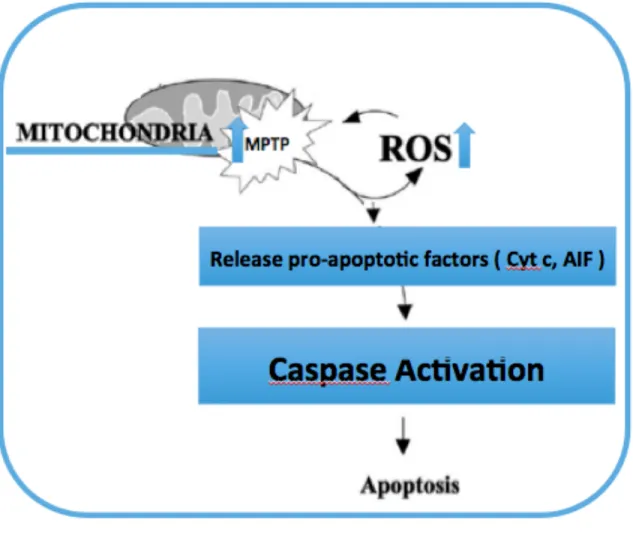
An abnormal MPTP opening can liberate Cyt C into the cytosol. Unusual release of Cyt C with some other mitochondrial pro-apoptotic proteins could be the immediate signal of apoptosis causing mitochondrial dysfunctions [137] as shown in schematic Fig. 12.
Schematic image of mitochondrial ROS’s roles in apoptosis. Figure 12.
Adapted from Qiu et al. [137]. This image demonstrates that increased ROS leads to increased permeability of the outer mitochondrial membrane and decreased mitochondrial membrane potential. This may cause apoptosis by activating a series of pro-apoptotic pathways.
During apoptosis, specific proteins are released from the mitochondria to promote cell death.
One of the main pro-apoptotic proteins activated in inflammation injury is Cyt C, a protein that is localized in the intermembrane space of the mitochondria in healthy cells and is essential to respiration [90, 91]. An abnormal MPTP opening can cause necrosis or apoptosis in cells. This translocation of Cyt C from the mitochondria to the cytosol starts
activating a series of caspases. Because of this cascade of caspases, the apoptotic signals release and finally provoke caspase-dependent apoptosis with activation of caspase-9, which then activates caspase-3. Unusual release of Cyt C with some other mitochondrial pro-apoptotic proteins could be the immediate signal of apoptosis causing mitochondrial dysfunctions [92].
MPTP malfunction due to high levels of ROS also initiates the release of AIF into the nucleus in the mature soluble form (AIFsol), where it provokes nuclear apoptosis in a caspase-independent manner and finally causes DNA damage [138]. High levels of ROS and increased expression of pro-apoptotic Bcl-2 family proteins, including Bax, can also cause cell death [90, 91]. In a study by Arab et al, to evaluate colonic apoptosis, the mRNA expression of Cyt C, Bax, Bcl-2 and caspase-3 was analyzed in trinitrobenezene sulphonic acid induced colitis in rats. This study showed increased mRNA expression of caspase-3, the pro-apoptotic Cyt C and Bax with downregulation of Bcl-2 [139].
In another study by Taha et al, the effect of oxidative stress on mitochondrial dysfunction in IBD was studied in the Caco-2/15 cell line. In this study Caco-2/15 cells were exposed to iron-ascorbate, which produces oxygen radicals and can contribute to lipid peroxidation in IBD. The result of this study indicated that the level of AIF and Cyt C proteins increased in Caco-2/15 cells after iron-ascorbate administration compared with the control group [112].
1.2.3.5. Ca2+ homeostasis and mitochondrial dysfunction in IBD
Ca2+ signaling) can be affected by ROS concentration, pro-inflammatory cytokines and also by the anti-apoptotic protein Bcl-2. Recently, Pinton et al. have suggested that the anti-apoptotic protein Bcl-2 could reduce ER Ca2+ levels, hence reducing mitochondrial Ca2+ uptake [77]. Alteration in ROS concentration can affect Ca2+ channel activity and change the concentration of mitochondrial Ca2+. Also, malfunctions in Ca2+ signaling and Ca2+ concentrations may lead to increased risk of cell damage and death [77]. Qureshi et al. noted that disorders in intracellular Ca2+ mobilization could cause increased NF-κB activity and result in dysmotility of colonic smooth muscles in murine models of experimental colitis [140]. In an investigation by Di Sabatino et al., it has been found that a reduction in pro-inflammatory cytokine release can occur by prevention of mitochondrial Ca2+ overload in inflamed intestinal cells. In addition, prevention of mitochondrial Ca2+ overload by activation of Ca2+ channel inhibitors can result in diminished mitochondrial ROS accumulation, increased mitochondrial energy production and correction of mitochondrial oxidative stress-mediated disorders [141].
1.3. Therapeutic intervention: antioxidants targeting
mitochondrial dysfunction
1.3.1. Antioxidant and anti-inflammatory agents
A phenolic antioxidant is a molecule with a strong capacity to reduce or stop the oxidation of lipids and other molecules and protect them against free radicals. In addition,
antioxidants play the main role in stabilizing or disabling free radicals before they become dangerous by attacking the closest cells and cellular components [142].
In recent years, flavonoids and other polyphenols, the so called bioactive molecules with antioxidant properties, have become the subject of many studies to determine their properties to prevent or cure oxidative damage caused by OxS, nitrogen species metabolism and other markers for oxidative damage [143, 144]. Many studies have shown that, in healthy and pathologic cell conditions, the endogenous antioxidants such as SOD, catalase [145] and GPx enzymes play an important role in keeping the balance between oxidative damage pro-oxidants and endogenous defense mechanisms. Non-enzymatic antioxidant substances are also responsible for the control of ROS overload and cell damages [146, 147].
Schematic illustration of oxidants and antioxidants imbalance, Figure 13.
oxidative damage, and chronic diseases. Adapted from Arulselvan et al. [148].
Endogenous enzymatic and non-enzymatic antioxidants also have an important role in decreasing OxS [149]. Recently, several studies have observed that natural antioxidants from several plant sources, including flavonoids and phenolic compounds play a preventive role in protecting against the generation of free radicals. In addition, flavonoids and phenolic compounds act as anti-inflammatory factors by inhibiting two main signalling pathways including NF-𝜅B and mitogen-activated protein kinases (MAPKs) that play a part in production of different proinflammatory mediators [149].
1.3.2. Polyphenols
1.3.2.1. Structural diversity and dietary sources of polyphenols
Polyphenol compounds are most commonly found in fruits, vegetables, cereals, and beverages [150]. These components have a simple structure and hold one or more benzene rings with 2 or more hydroxyl groups. In addition, polyphenols are considered the most available antioxidants in the human daily diet with important biological properties, including anti-inflammatory and immunomodulatory features. Presently, elegant studies have shown that more than five hundred different polyphenols are regularly found in daily foods while also providing important information about polyphenol consumption and bioavailability [151]. Dietary polyphenols from different sources, including apples have become a focus of research due to their potential
therapeutic effects and preventive properties against several chronic diseases, including cardiovascular disorders, degenerative diseases, diabetes, osteoarthritis and GI diseases. Polyphenols are classified into diverse groups according to their different chemical structures, degree of oxidation, amplitude of polymerization and substitutions of the basic skeleton [152]. The two principal subgroups of polyphenols are flavonoids and non-flavonoids, and each subgroup is divided into six different subclasses [151]. The two subgroups are categorized by the existence of different numbers of phenolic rings, along with two or more hydroxyl substitutions. In particular, the flavonoids contain two benzene rings linked by a linear three-carbon chain to form and produce different kinds of flavonoid subclasses. This characterization is based on the oxidation state of the central pyran ring in flavonoid molecules. Some common groups of flavonoids are flavones, isoflavones, flavonols, flavanones, flavanols, anthocyanins, and anthocyanidins. The nonflavonoids contain phenolic acids, lignans and stilbenes [152, 153].
“Aglycone”, the simple phenolic structure of polyphenols could be attached with carbohydrates and organic acids to produce “glycone” structures. Simple phenolic structure of polyphenols can also be attached with other polyphenols to produce “polymers”.
Polyphenols from food with simple phenolic compounds such as gallic acid, ellagic acid, catechin, eugenol, vanillin, caffeic acid, ferulic acid, apigenin, quercetin, gingerol, kaempferol, myricetin, resveratrol, rutin, naringenin, and cyaniding are factors that modify and prevent inflammatory response, as well as alter gut microbiota [154, 155].
1.3.2.2. Biological functions of polyphenols
Polyphenols play a key role in cell health through their antioxidant actions through two different pathways, ‘‘ROS-removing level’’ and ‘‘ROS formation level’’. The first mechanism shown in Fig.14 constitutes a direct pathway for ROS-inhibition. The ‘‘ROS-removing level’’ can act through 3 different mechanisms: ROS-scavenging (electron/hydrogen transfer), production of ROS-removing enzymes (e.g. SOD, catalase, GPx) and production of endogenous antioxidant-synthesizing enzymes (e.g. glutathione synthase). Furthermore, the second antioxidant mechanism of polyphenols is a direct action of polyphenols that inhibits the metal-dependent formation of free radicals (iron and copper) and decreases or controls ROS-forming enzymes (e.g., XO, NOX, LOX, MAO, iNOS) as shown in Fig.14.
Classification of the polyphenols antioxidant related action. Figure 14.
Adapted from Sandoval et al. [156]
The focus of this thesis is on the antioxidant and anti-inflammatory actions of polyphenols given their potential benefits in IBD. [157, 158].
1.3.2.3. Apple polyphenols
Apples are one of the richest sources of polyphenols. In recent years, different studies have shown that daily intake of apple polyphenols and apple-derived polyphenolic products may inhibit or decrease chronic diseases including OxS-associated disorders, cardiovascular diseases and arthritic diseases. Furthermore, they have shown to preserve the gut from drug damage [159].
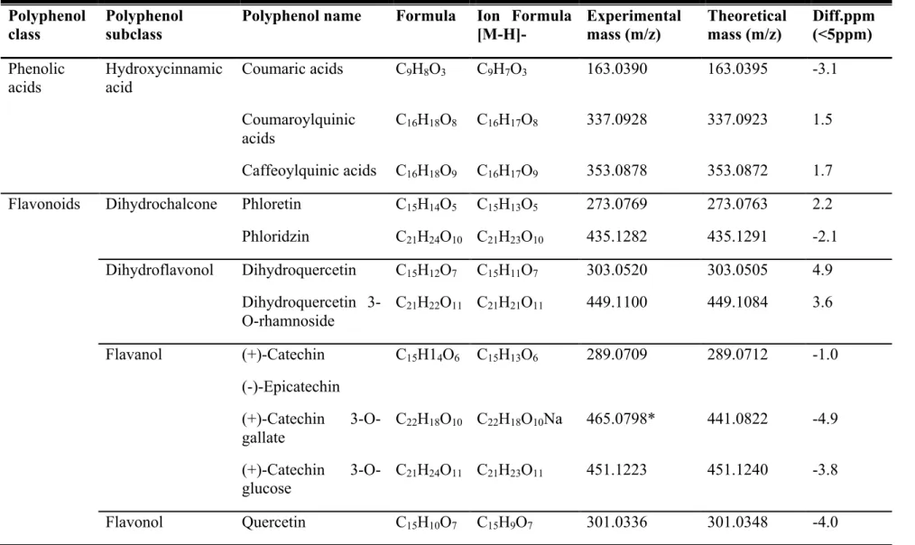
Apple polyphenols contain diverse structural models, such as hydroxycinnamic acids, flavonols (quercetin glycosides), dihydrochalcones (phloretin glycosides), chlorogenic acid, procyanidins, dihydrochalcones and flavan-3-ols (oligomericprocyanidins). The most prevalent groups of polyphenols present in apples are chlorogenic acid, procyanidins, and dihydrochalcones [160, 161].
Several preclinical studies have demonstrated that apple polyphenols have the potential to protect or treat peptic ulcer by numerous intracellular and molecular activities [11, 162]. Graziani et al. have observed that apple polyphenols inhibit expression of COX-2 mRNA and protein in gastric tissue. These polyphenols decreased the peroxidation of lipids in the tissue and caused a moderation in the level of MDA in gastric mucosa. In addition, apple polyphenols protected against xanthine-xanthine oxidase-induced damage and indomethacin-induced oxidative injury in human gastric epithelial cells by improving the antioxidant potential and preventing lipid peroxidation [11]. These results from human studies illustrate the powerful effects of polyphenols in reducing the risk of several chronic illnesses (e.g., cardiovascular disease, neurodegenerative disorders, diabetes, cancers, osteoarthritis, and GI diseases) or improving their symptoms. In this thesis our focus is to demonstrate the preventive and/or therapeutic potential of apple polyphenols in the management of IBD.
1.3.3. Apple polyphenols in intestinal inflammation
In different inflammatory diseases, including IBD, there is an increased expression of pro-inflammatory chemokines, cytokines, cell adhesion agents, and enzymes. In addition,
different studies have shown that both CD and UC are lifelong disorders that cause different challenges for the scientific and medical communities because of several unsolved problems, such as dramatically growing prevalence of IBD, absence of a cure, severe drug side effects, unresponsiveness to medical treatments, direct and indirect treatment costs and socio-economic burden as shown in Fig. 15 [163-165]. Recently, the illustration of biological and beneficial effects of polyphenols, in particular, apple polyphenols in the management of inflammation has captivated the attention and interest of researchers. These studies looked at the molecular mechanisms of action of the different natural elements [161, 166-168].
Schematic description of main reasons for treating IBD patients Figure 15.
The preclinical study by Jung et al. demonstrated that apple polyphenols can inhibit the expression of pro-inflammatory cytokines and enzymes in human colonic epithelial cells by the prevention of interferon-gamma-inducible protein-10, IL-8-promoter, and of NF-kB dependent signal transduction [161, 169]. Furthermore, apple polyphenols have also shown beneficial health effects in animal models of colitis by modulating the expression of MAPK, a signalling protein family. The modulation of this signalling pathway by polyphenols causes down-translation and down-transcription of proteins that are involved in the inflammatory response [128, 157].
Experimental aspirin-induced gastric ulcer presented gastroprotective effects of dietary intake of apple polyphenols. These natural polyphenols caused up-regulation of GSH in gastric mucosa of rats. As we know, GSH in gastric mucosa has antioxidant activity. Also, apple polyphenols elevate the expression of glutathione-S-transferase P1 that acts as an enzymatic antioxidant in gastric tissue. In Paturi’s study, they also found that apple polyphenols cause overexpression of mucin-2 and trefoil factor-2 genes, which assist in the protective function of the stomach barrier and help protect the gastric mucosa [162].
2 RESEARCH PROJECT
2.1. Hypothesis
We hypothesize that:
1. Dried apple peel powder (DAPP) has the potential to prevent or reduce OxS and inflammation via the modulation of mitochondrial functions or/and structure in experimental colitis.
2. The resulting beneficial actions may contribute to the preservation of intestinal epithelium and homeostasis.
2.2. Objectives
As mitochondria are the major source and target of free radicals, while exhibiting various important functions, the aims of our study are to determine:
• Alterations in prooxidant/antioxidant balance and the status of inflammatory factors in association with intestinal microbiota and in response to DSS-induced colitis;
• The contribution of DAPP to prevention and treatment of experimental colitis by fighting OxS, inflammation and mitochondrial derangements;
Apple peel polyphenols: A key player in the prevention and treatment of experimental inflammatory bowel disease
Marie-Claude Denis1,2, Denis Roy3, Pantea Rahmani Yeganeh1,2, Yves Desjardins3, Thibault Varin3, Nour Haddad1, Devendra Amre1,4, Alain Théophile Sané1, Carole Garofalo1, Alexandra Furtos5, Natalie Patey1,6, Edgard Delvin1,7, Eric Tremblay8, André
Marette3, Jean-François Beaulieu8, Emile Levy1,2,3,8
1Research Centre, CHU Sainte-Justine and 2Departments of Nutrition, 4Pediatrics, 5Chemistry, 6Pathology and 7Biochemistry, Université de Montréal, Montreal, Quebec, Canada, H3T 1C5
3Institute of Nutrition and FunctionalFoods (INAF), Université Laval, Quebec, Quebec, Canada, G1V 0A6
8Laboratory of intestinal physiopathology, Department of Anatomy and Cellular Biology, Faculty of Medicine and Health Sciences
Université de Sherbrooke, Sherbrooke, Quebec, Canada, J1H 5N4
Key words: Polyphenol; Peroxidation; Mitochondria; Inflammation
Running Head: Antioxidant and anti-inflammatory effects of apple polyphenols
Abbreviations: DAPP, dried apple peel powder; AIF, apoptosis-inducing factor; ATP,
adenosine triphosphate; CD, Crohn’s disease; COX-2, cyclooxygenase-2; DAI, disease activity index; DP, degree of polymerization; DSS, dextran sulfate sodium; FA, fatty acid; GPx, glutathione peroxidase; IBD, inflammatory bowel diseases; IL-6, interleukin-6; MDA, malondialdehyde; MPO, myeloperoxidase; NF-kB, nuclear factor-kappa B; Nrf-2, nuclear factor erythroid 2-related factor 2; OGG1, 8-oxoG-DNA glycosylase; OxS, oxidative stress; PGC-1α, peroxisome proliferator-activated receptor gamma coactivator-1-a; PGE2, prostaglandin E2; PPAR, peroxisome proliferator-activated receptor; PUFA, polyunsaturated fatty acid; ROS, reactive oxygen species; SOD, superoxide dismutase; TNF-α, tumor necrosis factor-α
Correspondence: Dr Emile Levy, GI-Nutrition Unit, Research Centre, CHU
Sainte-Justine, 3175 Sainte-Catherine Road, Montreal, Quebec, Canada H3T 1C5, Telephone: +1 (514) 345-7783; Fax: +1 (514) 345-4999; email: [email protected]