Université de Montréal
Role of Histone H3 Lysine 56 Acetylation in the
Response to Replicative stress
By
Jeanet Nersesian
Molecular Biology Program Faculty of Medicine
A thesis submitted to faculty of Medicine in partial fulfillment of the degree of
Master of Science in Molecular Biology
January, 2017
iii
Abstract
In Saccharomyces cerevisiae, histone H3 lysine 56 acetylation (H3K56ac) occurs on all newly synthesized histones H3 that are deposited behind DNA replication forks. H3K56ac plays critical role in chromatin assembly during DNA replication and repair. H3K56ac is also required for genome stability and stabilization of stalled replication fork. Cells lacking H3K56ac are sensitive to methyl methane sulfonate and other drugs that cause replicative stress.
In this thesis, we investigated the links between the replisome protein Ctf4 and the H3K56 acetyltransferase Rtt109. Deletion of CTF4 partially rescued the sensitivity of rtt109Δ cells to methyl methane sulfonate. Genetic analyses also showed that Ctf4, Rtt109, and the Rtt101-Mms1-Mms22 complex act in the same pathway to response to replicative stress. ctf4Δ and rtt109Δ cells displayed intense foci of the single-stranded DNA binding complex RPA during replicative stress, suggesting formation of excess single-stranded DNA regions at stalled replication forks, leading to hyper activation of DNA damage checkpoints. These mutants accumulated anaphase bridges and persistent foci of the homologous recombination proteins Rad51 and Rad52 in response to genotoxins, suggesting that abnormal DNA structure formed at stalled replisome may compromise their recovery. Deletion of HR genes (RAD51, RAD52, RAD54, RAD55 and MUS81) together with ctf4Δ and rtt109Δ presents synergistic sensitivity to MMS, suggesting that H3K56ac deficient cells use HR to repair the damages caused by replicative stress. Overall our results demonstrate that H3K56ac deficient cells cannot recover MMS- induced damages because HR is compromised in these mutants.
iv
Keywords:
Ctf4, Rtt109, MMS, histone H3 lysine 56 acetylation, chromatin structure, post-translational histone modifications, cell cycle, DNA replication, DNA replication coupled chromatin assembly, DNA damage response pathway, homologous recombination.v
Résumé
Chez la levure Saccharomyces cerevisiae, l’acétylation de l’histone H3 sur la Lysine 56 (H3K56ac) a lieu sur toutes les histones H3 nouvellement synthétisées qui sont déposées derrière les fourches de réplication. L’acétylation de H3K56 joue un rôle primordial dans l’assemblage de l’ADN lors la réplication et la réparation. L’acétylation de H3K56 joue également un rôle important dans la stabilité génomique et la stabilisation des fourches de réplication bloquée. En effet, les cellules dépourvues de H3K56ac sont sensibles au méthane sulfonate de méthyle (MMS) et à d’autres agents génotoxiques qui causent du stress réplicatif. Notre projet visait à investiguer les liens entre la protéine du réplisome Ctf4 et l’acétyltransférase d’histone Rtt109. Dans un premier lieu, la délétion de CTF4 a partiellement contré la sensibilité des cellules rtt109Δ au MMS. Notre analyse génétique a aussi montré que Ctf4, Rtt109, et le complexe Rtt101-Mms1-Mms22 agissent dans la même voie de réponse face à un stress réplicative. Nos résultats montrent que les cellules ctf4Δ et rtt109Δ présentent des foyers intenses du complexe de liaison à l'ADN simple-brin RPA en réponse au stress réplicatif, suggérant la formation excessive de régions d'ADN simple-brin aux fourches de réplication bloquées, ce qui conduit à une hyper activation des points de contrôle des dommages à l'ADN. Ces mutants présentent des ponts anaphase et des foyers persistants des protéines de recombinaison homologues Rad51 et Rad52 en réponse aux génotoxines, suggérant ainsi que la structure anormale des réplisomes bloqués peut compromettre leur récupération. Nos résultats indiquent également que la délétion des gènes de la RH (RAD51, RAD52, RAD54, RAD55 et MUS81) avec ctf4Δ et rtt109Δ respectivement, engendre une sensibilité synergique au MMS, suggérant que les cellules qui
vi
sont déficientes en H3K56 acétylation utilisent la RH pour réparer les dommages causés suite à un stress réplicatif. En conclusion, nos résultats suggèrent que les cellules déficientes en H3K56ac présentent des défauts de RH en réponse aux dommages à l’ADN induits par le MMS durant la phase S.
Mots-clés:
Ctf4, Rtt109, MMS, acétylation de la lysine 56 de l’histone H3, structure de la chromatine, modifications post traductionnelles des histones, cycle cellulaire, réplication de l'ADN, assemblage de la chromatine couplée à la réplication de l'ADN, voie de réponse aux dommages à l’ADN, recombinaison homologue.
vii
Table of contents
Abstract ... iii
Résumé ... v
Table of contents ... vii
List of figures ... x
List of tables ... xii
List of abbreviations ... xiii
Acknowledgements ... xvii
Introduction ... 1
1-1 Chromatin structure ... 1
1-2 Key stages of the cell cycle ... 2
1-2-1 The G1-S transition ... 3
1-2-2 DNA replication in S phase ... 4
1-3 Response to DNA damage during replicative stress ... 7
1-3-1 DNA damage and replication checkpoints ... 7
1-3-1-1 Apical checkpoint kinases: Mec1 and Tel1 ... 8
1-3-1-2 Checkpoint mediators: Rad9 and Mrc1 ... 9
1-3-1-3 Checkpoint effector kinases: Rad53, Dun1, Chk1 ... 9
1-3-2 MMS induced DNA Damages ... 10
1-3-3 DNA repair: Homologous recombination ... 12
viii
1-3-4 DNA damage tolerance pathways: post-replication repair ... 18
1-3-4-1 Ubiquitination ... 19
1-4 DNA replication coupled chromatin assembly ... 21
1-5 Histones post translational modifications ... 23
1-5-1 Histone Acetylation ... 25
1-5-1-1 Histone acetyltransferases ... 27
1-5-1-2 Histone deacetylases ... 29
1-5-1-3 Acetylation of newly synthesized free histones and their role in chromatin assembly and DNA damage response ... 31
1-6 Cellular functions of H3K56ac ... 32
1-7 Rtt101/Mms1/Mms22 ubiquitin ligase complex and its substrates ... 34
1-8 Rationale and objectives ... 35
Material and methods ... 37
2-1 Yeast strains and media ... 37
2-2 Cell synchronization and transient exposure to genotoxic agents ... 39
2-3 Cell viability assays ... 40
2-4 Immunoblotting ... 40
2-5 Fluorescence microscopy ... 41
2-6 Neutral 2-dimensional (2D) gel electrophoresis... 41
2-7 Automated evaluation of Rfa1-YFP foci intensity ... 42
ix
Results ... 43
3-1 CTF4 acts via an RTT109- and H3K56ac-dependent genetic pathway. ... 43
3-2 H3K56ac-deficient cells form persistent HR foci following transient exposure to MMS. ... 45
3-3 Cells lacking Ctf4 or Rtt109 present hyperactivation of the DNA damage checkpoint kinase Rad53 ... 56
3-4 Cells lacking Ctf4 or Rtt109 present anaphase bridge formation ... 57
3-5 Are Ctf4 and Rtt109 required for resolution of HR intermediate structures? ... 63
Discussion ... 67
Permissions ... 71
x
List of figures
Figure 1: Schematic of DNA replication in budding yeast. ... 6
Figure 2: DNA damage and repair during replication. ... 11
Figure 3: Diagram of the homologous recombination process. ... 15
Figure 4: Schematic diagram of the structural and functional domains of the three subunits of RPA in S. cerevisiae. ... 16
Figure 5: Rad52 and its role as a recombination mediator. ... 17
Figure 6: Rad51 protein. ... 18
Figure 7: Replication- coupled chromatin assembly. ... 23
Figure 8: Histones are subject to post-transcriptional modifications. ... 24
Figure 9: Lysine acetylation and deacetylation. ... 26
Figure 10: Ctf4 and Rtt109 act in a same pathway in response to replicative stress. ... 43
Figure 11: In response to MMS exposure during S, ctf4Δ and rtt109Δ mutations are epistatic. ... 44
Figure 12: Mutants of the H3K56 acetylation pathway display frequent spontaneous Rad52-YFP foci. ... 45
Figure 13: Transient exposure of ctf4Δ and rtt109Δ mutants to MMS results in persistent Rad52 foci. ... 47
Figure 14: Transient exposure of ctf4Δ and rtt109Δ mutants to MMS results in persistent Rad51 foci. ... 49
Figure 15: Transient exposure of ctf4Δ and rtt109Δ mutants to MMS results in persistent Rfa1 foci. . 51
Figure 16: The intensity of Rfa1 foci. ... 52
Figure 17: Rfa1-YFP foci intensity depends on ctf4Δ and rtt109Δ mutants. ... 55
Figure 18: Cells lacking Rtt109 or Ctf4 present hyperactivation of Rad53 in response to MMS exposure during S. ... 57
xi
Figure 19: Anaphase bridges are formed in rtt109Δ and ctf4Δ cells treated with MMS during replication. ... 58 Figure 20: Cells lacking Ctf4 or Rtt109 present anaphase bridge formation after MMS exposure during S. ... 59 Figure 21: Cells lacking Ctf4 or Rtt109 present persistent anaphase bridges and Rad52 foci after MMS exposure during S. ... 61 Figure 22: H3K56ac deficient cells do not prevent X-structure accumulation in sgs1Δ mutants. ... 65 Figure 23: Deletion of HR genes causes synthetic sensitivity to MMS in conjunction with rtt109Δ and
xii
List of tables
Table 1: Acetylated modification sites found in S. cerevisiae, in comparison to those found in and H.
sapiens. ... 25
Table 2: Different families of histone acetyltransferase in S. cerevisiae. ... 28
Table 3: Histone H3 acetylation and associated functions in S. cerevisiae. ... 29
Table 4: Yeast strains used in the research project ... 37
Table 5: Rfa1-YFP intensity of cells treated with MMS. ... 53
Table 6: Mean Rfa1-YFP intensity of cells treated with 0.02% MMS compared to WT treated with 0.06% MMS (Control). ... 54
Table 7: Anaphase bridges formation in rtt109Δ and ctf4Δ cells compared to WT in the presence of MMS. ... 60
Table 8: Anaphase bridges formation in rtt109Δ and ctf4Δ cells compared to WT in the presence of MMS. ... 60
Table 9: Anaphase bridges and Rad52-YFP foci formation in rtt109Δ and ctf4Δ cells compared to WT in the presence of MMS. ... 62
xiii
List of abbreviations
(H3-H4)2 H3-H4 Tetramer 3-mA 3-methyladenine 7-mG 7-methylguanine A Adenine Ac AcetylationAPC Anaphase Promoting Complex
Asf1 Anti-Silencing Function 1 Asf1 Anti-Silencing Factor 1 CAF1 Chromatin Assembly Factor-1 Cdc6 Cell Division Cycle 6
CDK Cycline-Dependent Kinase
Cdt1 Cdc10- Dependent Transcript 1 CPT
CTAB
Camptothecin
Cetyl Trimethyl Ammonium Bromide
CTD Carboxy-Terminal Domain
Ctf4 Chromosome Transmission Fidelity 4
DDK Dbf4-Dependent Kinase
DDR DDT
DNA Damage Response DNA Damage Tolerance
dHJ Double Holliday Junction
xiv
DSBs Double Strand Breaks
FACT Facilitates Chromatin Transcription
FHA Fork Head Associated Domain
GINS Go, Ichi, Ni, San
H3K56ac Histone H3 Lysine 56 Acetylation HAT Histone Acetyltransferase
Hda1 Histone Deacetylase 1 HDAC Histone Deacetylase enzyme Hif1 Hat1p-Interacting Factor-1
HJ Holiday Junction
HR Homologous Recombination
K Lysine
MCM Mini Chromosome Maintenance
MMS NAD+
Methyl Methane Sulfonate
Nicotinamide Adenine Dinucleotide NASP Nuclear Autoantigenic Sperm Protein
ORC 0rigin Recognition Complex
PCNA PH domain
Proliferating Cell Nuclear Antigen Pleckstrin Homology domain
Pol α DNA Polymerase Alpha
Pol δ DNA Polymerase Delta
Pol ε DNA Polymerase Epsilon
xv Pol
Pre-IC
DNA Polymerase Eta Pre Initiation
Pre-RC Pre-Replicative Complex PRR
PTM
Post Replication Repair
Post Translational Modification
RF Replication Fork
Rfa1 Replication Factor A1
RFC Replication Factor C
RNR Ribonucleotide Reductase
RPA Replication Factor A
RPA Replication Protein A
Rpd3 Reduced Potassium Dependency 3 Rtt109 Regulator of Ty1 Transposition 109 SC
SDS-PAGE
Synthetic Complete media
Sodium Dodecyl Sulfate Polyacrylamide Gel Electrophoresis Sir2 Silent Information Regulator 2
Sld Synthetically Lethal with Dpb11 ss- DNA Single Stranded DNA
SSBs Single Strand Breaks
T TCA TLS TS Thymine Trichloroacetic Transleison Synthesis Template Switching
xvi
Ub Ubiquitination
UV Ultraviolet
YFP Yellow Fluorescent Protein
xvii
Acknowledgements
I would first like to thank my research director Dr Hugo Wurtele for granting me the opportunity to pursue my master studies under his supervision. I sincerely want to say thanks for accepting me as his student and supporting me. His kindness and patience have always encouraged me throughout these years. I’m eternally grateful for his understanding.
Moreover, I would like to thank Dr Elliot Drobetsky for his helpful advice on my research project and Dr Santiago Costantino for his technical help.
I am also thankful to all previous and present members of our lab for their help and support; Antoine Simoneau, Ian Hammond-Martel, Jean-Philippe Angers, Kristelle Desfossés-Baron and Etienne Ricard, Edlie St-Hilaire, research assistant of our lab who was always available to teach me almost every technique learned throughout my master, to prepare all materials and start my long time courses. Rahma Sakouhi, my friend who was always pleased to help me and correct my French texts.
Finally, I must express my very profound gratitude to my spouse, Ishkhan Ishkhanian, who was always beside me. Without his unconditional love, patience, continuous encouragement and support I could never reach this milestone. To my lovely daughter Clara, for her beautiful and warm smile that gave me the courage and persistence to work hard. I am extremely grateful for having them in my life; my parents and siblings who encouraged me, even from far away, to continue this journey. This accomplishment would have not been possible without all of them.
1
Introduction
1-1
Chromatin structure
The eukaryotic genome is packaged into a structure known as chromatin [2]. The basic subunit of chromatin is called a nucleosome, which is formed by wrapping 147 base pairs of DNA around a histone octamer [3] [4]. An octamer is formed of a histone tetramer (H3+H4)2
and two histone dimers (H2A+H2B) [4]. Histones are classified into two main groups: core histones (H2A, H2B, H3, and H4) and linker histones (H1 and H5). Core histones are involved in the nucleosome structure, while the linker histones modulates higher levels of chromatin organization [4] [5] [6]. Linker DNA, approximately 20-90 base pairs length, connects nucleosomes together [5]. Histones permit DNA packaging by neutralizing the negative charge of the DNA backbone (formed of phosphodiester bonds), thereby reducing electrostatic repulsion between DNA molecules in the tiny confines of the eukaryotic nucleus [7]. Histones are rich in Arginine and Lysine amino acids that contain free amino group (NH2). Because of this, histones are basic, positively charged, and are able to interact with the
negatively charged DNA backbone [7]. These interactions permit approximatively two meters of human DNA to fit into a nucleus that is only a few micrometers wide [8].
Chromatin is a barrier for several cellular processes involving DNA, e.g. transcription, replication and DNA repair [9]. Eukaryotic cells have evolved different strategies to modify chromatin structure; these modifications facilitate the access of regulatory factors to the underlying DNA. Reversible covalent histone modification (PTM) is one of the major
2
mechanisms by which eukaryotic cells regulate chromatin structure and recruit remodeling enzymes[10].
In this thesis, we will discuss H3K56ac, a modification involved in DNA replication and repair. Therefore, before fruther describing histone PTMs, the following sections will provide an overview of the cell cycle, DNA replication, DNA repair and chromatin assembly.
1-2 Key stages of the cell cycle
In eukaryotes, the cell cycle is divided into four phases: G1 (G0), S, G2, M. Regulation of the cell cycle is critical for the survival of uni- or multicellular organisms and is highly controlled at a molecular level to prevent unregulated proliferation [11]. In the quiescent or senescent stage, the replication process is at rest and the cell status remains stable from a division standpoint. This stage is the “G0 phase”- or Gap 0 [11]. Actively proliferating cells usually do not present G0 phases. In the “G1 phase”, cell volume increases significantly and the G1 restriction checkpoint (see 1-2-1), first identified in S. cerevisiae as START, must be met in order for the cell to proceed with DNA replication in the synthesis phase- or “S phase”. Beyond the START switch, cells proceed to DNA synthesis, regardless of upstream signals or cell size [12]. A checkpoint, termed the “intra S checkpoint” can be activated to restrict S phase progression if DNA replication problems are encountered (see below). After DNA replication, cells enter G2, which occurs just prior to mitosis. In many cell types, an increase in cell volume and in protein synthesis takes place during G2. Mitosis- or “M phase” is the process by which a parent cell divides into two smaller daughter cells. Another checkpoint, the M checkpoint must be met in order to proceed with cell division [13].
3
In the case of actively dividing cells, the daughter cells can enter another interphase after the M phase. In other cases, like for some fully differentiated cells of multicellular organisms, the cells can also enter a prolonged period of quiescence (G0); this is the case for some highly differentiated neuron cells. Many other cell types in various tissues (e.g. lung, liver, kidneys) reach a transient period of quiescence and only enter another interphase in response to the detection of appropriate cell signals. In Saccharomyces cerevisiae, cells normally go through all stages every 90 minutes [11].
1-2-1 The G1-S transition
Despite evolutionary divergence in ortholog protein sequences, highly similar networks are present in S. cerevisiae and in mammalian cells to control the passage of START or the restriction point leading to the S phase [14]. This makes budding yeast cells a valid model to study these phenomena. The G1/S regulon, activating nearly 200 genes in S. cerevisiae [15], is under the control of 3 cyclins CLN1, -2 and -3 and several CDKs (cyclin-dependent kinases). CLN 1- and 2 exert a positive transcriptional feedback on each other in order to drive effective yeast cell budding, coherent G1/S regulon expression, and the activation of additional B-type cyclins [16]. The product of CLN3 initiates the transition to START by phosphorylating, in conjunction with the cyclin-dependent kinase (CDK) Cdc28, protein complexes bound to the promoter regions of transcription factors like MBF and SBF or repressor like Whi5, thereby modulating gene transcription [17]. Later in the cell cycle, mitotic cyclins will further inactivate SBF and MBF factors to effectively switch off the G1/S regulon and ensure a timely and sequentially regulated replication process [18]. S-phase includes an internal checkpoint to ensure the accurate replication of DNA and prevent stalling
4
of single strand DNA during replication that would also impair the progression of the replication fork (see section 1-3-1).
1-2-2 DNA replication in S phase
DNA replication takes place during S phase in several different steps. First, to initiate DNA replication, the two DNA strands should be unwound. This takes place at specific genomic regions called “origin of replication” [19]. The replication origins of S. cerevisiae are probably the best characterized eukaryotic origins. The 11 bp consensus initiation sequence, 5´-(A/T) TTTA (T/C) (A/G) TTT (A/T)-3´, is recognized by the hexameric origin recognition complex (ORC1-5) throughout the cell cycle. ORC acts as a platform to recruit other replication proteins [20]. During late M and early G1 phase, MCM2-7 are loaded to ORC-bound origins via the concerted action of Cdc6 and Cdt1, resulting in the formation of the “pre-replicative complex” (pre-RC) [21]. Assembly of pre-RC occurs only when cyclin-dependent kinase (CDK) activity is low. Indeed, at the onset of S phase, CDK becomes highly active thus leading to the degradation of Cdc6 and the nuclear export of Cdt1 [22] [23]. This degradation prevents DNA re-replication during a single cell cycle. The MCM hexamer has a globular shape with a central cavity through which DNA passes. Therefore, MCM remains bound to DNA even after degradation of Cdc6 and Cdt1 [24]. MCM complexes are not active during G1 phase; the helicase becomes active during the G1 to S phase transition (see below). Sld3, Sld7 and Cdc45 bind to the pre-RC in early-firing origins in G1 phase and with late firing origins in late S phase [25] [26]. These proteins need Dbf4/Cdc7 (Dbf4-dependent kinase or DDK) for their stable recruitment [25]. MCM (2-4-6) and Sld2-3 are phosphorylated in S phase by DDK and CDK, respectively [27, 28] which lead to recruitment of Dpb11/Top BP1 [29]. Dpb11 binds polymerase epsilon (Pol ε), the leading strand replicative DNA
5
polymerase, in S. cerevisiae [30]. Recruitment of a second helicase co-activator, the GINS complex (Go, Ichi, Ni, San, Japanese for 5, 1, 2, 3) which is composed of Psf1, Psf2, Psf3 and Sld5 to the pre-RC in early S phase results in the formation of the so-called CMG complex (Cdc45, MCM2-7, GINS) [31]. Formation of the active form of the CMG complex requires the addition of MCM10 which facilitates recruitment of the single stranded DNA (ssDNA) binding protein, RPA (replication factor A), precluding formation of ssDNA-containing secondary structure [32]. MCM10 also interacts with DNA polymerase α (Pol α) and Ctf4/And1 [33] [34]. Ctf4 (Chromosome transmission fidelity 4), one of replisome progression complex member [35], is required for efficient DNA synthesis, normal cell-cycle progression, genomic stability and sister chromatid cohesion. During replication, Ctf4 act as a scaffold to couple the CMG helicase with pol α within the replisome. The carboxy-terminal domain (CTD) of Ctf4 creates a trimer, which interacts with the amino-terminal tails of the catalytic subunit of Pol α and Sld5 from GINS complex [36].
After the helicase opens up the double stranded DNA at origins, and the replisome is assembled to the replication fork, a specific enzyme called primase together with Pol α catalyze the polymerisation of a short segment of RNA that acts as a primer for further DNA elongation by providing a free 3' OH to DNA polymerases [37]. Then Pol α switches to two conserved DNA polymerases to continue adding deoxyribonucleotides to the 3' end of the newly forming strand. This switching takes place with binding of replication factor C (RFC) to the 3' OH end of the nascent DNA strand, which loads the DNA processivity factor proliferating cell nuclear antigen (PCNA) at the junction between single-stranded and double-stranded DNA [38]. RFC dissociation allows binding of the replicative polymerases, DNA Polymerase delta (Pol δ) and Pol ε, which are the major lagging and leading strands DNA
6
polymerases in S. cerevisiae, respectively [39] [40] (Figure 1). Once the replicative machinery is set on DNA, it moves forward to generate a DNA replication fork.
Figure 1: Schematic of DNA replication in budding yeast. Figure is presented with permission [1].
7
1-3 Response to DNA damage during replicative stress
DNA is constantly under different stresses which could affect its integrity. The sources of these damages can be endogenous (e.g. DNA replication, reactive oxygen species) or exogenous (e.g. UV radiation, mutagenic chemicals). Below is a discussion of various aspect of the DNA damages response, including DNA damage-induced checkpoint activation and DNA repair.
1-3-1 DNA damage and replication checkpoints
The recognition of DNA damage activates “checkpoints”, leading to arrest in specific cell cycle phases. Checkpoints are well conserved between eukaryotes, indicating their significant role [41]. During of each cell cycle stages (G1, S and G2 phases), checkpoints can be activated to ensure that cells do not pass through the phase without repairing eventual DNA lesions. In S phase, the checkpoint action is most important because cells are replicating their DNA and each error might cause genomic instability and mutagenesis in a cell’s progeny. DNA damage during DNA replication activates the intra-S checkpoint signaling cascade which is initiated through signals that emanate are from the apical kinases (Mec1, Tel1) and relayed via mediators (Rad9, Mrc1) and effectors (Rad53, Chk1) (details are described below). This signaling pathway promotes stabilization of stalled replication forks, inhibition of late firing origins, eventual cell cycle arrest in G2, induction and modification of DNA damage response proteins, and DNA repair processes [42] [43] [44]. Overall, the intra S phase checkpoint promotes eventual completion of replication after DNA damage has been repaired [45] [46]. Mediators of the intra S phase checkpoint will be discussed in the following subsections.
8
1-3-1-1 Apical checkpoint kinases: Mec1 and Tel1
Most DNA lesions occurring during replication produce ssDNA, which is generated by uncoupling of the MCM helicase and the replicative DNA polymerases, as well as by the 5'-3' resection of the end of a DSB [47, 48]. This ssDNA is recognized by replication protein A (RPA). ssDNA-bound RPA signals DNA damages and activate checkpoint sensors during S phase [47] [49]. RPA coated ssDNA recruits Ddc2, which interacts with and permits the activation of Mec1, a protein kinase of the PIKK3 family (phosphoinositol-3-kinase-related kinase). Ddc2 and Mec1 are conserved from yeast to human and their homologs in human respectively are ATRIP and ATR [50]. ATR/Mec1 is the apical kinase of the intra S checkpoint response. Mec1 activation also requires the PCNA-like 9-1-1 complex (Ddc1, Mec3 and Rad17) which is loaded at stalled forks by the checkpoint clamp loader Rad24/RFC2-5 [52]. Likewise Dpb11, a DNA replication initiation protein, binds to Ddc2 and 9-1-1 to stimulate Mec1 activity [53] [54]. Once activated, Mec1-dependent phosphorylation of downstream proteins (checkpoint mediators) initiates a signal transduction cascade which culminates in the effects of the S phase checkpoint response.
Tel1 is another kinase in yeast S. cerevisiae that presents similarities to Mec1 and human ATM. Tel1/ATM is recruited to DNA damage sites via a DNA end-binding complex formed of Mre11-Rad50 and Xrs2 in yeast (MRX complex) or its human homolog MRN, (Mre11-Rad50 and Nbs1) [55]. Like Mec1, Tel1 phosphorylates checkpoint mediators to initiate a signaling cascade [56]. Tel1 is mostly active in response to DNA double-strand breaks and does not play critical roles in the S phase checkpoint [57].
9
1-3-1-2 Checkpoint mediators: Rad9 and Mrc1
Rad9 and Mrc1 act as adaptors to promote phosphorylation of downstream proteins by the Mec1 kinase in yeast S. cerevisiae. Hyper-phosphorylation of Rad9 by Mec1 causes activation of this scaffold protein, which permits it to mediate Mec1-dependent phosphorylation/activation of Rad53 (see below, section 1-3-1-3) [58] [59] [60] [61]. During replication, the activation of Rad53 is also promoted by Mrc1 that is present at replisome to pair DNA pol Ɛ with Cdc45 and MCM helicase [62] [63] [64]. Mrc1 plays a mediator function in the presence of stalled fork via its phosphorylation by Mec1. Phosphorylation of Mrc1 in SCD domain causes it to lose its interaction with pol Ɛ, which makes it able to recruit Rad53 and to promote its phosphorylation by Mec1 [64] [65].
1-3-1-3 Checkpoint effector kinases: Rad53, Dun1, Chk1
Rad53 (Chk2 in human cells) is recruited and activated at DNA damage site, which permits phosphorylation of downstream target proteins [66]. One of these targets is Dun1 kinase which, after DNA damage and during S phase, physically binds to Sml1. Sml1 is an inhibitor of ribonucleotide reductase (RNR). Dun1 binding leads to phosphorylation and degradation of Sml1 and consequent increased RNR function. RNR catalyze the conversion of ribonucleoside 5’- diphosphate (NDP) to 2’ deoxyribonucleoside 5’-diphosphate (dNDP). The activation of Dun1 and the decrease of Sml1 increase cellular pools of dNTP, presumably to facilitate DNA repair and replication [67]. Rad53 also phosphorylates Sld3 (CDK) and Dbf4 (DDK) which leads to inhibition of CDKs and DDKs-dependent pathways during replication. Phosphorylation of the C-terminal domain of Sld3 inhibits its interaction with Dbp11 and
10
Cdc45, whereas phosphorylated Dbf4 prevents the activation of the MCM helicase by Dbf4-Cdc7 complex [68].
Another checkpoint kinase, Chk1, was first identified in the fission yeast but is well conserved in different species including human. Chk1 functions mostly during G2/M in the presence of DNA damage; its activation leads to phosphorylation of Pds1 and inhibition of its ubiquitination. Pds1 ubiquitination causes its degradation via APC (anaphase promoting complex). Checkpoint-mediated accumulation of Psd1, in response to DNA damage, inhibits sister chromosomes segregation during mitosis, thereby preventing cell cycle progression [69].
1-3-2 MMS induced DNA Damages
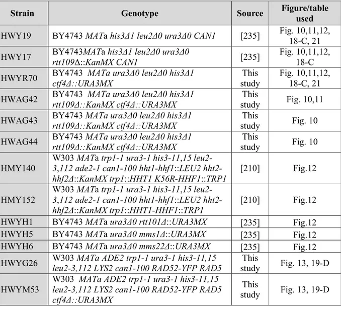
In our work, we frequently use methyl methane sulfonate (MMS), a DNA alkylating agent, to impede DNA replication progression. It is therefore appropriate to describe its action here. MMS cause DNA methylation on N7-deoxyguanine (N7-methylguanine; 7-mG) and N3-deoxyadenine (N3-methyladenine; 3-mA). Although 90% of total adducts induced by MMS is 7-mG, this lesion is nontoxic and non-mutagenic. The remaining 10% of adducts are 3-mA that are both toxic and mutagenic (Figure 2-a-b-c). 3-mA inhibit DNA replication fork progression (Figure 2-b), and cells are most sensitive to MMS during replication [70] [71]. During S-phase exposure to MMS, cells try to rescue stalled replication fork by different strategies such as restarting replication by firing dormant origins, repriming replication, reversing the stalled fork or activating the DNA damage tolerance (DDT) pathways (see section 1-3-4).
11
Figure 2: DNA damage and repair during replication. a) ssDNA nick in the leading strand template will directly lead to RF collapse and a one-ended DSB that could repair by BIR. b) DNA adducts can block replisome machinery which cause stalled fork or uncoupled synthesis. Then, fork reversal can take place leading to the formation of a chicken-foot structure. Restart can occur by HJ-cleavage followed by BIR but the RF can also undergo direct restart by HJ reversal to a fork after lesion bypass through template switching or lesion repair. c) Lesions can block the synthesis of only one DNA strand without inhibiting fork progression. In both cases, ssDNA gaps can be repaired by error-prone translesion synthesis (TLS) or by error-free HR [72]. The figure is presented with permission.
12
Repriming replication at the damage sites can leave ssDNA gaps, which could be a target for endonucleases, leading to DSBs [73]. Removal of the lesions by BER (base excision repair) can also generate single-strand breaks, which upon replication forms DSBs which can be repaired by HR (Figure 2-a) [74]. Stalled fork also could reverse to a chicken foot structure including Holiday Junction, again promoting HR-mediated repair (Figure 2-b) [73] [74] .
1-3-3 DNA repair: Homologous recombination
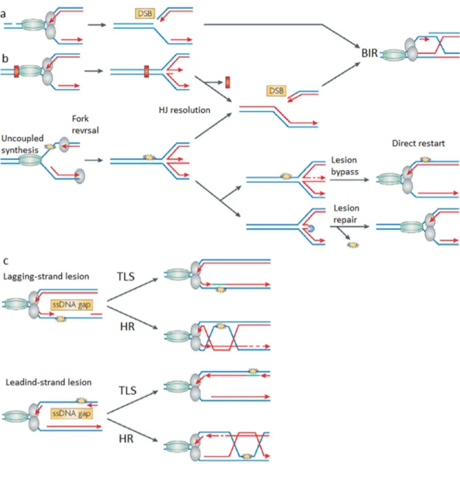
Impediments in replication forks progression can eventually lead to DNA double strand breaks (DSBs) [75]. DSBs, as its name indicate, occur when the phospho-sugar backbone of both complementary strands of DNA are broken at the same position or in sufficient proximity to allow detachment of the double helix into separate molecules [76]. Unrepaired DSBs lead to deletions, translocations and fusions in DNA; the resulting loss of genetic information or genomic rearrangements can cause cell death or diseases such as cancer. DSBs are repaired mostly by two different mechanisms; non-homologous end joining (NHEJ) and homologous recombination (HR) [76, 77]; since NHEJ is not a dominant mechanism during S phase, it will not be discussed further here. Homologous recombination (HR) permits the repair of double-strand breaks by using the genetic information present in homologous DNA sequences (sister chromatids or homologous chromosomes) as a template to repair the damage in an error-free manner [78]. Despite its complexity, HR is very well conserved in evolution. HR occurs mainly after DNA replication (during the S and G2 phases) in both yeast and human to repair DSBs, and also permits the restart of stalled DNA replication forks (discussed above; Figure 2) [79].
In the case of DSB repair by HR, the MRX complex (Mre11, Rad50 and Xrs2) first detect damaged DNA, binds the broken DNA ends, and recruits Tel1. Next, DSB ends undergo 5´to
13
3´ resection (Figure 3). MRX and Sae2 endonuclease activity generate a 3´ single stranded end. Then, additional proteins are recruited to the 3´ ends to promote more extensive resection, including the Exo1 and Dna2 exonucleases and the, Sgs1-Top3-Rmi1 helicase complex [80] [81, 82]. The newly-formed ssDNA is rapidly coated by heterotrimeric RPA to protect it from further degradation and to remove inhibitory secondary structures. RPA can at that point promote loading of Mec1-Ddc2 to trigger checkpoint activation and cell-cycle arrest [83]. RPA-coated ssDNA recruits recombinases to search and invade the homologous DNA sequences. Rad52 is loaded onto ssDNA which helps to recruit Rad51. Rad51 recombinase replaces RPA under the action of Rad52 [84], and forms a nucleoprotein filament on single-stranded DNA. This filament plays central roles in HR: the search for a homologous template DNA and the formation of a joint heteroduplex molecule between the damaged DNA and template [85]. Rad51 binds weakly to DNA; Rad55 and Rad57 which are Rad51 paralogs in S. cerevisiae, are required to stabilize and facilitate Rad51 binding and function [86] [87, 88]. Indeed, Rad55-Rad57 heterodimer stimulate the formation of Rad51nucleoprotein filament [87, 89]. Rad54 and Rdh54, which are DNA-dependent ATPase with translocase activity, then aid Rad51 filament to invade its homologous DNA either on its sister chromatid or, in diploid cells, on its homologous chromosome, forming a joint heteroduplex molecule [90]. At this point, the 3 'single stranded end can invade homologous DNA, hybridizes to the template strand and displaces the complementary strand. This forms a displacement loop (D-loop) (Figure 3). After strand invasion, a DNA polymerase extends the end of the invading 3' strand by synthesizing new DNA. This changes the D-loop to a cross-shaped structure known as a Holliday junction (HJ). A second HJ forms when the second 3' overhang (which was not involved in strand invasion) is captured to the extended D loop (Figure 3). After gap-repair
14
DNA synthesis and ligation, the structure is resolved at the HJs in a non-crossover or crossover mode (Figure 3).Chromosomal crossover will occur if one Holliday junction is cut on the crossing strand and the other Holliday junction is cut on the non-crossing strand. Alternatively, if the two Holliday junctions are cut on the crossing strands, then gene conversion without crossover will be produced [91]. These recombination products are generated by endonucleolytic cleavage by specific nucleases during resolution, for example Mus81-Mms4 and Yen1 in S. cerevisiae [92] [93]. Another HR pathway is single-strand annealing (SSA), which occurs when a DSB is closely flanked by direct repeats. SSA is a simple process which does not require Rad51 nor strand invasion of an intact duplex DNA, but annealing of two complementary ss-DNAs.
Some DSBs, such as those that can occur at telomeres or at broken replication forks, are single-ended. Break-induced replication (BIR) pathway could repair theses DSBs [94] [95]. The resection step of BIR is similar to the traditional HR but the invasion step is different, with only one end of the DSB invading the homologous duplex. Like DSBR, the presynaptic filament often invades the sister chromatid or homolog chromosome, which then leads to extensive DNA synthesis. Therefore, all three major replicative DNA polymerases and all essential components of DNA replication machinery, with the exception of pre-RC assembly factors, are required for BIR. This mechanism can re-form replisome and restart replication following fork collapse and DSB formation (Figure 2-a-b) [96] [97].
15
Figure 3: Diagram of the homologous recombination process. The figure is presented with permission [91].
1-3-3-1 HR proteins form foci at the site of DNA damage
Interestingly, many HR proteins accumulate into focal assemblies at the site of DNA damage. Such repair foci form at DSBs, sites of DNA replication stress, shortened telomeres, and
16
regions of single-stranded DNA in vivo. Foci are useful marker to follow ongoing HR. To visualize foci, microscopy coupled with indirect immunofluorescence or fluorescent protein fusions is used. DNA repair foci are highly dynamic structures. They can assemble and disassemble rapidly, which could help to repair and swift recovery from cell cycle arrest after completion of the repair reaction [98] [99] [100].
In this thesis, the formation of Rfa1, Rad52 and Rad51foci in the absence of Ctf4 and Rtt109 proteins has been investigated. We will therefore describe these proteins in more details.
Rfa1:
The Rfa1 protein is the large subunit of the yeast heterotrimeric RPA. Replication protein A (RPA) also known as replication factor A (RFA) is a highly conserved single-stranded DNA-binding protein. RPA from S. cerevisiae consists of three subunits; RFA1, RFA2 and RFA3 with approximately 70, 30, and 14 kDa molecular weight, respectively (Figure 4). All three members are crucial for cell viability and bind to ssDNA. RPA is a critical protein in several processes, including DNA replication, DNA repair and recombination [101] [102].Figure 4: Schematic diagram of the structural and functional domains of the three subunits of RPA in S. cerevisiae. The figure is presented with permission [102].
17
Rad52
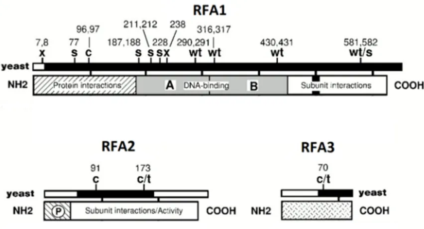
: Rad52 is one of the most important members of the RAD52 epistasis group in S. cerevisiae. Rad52 is a ring-shaped oligomer with DNA binding activity. Also it interacts with both Rad51 and RPA, physically and genetically (Figure 5). During HR, Rad52 promotes presynaptic filament assembly on RPA-covered ssDNA by recruiting of Rad51 (Figure 5), and stimulates ssDNA annealing in second end capture and SSA. Therefore, deletion of RAD52 (rad52Δ) in S. cerevisiae causes extreme sensitivity to a variety of DNA damaging agents (e.g. MMS) and also a general defect in HR pathways, such as DSBR, BIR and SSA [103] [104] [89].Figure 5: Rad52 and its role as a recombination mediator. The figure is presented with permission [94].
Rad51:
Rad51 (Figure 6) is another member of the RAD52 epistasis group. As mentioned before, Rad51 is a recombinase that binds DNA through Rad52 mediator activity (Figure 5). The formation of Rad51filament catalyzes homology search and strands pairing. Rad51 interacts with itself, with Rad52, and with other downstream proteins such as the Rad55-Rad57 heterodimer, Rad54 and Rdh54. Rad51 mutants are defective in the repair of DNA18
damage induced by ionizing radiation and MMS, in the maintenance of telomere length, in mitotic and meiotic recombination, and in mating-type switching [105] [106].
Figure 6: Rad51 protein. 8 sub-domains (B1, B2, C1, C2, D1, E1, E2, and F1) of Rad51 core region and ATP binding motifs A and B (dotted lines) are shown. The numbers immediately above sub-domains B1 and F1 are the first and last residues respectively, of the core domains of Rad51 protein. The figure is presented with permission [106].
1-3-4 DNA damage tolerance pathways: post-replication repair
In S phase, DNA lesions can be bypassed through DNA damage tolerance (DDT) pathways, in order to rapidly recharge the replicative polymerases on the DNA and to finalize the replication. DDT also is termed post replication repair (PRR). Cells via PRR pathways are allowed to continue DNA replication even in the presence of damage. PRR can occur in either an error-prone or error-free manner, which is regulated by posttranslational modifications of PCNA (ubiquitination is described below; section 1-3-4-1). Mono-ubiquitination of PCNA on lysine 164 triggers transleison synthesis (TLS) and its subsequent poly-ubiquitination leads to template switching (TS) [107].
During TLS, DNA is synthesized using damaged DNA template by translesion synthesis polymerases which can accommodate modified bases at their catalytic sites [108]. In yeast, TLS is initiated either by polymerase- (Pol η), encoded by the RAD30 gene or the
19
polymerase-ζ (Pol ζ). The latter is a heterodimer composed of the Rev3 (the catalytic subunit) and Rev7 (the regulatory subunit) [109]. The replacement of these polymerases with the replicative ones is stimulated through mono-ubiquitination of PCNA. Rad18, an E3 ubiquitin ligase, is recruited to RPA coated ss-DNA and forms a complex with Rad6, an E2 ubiquitin conjugating enzyme, which mono-ubiquitinates PCNA on lysine 164 [110] [111]. Mono-ubiquitinated PCNA interacts with Rev1, an essential component of TLS [112]. Rev1 mediates the interaction of pol ζ with the mono-ubiquitinated PCNA [112] [113]. The structural subunit of pol ζ, Rev7, interacts with both Rev1 and Rev3; in yeast, Rev7 stimulates the polymerase activity of Rev3 [113] [114]. TLS is an error prone DDT pathway which can incorporate incorrect nucleotide and generate point mutations [115]. Conversely, template switching is an error free bypass which works similarly to homologous recombination. Template switching uses undamaged sister chromatid as the template, which does not induce mutations, but may lead to chromosomal rearrangements [116] [117]. TS signals are initiated by the mono-ubiquitination of PCNA mediated by Rad6-Rad18 complex [118] and subsequent recruitment of the E3 ligase Rad5 to mono-ubiquitinated PCNA, which stimulates the E2 ubiquitin conjugating activity of the Mms2–Ubc13 complex [119]. This complex adds additional ubiquitin to the mono-ubiquitinated K164 PCNA to synthesize K63-linked poly-ubiquitin chains [120]. These modifications lead to the recruitment of HR factors such as Rad51 and Rad52 which allows the formation of TS intermediates [121].
1-3-4-1 Ubiquitination
Ubiquitin is a highly conserved 76 amino acid polypeptide which is conjugated to lysine residues on protein substrates through the sequential action of three enzymes; E1-activating,
20
E2-conjugating and E3-ligating enzymes. E1, ubiquitin-activating enzyme, in an ATP dependent reaction binds to ubiquitin and produces an ubiquitin-adenylate intermediate which is then transferred to an active site cysteine residue. E2 ubiquitin-conjugating enzyme binds to both ubiquitin and E1 to transfer ubiquitin from the E1 enzyme to the E2. E3 is generally referred to as the ubiquitin ligase, which recognizes the substrate proteins and directly or indirectly catalyzes its ligation to the ubiquitin [122]. E3 enzymes are classified in two major groups; the HECT (Homologous to the E6-AP Carboxyl Terminus) family and the RING (Really Interesting New Gene) family. The mechanisms that these two families use to transfer the ubiquitin to substrates are different. In HECT ligases, E2 transfers ubiquitin to the E3 then, the attachment of ubiquitin to the substrate is catalyzed directly by E3. This is in contrast to RING ligases, in which the E2 transfers ubiquitin to the E3-bound substrate. Of particular interst for this thesis, the cullin family of proteins is considered as RING E3s. Cullins bind to small RING proteins (Rbx1/Roc1or Hrt1/Roc2) to promote their ubiquitin ligase activity and form cullin-RING E3 ligase (CRL) [123] [124]. The S. cerevisiae genome encodes three cullins: Cul1/Cdc53, Cul3, and Cul8/Rtt101 [123] (Rtt101 is described below; section 1-7). A single lysine residue on the substrate protein can receive only one ubiquitin (mono-ubiquitination) or chains of ubiquitin (poly-(mono-ubiquitination) [125]. These modes of ubiquitination lead to different substrate fates. For example, mono- ubiquitination can regulate DNA damage bypass (discussed above; section 1-3-4) and poly-ubiquitination on lysine 48 residue often, but not always, promotes proteasomal degradation [126].
21
1-4 DNA replication coupled chromatin assembly
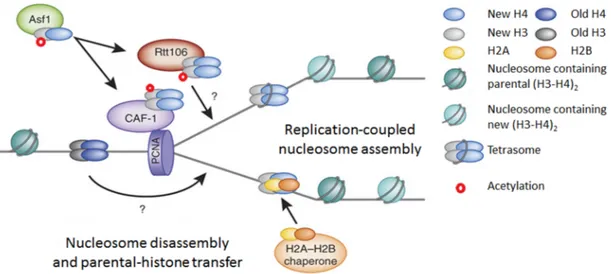
During DNA replication, parental histones are segregated on daughter chromatids, leading to the formation of nucleosome-free gaps that are filled in by assembly of new nucleosomes comprising newly synthesized histones [127]. Nucleosomes are assembled by two different pathways, replication-coupled assembly or replication-independent assembly; Replication-independent nucleosome assembly occurs during gene transcription and histone exchange [128]; this pathway is not relevant for this thesis and will not be discussed further here. Replication-coupled nucleosome assembly occurs during S phase and is closely coupled to passage of the replication fork [129]. We will focus on this latter pathway, specifically in S. cerevisiae.
During DNA replication, existing parental histones and newly synthesized histones areassembled into chromatin. Parental H3-H4 histones are segregated as tetramers (H3-H4)2
and are transferred onto replicated DNA. However, the molecular mechanism by which they are transmitted behind replication forks remain poorly understood [130]. During DNA replication-coupled chromatin assembly, new histone H3/H4 dimers are bound by the Asf1 histone chaperone. Asf1 presents H3/H4 dimers to Rtt109, which permits acetylation on H3K56 [131, 132]. The Rtt101–Mms1–Mms22 ubiquitin ligase complex influences the dissociation of histones from Asf1 (see section 1-7), thereby modulating availability of histones for Caf1 and Rtt106 (chaperones downstream of Asf1) [133]. Rtt106 and CAF1 then deposit H3-H4 onto newly replicated DNA (Figure 7). Recent studies suggest that Rtt106 binds to (H3-H4)2 tetramer [130] and CAF1 binds to two H3-H4 dimers or a single
22
replication machinery, through their interactions with PCNA [135] [136], and in the case of Asf1, the PCNA loader RFC (replication factor C) [137].
After (H3-H4)2 deposition, H2A-H2B dimers are incorporated on either side of (H3-H4)2
tetramer, to form a histone octamer. FACT (facilitates chromatin transcription) and Nap1 chaperones both are important for the transfer and deposition of H2A-H2B dimers [138] [139]. In S. cerevisiae, FACT interacts with Nap1 and RPA. FACT consists of two conserved subunits; Spt16 (Suppressor of ty 16) and SSRP1 (structure-specific recognition protein 1). Through these subunits, FACT can bind to both (H3-H4)2 tetramer and H2A-H2B dimer. In
budding yeast, SSRP1 is separated into two proteins Pob3 and Nhp6 [140]. Pob3 contains tandem PH domains capable of binding histones, a motif also found in Rtt106. The N terminus of Spt16, in budding yeast, has also been shown to bind H3–H4 in vitro [141] [142]. The classic H2A–H2B chaperone, Nap1, shares sequence homology with a large class of histone chaperones, such as Vps75 in yeast. In vitro, Nap1 binds to (H3-H4)2 tetramer and
H2A-H2B dimer, with same affinity whereas in vivo, it appears to bind H2A-H2B dimer preferentially. Nap1 is a nucleocytoplasmic shuttling protein which enables H2A-H2B import. Nap1 also promotes nucleosome formation by disassembling nonproductive histone-DNA interactions, and directly deposits histones onto DNA [141].
Histone H1 is the last histone to be assembled onto the chromatin after replication. Hif1/NASP (nuclear auto antigenic sperm protein) chaperone could incorporate H1 into DNA. In S. cerevisiae, Hif1 (Hat1p-interacting factor-1) demonstrate H3-H4 and H1 chaperone activity. Hif1 physically interacts with Hat1 and Hat2 and functions in the acetylation of newly synthesized histone H4 [143].
23
Figure 7: Replication- coupled chromatin assembly. The histone chaperones (Asf1, Rtt106 and CAF1), acetylated and deacetylated H3K56 are shown. The figure is presented with permission [141].
1-5 Histones post translational modifications
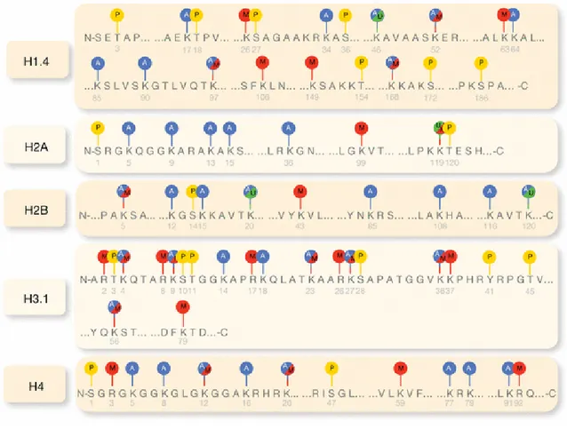
Histones are the most important proteins in the chromatin structure [144] and are remarkably well conserved throughout evolution [145]. Histones contain two domains; N-terminal tail and central domain (histone fold domain). The latter is formed from three α-helices which are separated by two loop regions, and is involved in the interactions of histone-histone and histone- DNA [146] [147] [148]. Both of these domains are subjected to post translational modifications by adding or removing small chemical groups, such as acetyl, methyl, phosphate or proteins like ubiquitin and SUMO. These enzymatic modifications alter the interactions between histones and DNA which leads to chromatin structural changes; dynamic PTMs can also modulate the recruitment of proteins and complexes with specific enzymatic activities [149] by increasing affinity for protein recognition modules. For example,
24
acetylated lysines are bound by bromodomains [150, 151] and methylated lysines or arginines by chromodomains, Tudor domains and PHD domains [152] [153]. Ongoing studies are describing an ever expanding list of histone PTMs (Figure 8) highlighting the important roles of these modifications in several vital cellular processes such as replication, transcription and DNA repair [133] [154] [155] [156].
Figure 8: Histones are subject to post-transcriptional modifications. Well characterized histone post-transcriptional modifications are depicted in this figure: acetylation (blue), methylation (red), phosphorylation (yellow) and ubiquitination (green). The number in gray under each amino acid represents its position in the sequence [157]. Figure is reproduced with permission.
25
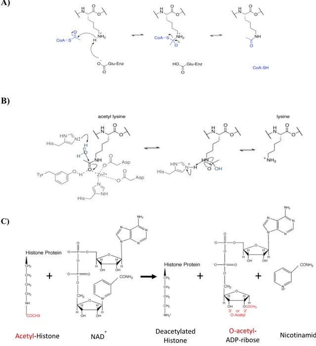
1-5-1 Histone Acetylation
Addition of an acetyl function to the Ɛ-amino group of specific lysine residues on the histone tails or globular histone core is catalyzed by enzymes called Histone acetyltransferases (HATs) [158] and removed by histone deacetylases (HDACs) (Figure 9) [159].
Histone acetylation affects several nuclear processes (Table 3) through different mechanisms. By neutralizing the positive charge of Lysine, histone acetylation leads to a decrease in the affinity of histones for negatively charged DNA molecules. Therefore, acetylation at certain lysine residues causes a loosening of chromatin structure and promotes transcription [160, 161]. Lysine acetylation also promotes its interaction with certain protein recognition modules, including bromodomains [151]. For example, Gcn5 (Histone acetyltransferase belongs to GNATs family) in yeast and human contains bromodomains which mostly binds to acetylated histone H3 residues (e.g. H3K9/K14) [150] [162] [163] [164] [165] [166]. In addition, acetylated lysines are able to interact with other protein motif such as PH-like domain, e.g., S. cerevisiae Rtt106 interacts with K56 acetylated histone H3 through its PH-like domain [167].
Histone Acetylation site in S. cerevisiae Acetylation site in H. sapiens
H2A K4, K7, k14 [168] K5, K9 H2B K3, K6, K11, K16, K21, K22, K49 [169] K5, K11, K12, K15, K16, k20, K23,K46, K120 [170] H3 K4, K9, K14, K18, K23, K27, K36, K56 K4, K9, K14, K18, K23, K27, k36, K56 H4 N-terminus, K5, K8, K12, K16 N-terminus, K5, K8, K12, K16, K91 Table 1: Acetylated modification sites found in S. cerevisiae, in comparison to those found in and H. sapiens. The most conserved sites are found in histone H3 [171]. H3K56 is well conserved from S. cerevisiae to human.
26 A)
B)
C)
Figure 9: Lysine acetylation and deacetylation. A) HATs catalyze the transfer of the acetyl group (from Acetyl-CoA) to the Ɛ-amino group of specific lysine residues on the histone. Figure A is created by Trcum and is published in Wikipedia. Sharing the image does not require permission. B) HDACs remove the acetyl group from acetylated lysine in different mechanisms. This figure shows deacetylation of acetyl lysine by class I, II and IV histone deacetylases. Figure B is presented with a permission from Epigenetics (ATDBio).C) The global mechanism of sirtuins (class III HDACs). These HDACs are more relevant to H3K56ac. The figure is created by Dr Wurtele.
+
+
+
27
The role of histone acetylation in chromatin folding and transcriptional regulation is the focus of vigorous research. In addition, its role in DNA replication and repair remains poorly understood [172]. In this thesis, the acetylation of specific lysine residue on histone H3 (H3K56ac) in Saccharomyces cerevisiae and its role in the cellular response to DNA damage will be discussed. H3K56ac is conserved from yeast to human although it remains unclear whether this modification plays similar roles in both organisms (Table 1).
1-5-1-1 Histone acetyltransferases
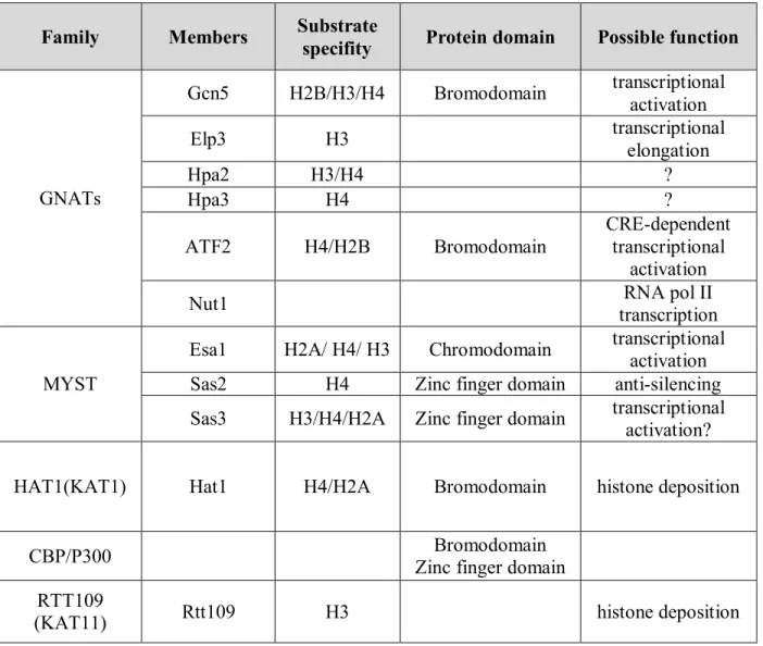
HATs come in two major classes based on their subcellular localization and substrate specificity; type-A and type-B. Type-A HATs are found in the nucleus and are able to acetylate histones in the context of nucleosomes, whereas type-B HATs are cytoplasmic and acetylate free histones before/during chromatin assembly [173]. In reality, HATs diversity does not allow them all to be strictly classified within these two groups; HAT can therefore be categorized in several families based on their sequence homology, structural features and functional roles; GNATs, MYST, HAT1, CBP/p300 and RTT109 (Table 2).
Rtt109 catalyzes H3K56ac in yeast, and its structural homolog CBP/P300 does the same in human cells [174] [175]. Rtt109 (named for its initial identification as a yeast regulator of Ty1 transposition gene product 109), also known as KAT11, is a fungal-specific HAT [158]. Rtt109 associates with either of two histone chaperone proteins Asf1 or Vps75, which stimulates its acetyltransferase activity [176] [177] [131]. The interaction of Rtt109 with these chaperones is distinct; Rtt109-Vps75 forms an almost stoichiometric complex [178] which promotes the stability of Rtt109 in vivo, while Rtt109-Asf1 forms a rather loose and transient one [131]. Importantly, formation of these two complexes is an important determinant of Rtt109’s substrate specificity. Rtt109 paired with the Vps75 chaperone associates with either
28
H3-H4 dimers or (H3-H4)2 heterotetramers [179] [180] and acetylates histone H3 on its
N-terminal lysine 9 and 27 residues. In contrast, Rtt109-Asf1 can only bind H3-H4 dimers and acetylates H3K56 [179] [181] [182] [183] [184] [185].
Family Members Substrate
specifity Protein domain Possible function
GNATs Gcn5 H2B/H3/H4 Bromodomain transcriptional activation Elp3 H3 transcriptional elongation Hpa2 H3/H4 ? Hpa3 H4 ? ATF2 H4/H2B Bromodomain CRE-dependent transcriptional activation
Nut1 RNA pol II
transcription
MYST
Esa1 H2A/ H4/ H3 Chromodomain transcriptional activation Sas2 H4 Zinc finger domain anti-silencing Sas3 H3/H4/H2A Zinc finger domain transcriptional
activation?
HAT1(KAT1) Hat1 H4/H2A Bromodomain histone deposition
CBP/P300 Bromodomain
Zinc finger domain RTT109
(KAT11) Rtt109 H3 histone deposition
Table 2: Different families of histone acetyltransferase in S. cerevisiae. HAT families along with their associated members, histone substrates, and protein domains in S. cerevisiae [158] [173] [186] [187] [188] [185] [189] [190] [191] [192]. Protein domains are parts of a polypeptide chain with similar sequence and structure which can fold independently of the rest of the protein chain [193].
29
Site Domain
Histone acetyltransferase
Histone
deacetylase Proposed function Ref.
K4 N-terminal tail Gcn5, Rtt109 Hst1, Sir2 Transcriptional activation [194] K9 N-terminal tail Gcn5, Rtt109 Sir2, Rpd3 Transcriptional activation, Histone deposition [195] [196, 197] K14 N-terminal tail Gcn5, Hat1, Esa1, NuA3 Sir2, Rpd3 Transcriptional activation, DNA repair [196] [198, 199] [197, 200, 201] K18 N-terminal tail Gcn5 Hda1, Rpd3 Transcriptional activation [202] [197, 203] K23 N-terminal
tail Gcn5, Sas3, Rpd3 DNA repair
[199, 204] [197, 205] K27 N-terminal tail Gcn5, Rtt109 Rpd3 Histone deposition, DNA repair [197, 206] K36 N-terminal tail Gcn5 Rpd3 Transcriptional activation [207, 208] K56 Fold domain Spt10, Rtt109 Hst3/Hst4 Transcriptional activation, DNA repair, Histone deposition [209, 210] Table 3: Histone H3 acetylation and associated functions in S. cerevisiae. H3K56ac occurs in the core domain of histone H3, most of histone H3 acetylation residues are situated on the N-terminal tail.
1-5-1-2 Histone deacetylases
Histone deacetylase enzymes (HDAC) reverse the effect of HATs and remove the acetyl group from lysine residues. HATs and HDAC act in concert to regulate the acetylation level which is important in numerous cell processes such as transcription, replication and DNA repair [159]. HDACs are broadly divided in two categories: so-called “classical” HDACs; (Class I, II, and IV) and Sir2-related (sirtuin; Class III). Based on sequence homology and cofactor dependency, these two families of HDAC are further subdivided in four different classes as indicated above. In humans, the classical HDAC family includes class I (HDAC 1,
-30
2, -3, -8) and class II (HDAC 4, -5, -6, -7, -9, -10) and class IV (HDAC 11). They share sequence similarity and require Zn2+ for deacetylase activity. The sirtuin family contains class III enzymes (human SIRT 1-7). Sirtuins present no sequence similarity to members of the classical family and use nicotinamide adenine dinucleotide (NAD+) as a cofactor for catalysis [211, 212].
Human HDACs are homolog to archetypal yeast enzymes: class I HDACs are most closely related to the yeast (Saccharomyces cerevisiae) transcription regulator (Rpd3); histone deacetylase 1 (Hda1), corresponds to Class II human enzymes, while silent information regulator 2 (Sir 2), corresponds, to Class III [213]. The S. cerevisiae genome encodes five sirtuins: Sir2p, Hst1p, Hst2p, Hst3p and Hst4p. Deacetylation of H3K56 in the chromatin of S. cerevisiae depends on Hst3 and Hst4 activity, and these enzymes appears at least partially redundant in this regard. Indeed, while single hst3 or hst4 mutants present mild phenotypes and relatively minor modulation of H3K56ac levels, lack of both of these sirtuins (in hst3∆ hst4∆ mutants) causes defects in cell cycle progression, chromosome missegregation, elevated rates of mitotic recombination, reduced viability, telomeric silencing, spontaneous DNA damage, also severe sensitivity to genotoxic agents, thermosensitivity and synthetic UV sensitivity [214] [215]. All these phenotypes appear to mostly results from constitutive H3K56 hyper acetylation throughout the cell cycle since mutations that abolish this modification, e.g. H3K56R, causes partial phenotypic rescue [216] [217].
Hst3 and Hst4 are expressed in G2/M and M/G1, respectively and they deacetylate H3K56. Deacetylation of H3K56 occurs largely in G2 when the expression of Hst3 is at the peak, suggesting the remarkable role of Hst3 in this act [218]. In the double mutant (hst3Δhst4Δ), virtually all H3K56 remain acetylated throughout cell cycle [215]. These deletions
31
(hst3Δhst4Δ) have no effect on the other H3/H4 residues, indicating that these enzymes display remarkable substrate specificity in vivo [219] [217].
1-5-1-3 Acetylation of newly synthesized free histones and their role in
chromatin assembly and DNA damage response
New histones are synthesized in the cytoplasm and need to be transferred to the nucleus to perform their essential functions. In S. cerevisiae, histone H3-H4 associated with the chaperone Hif1 is acetylated at H4K5 and H4K12 [220] by Hat1 and Hat2 (histone acetyltransferases) [221] [222]. Asf1, in association with karyopherins, receive histones H3-H4 (K5ac, K12ac) and escort them toward the nucleus [223]. Rtt109 forms a complex with Asf1 to acetylate H3K56 [210] [44] [112], while Gcn5 mediates the acetylation of lysines on the N-terminus of H3, including H3K9ac and H3K27ac [165]. H3K56ac increases the affinity of both Rtt106 and CAF1 for H3-H4 [167]. While the reasons explaining increased affinity of CAF1 for acetylated H3K56 remain unclear, Rtt106 contains two tandem PH domains connected by a disordered loop, and interacts with dsDNA and H3K56ac-H4 via these PH domains [224] [225]. H3K9ac and H3K27ac also increase the binding affinity for CAF1 [206].
After deposition of (H3-H4)2 tetramer on DNA, two H2A-H2B dimers incorporate on either
sides of tetramer to form an octamer. Lysine 91 residue of histone H4 is a core domain acetylation site which controls the interaction between H2A/H2B dimers and H3/H4 tetramers [226]. Newly synthesized histone H2A and H2B have not been shown to present any particular pattern of acetylation [227]. Overall, the evidence indicates that PTMs on newly synthesized histones play an important role in the formation of nucleosomes on newly
32
replicated DNA. Furthermore, the acetylation of these sites can be critical in DNA damage response signaling and viability of cells [228].
In Saccharomyces cerevisiae, cells lacking H3K56 acetylation (H3K56R mutant) are sensitive to DNA-damaging agents (see section 1-6) [210]. Lack of H4K91ac also cause defects in silent chromatin formation and sensitivity to DNA-damaging agents in yeast [229]. In addition, mutations in all three sites of acetylation on newly synthesized histone H4 (triple mutants of H4K91Q, H4K5R and H4K12R) result in sensitivity to DNA- damaging agents and DNA replication stress; in addition, combining of these mutations together with H3K56R are synthetically lethal [228]. Therefore, H3K56ac, H4K5ac, H4K12ac, and H4K91ac appear at least partially functionally redundant in yeast [228].
Finally, after the repair of HO-mediated DNA double-strand breaks, H3K56 mutants are not able to reassemble chromatin around the break with proper kinetics [230]. The same issue exists in triple mutants of H4K91Q, H4K5R and H4K12R. Thus H3K56ac and H4 acetylation sites on the newly synthesized histones can be important for chromatin assembly in DNA damage signaling [230] [228].
1-6 Cellular functions of H3K56ac
In S. cerevisiae, H3K56ac has been involved in several important cellular functions including replication-coupled nucleosome assembly, replication-independent histone deposition, regulation of gene expression and the DNA damage response [167] [231] [230]. This thesis examines the role of this modification in DNA replication and repair, which will be the focus of this section.
33
In S phase, H3K56ac promotes replication-coupled nucleosome assembly by increasing the affinity of Rtt106 and CAF-1 for histones [167], as described above (see section 1-4). After deposition of histones onto DNA and completion of replication, the acetylation is removed from H3K56. This deacetylation is critical for cell viability and the response to replicative stress; indeed, mutants that cannot deacetylate H3K56ac (for example those lacking both Hst3 and Hst4), present very severe phenotypes as mentioned in section 1-5-1-2. Indeed, proper regulation of the level of H3K56ac and by Rtt109 and Hst3/4 is important to maintain genomic stability. Unregulated level of H3K56ac gives rise to spontaneous DNA damage [232] [233]. Cells lacking H3K56ac, (i.e. rtt109∆ or H3K56R mutants) are sensitive to several genotoxic agents such as methyl methane sulfonate (MMS) and camptothecin (CPT), which induce replicative stress [234]. These mutants are unable to complete DNA replication and display persistent foci of the homologous recombination (HR) protein, Rad52, after MMS-induced replicative stress [235]. This persistence of DNA repair foci is consistent with published results indicating that HR-mediated sister-chromatid exchange is defective in cells lacking H3K56ac in response to MMS, and suggests that HR defects may account in part for the sensitivity to genotoxic stress of cells that are devoid of H3K56ac [236] [237]. Moreover, Lack of H3K56ac shows continual activation of the DNA damage checkpoint [230], which is consistent with deactivation of DNA damage checkpoints requiring proper chromatin reassembly in the presence of H3K56ac after completion of DNA repair [230] [238].
Finally, hyper-acetylation of H3K56 in hst3∆ hst4∆ deacetylases mutants generates exquisite sensitivity to genotoxic agents and causes cell death in the human fungal pathogen Candida albicans [239], and cause defects in sister chromatid cohesion and recombination in budding
![Figure 1: Schematic of DNA replication in budding yeast. Figure is presented with permission [1].](https://thumb-eu.123doks.com/thumbv2/123doknet/2045014.4997/23.918.253.730.206.957/figure-schematic-replication-budding-yeast-figure-presented-permission.webp)
![Figure 3: Diagram of the homologous recombination process. The figure is presented with permission [91]](https://thumb-eu.123doks.com/thumbv2/123doknet/2045014.4997/32.918.183.740.138.765/figure-diagram-homologous-recombination-process-figure-presented-permission.webp)
![Figure 5: Rad52 and its role as a recombination mediator. The figure is presented with permission [94].](https://thumb-eu.123doks.com/thumbv2/123doknet/2045014.4997/34.918.168.752.468.731/figure-rad-role-recombination-mediator-figure-presented-permission.webp)