HAL Id: hal-02888753
https://hal.sorbonne-universite.fr/hal-02888753
Submitted on 3 Jul 2020
HAL is a multi-disciplinary open access
archive for the deposit and dissemination of
sci-entific research documents, whether they are
pub-lished or not. The documents may come from
teaching and research institutions in France or
abroad, or from public or private research centers.
L’archive ouverte pluridisciplinaire HAL, est
destinée au dépôt et à la diffusion de documents
scientifiques de niveau recherche, publiés ou non,
émanant des établissements d’enseignement et de
recherche français ou étrangers, des laboratoires
publics ou privés.
To cite this version:
Nicolo Tonali, Isabelle Correia, Jacopo Lesma, Guillaume Bernadat, Sandrine Ongeri, et al..
In-troducing sequential aza–amino acids units induces repeated β–turns and helical conformations in
peptides. Organic and Biomolecular Chemistry, Royal Society of Chemistry, 2020, 18, pp.3452-3458.
�10.1039/c9ob02654a�. �hal-02888753�
Introducing sequential aza-‐amino acids units induces repeated β-‐turns and helical
conformations in peptides
Nicolo Tonali,a Isabelle Correia,b Jacopo Lesma,a Guillaume Bernadat,a Sandrine Ongeri,*a Olivier Lequin*b
a Université Paris-‐Saclay, CNRS, BioCIS, 92290 Châtenay-‐Malabry, France. E-‐mail: sandrine.ongeri@universite-‐paris-‐saclay.fr
b Sorbonne Université, Ecole Normale Supérieure, PSL University, CNRS, Laboratoire des Biomolécules, 4 place Jussieu, 75252 Paris Cedex 05, France. Email: olivier.lequin@sorbonne-‐universite.fr
Electronic Supplementary Information (ESI) available:
Synthesis and characterization of 1, 3 and 5. NMR data of compounds 1-‐5. Crystallographic data for 2. Molecular mechanics and DFT calculations. See DOI: 10.1039/c9ob02654a
A major current issue in medicinal chemistry is the design of small peptide analogues resistant to proteolysis and able to adopt preferential conformations, while preserving the selectivity and efficiency of natural peptides. Whereas the introduction of one aza-‐Gly in peptides has proven numerous biological and structural interest, the conformational effect of sequential aza-‐Gly or aza-‐amino acids bearing side chains has not been investigated. In this work, experimental NMR and X-‐ray data together with in silico conformational studies reveal that the introduction of two consecutive aza-‐amino acids in pseudotripeptides induces the formation of stable hydrogen-‐bonded β-‐turn structures. Notably, this stabilization effect relies on the presence of side chains on aza-‐amino acids, as more flexible conformations are observed with aza-‐Gly residues. Remarkably, a longer aza/aza/α/aza/aza/α pseudohexapeptide containing substituted aza-‐amino acids adopts repeated β-‐turns conformations which interconvert with a fully helical structure mimicking a 310 helix.
Introduction
Peptide-‐based drugs have gained tremendous interest in the last two decades, reaching more than 60 approvals as commercial drugs and about 140 in clinical trials.1 However, one major current issue is the design of small peptide
analogues able to adopt preferential conformations while possessing side chains similar to natural peptides in order to preserve their unique selectivity. The introduction of one aza-‐aminoacid in peptides, in order to afford the so-‐ called aza-‐peptides, has shown significant success in providing biologically active peptides.2-‐6 Indeed, the
substitution of the α–CH(R) of at least one amino-‐acid residue by a nitrogen atom can increase drug efficiency, selectivity, and stability towards proteolytic degradation compared to the parent natural peptide.2-‐6 Among the
numerous examples of peptides containing one aza-‐amino acid, Goserelin (Zoladex) has been approved as drug for the treatment of prostate cancer. It contains an aza-‐glycine (aGly), as most often encountered in representative aza-‐ peptides. Although peculiar conformational properties of aza-‐amino acids are claimed, a limited number of computational and experimental studies have been reported. The introduction of an α-‐nitrogen atom generates two structural elements, hydrazine and urea. Computational analysis of 1,2-‐diformylhydrazine indicated that the stable structures are non-‐planar at nitrogen and show twisted conformation around the N-‐N bond, with a favored φ dihedral angle around ±90°.7 The planar arrangement of the urea moiety constrains the ψ dihedral angle around 0° or 180° (Figure 1). The propensity of small peptides containing one aza-‐amino acid to adopt a β-‐turn conformation has been mainly described.8 Zhou et al investigated the capacity of oligomers of mixed α/aza/α
pseudotripeptides to adopt a i, i + 2 or i, i + 3 hydrogen-‐bonded helical conformation.9 Otherwise Zhang et al.
demonstrated that the substitution of Gly with aGly in collagen provides an extra H-‐bond donor that can lead to a hyperstable collagen triple helix.10
Fig. 1 Structure of aza-‐amino acid residues showing backbone dihedral angles and two rotameric states of the urea group.
N H N O O R HN R=H aGly R=Me aAla R=iPr aVal N H N HN O R O φ ψ ψ = 0° ψ = 180° E Z Z Z ω
Fig. 2 Structure of the tripeptide containing three natural L-‐α-‐amino acids 1, of the aza/aza/α pseudotripeptides 2-‐4, and of the aza/aza/α/aza/aza/α pseudohexapeptide 5.
Results and discussion
The synthesis of the tripeptide analogues containing two consecutive aza-‐amino acids Boc-‐aVal-‐aAla-‐Val-‐NH2 2 and
Boc-‐aGly-‐aGly-‐Val-‐NH2 4 has been recently reported by us.11,12 Only very rare examples of the incorporation of two
or more consecutive aza-‐amino acids were reported to date, and we recently established some general guidelines regarding the activation and the coupling of alkyl-‐hydrazides either mutually or with a natural amino acid.11 While
the incorporation of aGly is rather efficient from hydrazine moieties, the introduction of aza-‐amino acids bearing side chains must take into account the nucleophilicity and the nature of the alkyl-‐hydrazides. The syntheses of Boc-‐ Val-‐Ala-‐Val-‐NH2 1, Boc-‐aVal-‐aAla-‐Val-‐OBn 3, and Boc-‐aVal-‐aAla-‐Val-‐aVal-‐aAla-‐Val-‐NH2 5 are described in detail in
supplementary information. Boc-‐Val-‐Ala-‐Val-‐NH2 1 was prepared using classical peptide coupling methods. Boc-‐
aVal-‐aAla-‐Val-‐OBn 3 was prepared following the same procedure as for the synthesis of its C-‐terminal amide analogue 2 described in ref 11. It is based on the activation of Cbz-‐1-‐methyl-‐hydrazine using 4-‐ nitrophenylchloroformate (93% yield) and on the subsequent reaction with Boc-‐2-‐isopropyl-‐hydrazine in order to obtain the diazapeptide in good yield (80%). The successive cleavage of the Cbz group followed by the reaction with the activated Val-‐OBn (using 4-‐nitrophenylchloroformate) afforded compound 3 in satisfactory yield (66%, Scheme S2). Boc-‐aVal-‐aAla-‐Val-‐OBn 3 was then either deprotected at its C-‐terminus by hydrogenolysis or at its N-‐terminus by acid cleavage to obtain the two intermediates that were coupled to give, after the replacement of the benzyl ester group by the C-‐terminal amide, the desired pseudohexapeptide Boc-‐aVal-‐aAla-‐Val-‐aVal-‐aAla-‐Val-‐NH2 5
(Scheme S2).
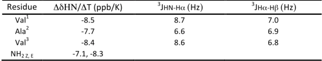
As some compounds exhibited low solubility in water, NMR conformational analyses were run in CD3OH. This protic
solvent allowed us to get as close as possible to an aqueous environment and also to record NMR spectra at low temperatures (248–273 K range), in order to better characterize slowly interconverting conformers. Different NMR parameters were examined to analyze the conformational propensities of compounds 1-‐5, in particular interproton ROE correlations, temperature coefficient (ΔδHN/ΔT) of the amide protons, and vicinal 3JHN-‐Hα coupling constants of
the natural amino acids.
As expected for a short linear peptide containing only natural α-‐amino acids, tripeptide Boc-‐Val-‐Ala-‐Val-‐NH2 1 was
found to be highly disordered. Vicinal 3J
HN-‐Hα coupling constant analysis shows that the central Ala residue is flexible
(3J
HN-‐Hα of 6.6 Hz) while the two flanking Val residues preferentially adopt extended conformations (3JHN-‐Hα of 8.7 Hz
residues (Table S3). Strong sequential and medium intraresidual Hα-‐HN ROE correlations also support mostly
extended conformations (Figure S3). The highly negative temperature coefficients of all amide protons (Table 1) confirms solvent exposure and the absence of stable intramolecular hydrogen bonds.
Fig. 3 1D 1H NMR spectra of compounds 2, 3 and 5 in methanol at 273 K, showing the region of HN signals. The assignments of the two most-‐populated forms for each compound are indicated by one-‐letter amino acid code (aX corresponds to aza-‐amino acids).
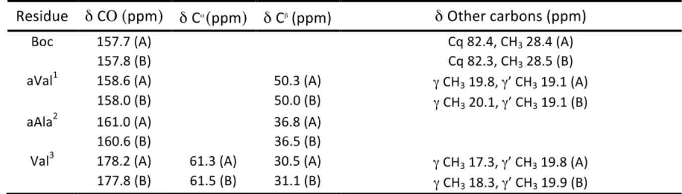
Table 1. Amide proton temperature coefficients ΔδHN/ΔT of compounds 1-‐5 in methanol. The two most-‐populated conformers observed
for compounds 2, 3 and 5 are indicated by A and B letters. Temperature coefficients above -‐4.5 ppb/°C are indicated in bold.
Res 1 Res 2A 2B Res 3A 3B Res 4 Res 5A 5B
Val1 -‐8.5 aVal1 -‐7.3 -‐7.1 aVal1 -‐7.0 -‐6.9 aGly1 -‐9.4 aVal1 -‐6.1 -‐6.5 Ala2 -‐7.7 aAla2 -‐6.6 -‐6.8 aAla2 -‐7.8 -‐7.7 aGly2 -‐6.3 aAla2 -‐5.0 -‐6.3 Val3 -‐8.4 Val3 -‐2.6 -‐2.3 Val3 -‐3.9 -‐3.9 Val3 -‐4.3 Val3 -‐2.9 -‐3.0
NH2 Z -‐7.1 NH2 Z -‐8.4 -‐7.3 NH2 Z -‐8.6 aVal4 -‐6.3 -‐4.5 NH2 E -‐8.3 NH2 E -‐3.3 -‐6.2 NH2 E -‐6.1 aAla5 -‐2.8 -‐3.6
Val6 -‐1.8 -‐2.2
NH2 Z -‐5.5 -‐8.8
NH2 E -‐5.8 -‐3.8
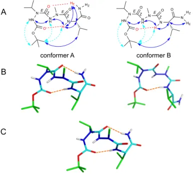
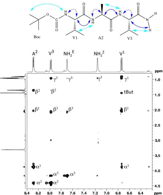
Unlike natural peptide 1, the diaza-‐peptide analogue Boc-‐aVal-‐aAla-‐Val-‐NH2 2 was characterized by a markedly
different conformational space. An equilibrium between two major conformers (45% population for each) was observed by NMR in methanol (Figures 3 and S6). Two additional minor forms were also observed (population < 5%) and were ascribed to the cis-‐trans isomerization of the Boc protective group (a feature that was also observed for tripeptide 1). Both major conformers show similar long-‐range ROEs between the tert-‐butyl protons of Boc with the amide proton and side chain methyl protons of the Val residue, indicative of folded conformations (Figure 4A). In both conformers, the amide protons of aVal1 and aAla2 residues exhibited strong variations of their temperature coefficient (Table 1), as expected for solvent-‐exposed groups, whereas the NH of Val3 has a small temperature dependence (ΔδHN/ΔT higher than -‐4.5 ppb/°C) suggesting its engagement in a stable H-‐bond. One of the two
5
2
3
aV1 aA2 V3 aV4 aA5 V6 Z NH2 E aV1 aA2 V3 aV4 aA5 V6 aV1 aV1 aA2 aA2 aV1 aA2 aV1 aA2 V3 V3 Bz NH2 Z E E Z 10 9 8 7 6 11 δ 1H (ppm) Z NH2 E V3 V3consecutive aza-‐amino acids adopt similar signs for their φ angle. NMR structure calculations refined by DFT (Figure 4B) revealed that conformer A corresponds to a double turn structure in which all residues have negative φ angles. The alternative conformer B is characterized by positive φ values for the two aza-‐amino acids and a negative φ value for Val3. Thus the conformational equilibrium observed by NMR corresponds to slow exchange within the two aza-‐ amino acid units, interconverting from –/– to +/+ φ angles. Forms A and B have conformations close to regular types I and I’ β−turns, respectively. When all residues have negative φ values (conformer A), the backbone folds in a right-‐ handed helical turn allowing the carboxamide group to be engaged in an additional H-‐bond with aVal1 carbonyl group. This H-‐bond formation is no longer possible when the two aza-‐aminoacid units change handedness (conformer B), as the L-‐Val3 residue still adopts a favored negative φ angle.
Fig. 4 Conformations of Boc-‐aVal-‐aAla-‐Val-‐NH2 2. A) NMR conformational parameters in methanol: schematic representation of ROEs (blue
and dashed cyan arrows correspond to strong, and weak intensity ROEs, respectively), and of hydrogen bonds (in red) as inferred from amide proton temperature coefficients. B) Corresponding NMR structures calculated from distance restraints using AMBER and refined by DFT with methanol PCM solvation model. The (φ, ψ) angles of aVal1 and aAla2 are (–73°, –10°) and (–64°, –16°) in conformer A, and (+71°, +16°) and (+81°, +7°) in conformer B. C) X-‐ray structure showing one of the two chains of the asymmetric unit. The average (φ, ψ) angles of X-‐ray chains are: (–62°, –19°) for aVal1, (–77°, –7°) for aAla2 and (–65°, –23°) for Val3. The Nα atoms have a small pyramidal character, as assessed by the 11° angular deviation between N-‐Cβ-‐C’ and N-‐Cβ-‐Nα planes. The DFT refinement of NMR structures yielded good description of the pyramidal character, with an angular deviation of 12° (vs. 3° before refinement).
Diaza-‐peptide 2 was amenable to crystallisation and X-‐ray diffraction data revealed a double turn structure stabilized by two intramolecular hydrogen bonds, which is very similar to conformer A characterized by NMR in solution (Figures 4C, S16 and S17). Structures of compounds were also studied by Monte Carlo conformational search using GBSA water solvation model, followed by refinement using DFT. The objective of the simulations was double: firstly, evaluate our ability to predict the conformations of peptides containing aza-‐amino acids and the rotameric states of urea moieties by in silico tool; secondly, better anticipate the peptide conformations in a solvent closer to physiological conditions by using a water solvation model. The DFT refinement enabled us to better take into account the weak pyrimidalization of the nitrogen atoms in aza-‐amino acids that was slightly underestimated in
A
B
C
conformer A conformer B O O HN N O N H N O N N O H HE HZ E Z E Z O O HN N O N H N O N N O H HE HZ E E Z Z
molecular mechanics force fields. Computational studies were fully consistent with the NMR experimental data, with a predominance of conformers showing extended or semi-‐folded structures for the natural peptide 1 (Tables S17 and S18), and folded turn structures for diaza-‐peptide 2. A very good agreement was observed with the X-‐ray structure of 2 (Figure S19).
We next studied the diaza-‐peptide analogue Boc-‐aVal-‐aAla-‐Val-‐OBn 3 having a C-‐terminal benzyl ester instead of a carboxamide as in 2, which therefore lacks a second H-‐bond donor group. Diaza-‐peptide 3 exhibits a similar conformational equilibrium in solution and adopts single hydrogen bonded β-‐turn structures, as revealed by ROEs and amide bond temperature coefficients (Table 1, Figure S8). However, the equilibrium is shifted toward one conformer (7:3). This shows that, irrespective of H-‐bond formation, the presence of a chiral L-‐amino acid induces a conformational preference for one β-‐turn folding type (I vs I’) within the achiral diaza-‐peptide unit. Computational studies suggest that the most stable conformation is type I’ β-‐turn, corresponding to +/+ φ angles for aza-‐amino acids (Table S16).
The influence of aza-‐amino acid side chain was investigated by examining tripeptide Boc-‐aGly-‐aGly-‐Val-‐NH2 4. NMR
spectra showed faster backbone rotation of aGly units with respect to substituted aza-‐amino acids as a single set of resonance was observed at room temperature. However conformational isomers in slow exchange could be detected by lowering the temperature. The amide proton of Val3 has a medium temperature coefficient (Table 1), and a weak ROE with Boc protons, indicating a weak propensity to populate β-‐turn conformations. MM/DFT calculations of Boc-‐aGly-‐aGly-‐Val-‐NH2 4 (Tables S17-‐S18) showed a wider conformational space that encompasses
more extended conformations. In particular, the absence of side chain allows aGly residue to populate two minima corresponding to ψ dihedral angles around 0° or 180°. In contrast, in substituted aza-‐amino acids, the urea motif has a preferred E,Z geometry in all low energy conformers, with ψ dihedral angles around 0°.
Thus, unlike the corresponding natural tripeptide 1 and pseudotripeptide 4 containing two aGly which turned out to be preferentially extended, aza/aza/α pseudotripeptides containing aza-‐amino acids with side chains have the tendency to adopt 10-‐membered β-‐turn conformations, with a hydrogen bond between the carbonyl group of the Boc group, mimicking a residue i and the amide proton of residue i+3. The C-‐terminal primary amide, mimicking the possible presence of an amide bond with a fourth residue, imparts to the backbone of the aza/aza/α pseudotripeptides the possibility to establish a second i, i+3 hydrogen bond offering two 10-‐membered cycles, provided that aza-‐amino acids adopt negative φ angles like the L-‐amino acid.
As the helical conformation propensity of Boc-‐aVal-‐aAla-‐Val-‐NH2 2 was demonstrated, we envisioned the possibility
to form a pseudo 310-‐helix conformation with its dimer analogue Boc-‐aVal-‐aAla-‐Val-‐aVal-‐aAla-‐Val-‐NH2 5. The NMR
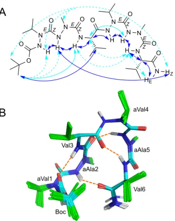
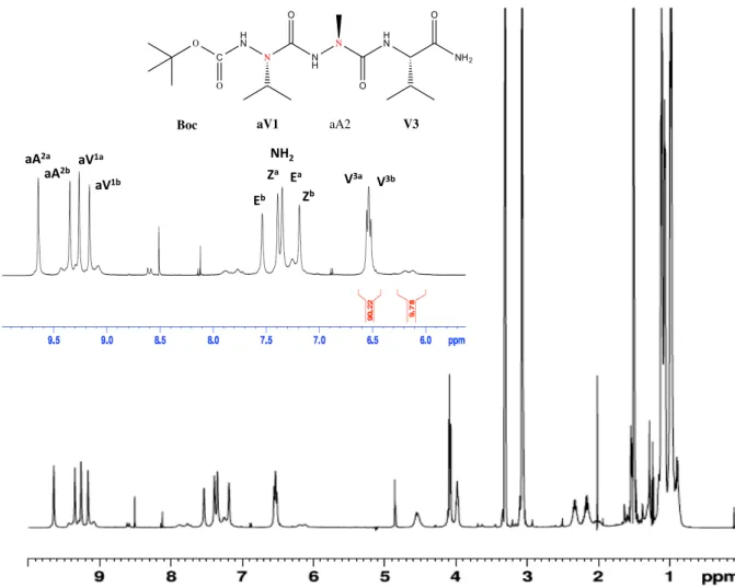
spectra of pseudo hexapeptide 5 in methanol exhibited very high complexity. Indeed, at least eight sets of resonances could be detected, corresponding to different conformational isomers in slow exchange (Figure 3). The population of the two most populated forms reaches 55% and 15%, the other minor forms being far less populated (7% or less).
All aza-‐amino acids in the different forms have strong sequential HNi/HNi+1 ROEs with the following residue, compatible with (φ, ψ) angles around (±90°, 0°). This suggests that the conformational equilibria likely correspond to slow rotation between –90° and +90° φ dihedral angles. The temperature coefficients of amide protons could be measured for the two most populated species (Table 1). Interestingly, several amide protons have small temperature dependence of their chemical shifts, supporting the formation of H-‐bonded turns in the different conformational isomers of hexapeptide 5. In particular, the major form shows evidence of H-‐bond involvement for HN groups of Val3, aAla5 and Val6, but not aVal4. Accordingly, this conformer shows several i, i+2 and i, i+3 ROE connectivities, consistent with the formation of several turn structures along the backbone (Figure 5A). NMR structure calculations converged toward a low energy conformer in which the aza-‐amino acids have φ dihedral angles around +90°, corresponding to φ signs +/+/–/+/+/– for the six residues (Figure 5B). The hexapeptide forms three H-‐bonded turns, two type I’ β-‐turns centered on diaza-‐peptide segments and a type II β-‐turn centered on Val3-‐aVal4. The repeated β-‐turn structure leads to a compact hairpin shape with close proximity of residues aAla2 and Val6 at the N-‐ and C-‐extremities, mimicking an α-‐turn geometry. This compact conformation is fully consistent with the observation of several long-‐range ROEs between carboxamide HZ proton of Val6 with Boc, aVal1 and aAla2
protons. The calculated structure is stabilized by an additional H-‐bond between the CO group of Val6 and HN of aAla2. However, this amide proton has average temperature dependence, suggesting a more transient H-‐bond engagement.
Fig. 5 Conformation of the major conformer of Boc-‐aVal-‐aAla-‐Val-‐aVal-‐aAla-‐Val-‐NH2 5A. A) ROEs observed in methanol: plain blue, plain cyan and dashed cyan arrows correspond to strong, medium and weak intensities, respectively. B) NMR structure showing the repeated turns structure. Hydrogen bonds are indicated by dashed orange lines.
Fig. 6 Conformation of the second most-‐populated conformer of Boc-‐aVal-‐aAla-‐Val-‐aVal-‐aAla-‐Val-‐NH2 5B. A) ROEs observed in methanol. B)
NMR structure showing 310-‐type helical folding. Hydrogen bonds are indicated by dashed orange lines.
B
aVal1 aVal4 Val6 aAla2 Val3 aAla5 Boc O N O N H O N H E N N H N H N N N H N O H O N E E HE HZ Z Z Z ZAzapeptide hexamérique JL54
A
B
aVal1 aVal4 Val6 aAla2 Val3 aAla5 Boc NH N N H N N H NH N N H N N H N O O O O O O HZ HE O O Z E Z E Z E Z E
Interestingly, the second most-‐populated form is characterized by small temperature coefficients of HN protons for all residues within 3–6 segment (Table 1), supporting an uninterrupted H-‐bond network, in contrast with the major conformer. This is consistent with full helix formation, which is confirmed by a sequential ROE between HN protons of Val3 and aVal4, which was not observed in the most-‐populated conformer (Figure 6A), and a smaller 3J
HN-‐Hα
coupling constant for Val3 in agreement with a helical conformation (5.2 Hz versus 6.5 Hz in the major conformer, Table S13). NMR structure calculation indicates that all residues adopt negative φ angles (–/–/–/–/–/– conformer). The fold is close to that of a regular 310 helix with i, i+3 hydrogen bonding (Figure 6B).
The high complexity and spectral overlap of NMR spectra precluded complete assignment and full characterization of the minor conformational isomers of compound 5. However, structure calculations with a limited set of NMR distance restraints suggest that the main source of conformational heterogeneity arises from the interconversion of diaza-‐peptide units between +/+ and –/– φ angles. Four main topologies can be described (Figure S18), consisting of repeated β-‐turns of types I, I’, or II, and potentially leading to partial or full helical fold.
Conclusions
In conclusion, this work brings the first structural data on peptides containing consecutive aza-‐amino acids, from combined NMR, X-‐ray and in silico MM/DFT studies. The twisted conformation around the N-‐N bonds of aza-‐amino acids induces favored φ dihedral angles around ±90°. The presence of a side chain (aVal, aAla) further restricts the ψ dihedral angles around 0°. In contrast to peptides made of α-‐amino acids, these conformational restrictions confer unique properties on the aza/aza/α pseudotripeptides giving them a strong tendency to adopt 10-‐membered β-‐turn conformations. Due to the absence of chirality, the aza-‐aza moiety can adopt type I or the mirror image type I’ β-‐ turns, corresponding to –/– or +/+ φ angles, respectively. However, the equilibrium between types I and I’ conformations is influenced by the chiral α-‐amino-‐acid. The slow exchange within the two aza-‐amino acid units, interconverting from –/– to +/+ φ angles observed in solution is displaced in favor of one conformer at the solid-‐ state as demonstrated in the X-‐ray diffraction data. Similarly, it can be assumed that this conformational equilibrium can be shifted upon interaction with a biological target. We have then explored the potentiality of aza/aza/α bricks to adopt hydrogen-‐bonded structures in longer peptides, using a pseudohexapeptide foldamer aza/aza/α/aza/aza/α. This compound was proven to adopt repeated β-‐turns or fully helical structures. Similar i, i+3 C=O·∙·∙·∙H-‐N hydrogen-‐bonded helices were reported by S. Gellman et al. using backbones that contain pure β-‐residue oligomers (12-‐helix), and oligomers containing a 1:1 α:β repeat (11-‐helix),13 and either a 2:1 or a 1:2 repeating pattern of α-‐ and β-‐amino acid residues (12/11 or 10/11 type helix).14 12-‐helix, 13-‐helix and α-‐helix-‐like conformation were also observed by S. Gellman et al. in α/γ, β/γ and α/β/γ-‐peptides respectively.15,16,17 However, the 310 helix observed in aza/aza/α/aza/aza/α is more comparable to i, i+3 hydrogen-‐bonded helices formed by
pure α-‐residue oligomers. Interestingly also, a hairpin structure was observed in the major conformer mimicking an α-‐turn geometry. Very few molecules have been described to stabilize isolated α-‐turn conformations and the strategy is essentially to replace the i, i+4 hydrogen bond by a covalent bond.18 As far as we know, this work provides the proof of concept that foldamers based on diaza-‐amino acids units might resolve a major issue in the use of peptides as drugs, by stabilizing turns or helical conformations similar to natural peptides and retaining the selectivity due to the lateral chains. This new class of foldamers provides valuable opportunities to explore them in many different areas and applications. In particular, the number of aza/aza/α bricks and the nature of the side chains can be modulated and adapted to further promote helical structures, and to design helical mimetics as ligands of helical structures of major biological targets. We can explore them for example as selective inhibitors of protein-‐protein interactions.19,20 The adaptive chirality of the aza-‐aza subunit can be an advantage as it is recognized that compounds adopting several kinetically and thermodynamically accessible conformations might be much more powerful inhibitors of protein-‐protein interactions with respect to rigid ones.21,22 Helical mimetics can also afford various applications such as membrane active peptides possessing antimicrobial, cytolytic, or cell penetrating properties.23,24,25
room temperature TXI probe (compounds 1-‐4) or a cryogenic TCI probe (compound 5). NMR data were processed with Topspin 3.2 (Bruker) and 2D experiments were analysed with NMRFAM-‐SPARKY program. 1H and 13C chemical shifts were referenced to the solvent signal (residual protonated CHD2OH at 3.31 ppm and deuterated 13CD3OH at 49.0 ppm, respectively) and 15N chemical shifts were referenced
indirectly. 1H, 13C and 15N resonance assignments were obtained from the analysis of 2D 1H-‐1H TOCSY (mixing time of 70 ms), 2D 1H-‐1H ROESY (mixing times of 0.5-‐0.8 s), 2D 1H-‐13C HSQC, 2D 1H-‐13C HMBC, and 2D 1H-‐15N HSQC spectra. The temperature gradients of the amide proton chemical shifts were derived from a series of 1D 1H spectra recorded between 258 K and 313 K for compounds 1-‐4, and from 2D 1H-‐
15N HSQC spectra recorded between 272 K and 298 K for compound 5.
NMR structure calculation
Aza-‐amino acids were parameterized using gaff forcefield atom types and partial charges were computed with AM1-‐BCC method implemented in Antechamber. Structures of compounds 2 and 5 were calculated using Amber14 program and ff14SB forcefield, using NMR-‐restrained molecular dynamics, as previously described.26 Selected structures were refined without restraints by DFT at the B3LYP-‐ D3/cc-‐pVTZlevel in Gaussian 16 (see below), using PCM solvation model matched for methanol.
Molecular mechanics and DFT calculations
Arbitrary initial conformations of compounds 1–5 were energy-‐minimized according to the conjugate gradient method27 in the OPLS-‐
2005 force field28 combined with GBSA implicit water solvation model29 as implemented in the MacroModel v10.2 software package. 30 The convergence criterion was set to 0.05 kJ·∙mol−1·∙Å−1 on the energy gradient. Resulting coordinates were then individually subjected to an
unconstrained conformational search according to the MCMM (Monte Carlo Multiple Minima) method31 with similar parameters. 100,000 conformations were calculated and clustered by the centroid linkage method with a 0.66 Å merge distance threshold.
Geometries of selected models were then optimized without constraint in the framework of the density functional theory32 using Becke
three-‐parameter Lee–Yang–Parr exchange-‐correlation functional,33 Dunning’s correlation consistent basis set of triple ζ valence quality,34 and the D3 version of Grimme’s dispersion correction,35 in conjunction with the polarizable continuum solvation model parametrized for water,36 as implemented in the Gaussian 16, revision B.01 software package.37 Stationary points were qualified as energy minima by checking for the absence of imaginary frequency after performing vibrational analysis at the same level of theory upon geometrical convergence. Thermodynamic quantities at 298.15 K were calculated using the zero-‐point and thermal energy corrections derived from unscaled frequencies.
Conflicts of interest
There are no conflicts to declare.
Acknowledgements
The French Ministère de l’Enseignement Supérieur et de la Recherche (MESR) is thanked for the PhD fellowships for N. Tonali and J. Lesma. Karine Leblanc (Service d'Analyses–HPLC-‐Masse BioCIS, Univ. Paris Saclay) is thanked for HPLC and HRMS analyses. The DRX Platform of Sorbonne Université is thanked for the crystallographic data. The Laboratory BioCIS is a member of the Laboratory of Excellence LERMIT supported by a Grant from ANR (ANR-‐10-‐LABX-‐33).
References
1 (a) K. Fosgerau and T. Hoffmann, Drug Discov Today, 2015, 20, 122; (b) J. L. Lau and M.K. Dunn, Bioorg. Med. Chem., 2018, 26, 2700.
2 (a) C. Proulx , D. Sabatino , R. Hopewell, J. Spiegel, Y. García Ramos and W. D. Lubell, Future Med Chem., 2011, 3, 1139-‐1164; (b) A. Zega, Curr. Med. Chem., 2005, 12, 589-‐597; (c) A. Begum, D. Sujatha, K.V.S.R.G. Prasad and K. Bharathi, Asian J. Chem., 2017,
29,1879.
3 (a) J. Lee and M. Bogyo, Bioorg. Med. Chem. Lett., 2012, 22, 1340; (b) A. J. Kasznel, Y. Zhang, Y. Hai, D. M. Chenoweth, J. Am.
Chem. Soc., 2017, 139, 9427.
4 (a) R. Chingle, C. Proulx and W. D. Lubell, Acc. Chem. Res. 2017, 50, 1541; (b) F. M. Mir, N. D. Prasad Atmuri, C. B. Bourguet, J. Rodon Fores, X. Hou, S. Chemtob and W. D. Lubell, J. Med. Chem., 2019, 62, 4500.
5 J. T. Randolph, X. Zhang, P. P. Huang, L. L. Klein, K. A. Kurtz, A. K. Konstantinidis, W. He, W. M. Kati and D. J. Kempf, Bioorg. Med.
Chem. Lett., 2008, 18, 2745.
6 M. Galibert, M. Wartenberg, F. Lecaille, A. Saidi, S. Mavel, A. Joulin-‐Giet, B. Korkmaz, D. Brömme, V. Aucagne, A. F. Delmas and G. Lalmanach, Eur. J. Med. Chem. 2018, 144, 201.
7 (a) M. Thormann and H.-‐J. Hofmann, J. Mol. Struct. (THEOCHEM), 1999, 469, 63; (b) F. Ramondo and L. Bencivenni, J. Chem. Soc.,
Perkin Trans. 2, 1995, 1797; (c) C. H. Reynolds and R. E. Hormann, J. Am. Chem. Soc., 1996, 118, 9395.
8 (a) H.-‐J. Lee, I.-‐A. Ahn, S. Ro, K.-‐H. Choi, Y.-‐S. Choi and K.-‐B. Lee, J. Peptide Res., 2000, 56, 35; (b) H.-‐J. Lee, H. M. Park and K.-‐B. Lee, J. Mol. Struct., 2001, 569, 43; (c) H.-‐J. Lee, K.-‐H. Choi, I.-‐A. Ahn, S. Ro, H. G. Jang, Y.-‐S. Choi and K.-‐B. Lee, Biophys. Chem., 2007, 125, 117; (d) C. Abbas, G. Pickaert, C. Didierjean, R. Vanderesse and B. Jamart-‐Grégoire, Tetrahedron Lett., 2009, 50, 4158. 9 Z. Zhou, C. Deng, C. Abbas, C. Didierjean, M.-‐C. Averlant-‐Petit, J. Bodiguel, R. Vanderesse and B. Jamart-‐Grégoire, Eur. J. Org.
Chem., 2014, 7643.
10 Y. Zhang, R. M. Malamakal, D. M. Chenoweth, J. Am. Chem. Soc., 2015, 137, 12422.
11 F. Bizet, N. Tonali, J.-‐L. Soulier, A. Oliva, J. Kaffy, B. Crousse and S. Ongeri, N. J. Chem. 2018, 42, 17062.
12 L. Dufau, A. S. Marques Ressurreição, R. Fanelli, N. Kihal, A. Vidu, T.Milcent, J.-‐L. Soulier, J. Rodrigo, A. Desvergne, K. Leblanc, G. Bernadat, B. Crousse, M. Reboud-‐Ravaux and S. Ongeri, J. Med. Chem., 2012, 55, 6762.
13 M. A. Schmitt, S. H. Choi, I. A. Guzei and S. H. Gellman, J. Am. Chem. Soc. 2005, 127, 13130. 14 S. H. Choi, I. A. Guzei, L. C. Spencer, S. H. Gellman, J. Am. Chem. Soc. 2009, 131, 2917.
15 L. Guo, Y. Chi, Almeida, A. M.; Guzei, I. A.; Parker, B. K.; Gellman, S. H. J. Am. Chem. Soc. 2009, 131, 16018.
16 Guo, L.; Almeida, A. M.; Zhang, W.; Reidenbach, A. G.; Choi, S. H.; Guzei, I. A.; Gellman, S. H. J. Am. Chem. Soc. 2010, 132, 7868. 17 Y-‐H. Shin, D. E. Mortenson, K. A. Satyshur, K. T. Forest, S. H. Gellman, J. Am. Chem. Soc. 2013, 135, 8149.
18 L. Wang, P. Coric, K. Zhu, W-‐Q. Liu, M. Vidal, S. Bouaziz, S. Broussy, Org. Biomol. Chem. 2018, 16, 459.
19 M. Pelay-‐Gimeno, A. Glas, O. Koch and T. N. Grossmann, Angew. Chem. Int. Ed., 2015, 54, 8896. 20 J. Laxio-‐Arenas, J. Kaffy and S. Ongeri, Curr. Opin. Chem. Biol. 2019, 52, 157.
21 (a) X. Li, J. Taechalertpaisarn, D. Xin, K. Burgess Org. Lett., 2015, 17, 632; b) E. Ko, A. Raghuraman, L. M. Perez, T. R. Ioerger, K. Burgess J. Am. Chem. Soc. 2012, 135, 167.
22 S. Pellegrino, N. Tonali, E. Erba, J. Kaffy, M. Taverna, A. Contini, M. Taylor, D. Allsop, M. L. Gelmi, S. Ongeri, Chem. Sci., 2017, 8, 1295.
23 J. Li, J.-‐J. Koh, S. Liu, R. Lakshminarayanan, C. S. Verma and R. W.Beuerman, Front. Neurosci., 2017, 11, 73. 24 M. R. Felício, O. N. Silva, S. Gonçalves, N. C. Santos and O. L. Franco, Front. Chem., 2017, 5, 5.
25 D. Kalafatovic and E. Giralt, Molecules, 2017, 22, 1929.
26 C. Byrne, M. Belnou, E.-‐E. Baulieu, O. Lequin and Y. Jacquot, Pept. Sci., 2019, e24113.
27 E. Polak and G. Ribière, Revue Française d’Informatique et de Recherche Opérationnelle, 1969, 16, 35.
28 J. L. Banks, H. S. Beard, Y. Cao, A. E. Cho, W. Damm, R. Farid, A. K. Felts, T. A. Halgren, D. T. Mainz, J. R. Maple, R. Murphy, D. M. Philipp, M. P. Repasky, L. Y. Zhang, B. J. Berne, R. A. Friesner, E. Gallicchio and R. M. Levy, J. Comp. Chem., 2005, 26, 1752. 29 W. C. Still, A. Tempczyk, R. C. Hawley and T. Hendrickson, J. Am. Chem. Soc. 1990, 112, 6127.
30 MacroModel, version 10.2, Schrodinger, LLC, New York, NY (USA) 2013. 31 G. Chang, W. C. Guida and W. C. Still, J. Am. Chem. Soc., 1989, 111, 4379.
32 (a) P. Hohenberg and W. Kohn, Phys. Rev., 1964, 136, B864–B871; (b) W. Kohn and L. J. Sham, Phys. Rev., 1965, 140, A1133. 33 (a) C. Lee, W. Yang and R. G. Parr, Phys. Rev. B, 1988, 37, 785–789; (b) A. D. Becke, J. Chem. Phys., 1993, 98, 5648.
34 R. A. Kendall, T. H. Dunning Jr. and R. J. Harrison, J. Chem. Phys., 1992, 96, 6796. 35 S. Grimme, J. Antony, S. Ehrlich and H. Krieg, J. Chem. Phys., 2010, 132, 154104. 36 J. Tomasi, B. Mennucci and R. Cammi, Chem. Rev., 2005, 105, 2999.
37 M. J. Frisch, G. W. Trucks, H. B. Schlegel, G. E. Scuseria, M. A. Robb, J. R. Cheeseman, G. Scalmani, V. Barone, G. A. Petersson, H. Nakatsuji, X. Li, M. Caricato, A. V. Marenich, J. Bloino, B. G. Janesko, R. Gomperts, B. Mennucci, H. P. Hratchian, J. V. Ortiz, A. F. Izmaylov, J. L. Sonnenberg, D. Williams-‐Young, F. Ding, F. Lipparini, F. Egidi, J. Goings, B. Peng, A. Petrone, T. Henderson, D. Ranasinghe, V. G. Zakrzewski, J. Gao, N. Rega, G. Zheng, W. Liang, M. Hada, M. Ehara, K. Toyota, R. Fukuda, J. Hasegawa, M. Ishida, T. Nakajima, Y. Honda, O. Kitao, H. Nakai, T. Vreven, K. Throssell, J. A. Montgomery, Jr., J. E. Peralta, F. Ogliaro, M. J. Bearpark, J. J. Heyd, E. N. Brothers, K. N. Kudin, V. N. Staroverov, T. A. Keith, R. Kobayashi, J. Normand, K. Raghavachari, A. P. Rendell, J. C. Burant, S. S. Iyengar, J. Tomasi, M. Cossi, J. M. Millam, M. Klene, C. Adamo, R. Cammi, J. W. Ochterski, R. L. Martin, K. Morokuma, O. Farkas, J. B. Foresman and D. J. Fox, Gaussian 16, Revision B.01, Gaussian, Inc., Wallingford CT (USA) 2016.
E-mail: sandrine.ongeri@universite-paris-saclay.fr
[b] Sorbonne Université, Ecole Normale Supérieure, PSL University, CNRS, Laboratoire des Biomolécules, 4
place Jussieu, 75252 Paris Cedex 05, France. Email: olivier.lequin@sorbonne-universite.fr
Table of contents
Pages S2-S11 Synthesis and characterization of Boc-Val-Ala-Val-NH2 1, Boc-aVal-aAla-Val-OBn 3, and
Boc-aVal-aAla-Val-aVal-aAla-Val-NH2 5
Pages S12-S27 NMR data of compounds 1-5: assignment tables, conformational parameters and NMR spectra
Page S28 Crystallographic data for Boc-aVal-aAla-Val-NH2 2
Pages S29-S31 NMR structures
Pages S32-S36 Ab initio conformational search: molecular mechanics and DFT calculations Pages S37-S61 Atomic coordinates of molecular models refined by DFT
Synthesis and characterization of Boc-Val-Ala-Val-NH
21, Boc-aVal-aAla-Val-OBn 3,
and Boc-aVal-aAla-Val-aVal-aAla-Val-NH
25
General Experimental Methods
Usual dry solvents were purchased from commercial sources. 4-nitrophenyl chloroformate, N,N,N′,N′-Tetramethyl-O-(1H-benzotriazol-1-yl)uronium hexafluorophosphate (HBTU), 4-(4,6-dimethoxy-1,3,5-triazin-2-yl)-4-methylmorpholinium chloride (DMTMM(Cl-)), 1-hydroxybenzotriazole (HOBt), [Bis(dimethylamino)methylene]-1H-1,2,3-triazolo[4,5-b]pyridinium 3-oxid hexafluorophosphate (HATU), 1-Hydroxy-7-azabenzotriazole (HOAt), Boc-NH-Val-OH, L-Ala-OCH3, L-Val-NH2 and L-Val-OBn were
purchased from commercial sources. Boc-aVal-aAla-Val-NH2 2 and Boc-aGly-aGly-Val-NH2 4 were prepared
according to our published methods (F. Bizet, N. Tonali, J.-L. Soulier, A. Oliva, J. Kaffy, B. Crousse, S. Ongeri, N. J. Chem. 2018, 42, 17062-17072; L. Dufau, A. S. Marques Ressurreição, R. Fanelli, N. Kihal, A. Vidu, T.Milcent, J.-L. Soulier, J. Rodrigo, A. Desvergne, K. Leblanc, G. Bernadat, B. Crousse, M. Reboud-Ravaux,S. Ongeri, J. Med. Chem. 2012, 55, 6762–6775). Pure products were obtained after liquid chromatography using Merck silica gel 60 (40−63 µm). TLC analyses were performed on silica gel 60F-250 (0.26 mm thickness) plates. The plates were visualized with UV light (λ = 254 nm) or revealed with a 5% solution of phosphomolybdic acid in EtOH or with a solution of ninhydrin in EtOH. Melting points were determined on a Kofler melting point apparatus. Element analyses (C, H, and N) were performed on a PerkinElmer C, H, N Analyzer 2400 at the Microanalyses Service of the Faculty of Pharmacy at Châtenay-Malabry (BioCIS, France). NMR spectra were recorded on an Ultrafield Bruker AVANCE 300 (1H, 300 MHz,
13C, 75 MHz) or on a Bruker Avance 400 (1H, 400 MHz, 13C, 100 MHz), or on a Bruker NMR spectrometer
operating at a 1H frequency of 500.3 MHz and equipped with either a room temperature TXI probe or a cryogenic TCI probe. Chemical shifts δ are in ppm, and the following abbreviations are used: singlet (s), broad singlet (bs), doublet (d), doublet of triplet (dt), triplet (t), multiplet (m). IR spectra were recorded on a Bruker Vector 22 FT-IR spectrometer. HRMS were obtained using a TOF LCT Premier apparatus (Waters), with an electrospray ionization source. The purity of compounds was determined by HPLC using the 2695 Alliance system (Waters) and a XBridge Select (C18, 3.5 µm, 150 mm × 2.1 mm); mobile phase, MeCN/H2O
+ 0.1% formic acid from 5 to 100% in 20 min; detection at 220 nm or 245 nm; flow rate 0.25 mL/min.
under reduced pressure. Purification by column chromatography on silica gel using Cyclohexane/EtOAc 7:3 as eluent afforded compound 6 (564 mg, 1.87 mmol, 81%) as a white solid. Rf = 0.35 (Cyclohexane /EtOAc 7:3); 1H NMR (DMSO-d6, 300 MHz): 8.29 (1H, d, J = 6.5 Hz); 6.61 (1H, d, J = 9.0 Hz); 4.25 (1H, m); 3.78
(1H, m); 3.60 (3H, s); 1.92 (1H, m); 1.37 (9H, s); 1.27 (3H, d, J = 7.3 Hz); 0.86 (3H, d, J = 6.7 Hz);0.81 (3H, d, J = 6.7 Hz) 13C NMR (DMSO-d6, 75 MHz): 172.9; 171.3; 155.4; 78.0; 59.1; 51.8; 47.5; 30.6; 28.2; 19.1,
18.0; 16.9; mp = 142-144 °C; IR: 3310 cm-1; 2976 cm-1; 1751, 1682, 1649 cm-1; 1557, 1524 cm-1; 1160 cm-1. HRMS (TOF ESI, ion polarity positive): m/z 325.1739 calc. for [C14H26N2O5 + Na]+, found 325.1746.
tert-butyl N-[(1S)-1-[[(1S)-2-[[(1S)-1-carbamoyl-2-methyl-propyl]amino]-1-methyl-2-oxo-ethyl]carbamoyl]-2-methyl-propyl]carbamate (1): Compound 6 (495 mg, 1.64 mmol, 1.0 eq.) was
dissolved in MeOH (20.0 mL) and an aqueous solution of NaOH 2 M (4.1 mL, 8.19 mmol, 5.0 eq.) was added dropwise to the solution. The reaction was stirred at 60° C for 1 h. The volatiles were evaporated and the solid obtained was solubilized in water. The mixture was acidified with 10% aqueous KHSO4 solution
until pH = 2-3. The product was extracted from the water phase with DCM. The organic phase was dried over Na2SO4 and concentrated under vacuum to afford the free carboxylic acid (417 mg, 1.45 mmol, 89%)
that was used without any further purification and dissolved in anhydrous DMF (10mL). The solution was cooled at 0°C and HOBt (245 mg, 1.6 mmol, 1.1 eq.) and HBTU (607 mg, 1.6 mmol, 1.1 eq.) were added. The reaction mixture was stirred at 0°C for 30 min and the solution of HCl·NH2-Val-NH2 (221 mg, 1.45 mmol,
1.0 eq.) and DIPEA (0.74 mL, 4.35 mmol, 3.0 eq.) in DMF (10.0 mL) were added. The reaction was carried at room temperature overnight. After evaporation of the volatiles under vacuum, the oily residue was taken up with EtOAc. After few minutes a white solid precipitated which after filtration afforded compound 1 (440 mg, 1.14 mmol,79% so 70% for two steps from compound 6) as a white solid. 1H NMR (DMSO-d6, 400 MHz):
7.92 (1H, d, J = 7.2 Hz); 7.69 (1H, d, J = 8.9 Hz); 7.36 (1H, s); 7.01 (1H, s); 6.71 (1H, d, J = 8.9 Hz); 4.38 (1H, m); 4.09 (1H, dd, J = 8.8, 6.5 Hz); 3.80 (1H, m); 1.94 (2H, m); 1.37 (9H, s); 1.19 (3H, d, J = 6.9 Hz); 0.82 (12H, m); 13C NMR (DMSO-d6, 400 MHz): 172.7; 171.8; 170.9; 155.4; 78.0; 59.5; 57.2; 48.0; 30.5, 30.3; 28.2;
19.2; 18.1; 17.9; 17.8;; mp = 266-270 °C; IR: 3293 cm-1; 2960 cm-1; 1671, 1626 cm-1; 1529 cm-1. HRMS (TOF ESI, ion polarity positive): m/z 387.2607 calc. for [C18H35N4O5+ H]+ and 409.2427 calc. for
[C18H35N4O5+ Na]+, found 387.2602 and 409.2427; Elemental analysis: C18H34N4O5 + 1.5 H2O calcd C 52.28,
H 9.04, N 13.55; found C 52.37, H 8.24, N 13.60. HPLC purity (XBridge Select C18, 3.5 µm, H2O + 0.1%
form. ac./ACN – grd 5-100% in 20 min, detection at 245 nm): TR = 11.83 min, 100%.
1.49 (9H, s); 1.12 (6H, m).0.93 (6H, m); 13C NMR ( 100 MHz, DMSO-d6): 173.0; 158.6; 151.9; 144.5; 135.9;
128.6, 128.3; 82.9; 66.5; 58.7; 48.9; 36.2; 31.1; 28.1; 19.2; 18.9; mp = 174 – 176 °C; IR: 3391, 3296 cm-1 (N-H stretch); 2975 cm-1 (C-H stretch); 1740, 1725, 1673, 1643 cm-1 (C=O stretch); 1536, 1494 cm-1 (N-H bend); 1272, 1243 cm-1 (C-N stretch); 1179 cm-1 (C-C(O)-C stretch); HRMS (TOF ESI, ion polarity positive): m/z 480.2822 calc. for [C23H38N5O6+ H]+, found 480.2823; HPLC purity (XBridge Select C18, 3.5 µm, H2O + 0.1%
form. ac./ACN – grd 5-100% in 20 min): TR = 17.63 min, 97%
(2S)-2-[[[[(tert-butoxycarbonylamino)-isopropyl-carbamoyl]amino]-methyl-carbamoyl]amino]-3-methyl-butanoic acid (8): 3 (100 mg, 0.21 mmol, 1.0 eq) was dissolved in methanol (3 mL) and then Pd/C
10% was added (10 mg). The reaction mixture was stirred at room temperature under hydrogen atmosphere. After complete disappearance of the starting material, the mixture was filtered through a celite pad and after evaporation of the volatiles the desired product 8 was obtained (82 mg, 0.21 mmol, 100 %) and used without any further purification. HRMS (TOF ESI, ion polarity positive): m/z 390.2308 calc. for [C16H31N5O6+ H]+ and
412.2172 calc. for [C16H31N5O6+ Na]+, found 390.2345 and 412.2177.
Benzyl (2S)-2-[[[[amino(isopropyl)carbamoyl]amino]-methyl-carbamoyl]amino]-3-methyl-butanoate
(9):
To a solution of 3 (100.0 mg, 0.21 mmol, 1.0 eq) in dioxane (3 mL), a 4 N solution of HCl in dioxane (1.7 mL, 40.0 eq.) was added at 0°C. The mixture was stirred at room temperature until complete disappearance of the starting material (monitored by TLC). The desired product 9 (79.7 mg, 0.21 mmol, 100 %) was isolated as its hydrochloride salt by precipitation in diethyl ether and used for the next step without any further purification. HRMS (TOF ESI, ion polarity positive): m/z 380.2253 calc. for [C17H28N4O6+ H]+ and 402.2117
calc. for [C17H28N4O6+ Na]+, found 380.2292 and 402.2139.
Benzyl
(2S)-2-[[[[[[(2S)-2-[[[[(tert-butoxycarbonylamino)-isopropyl-carbamoyl]amino]-methyl- carbamoyl]amino]-3-methyl-butanoyl]amino]-isopropyl-carbamoyl]amino]-methyl-carbamoyl]amino]-3-methyl-butanoate (10): 8 (79.7 mg, 0.21 mmol) was dissolved in DMF (2 mL) under hydrogen
atmosphere and cooled at 0°C. At these time HOAt (32 mg, 0.23 mmol, 1.1 eq) and HATU (88mg, 0.23 mmol, 1.1 eq) were added and the mixture was stirred for 1 hour. Then 9 (81.8 mg, 0.21mmol) and DIPEA (146 µL, 0.84 mmol, 4.0 eq) were added and the mixture was stirred at room temperature overnight. The volatiles were evaporated under reduced pressure, the residue was taken up with AcOEt and washed with citric acid (10% in water), saturated NaHCO3 and brine, dried over Na2SO4 filtered and concentrated under
vacuum. The crude was purified by column chromatography on a silica gel using AcOEt/MeOH 95:5 as eluent to afford 10 (24 mg, 0.031 mmol, 16 %) Rf= 0.2 (AcOEt/MeOH 95/5) 1H NMR (300 MHz, CDCl3): δ =
8.78 (1H, bs); 8.18 (1H, bs); 8.11 (1H, bs); 7.96 (1H, bs); 7.28 (5H, m); 6.61 (1H, bs); 6.10 (1H, bs); 5.14 (2H, m); 4.62 (2H, m); 4.37 (2H, m); 3.20 (6H, s); 2.90 (2H, m); 1.39 (9H, s); 1.20- 0.90 (24H, m); 13C NMR (75 MHz, CDCl3): 174.0; 157.5; 152.1; 142.1; 136.0; 128.7, 127.0; 82.9; 66.5; 59.1; 48.9; 48.8; 36.4; 31.1;
30.9; 28.3; 20.0; 19.6; 18.2; HRMS (TOF ESI, ion polarity positive): m/z 751.4466 calc. for [C34H58N10O9+ H]+
and 773.4286 calc. for [C34H58N10O9+ Na]+, found 751.4482 and 773.4291;
tert-butyl N-[[[(1S)-1-[[[[(1S)-1-carbamoyl-2-methyl-propyl]carbamoyl-methyl-amino]carbamoyl-
isopropyl-amino]carbamoyl]-2-methyl-propyl]carbamoyl-methyl-amino]carbamoyl-isopropyl-amino]carbamate (5): To a solution of 10 (24.0 mg, 0.031 mmol,1.0 eq) in methanol (1 mL) was added
Pd/C 10% (10.0 mg, 0.009 mmol, 0.3 eq). The reaction mixture was stirred at room temperature under hydrogen atmosphere. After complete disappearance of the starting material, the mixture was filtered through a celite pad. After evaporation of the volatiles under reduced pressure the white solid was dissolved in isopropanol (1 mL) and DMTMM·HCl (12.5 mg, 0.045mmol, 1.5 eq) was added. At the resulting suspension, a solution of NH3 in methanol (13 µL, 0.087 mmol, 3 eq) was slowly dropped. The mixture was
AcOEt and the organic layer was washed with saturated aqueous NaHCO3, dried over Na2SO4, filtered and
concentrated under vacuum. Purification by column chromatography on silica gel AcOEt/MeOH 95/5 and then 80/20 afforded compound 5 (6.3 mg, 9.6 µmol, 33%) as a white solid. Rf= 0.1 (AcOEt/MeOH 95/5). For
1H and 13C NMR, see Tables S13-S14. HRMS (TOF ESI, ion polarity positive): m/z 660.4157 calc. for
[C27H53N11O8+ H]+ and 682.3976 calc. for [C27H53N11O8+ Na]+ found 660.4157 and 682.3978; HPLC purity
(XBridge Select C18, 3.5 µm, H2O + 0.1% form. ac./ACN – grd 5-100% in 20 min): TR = 15.12 min.
NMR data of compounds 1-5
NMR data for peptide 1 (Boc-Val-Ala-Val-NH
2)
Table S1.
1H NMR chemical shifts of peptide 1 (4 mM) in CD
3
OH at 293 K
Residue
δ HN (ppm),
3J (Hz)
δH
α(ppm),
3J (Hz)
δ H
β(ppm)
δ Other protons (ppm)
Boc CH3 1.44 Val1 6.62 (d, J = 8.7) 3.87 (dd, J = 8.7, 7.0) 2.03 γ CH3 0.95, γ’ CH3 0.91 Ala2 8.18 (d, J = 6.6) 4.41 (q, J = 6.9) 1.36 Val3 7.86 (d, J = 8.6) 4.17 (dd, J = 8.6, 6.8) 2.07 γ CH3 0.96, γ’ CH3 0.95 NH2 Z, E 7.04 (s), 7.60 (s)
Table S2.
13C NMR chemical shifts of peptide 1 (4 mM) in CD
3
OH at 293 K
Residue δ CΟ (ppm) δ C
α(ppm) δ C
β(ppm) δ Other carbons (ppm)
Boc 158.0 Cq 80.5, CH3 28.6
Val1 174.3 61.4 32.0 γ CH3 19.74, γ’ CH3 18.2
Ala2 174.7 50.4 17.9
Val3 176.1 59.8 31.8 γ CH3 19.69, γ’ CH3 18.3
Table S3. NMR conformational parameters for peptide 1 in CD
3OH at 293 K
Residue
ΔδΗΝ/ΔT (ppb/K)
3J
HN-‐Hα(Hz)
3J
Hα-‐Hβ(Hz)
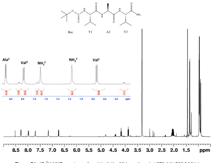
Val1 -‐8.5 8.7 7.0 Ala2 -‐7.7 6.6 6.9 Val3 -‐8.4 8.6 6.8 NH2 Z, E -‐7.1, -‐8.3Figure S1: 1D 1H NMR spectrum of peptide 1 (4 mM) in methanol at 278.4 K (500.3 MHz)
Figure S2: 1D 13C DEPTQ NMR spectrum of peptide 1 (4 mM) in methanol at 278.4 K (125.8 MHz)
Ala Val3% NH 2E% NH2 Val 1" Figure%S5:"1D"1H"NMR"spectrum"of"pep3de"1%(4mM)"in"methanol"at"278.4"K"(500.3"MHz)" Figure%S6:"1D"13C"DEPTQ"NMR"spectrum"of"pep3de"1%(4"mM)"in"methanol"at"278.4"K"(125.8"MHz)" C NH2 H N O V1 N H O A2 H N O V3 O O Boc