HAL Id: dumas-02383470
https://dumas.ccsd.cnrs.fr/dumas-02383470
Submitted on 27 Nov 2019HAL is a multi-disciplinary open access archive for the deposit and dissemination of sci-entific research documents, whether they are pub-lished or not. The documents may come from teaching and research institutions in France or abroad, or from public or private research centers.
L’archive ouverte pluridisciplinaire HAL, est destinée au dépôt et à la diffusion de documents scientifiques de niveau recherche, publiés ou non, émanant des établissements d’enseignement et de recherche français ou étrangers, des laboratoires publics ou privés.
Rehabilitation in patients with pulmonary arterial
hypertension and chronic thromboembolic pulmonary
hypertension: results from the Grenoble experience
Thomas Barret
To cite this version:
Thomas Barret. Rehabilitation in patients with pulmonary arterial hypertension and chronic throm-boembolic pulmonary hypertension: results from the Grenoble experience. Human health and pathol-ogy. 2019. �dumas-02383470�
AVERTISSEMENT
Ce document est le fruit d'un long travail approuvé par le
jury de soutenance et mis à disposition de l'ensemble de la
communauté universitaire élargie.
Il n’a pas été réévalué depuis la date de soutenance.
Il est soumis à la propriété intellectuelle de l'auteur. Ceci
implique une obligation de citation et de référencement
lors de l’utilisation de ce document.
D’autre part, toute contrefaçon, plagiat, reproduction illicite
encourt une poursuite pénale.
Contact au SID de Grenoble :
bump-theses@univ-grenoble-alpes.fr
LIENS
LIENS
Code de la Propriété Intellectuelle. articles L 122. 4
Code de la Propriété Intellectuelle. articles L 335.2- L 335.10
1
UNIVERSITÉ GRENOBLE ALPES UFR DE MÉDECINE DE GRENOBLE
Année : 2019
LA READAPTATION CARDIAQUE DANS LA PRISE EN CHARGE DE L’HYPERTENSION ARTERIELLE PULMONAIRE ET DE L’HYPERTENSION
PULMONAIRE THROMBOEMBOLIQUE CHRONIQUE : RÉSULTATS DE L’EXPÉRIENCE GRENOBLOISE
THÈSE
PRÉSENTÉE POUR L’OBTENTION DU TITRE DE DOCTEUR EN MÉDECINE DIPLÔME D’ÉTAT
Thomas BARRET
THÈSE SOUTENUE PUBLIQUEMENT À LA FACULTÉ DE MÉDECINE DE GRENOBLE
Le 25 Novembre 2019
DEVANT LE JURY COMPOSÉ DE Président du jury :
M. le Professeur Bruno DEGANO Membres :
Me le Docteur Hélène BOUVAIST, directrice de thèse M. le Professeur Gérald VANZETTO
M. le Professeur Christophe PISON Me le Docteur Cécile ROCCA
L’UFR de Médecine de Grenoble n’entend donner aucune approbation ni improbation aux opinions émises dans les thèses ; ces opinions sont considérées comme propres à leurs auteurs.
6
Remerciements
Monsieur le Professeur Bruno Degano,
Vous me faites l’honneur d’être le président de ma thèse. Merci d’avoir accepté de piloter ce travail, et d’avoir consacré de votre temps pour m’aiguiller. Je reste impressionné par la pertinence de vos remarques durant nos échanges. Votre savoir et votre pédagogie forcent le respect. Veuillez trouver ici le témoignage de toute ma reconnaissance.
Madame le Docteur Hélène Bouvaist,
Vous m’avez fait l’honneur de me confier ce sujet et je vous en remercie. Je voudrais vous remercier également pour vos conseils avisés pendant ce travail. Depuis mon premier jour en post USIC où je vous ai rencontrée, vous avez su garder votre sourire et votre bienveillance en toutes circonstances. J’ai la chance de travailler à vos côtés et je suis admiratif devant votre implication et vos connaissances dans tous les domaines de la cardiologie et bien au-delà.
Monsieur le Professeur Gérald Vanzetto,
Notre premier contact fut pour le moins tumultueux, pour autant vous ne m’en avez jamais tenu rigueur. Au contraire, durant ces années d’internat, j’ai eu la chance de bénéficier de votre bienveillance et cela a énormément compté pour moi. Votre pédagogie, connue et reconnue par tous, m’a beaucoup apporté. Vos qualités humaines, vos connaissances et votre maîtrise technique me tiennent en admiration. C’est un honneur que vous me faîtes en faisant partie de mon Jury de Thèse.
Monsieur le Professeur Christophe Pison,
Je voudrais sincèrement vous remercier d’avoir accepté de faire partie de mon jury de thèse. Vous écouter, en cours ou au colloque HTAP, est d’un grand enseignement et source d’un immense plaisir. Votre engagement et votre enthousiasme force l’admiration. Je tenais à vous exprimer mon profond respect.
Madame le Docteur Cécile Rocca,
Merci de me faire l’honneur de juger ce travail. Tes remarques pertinentes m’ont grandement aidé. Merci de m’avoir accueilli de ton service, où c’est avec grand plaisir que je travaille à tes côtés. Je te remercie de la confiance que tu m’as renouvelée. Tu as toute ma
7
A mes maitres en cardiologie,
Bien sûr ceux que j’ai cités précédemment.
Au Dr Pierre-Vladimir Ennezat, pour votre sourire au quotidien et votre engagement auprès de vos patients et de la jeune génération de médecins. Merci de continuer à vous battre pour éveiller notre sens critique.
Au Dr Peggy Jacon, pour tes compétences et ton humilité. Au Dr Stéphanie Marlière, pour ton dynamisme et ton efficacité.
Au Dr Elisabeth Borrel, pour ton investissement et tes qualités humaines. Au Dr Raoul Bacquelin, pour ta science et ta modestie.
Au Dr Aude Boignard, pour ta générosité et ta rigueur. Au Dr Carole Saunier, pour ton enthousiasme et ton humour.
Au Pr Gilles Barone-Rochette, pour votre enseignement et vos anecdotes. Et tant d’autres.
Aux médecins et services qui m’ont accompagné pendant mes études :
A l’équipe de cardiologie du Saint-Luc Saint-Joseph. Au Dr Jean-François Aupetit. De mes petits yeux de jeune externe, vous êtes un grand homme et un grand médecin. Vous avez certainement fait un peu plus pencher la balance en faveur de la cardiologie.A l’équipe de radiologie d’Auxerre : au Dr Maher Yatim et au Dr Thomas Germain, merci de m’avoir tant appris et de m’avoir soutenu quand je vous ai fait part de mes remords.
A l’équipe du 8eme A, qui m’a assisté, accompagné et aidé durant mes premiers pas en cardiologie : A Christelle, Florence, Morgane (et notre virée sushis au « lac »), Maryline, Emmanuelle, Greg, Dimitri, Raphaëlle, David, Morgane, Souhaila, Noémie, Elodie, Laura, Aurore… et tous ceux que j’oublie. Vous m’avez énormément apporté.
A l’équipe du 8eme B. A Gaël.
A l’équipe de néphrologie et de dialyse. Au Dr Thomas Jouve. A Guy. A l’équipe de réanimation médicale. Au Pr Carole Schwebel.
A toute l’équipe du CH du Médipôle Savoie. Merci au Dr Vincent Descotes-Genon et à tous les autres médecins de Chambéry qui m’ont fait confiance.
A l’équipe du service de rééducation cardiaque : Dr Marianne Noirclerc, Dr Merel dit Greg, Dr Laurence Poletti, Sébastien, Christophe, Audrey, Sandrine, et toutes les infirmières. Merci pour votre bonne humeur et vos qualités humaines et professionnelles. Merci de m’accueillir à nouveau. A l’équipe d’explorations fonctionnelles cardiovasculaires à Lyon. Au Pr Hélène Thibault, Dr Martine Barthelet, Dr Sophie Thivolet pour votre bienveillance et vos enseignements précieux dans l’art de l’échographie cardiaque. Au Dr Marion Rosset, Dr Sophie Lamoureux.
A Marie, merci pour toute l’aide apportée afin de finaliser mes recueils de données.
Aux équipes médicales et paramédicales d’Oulan-Bator, de Montréal et celles du monde entier qui œuvrent pour le bien des patients.
8
A mes co-internes, sans qui la vie hospitalière n’aurait pas la même saveur,
A ma promo. Je ne sais pas si l’on peut en rêver de meilleure :A Antoine, dit Michel, grâce à toi la visite à l’USIC n’a pas son pareil. Polyvalent, tu es le meilleur dribbleur de Côte d’Or que je connaisse, et tu portes la nuque longue comme personne. « Sacré Michel ! Toujours le mot pour rire ».
A Anne, tu as été un soutien primordial dans les moments de galère des premiers mois d’internat. J’ai hâte de pouvoir répondre à ton invitation et découvrir enfin celui qui fait ton bonheur à la maison. A Lauriane, tu es la seule à avoir le droit de m’appeler « tomtom » et la seule capable de faire une relève entière à l’USIC en me montrant des photos de ta fille. Tu es un peu spéciale pour moi et c’est un vrai plaisir à chaque fois de te revoir.
A Léa. Si j’ai un regret c’est celui de n’avoir jamais été dans le même stage que toi. Mais ça ne m’a pas empêché d’apprécier tes qualités humaines et professionnelles. Je te souhaite que du bonheur dans ta nouvelle vie de docteur.
A ceux qui m’ont montré la voie, aujourd’hui devenus d’éminents cardiologues : Nicolas, Adrien, Katell, Thomas, Johanne, Marjorie, Océane, Wassima, Lisa…
A ceux qui suivent : Victor (dit professeur Mathieu), Sara, Elodie, Lucie, Arnaud, Benjamin, Estelle, Guillaume, Antoine, Charles-Eric, Laura, Robin, Lou, Rémi, …
A Olivier et Emilien venus se perdre en cardiologie adulte.
Aux internes de l’internat. A Elsa, pour être simplement qui tu es. A Valentine. A Maïty, Evelyne, Donatien pour m’avoir fait découvrir la kiz, et la via ferrata.
A Catherine, Antoine, Morgane, pour votre bonne humeur dans ce monde mystérieux où les glomérules peuvent prendre la forme de « pain à cacheter », et où l’on sait se serrer les coudes. A Alison, Louis, Quentin, Florian, Sophie, Hélène, Salomé, Malik. Pour ces moments inoubliables à vivre une montagne russe d'émotions, avec des rires le plus le souvent. Je t’en veux toujours, Florian, d’avoir eu, à une relève, cette vision qui t’a permis de voir une idylle naissante, avant tout le monde. A Carolina, Pernelle, un jour on sera des pro de l’impro c’est sûr ! A Alexis, plus attiré par le bloc d’escalade que par le bloc note ou le bloc opératoire.
A Ahmad, tu forces le respect par ta gentillesse, tes compétences et ton travail. A Gaultier, et nos colloques improvisés sur le Strain de l’oreillette gauche.
A Jessica, délicate et adorable, tu as su mener de front la thèse et le mariage, et je te souhaite le meilleur pour la suite.
A mes co-internes de Dijon, Chalon-sur-Saône ou Auxerre. A Grégoire, Pauline, Maxime, Julien, Lucie que j’ai quittés trop vite. A Alexis, Carole, Céline, Marion, Emilie (et à nos pauses thé-Mario Kart), Karim, Pierre, Michel, Jérémy, …
Aux externes, Florian, Quentin (pour cette virée à Lyon), Nathan, Quentin (pour avoir égayé votre passage à l’USIC), Jade(-Marie) (merci d’avoir joué les entremetteuses), Ariane (et Adrien) et à nos parties d’escape futures, Léa (l’alliance parfaite entre les potins et la cardiologie), Safa (pour avoir amené un peu de philosophie dans nos discussions), Léa, et tant d’autres… A Sven, ouvert, érudit, sportif et sans doute la plus belle personne que j’ai jamais connue ; je pense à toi là où tu es.
9
A mes amis de fac et plus encore,
A Johan. Parfois la vie force des rencontres improbables. Tu avais écrit un jour que tu seras toujours de mon côté ; saches que tu peux me compter à tes côtés éternellement. Même si je n’ai pas souvent appliqué tes conseils à ton grand désespoir, c’est un bonheur les écouter et de pouvoir discuter avec un esprit éclairé. A Barcelone, à Montélimar, aux Cowboys ou au ski, je n’ai que des bons souvenirs en tête. Et j’ai hâte d’en créer d’autres avec toi.
A tes parents. A Pauline, et sa bonne humeur rayonnante. A Rachid, vent de fraîcheur s’il en est.
A Antoine. Je ne te l’ai jamais caché, tu as toujours été un modèle pour moi. En toutes circonstances, tu sais te dépasser : que ça soit à la fac, au surf, aux jeux de société, … Ton enthousiasme est
communicatif, et c’est un vrai plaisir de partager des moments avec toi au goûter, en soirée ou en vacances (même si tu parles quand même toujours autant à la coinche). Je te souhaite le meilleur dans votre nouvelle maison et cette nouvelle vie qui s’offre à vous.
A Juliette, toujours charmante et accueillante ; Antoine a bien de la chance de t’avoir à ses côtés. Les yeux dans les yeux, vous croquez la vie à pleines dents tendrement, et ça fait chaud au cœur.
A Jonathan. Ta disponibilité, ton humour, ton goût pour les mots et le sport sont le cocktail idéal pour passer de bons moments. Toujours partant pour de nouvelles expériences culinaires ou culturelles, c’est un plaisir de les partager avec toi. Après tant d’années à Lyon, il te suffirait d’adopter le quart d'heure lyonnais pour vivre comme les locaux (mais je ne suis pas sûr que ça change). Dove fare un
buon aperitivo a Roma ? Maintenant je sais : à Trastevere avec toi. A presto.
A Geoffrey, pour ta franchise et ton authenticité. A un moment donné, je suis quand même bien obligé de remercier la Drôme de me permettre des rencontres aussi sincères qu’extraordinaires.
A Thomas, dit « Nounours », pour ta gentillesse et nos poutines en sortant de gynéco. A mon groupe de sous-conf salvateur : Guillaume, Anne, Camille.
A Audric, pour le temps passer à l’ENS à réviser.
A Bénédicte. Merci de m’avoir supporté pendant deux années probablement parmi les plus déstabilisantes.
10
A ma famille, ou tout comme,
A mes parents. Merci pour votre amour et votre soutien sans faille. Vous avez bien voulu me
supporter depuis le début et jusqu’au bout (et même durant mon dernier semestre à Lyon). Si j’en suis là aujourd’hui c’est aussi grâce à vous. Je suis fier d’être votre fils.
A Noémie. Parfois, j’ose imaginer avoir été un modèle pour toi, mais ce serait trahir ton intelligence et ta personnalité. Même si tu restes ma petite sœur chérie, le temps passe si vite et je suis fière de ton parcours.
A Laurie. Depuis le début, tu as toujours été là quand il le fallait, et je ne t’en remercierai jamais assez. Ta sensibilité, ta rigueur et ta combativité, me laissent admiratif. Tu es indispensable. A Simon. Enfant, je me suis longtemps efforcé de te ressembler, en vain. Aujourd’hui quand je te vois, j’ai encore mes yeux de petit frère et je reste impressionné par le chemin que tu as parcouru. Merci aussi à Elodie, ta super femme, qui contribue grandement à votre bonheur.
A Magali. A Julien. Merci de compléter cette formidable fratrie. Merci d’être là, même quand vous êtes loin. A Lionel. A Mélisa. Merci pour votre bonne humeur et votre gentillesse.
A mes neveux et nièces : Laly, Flore et Adam qui m’ont soutenu psychologiquement jusque dans la dernière ligne droite, en acceptant volontiers de jouer à cache-cache, miaou-miaou ou mistigri avec moi. Cybélia, Macéo, Goran, Guilhem, et leur accent chantant.
A mes grands-parents, qui sont et resteront un modèle pour moi.
A ma marraine Christelle. Passionnée et passionnante, ton ouverture d’esprit et ton ouverture aux autres font de toi une personne en or.
A mon parrain Luc. Une personne extraordinaire, que je connais trop peu. Malgré tout, je sais que je peux compter sur toi, et j’aimerais passer plus de temps avec toi. A Paulette
A Amandine. Je mesure la chance que j'ai de t’avoir rencontrée, et plus encore de te savoir auprès de moi. Merci pour tes sourires, ta patience, ton soutien sans faille, et ton enthousiasme. Chaque moment partagé ensemble est un bonheur de plus. Tu m’as déjà tellement apporté, pourtant je me dis que le meilleur est à venir.
11
« Privé de nourriture, tu t’imagines bien qu’au troisième jour de marche… mon cœur, ça n’allait plus très fort… Eh bien ! le long d’une pente verticale, sur laquelle je progressais, suspendu au-dessus du vide, creusant des trous pour loger mes poings, voilà que mon cœur tombe en panne. Ça hésite, ça repart. Ça bat de travers. Je sens que s’il hésite une seconde de trop, je lâche. Je ne bouge plus et j’écoute en moi. Jamais, tu m’entends ? Jamais en avion je ne me suis senti accroché d’aussi près à mon moteur, que je ne me suis senti, pendant ces quelques minutes-là, suspendu à mon cœur. Je lui disais : Allons, un effort ! Tâche de battre encore… Mais c’était un cœur de bonne qualité ! Il hésitait, puis repartait toujours… Si tu savais combien j’étais fier de ce cœur ! »
Dans la chambre de Mendoza où je te veillais, tu t’endormais enfin d’un sommeil essoufflé. Et je pensais : « Si on lui parlait de son courage, Guillaumet hausserait les épaules. Mais on le trahirait aussi en célébrant sa modestie. Il se situe bien au-delà de cette qualité médiocre. S’il hausse les épaules, c’est par sagesse. Il sait qu’une fois pris dans l’événement, les hommes ne s’en effraient plus. Seul l’inconnu épouvante les hommes. Mais, pour quiconque l’affronte, il n’est déjà plus l’inconnu. Surtout si on l’observe avec cette gravité lucide. Le courage de Guillaumet, avant tout, est un effet de sa droiture. » Sa véritable qualité n’est point là. Sa grandeur c’est de se sentir responsable. Responsable de lui, du courrier et des camarades qui espèrent. Il tient dans ses mains leur peine ou leur joie. Responsable de ce qui se bâtit de neuf, là-bas, chez les vivants, à quoi il doit participer. Responsable un peu du destin des hommes, dans la mesure de son travail. Il fait partie des êtres larges qui acceptent de couvrir de larges horizons de leur feuillage. Être homme, c’est précisément être responsable. C’est connaître la honte en face d’une misère qui ne semblait pas dépendre de soi. C’est être fier d’une victoire que les camarades ont remportée. C’est sentir, en posant sa pierre, que l’on contribue à bâtir le monde.
Antoine de Saint Exupéry, Terre des hommes. A propos de son ami Henri Guillaumet qui est arrivé à regagner la civilisation, après un atterrissage forcé dans la Cordillère des Andes et six jours éprouvants passés dans le froid et la neige
12
PART I:
PULMONARY ARTERIAL HYPERTENSION, CHRONIC
THROMBOEMBOLIC PULMONARY HYPERTENSION AND
13
Table of contents
1. INTRODUCTION ... 16
2. DEFINITION OF PULMONARY HYPERTENSION ... 16
3. DIFFERENT GROUPS OF PULMONARY HYPERTENSION ... 17
4. EPIDEMIOLOGIC DATA... 18 A. PAH ... 19 B. CTEPH ... 19 5. RISKS FACTORS ... 20 A. PAH ... 20 B. CTEPH ... 20 6. PATHOPHYSIOLOGY ... 20 A. PAH ... 21 B. CTEPH ... 21 7. CLINICAL PRESENTATION ... 22 8. DIAGNOSIS ... 22 A. PAH ... 23 B. CTEPH ... 23 9. TREATMENT ... 23 A. PAH ... 23 B. CTEPH ... 25
10. PROGNOSIS AND RISK ASSESSMENT... 26
A. PAH ... 26
B. CTEPH ... 27
11. EXERCISE TRAINING AND PULMONARY HYPERTENSION ... 28
A. EXERCISE TRAINING AND REHABILITATION ... 28
B. MECHANISMS OF EXERCISE TRAINING IN PAH PATIENTS ... 29
C. DIFFERENT COMPONENTS OF THE REHABILITATION PROGRAMME WITH PULMONARY ARTERIEL HYPERTENSION (PAH) ... 30
D. EVIDENCE AND EFFECTS OF REHABILITATION IN PULMONARY HYPERTENSION PATIENTS ... 31
E. SAFETY AND ADVERSE EVENTS ... 33
F. OTHER COMPONENTS OF REHABILITATION ... 34
14
ABBREVIATIONS
6MWD, six-minute walk distance 6MWT, six-minute walking test ALK1, activin receptor-like kinase 1
BMPRII, bone morphogenetic protein receptor type II BNP, brain natriuretic peptide
BPA, balloon pulmonary angioplasty CPA, conventional pulmonary angiography
CTEPH, chronic thromboembolic pulmonary hypertension CTPA, computed tomography pulmonary angiography ENG, endoglin 1
ERA, endothelin receptor antagonist ERS, european respiratory society ESC, european society of cardiology ET, exercise training
FC, functional class
HIV, human ımmunodeficiency virus HR, heart rate
IPAH, idiopathic pulmonary arterial hypertension LVEF, left ventricular ejection fraction
mPAP, mean pulmonary artery pressure MRI, magnetic resonance imaging PAH, pulmonary arterial hypertension PAWP, pulmonary arterial wedge pressure PAOP, pulmonary arterial occlusion pressure PCH, pulmonary capillary haemangiomatosis PDE5, phosphodiesterase-5
15
PEA, pulmonary endarterectomy PH, pulmonary hypertension
PVOD, pulmonary veno-occlusive disease PVR, pulmonary vascular resistance RAP, right atrial pressure
RCTs, randomized control trials SF-36, Short Form 36
sGC, soluble guanylate cyclase
SMAD9, Mothers against decapentaplegic homolog 9 UK, United Kingdom
USA, United States of America WHO, World Health Organization
WSPH, World Symposium on Pulmonary Hypertension WU, Wood units
16
1. Introduction
Pulmonary arterial hypertension (PAH) and chronic thromboembolic pulmonary hypertension (CTEPH) are subtypes of pre-capillary pulmonary hypertension (PH). In these conditions, remodeling of the pulmonary vascular bed with an obliterative vasculopathy is responsible for the rise in pulmonary arterial pressure and pulmonary vascular resistance (PVR), and without treatment ultimately leads to right ventricular failure and death. Despite advances in drug therapy with improvements in the short-term survival of patients with PAH, the condition remains incurable. The recent development of effective medical therapies and the use of combination therapy have considerably improved the prognosis of patients, but also functional capacity and haemodynamics. By contrast, CTEPH is potentially curable.
For decades, exercise training has not been recommended for patients with pulmonary arterial hypertension (PAH) due to the fear of a possible risk of sudden cardiac death, arising from worsening hemodynamics and deterioration in right heart function during exercise. It was commonly believed that physical training may contribute negatively to the evolution and progression of PH (1).
Because of a lack of efficacy data, supervised exercise rehabilitation was first recommended from 2009 ESC/ERS pulmonary hypertension Guidelines but only in deconditioned PAH patients in a highly supervised setting with avoidance of excessive physical activity (exercise training (ET) remained in class IIa recommendation with a level of evidence B) (2). And yet, reduced exercise capacity in PH is common and associated with anxiety disorders, depression, skeletal muscle dysfunction and compromised quality of life (3,4).
Since then, more studies in rather small cohorts of patients with PAH and CTEPH have shown beneficial effects of exercise training as add-on to medical therapy by improving symptoms, exercise capacity and quality of life, with reassuring data on safety (5–8).
2. Definition of pulmonary hypertension
Normal mean pulmonary artery pressure (mPAP) at rest is 14.0±3.3 mmHg; this value is slightly influenced by age and posture, and independent of sex and ethnicity (9)
17
Since the 1st World Symposium on Pulmonary Hypertension (WSPH) organised by the WHO in 1973, pulmonary hypertension is defined by a resting mPAP ≥25 mm Hg measured by right heart catheterisation in the supine position (10–12).
During the 6th WSPH in 2018, the task force members suggested mPAP >20 mmHg as above the upper limit of normal (above the 97.5th percentile), and the need for PVR ⩾3WU to define the presence of pre-capillary PH (groups 1, 3 and 4, some patients from group 5, and exceptionally patients from group 2 with combined pre- and post-capillary PH) (13).
3. Different groups of pulmonary hypertension
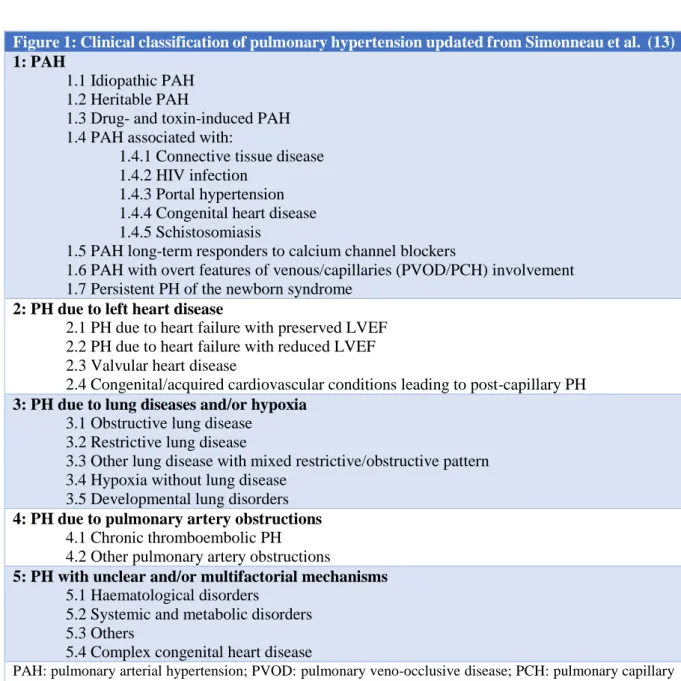
PH is classified into five groups based on the aetiology, or pathophysiological features:
- Group 1, pulmonary arterial hypertension (PAH) comprises patients with precapillary pulmonary hypertension due to distinct underlying disorders who share a similar angioproliferative vasculopathy in the pulmonary arterioles, leading to endothelial and smooth muscle proliferation and dysfunction, inflammation and thrombosis (14,15). PAH comprises various different pathologies, such as idiopathic, heritable, drug-induced PAH (e.g. anorexigenic agents), as well as PAH associated with concomitant diseases such as connective tissue disease, HIV infection, portal hypetension, or congenital heart disease. Since the 3rd WSPH held in 2003, pre-capillary PH of group 1 (PAH) has been defined by the presence of mPAP ⩾25 mmHg with a normal pulmonary artery wedge pressure (PAWP)⩽15 mmHg and elevated pulmonary vascular resistance (PVR) ⩾3 Wood Units (WU) and the absence of other causes of pre-capillary PH, (e.g. hypoxemia or chronic parenchymal lung diseases or chronic thromboembolic disease) (10,11).
- Group 2 is classified as pulmonary hypertension due to left heart disease. - Group 3 includes PH due to lung diseases and/or hypoxia.
- Group 4 includes those with PHdue to pulmonary artery obstructions, such as chronic thromboembolic PH.
- Group 5 is pulmonary hypertension due to miscellaneous disorders that cannot be attributed to one other group (for example pulmonary hypertension in the context of haematological disorders).
18
Figure 1: Clinical classification of pulmonary hypertension updated from Simonneau et al. (13)
1: PAH
1.1 Idiopathic PAH 1.2 Heritable PAH
1.3 Drug- and toxin-induced PAH 1.4 PAH associated with:
1.4.1 Connective tissue disease 1.4.2 HIV infection
1.4.3 Portal hypertension 1.4.4 Congenital heart disease 1.4.5 Schistosomiasis
1.5 PAH long-term responders to calcium channel blockers
1.6 PAH with overt features of venous/capillaries (PVOD/PCH) involvement 1.7 Persistent PH of the newborn syndrome
2: PH due to left heart disease
2.1 PH due to heart failure with preserved LVEF 2.2 PH due to heart failure with reduced LVEF 2.3 Valvular heart disease
2.4 Congenital/acquired cardiovascular conditions leading to post-capillary PH 3: PH due to lung diseases and/or hypoxia
3.1 Obstructive lung disease 3.2 Restrictive lung disease
3.3 Other lung disease with mixed restrictive/obstructive pattern 3.4 Hypoxia without lung disease
3.5 Developmental lung disorders 4: PH due to pulmonary artery obstructions
4.1 Chronic thromboembolic PH 4.2 Other pulmonary artery obstructions
5: PH with unclear and/or multifactorial mechanisms 5.1 Haematological disorders
5.2 Systemic and metabolic disorders 5.3 Others
5.4 Complex congenital heart disease
PAH: pulmonary arterial hypertension; PVOD: pulmonary veno-occlusive disease; PCH: pulmonary capillary haemangiomatosis; LVEF: left ventricular ejection fraction.
4. Epidemiologic data
Because the diagnosis requires an invasive procedure (right heart catheterization), the epidemiology of pulmonary hypertension is difficult to study. Therefore, large-scale population studies are based on echocardiography because invasive tests for epidemiological studies would not be ethical.
19
a. PAH
The reported incidence of pulmonary arterial hypertension in the developed world is 1,1–7,6 per million adults per year; the prevalence of pulmonary arterial hypertension is 6,6–26,0 per million adults (16–19)
Pulmonary arterial hypertension has been thought to affect mainly younger people, mostly females (20). This consideration is particularly true for heritable PAH, which affects twice as many women as men (21) and the average age at the time of diagnosis was approximately 50 years (22).
More recently, in Germany the average age of patients at the time of diagnosis was 64 years (22), and in UK 29% of the patients with newly diagnosed PAH were over the age of 70 years (23); reinforcing the notion that the phenotype and the age of patients with PAH were continuous to change. Advancing age has been identified as an independent risk factor for mortality in this population (24).
b. CTEPH
In the various registries, CTEPH is mostly diagnosed between 50 and 70 years-old, with males and females being equally affected (25–27).
Studies determined that the incidence of CTEPH was 1,0–8,8% of all survivors of acute pulmonary embolism (28–31), with a calculated average incidence of 4% in Europe and USA (32).
In European registries, the reported annual chronic thromboembolic pulmonary hypertension incidence rates is 0,9-5,7 cases per million adults per year (16,19,33,34). But this number is much fewer than the expected annual incidence (16).
Up to 25% of patients with CTEPH have no history of acute pulmonary embolism (35), the symptoms are relatively unspecific, and physicians are ill-informed about the disease; this is why CTEPH is certainly underdiagnosed (36). The calculated incidence including diagnosed and undiagnosed disease ranges from 30 to 50 cases per million inhabitants per year in the USA and Europe. The projection model indicated that incidence of CTEPH will continue to increase over the next decade (37).
20
5. Risks factors
a.
PAH
The idiopathic form of the disease concerns mostly women of childbearing age. There is an inherited form of PAH. Several gene mutations have been identified; the most common coding for bone morphogenetic protein receptor type II (BMPR2) cause up to 80% of the heritable PAH population (despite an incomplete penetrance about 20%), and 20% of cases of sporadic idiopathic disease (38,39). In comparison, activin receptor-like kinase 1 (ALK1), mothers against decapentaplegic homolog 9 (SMAD9) and endoglin 1 (ENG) concern only 5% of cases of heritable disease (15).
Lupus, scleroderma, cirrhosis, and HIV infection are strongly associated with PAH. Some drugs, methamphetamine and cocaine use increase the risk for developing this disease (40).
b.
CTEPH
Multiple risk factors have been linked to CTEPH, such as priorsplenectomy, autoimmune or hematological disorders (e.g., lupus or antiphospholipid syndrome), cancer, ventriculoatrial shunts, chronic inflammatory disorders (such as osteomyelitis and Crohn’s disease), thyroid replacement therapy, and non-O blood group (41–45).
The higher incidence of chronic thromboembolic disease in Japanese patients compared with patients from USA, and the predominance of CTEPH among women, suggest that race, sex, or environmental exposure may play a key role in this condition (46).
6.
Pathophysiology
Pulmonary arterial hypertension (PAH) and chronic thromboembolic PH (CTEPH) share similar physiological outcome, in which remodelling of the pulmonary vascular bed is responsible for the rise in pulmonary vascular resistance (PVR), leading to progressive right ventricular failure and functional decline.
21
a. PAH
The pathobiology of PAH is complex and incompletely understood. Latest advances have led to a better understanding of the key components underlying this inadequate accumulation of pulmonary vascular cells within the arterial walls leading to vascular remodeling causing a partial occlusion of small pulmonary arteries, and high pulmonary arterial pressure (15).
Among the under lying molecular and cellular mechanisms, some play critical roles in the pathogenesis of the disease (47,48) :
- pulmonary endothelial dysfunction, in particular vascular stiffness (by increased wall thickness, smooth muscle cells proliferation and changes in the type and quantity of extracellular matrix proteins (49)), and endothelial-to-mesenchymal transition (involved by the formation of neo-intimal vascular lesions in the pulmonary arteries caused by increased proliferation of endothelial cells (EC) and smooth muscle cells (SMC) (50)).
- alterations of the inter-cell communications within the pulmonary arterial walls, - alteration of the inflammatory component: vascular cells can respond to
inflammatory stimuli by enhanced proliferation and migration and reduced apoptosis; responsible for the exaggerated perivascular infiltration of inflammatory cells (B- and T-lymphocytes, mast cells, dendritic cells, macrophages, etc.) (51).
- loss of BMPR2 (bone morphogenic protein receptor type 2) activity increases apoptosis in vascular cells, leading to vascular remodeling and ultimately PAH (15,52)
However, there are many other complex and often interacting pathways involved in the development of PAH.
b. CTEPH
The pathophysiology of CTEPH is complex and multifactorial; including pulmonary artery obstruction by non-resolved thrombi, and pulmonary microvasculopathy. But it remains still not completely understood.
The following factors have been related to failure of thrombus resolution, and development of CTEPH: inflammation; genetic factors leading to fibrinolysis resistance; reduced fibrinolytic capacity; increased hypercoagulability; impaired angiogenesis; small vessel disease (53,54).
22
7. Clinical presentation
The cardinal symptom of PH isprogressive exercise dyspnea (11); but symptoms of PH remain non-specific: dyspnea upon exertion, fatigue, weakness, chest pain, lightheadedness/syncope with exertion, anorexia and, less frequently, cough. Progressive right-sided heart failure occurs in late stage of disease. Rarely, haemoptysis, hoarseness (unilateral vocal chord paralysis) and arrhythmias may characterise PH (55).
Physical examination is often without abnormalities, when PH is compensated. Findings may include loud heart sound (P2 component), murmur of tricuspid regurgitation, and signs of right-sided heart failure as jugular venous distension, hepatojugular reflux, ascites, hepatomegaly and splenomegaly, peripheral oedema (55). In some cases, clinical examination may suggest an underlying cause of PH.
Regarding CTEPH, about 25% of patients have no history of acute pulmonary embolism (35), and there are no specific symptoms of CTEPH. For this reason, CTEPH diagnosis is often delayed : mean delay from onset of symptoms to diagnosis of CTEPH was 18 ± 26 months (56). Thus, patients may be asymptomatic for several years before presenting progressive dyspnoea on exertion, exercise intolerance, fatigue or depression (56) ; then syncope and oedema occurs in late stages of disease progression. (36).
8. Diagnosis
In the case of clinical suspicion of pulmonary hypertension, an echocardiography is performed in search of elevation of peak tricuspid regurgitation velocity over 3.4m/s or over 2.8m/s associated with right heart overload. But to confirm the diagnosis of pulmonary hypertension right heart catheterisation is mandatory. Diagnostic criteria for PAH and CTEPH include a resting mean pulmonary arterial pressure greater than or equal to 25 mmHg. Measurement of pulmonary arterial occlusion pressure (PAOP) is necessary to exclude post-capillary pulmonary hypertension.
23
a.
PAH
In case of PH suspicion, the diagnosis process have to rule out all other causes of PH, in particular most common clinical groups of PH [group 2 and group 3], then distinguishes group 4, and finally concluded the PAH’s accountability (26).
Indeed, if transthoracic echocardiography is compatible with PH, the clinician needs to collect clinical history, symptoms, physical signs, ECG, chest radiograph, pulmonary function tests and chest CT to identify the presence of group 2 (left heart diseases) or group 3 (lung diseases) PH. If the diagnosis of left heart or lung diseases is confirmed, the appropriate treatment for these illnesses should be considered.
In the contrary case, a V/Q lung scan should be performed for the differential diagnosis between CTEPH and PAH.
b.
CTEPH
Even though there is no clear recommendation, a screening by echocardiography could be considered up to 2 years after acute pulmonary embolism (46).
CTEPH is defined as symptomatic pulmonary hypertension with persistent pulmonary perfusion defects despite a 3-month anticoagulant therapy (37).
Imaging plays a central role in CTEPH diagnosis: ventilation/perfusion lung scintigraphy (V/Q scan) shows segmental perfusion defects without matching ventilation abnormalities. Thereafter presence of specific imaging findings on computed tomography pulmonary angiography (CTPA), cardiovascular magnetic resonance imaging (MRI) or conventional pulmonary angiography (CPA) will define the diagnosis of CTEPH (26,57)
9. Treatment
a.
PAH
Treatment includes supportive therapies (as anticoagulants when indicated, diuretics, supplemental oxygen, psychosocial support, genetic counselling, appropriate birth control,
24
supervised physical exercise) (15,26), and disease-targeted drugs, consisting of vasodilators agents (5).
Until 2001, epoprostenol was the only medicaton available to treat PAH, and mainly used as a bridge to transplantation (58).
The pathogenesis of PAH involves dysfunction of three key pathways: the nitric oxide pathway, the prostacyclin pathway, and the endothelin pathway (59), cf. annex 1.
This three relevant signaling pathways represent therapeutic targets for the medical treatment options (60), with several drugs currently approved for the treatment of pulmonary arterial hypertension (61):
- the endothelin receptor antagonists ambrisentan, bosentan, and macitentan; - the phosphodiesterase-5 inhibitors sildenafil and tadalafil;
- the soluble guanylate cyclase stimulator riociguat;
- the prostacyclin analogues beraprost (in Japan and South Korea only), epoprostenol, iloprost and treprostinil;
- the prostacyclin receptor agonist selexipag.
Adverse effects occur with all of the specific medication (59).
Over the past few decades, the development of effective medical drugs and the use of combination therapyto target multiple pathogenic pathways simultaneously have considerably improved the prognosis of patients with PAH (62), and also improved symptoms, exercise capacity, and quality of life (66).
Whereas previous guidelines recommended initial monotherapy, followed by regular assessments, and initiation of combination therapy when treatment goals were not met (61), the current ESC/ERS guidelines recommend initial combination therapy:
- Newly diagnosed PAH patients with low or intermediate risk receive initial or early combination therapy comprising an endothelin receptor antagonist (ERA) with a phosphodiesterase-5 (PDE5) inhibitor or a soluble guanylate cyclase (sGC) stimulator (63–65).
- High-risk PAH patients receive a recommended triple treatment with an ERA, a PDE5 inhibitor or an sGC stimulator, and an intravenously administered prostacyclin analog (26).
25
The treatment response and clinical assessment must performed every 3-6 months (26). The treatment strategy depends on the individual response: if the patient has not achieved the primary treatment goal, that is attainment of the low-risk category after the initial treatment, the next step is dual or triple combination therapy (11).
Sitbon and al. showed that initial oral dual combination therapy of an ERA plus a PDE5 inhibitor was associated with significant improvements in functional class, exercise capacity and dyspnea (58). Simultaneously, signs of right heart failure and levels of brain natriuretic peptide (BNP) were significantly reduced, suggesting a reduction in clinical worsening. This strategy was also accompanied by significant improvements in 6MWD and haemodynamic parameters, including reductions in pulmonary vascular resistance (PVR) and mPAP.
In patients with severe PAH, the triple combination therapy is a way to improve the long-term benefits (66).
Despite the currently available treatment, many patients remain symptomatic with reduced exercise capacity, quality of life, or develop right heart failure (67).
Current specific PAH therapy do not specifically target inflammation and pulmonary vascular remodeling, which are likely new targets of the next therapeutic innovation. It requires a better identification of the pathobiological mechanisms underlying,
b.
CTEPH
Surgical pulmonary endarterectomy is the treatment of choice for patients with chronic thromboembolic pulmonary hypertension (26). Among them 50 to 70% are operable (35). The perioperative mortality at experienced centres is 2 to 4% (68,69). Approximately half of the patients had a post-operative mPAP ≤25 mmHg (69). In around 20% of patients treated surgically, the residual pulmonary hypertension is clinically significant and has to be treated (69).
For patients who are not candidates for surgery, interventional balloon pulmonary-angioplasty is an emerging treatment option to recanalize obliterated pulmonary vessels at subsegmental level (25).
Riociguat is the only drug approved for patients with inoperable chronic thromboembolic pulmonary hypertension or residual postoperative disease. Other classes of drugs might be used with a lower grade of recommendation. (61)
26
10.
Prognosis and risk assessment
PH is characterized by a progressive increase in pulmonary vascular resistance, leading to right heart failure and premature death (70,71).
a.
PAH
Over the past decades, the life expectancy of PAH patients has increased with a 3-year survival rate of 70 to 80% now (24), compared with 40% in the 1980s (11). Before the late 1990s, treatments for PAH patients consisted of supportive therapy, and was focused on symptoms related to right heart failure, with a median survival of 2.8 years in 1986 (95% Cl, 1.9 to 3.7 years) (72). Recent data from the United States-based registry assessing pulmonary arterial hypertension disease management (REVEAL registry) showed a median survival of 7 years presumably due to a combination of significant advances in treatment strategies and patient support strategies (15,62). Despite improvements in the short-term survival of patients with PAH, the condition remains incurable (26).
The 2015 ESC/ERS (European Society of Cardiology/European Respiratory Society) guidelines (26) and the 2018 Proceedings of the 6th World Symposium on Pulmonary Hypertension (73) recommend regular risk assessment at expert PH centres using a series of variables to stratify patients into low, intermediate and high-risk categories (corresponding to an estimated one-year mortality rate of <5%, 5–10% and >10% respectively), cf Annex 2. This assessment is meant to be applicable in patients with newly diagnosed PAH and at any time during the course of the disease, and aims to facilitate treatment decisions. It includes clinical features (e.g. World Health Organization (WHO) functional class (FC), right ventricular function), exercise testing (e.g., 6-minute walking test (6MWT), cardiopulmonary exercise testing), imaging (echocardiography, cardiac magnetic resonance imaging), haemodynamics (right atrial pressure, cardiac index, mixed venous oxygen saturation), and biomarkers (BNP and NT-proBNP).
According to these guidelines, certain low-risk features are associated with better prognosis in PAH, such as no sign right heart failure, a WHO FC I or II, a 6MWD >440 m, an haemodynamic assessment with a right atrial pressure (RAP) <8 mmHg and a cardiac index ⩾2.5 L/min/m². Also, BNP levels <50 ng/L or NT-proBNP <300 ng/L are considered to be low-risk features.
27
During follow-up, achieving and maintaining a low-risk profile is the recommended goal of PAH treatment. However, the accuracy of this risk assessment has not been fully established and most of cutoffs are based on expert opinion, with low levels of evidence.
Since then, abbreviated versions of the 2015 ESC/ERS risk stratification strategy have been evaluated retrospectively in newly diagnosed PAH (74–77):
- Boucly et al. (2017) studied a retrospective incident cohort of patients with PAH, and showed the risk of death or lung transplantation was accurately determined using a simple quantification of the number of low-risk criteria present at diagnosis, and, most importantly, achieved during the first year of follow-up for WHO FC, 6MWD, right atrial pressure (RAP) and cardiac index. In the analysis, attaining only two or less low-risk criteria at follow-up had a worse long-term prognosis than those with three or four low-risk criteria (74).
- Kylhammar et al. (2017) proposed a method whereby 8 criteria (6MWD, WHO FC, NT-proBNP, right atrial area, pericardial effusion, RAP, cardiac index, and SvO2) are graded from 1 to 3 (1=low, 3=high) using the ESC/ERS risk stratification. The rounded mean of these grades is then used to define patient risk status (76).
- Hoeper et al. (2017) evaluated an abbreviated version of the risk stratification model in newly diagnosed PAH patients (from the COMPERA web-based PH registry) using 6 variables (6MWD, WHO FC, NT-proBNP/BNP, RAP, cardiac index, and SvO2) and the same analysis method as Kylhammar et al. Among them, 6MWD, WHO FC, BNP/NT-proBNP and SvO2 were closely linked to the mortality risk (75).
Two studies in PH have demonstrated that the presence of anaemia is indicative of a poor prognosis (78,79). Furthermore, iron deficiency is prevalent in patients with IPAH and is associated with disease severity (80,81).
b.
CTEPH
Unlike others forms of PH, CTEPH is potentially curable. Pulmonary endarterectomy (PEA) is the treatment choice for patients who are surgical candidates (82) : however, up to 50% of patients are considered inoperable because of an inaccessible and/or distal thromboembolism, or serious comorbidities (34).
28
31% to 50% develop persistent/recurrent pulmonary hypertension after undergoing PEA (77,82). More recently, balloon pulmonary angioplasty (BPA) may be considered in patients who are technically non-operable or carry an unfavourable risk/benefit ratio for PEA (26).
However, without surgical intervention survival is poor and proportional to the degree of pulmonary hypertension and right ventricular dysfunction: in 1982, CTEPH patients had an estimated 5-year survival of 30% if mPAP was greater than 40 mmHg, and 10% if mPAP was more than 50 mmHg (83).
With new treatment options, including BPA, the prognosis of non-operated patients with CTEPH improved. Indeed, recent studies estimate a 1-year survival over 90%, a 3-year survival of 85%, (84,85), and a 5-year survival around 60% (84).
In these patients, baseline NYHA FC, RAP, 6MWD, BPA therapy and diagnosis period (2006−2012 vs 2013−2016) were predictors of survival (85).
There is currently no established risk assessment strategy to guide treatment decisions in patients with CTEPH. However, a recent study suggested thatthat an abbreviated version of the ERS/ESC risk stratification may be applicable (with the method from Kylhammar et al.) to predict mortality in non-operated and medically treated CTEPH patients (84). In a similar way as PAH mortality risk, variables closely linked to the mortality risk were 6MWD, WHO FC, BNP/NT-proBNP.
11.
Exercise training and pulmonary hypertension
a.
Exercise training and rehabilitation
Chronic cardiorespiratory disorders as PAH lead to physical inactivity, skeletal muscle dysfunction and compromised quality of life (4). In addition, patients with PAH were advised, for decades, to avoid physical exercise which has been thought to carry a high risk of sudden cardiac death and exacerbate right heart failure. Indeed, it was commonly believed that physical activity may have a negative impact on patients by increasing pulmonary remodeling and contributing to the progression of PH (1). In PH population reduced exercise capacity is associated with depression and anxiety disorders (3).
29
In the guidelines of 2009, because of a lack of efficacy data, rehabilitation was considered only in special circumstances, such as in deconditioned PAH patients, in a highly supervised setting with avoidance of distressing and excessive physical activity (2).
However, there is some evidence from left heart failure that regular physical activity improve endothelial function, exercise capacity, and quality-of-life parameters, and may be beneficial even in severe disease (86). Physical training reduce vascular oxidative stress and exert beneficial vascular effects in part by increasing activity of endothelial nitric oxide synthase (87).
Since then, additional studies have been performed that provided evidence of improvements of symptoms and exercise performance with reassuring data on safety (6,7). However, it must be emphasised that the patients in these studies were all in a stable condition on advanced drug therapy, under close supervision in PAH expert centres and the training was specially designed for PAH patients.
In the updated guidelines of 2015 (26), specialised PAH rehabilitation is recommended for patients in stable clinical condition with the best standard of pharmacological treatment, with the notion that supervised rehabilitation should be implemented by centres experienced in both PAH patient care and rehabilitation of compromised patients (class IIa recommendation, evidence level B). Optimised pharmacological treatment is defined as single or combination therapy with PAH-targeted drug therapy such as calcium-channel blockers, prostacyclin analogues, endothelin receptor antagonists, phosphodiesterase type 5 inhibitors, and guanylate cyclase stimulator. The guidelines also clearly suggest that such an expert centre should consist of a multidisciplinary team including cardiology and respiratory medicine physicians, clinical nurse specialist, radiologists, psychological and social work support. Furthermore, it is recommended for expert referral centres to have a direct link to adult congenital heart disease units, pulmonary endarterectomy surgeons and a lung transplantation programme. Expert referral centres are requested to follow at least 50 patients with PAH or chronic thromboembolic pulmonary hypertension (CTEPH), and should have two new referrals per months.
b.
Mechanisms of exercise training in PAH patients
The exact pathophysiological mechanisms by which exercise leads to beneficial effects are not yet fully understood.
30
In mice, exercise training was able to prevent hypoxia-induced pulmonary vascular remodeling, and improved exercise capacity and pulmonary haemodynamics (88). The effect of exercise training on physical capacity was more important in mice who received drug treatment (sildenafil). The effects were not associated with changes in the expression of the nitric oxide, phosphodiesterase 5, or soluble guanylate cyclase pattern, which belongs to targeted pathways for PAH therapy. These results suggest that exercise training might be complementary to pharmacological treatment, affecting molecular pathways not yet identified.
In other rat and mice experiments, it has been shown an attenuation of acute vasoconstriction in response to hypoxia, a reduction in right ventricular pressure and pulmonary artery diameter, and a better right ventricle capillary density (89,90)
In patients with chronic heart failure and PAH, exercise training has been shown to reduce inflammation and TNF-level, which is involved in the development of PAH (91–93).
In PAH patients, skeletal muscle dysfunction has been reported with muscle atrophy, reduced capillary density, decreased oxidative enzyme activity, reduced exercise capacity and muscle strength, fewer type IIa fibres. In few studies, exercise training significantly improve these alterations in muscle tissue and muscle function (8,94,95).
Exercise training also improves lung performance with better pulmonary perfusion, oxygen uptake and inhibition of smooth muscle cells (95). In addition, it decreases insulin resistance that have been reported more prevalent in PAH population (96).
A recent study showed that aerobic exercise promotes a significant increase in H2O2/VEGF/p-Akt signal for pulmonary physiological angiogenesis (97).
In summary, exercise training impacts angiogenesis, muscular strengh, right ventricle contractility, ventilatory efficiency, hormonal, and immune system, and may improve symptoms, exercise capacity, quality of life and hemodynamics.
c.
Different components of the rehabilitation
programme with pulmonary arteriel hypertension
(PAH)
Since 2006, several published studies conducted rehabilitation in PAH patients with different training protocols, but there is no consensus on how to train PH patients.
31
Most published studies were conducted in the PAH centre of Heidelberg in Germany (5,98): they established a training programme, which started as a 3-week inpatient training followed by a home-based training with individualised instructions and supervised by the respective expert centre. Before the rehabilitation and right after, patients are evaluated in the PAH centre and were stable and compensated under optimized medical therapy (such as endothelin antagonists, iloprost, sildenafil, calcium channel blockers, anticoagulants, diuretics, and supplemental oxygen) for at least 2-3 months before entering the study. The initial exercise training intensity was also assessed. During the following 3 weeks of in-hospital rehabilitation, the physicians for rehabilitation stay in contact with the PAH experts. The training consists of interval bicycle ergometer training with a lower workload (0 to 10W) for 30 seconds and a higher workload for 1 minute (20 to 35W) for 10 to 25 minutes per day in a supervised and monitored setting. The training intensity was increased (eg, up to 35 to 50 W) with respect to the individual tolerability and improvement. Training intensity was limited by peak heart rate (HR) (not more than 120 bpm), oxygen saturation (not less than 85%), and subjective physical exertion. If needed training was reduced. Furthermore, 60 minutes of walking, 30 minutes of dumbbell training of single muscle groups with low weights (500 to 1000 g) and 30 minutes of respiratory training were performed 5 days a week. Patients was also educated in perceiving their physical limits, and all patients were advised to avoid heavy exercise.Other centres have also used treadmill walking for 30 to 45 minutes under similarly intensive supervision (99).
And thus, most published studies used aerobic exercise training with low workload (intensity titrated between 40 to 80% of peak exercise capacity or about 60–80% of the HR achieved during a baseline exercise test) (95) under close supervision by PAH experts, in patients who were clinically stable and treated with optimized PAH drug therapy without signs of overt right heart failure.
d.
Evidence and effects of rehabilitation in pulmonary
hypertension patients
In healthy people and chronically ill patients, a higher daily physical activity lead to a better survival than less active controls (80,81).
Previous studies in rather small cohorts of patients with idiopathic pulmonary arterial hypertension (IPAH) and inoperable chronic thromboembolic pulmonary hypertension
32
(CTEPH) have shown beneficial effects of exercise training as add-on to disease targeted medical therapy by increasing exercise capacity and quality of life (5,8). The effects were observed in different forms of pulmonary vascular diseases, such as idiopathic PAH, CTEPH and PH due to respiratory or left heart diseases (7). The improvement in exercise capacity was recorded with an improvement in 6MWD, VO2 peak, cardiac output (100), muscle strength and muscle endurance. Also, physical and mental quality of life scores measured with the SF-36 questionnaire improved significantly, especially in the domains of physical functioning, general health perception, vitality, social functioning, role emotional and mental health (6).
A meta-analysis in 2015, including 16 prospective intervention studies with 469 PH participants, observed a 53.3 m improvement in 6MWD at 15 weeks with exercise training on pooled analysis (weighted mean difference, 53.3 m; 95% CI, 39.5–67.2). Exercise training was also associated with significant improvement in pulmonary arterial systolic pressure (weighted mean difference, −3.7 mm Hg; 95% CI, −5.4 to −1.9), peak oxygen uptake (weighted mean difference, 1.8 mL/kg per minute; 95% CI, 1.4–2.3), and quality of life as measured on SF-36 questionnaire subscale scores. (99)
In 2017, a review of randomized controlled trials (RCTs) assesses the efficacy and safety of exercise-based rehabilitation for people with PH (most participants were Group I pulmonary artery hypertension) (101). They study 6 RCTs with 206 participants included. Study duration ranged from 3 to 15 weeks. Exercise programmes included both inpatient- and outpatient-based rehabilitation. The mean 6MWD following exercise training was 60.12 metres higher than control (30.17 to 90.07 metres, n = 165, 5 RCTs), the mean peak oxygen uptake was 2.4 ml/kg/minute higher (1.4 to 3.4 ml/kg/min, n = 145, 4 RCTs). The health-related quality of life also improved with a mean change for the SF-36 physical component score 4.63 points higher (0.80 to 8.47 points, n = 33, 2 RCTs) and for the SF-36 mental component score 4.17 points higher (0.01 to 8.34 points; n = 33; 2 RCTs).
Ehlken et al. (102) evaluated the effect of training on morbidity and mortality of PAH patients (n=58) compared with a retrospective control group (n=46) treated with usual care and optimized PAH therapy. During a mean follow-up of 24 ± 12 months, 25.8% of the patients receiving exercise training had adverse events, a rate much lower when compared to the control patients (54,4%). The incidence of clinical worsening was also lower. The 1- and 2-year survival rates were better in the exercise training group compared to the control group (100% and 95% vs. 93% and 88%, respectively; P<0.05).
33
e.
Safety and adverse events
The rapid deterioration of right ventricle function leading to hemodynamic impairment, the occurrence of arrhythmias and syncope have been considered as the major risks of an exercise programme in patients with severe PAH (103,104).
Side effects in specialist centres under expert supervision were relatively rare. Actually, no study has reported an increased incidence of major adverse events in PAH patients undergoing a monitored and personalised exercise training (105)
Of the 6 RCTs analyzed by Morris et al., one study reported a single adverse event, where a participant stopped exercise training due to lightheadedness (101). In one of the largest studies (183 patients), a total of 13.6% of their participants had adverse events. Almost half of these patients suffered acute respiratory infections that had to be treated with antibiotics and forced the transitory suspension of the training programme. Severe adverse events, such as syncope/pre-syncope or supra-ventricular arrhythmias, occurred only in 4.4% of the patients. No signs of clinical worsening of right heart failure were detected during their hospitalisation, and no severe adverse episodes occurredwhen the patients continued the exercise training at home (between hospital discharge and the planned follow-up, 15 weeks later) (7). The meta-analysis including 16 prospective intervention studies with 469 PH patients, reported dizziness, presyncope, palpitation, hypotension and oxygen desaturation in 4,7% of training patients (one-third were not related to exercice). No major adverse events, such as progression of symptoms, right heart failure, or death, were observed among the patients during the training period.On median follow-up of 29.5 months (21–36 months), the survival rate was 91%, and the transplant-free survival rate was 89%. (99)
PH patients are often not aware of their own physical limits and may tend to overexercise. They have to be supervised and taught to recognise their physical limits, to prevent dangerous adverse effects of exercise. To minimize the risk, some safety precautions must be adopted when starting an exercice training programme. In the previous studies, it is required that PH patients are in clinically stable condition on optimised PH targeted therapy for at least 2 months (98). Training should be initiated with very low workloads (between 40 and 80% of peak exercise capacity), as well, dumbbell training must be practiced with low weights involving single muscle groups only. Oxygen saturation and heart rate have to be closely monitored (SpO2 >85%, HR <120 per minute). Especially for severe patients, it is recommended to start the
34
rehabilitation programme in an inpatient setting, where both supervision during training and observation just after is possible (95).
f.
Other components of rehabilitation
Patients with PH have to face many life stress factors, such as physical limitation, contraindications to have a pregnancy, high cost of treatment, unemployment, etc. Mental disorders (such as anxiety and depression) are more prevalent in this population, and closely related to the functional disability: the percentages of major depression higher in WHO FC IV patients (45%) than for the WHO FC I (7.7%) (3). That is why, goals of PH treatment should include quality of life as a major targets to reach (106), and many studies have pointed out that the rehabilitation programme, in addition to medical therapy, may improve depression and anxiety symptoms in patients with PH (5,105,107–110).
Rehabilitation is a global approach that not only consists of exercise training, but also includes a multidisciplinary team (including cardiology and respiratory medicine physicians, specialised nurses, radiologists, access to psychological and social work support), which provides informations, counselling and therapeutic interventions, during individual or small groups. It is a way to improve physical and emotional status. Moreover, during theirs stays patients may benefit from specific PH-drug titration: side effects are not uncommon, and could limit an increase in the dose if the titration is feared or not well tolerated by outpatients. A progressive titration when appropriate with management of side effects is a way to achieve optimal medical therapy.
35
12.
Conclusions
Supervised rehabilitation in PH patients should be recommended, in addition to optimal medical treatment with PH targeted drugs, if performed in a highly experienced team under expert supervision. The pathomechanisms are not fully understood yet, but many studies agree in the favorable effects of training programme: it leads to significant improvements in symptoms, exercise capacity and quality of life.
This programme should be implemented by centres experienced in both PH patient care and rehabilitation. Some safety precautions must be adopted in order to avoid adverse events: among other things, patients have to be in stable condition on optimised medical therapy, and closely monitored during the exercise training. Despite the few studies so far, there is no published guidelines available detailing how exercise rehabilitation should be carried out.
36
ANNEXES
Annex 1 from Humbert et al. (59)
37
BIBLIOGRAPHY
1. Gaine SP, Rubin LJ. Primary pulmonary hypertension. Lancet Lond Engl. 1998 Aug 29;352(9129):719–25.
2. Galiè N, Hoeper MM, Humbert M, Torbicki A, Vachiery J-L, Barbera JA, et al. Guidelines for the diagnosis and treatment of pulmonary hypertension: the Task Force for the Diagnosis and Treatment of Pulmonary Hypertension of the European Society of Cardiology (ESC) and the European Respiratory Society (ERS), endorsed by the International Society of Heart and Lung Transplantation (ISHLT). Eur Heart J. 2009 Oct;30(20):2493–537.
3. Löwe B, Gräfe K, Ufer C, Kroenke K, Grünig E, Herzog W, et al. Anxiety and depression in patients with pulmonary hypertension. Psychosom Med. 2004 Dec;66(6):831–6.
4. Osadnik CR, Rodrigues FMM, Camillo CA, Loeckx M, Janssens W, Dooms C, et al. Principles of Rehabilitation and Reactivation. Respiration. 2015;89(1):2–11.
5. Mereles D, Ehlken N, Kreuscher S, Ghofrani S, Hoeper MM, Halank M, et al. Exercise and respiratory training improve exercise capacity and quality of life in patients with severe chronic pulmonary hypertension. Circulation. 2006 Oct 3;114(14):1482–9.
6. Grünig E, Ehlken N, Ghofrani A, Staehler G, Meyer FJ, Juenger J, et al. Effect of Exercise and Respiratory Training on Clinical Progression and Survival in Patients with Severe Chronic Pulmonary Hypertension. Respiration. 2011;81(5):394–401.
7. Grünig E, Lichtblau M, Ehlken N, Ghofrani HA, Reichenberger F, Staehler G, et al. Safety and efficacy of exercise training in various forms of pulmonary hypertension. Eur Respir J. 2012 Jul;40(1):84–92.
8. de Man FS, Handoko ML, Groepenhoff H, van ’t Hul AJ, Abbink J, Koppers RJH, et al. Effects of exercise training in patients with idiopathic pulmonary arterial hypertension. Eur Respir J. 2009 Sep 1;34(3):669–75.
9. Kovacs G, Berghold A, Scheidl S, Olschewski H. Pulmonary arterial pressure during rest and exercise in healthy subjects: a systematic review. Eur Respir J. 2009 Oct;34(4):888–94.
10. Hoeper MM, Bogaard HJ, Condliffe R, Frantz R, Khanna D, Kurzyna M, et al. Definitions and diagnosis of pulmonary hypertension. J Am Coll Cardiol. 2013 Dec 24;62(25 Suppl):D42-50. 11. M. Hoeper M, Ghofrani H-A, Grünig E, Klose H, Olschewski H, Rosenkranz S. Pulmonary
Hypertension. Dtsch Ärztebl Int. 2017 Feb;114(5):73–84.
12. Hatano S, Strasser T, Organization WH. Primary pulmonary hypertension : report on a WHO meeting, Geneva, 15-17 October 1973 [Internet]. World Health Organization; 1975 [cited 2019 Aug 26]. Available from: https://apps.who.int/iris/handle/10665/39094
13. Simonneau G, Montani D, Celermajer DS, Denton CP, Gatzoulis MA, Krowka M, et al. Haemodynamic definitions and updated clinical classification of pulmonary hypertension. Eur Respir J. 2019 Jan;53(1):1801913.
14. Tuder RM, Archer SL, Dorfmüller P, Erzurum SC, Guignabert C, Michelakis E, et al. Relevant issues in the pathology and pathobiology of pulmonary hypertension. J Am Coll Cardiol. 2013 Dec 24;62(25 Suppl):D4-12.
15. Lan NSH, Massam BD, Kulkarni SS, Lang CC. Pulmonary Arterial Hypertension: Pathophysiology and Treatment. Diseases [Internet]. 2018 May 16 [cited 2019 Oct 5];6(2). Available from: https://www.ncbi.nlm.nih.gov/pmc/articles/PMC6023499/
16. Hoeper MM, Humbert M, Souza R, Idrees M, Kawut SM, Sliwa-Hahnle K, et al. A global view of pulmonary hypertension. Lancet Respir Med. 2016;4(4):306–22.