R ES EAR CH A R T I C LE
Open Access
Protein interactions of the transcription
factor Hoxa1
Barbara Lambert
1, Julie Vandeputte
1, Sophie Remacle
1, Isabelle Bergiers
1, Nicolas Simonis
2, Jean-Claude Twizere
3,
Marc Vidal
4,5and René Rezsohazy
1*Abstract
Background: Hox proteins are transcription factors involved in crucial processes during animal development. Their mode of action remains scantily documented. While other families of transcription factors, like Smad or Stat, are known cell signaling transducers, such a function has never been squarely addressed for Hox proteins.
Results: To investigate the mode of action of mammalian Hoxa1, we characterized its interactome by a systematic yeast two-hybrid screening against ~12,200 ORF-derived polypeptides. Fifty nine interactors were identified of which 45 could be confirmed by affinity co-purification in animal cell lines. Many Hoxa1 interactors are proteins involved in cell-signaling transduction, cell adhesion and vesicular trafficking. Forty-one interactions were detectable in live cells by Bimolecular Fluorescence Complementation which revealed distinctive intracellular patterns for these interactions consistent with the selective recruitment of Hoxa1 by subgroups of partner proteins at vesicular, cytoplasmic or nuclear compartments.
Conclusions: The characterization of the Hoxa1 interactome presented here suggests unexplored roles for Hox proteins in cell-to-cell communication and cell physiology.
Keywords: Hox, Hoxa1, ORFeome, Interactome Background
The conserved family of homeodomain Hox transcrip-tion factors is critically involved in patterning the body plan of bilaterian embryos by controlling multiple mor-phogenetic and organogenetic processes during animal development [1-4]. Modifications in Hox protein expres-sion and activity have likely contributed to the evolution-ary diversification of animal forms [5,6]. Misregulation or mutation of several Hox proteins has been associated with pathologies like cancer or neuropathies [7,8].
Hox proteins are transcription factors which regulate expression of target genes and chromatin remodeling [9]. A handful of proteins that interact with Hox pro-teins have been identified so far, and these are almost ex-clusively transcription factors, like the well-characterized Three Amino acid Loop Extension (TALE) homeodo-main proteins Pbx/Exd and Prep/Meis/Hth [10], TFIIEβ
[11], TATA Binding Protein (TBP) [12], Gli3 [13], Maf [14], Smad [15,16], High Mobility Group protein 1 (HMG1) [17], or transcriptional coregulators like CREB Binding Protein (CBP)/p300 [18-20]. Hox proteins may also form complexes with the translation initiation factor eIF4E to control the translation of target mRNAs [21]. Some Hox-like homeodomain proteins can be secreted into the extracellular compartment and translocate through the cell membrane to gain access to the cytosol and nucleus of neighboring cells, so it has been pro-posed that Hox proteins could display a paracrine tran-scriptional activity [22,23].
Numerous transcription factors, involved in critical developmental processes, like Smad, STAT, β-catenin or NFκB, are primarily signal transducers. Though primar-ily cytoplasmic, upon activation these can translocate to the nucleus, where they convey signaling by affecting gene regulation. As signal transducers these transcrip-tion factors can interact with enzymatically active mem-brane receptors, adaptor proteins, signal transducing kinases, or ubitiquin ligases. Possibly, Hox transcription factors could similarly fulfill pivotal roles at the heart of
* Correspondence:rene.rezsohazy@uclouvain.be
1Molecular and Cellular Animal Embryology group, Life Sciences Institute
(ISV), Université Catholique de Louvain, Louvain-la-Neuve 1348, Belgium Full list of author information is available at the end of the article
© 2012 Lambert et al.; licensee BioMed Central Ltd. This is an Open Access article distributed under the terms of the Creative Commons Attribution License (http://creativecommons.org/licenses/by/2.0), which permits unrestricted use, distribution, and reproduction in any medium, provided the original work is properly cited.
Lambert et al. BMC Developmental Biology 2012, 12:29 http://www.biomedcentral.com/1471-213X/12/29
developmental processes, acting at the crossroads be-tween cell-to-cell communication and cell fate deter-mination. To our knowledge no exhaustive interaction screen has been performed to detect functional connec-tions for a Hox protein.
Here, we conducted a proteome-wide screening for candidate interactors of Hoxa1. Hoxa1 is one of the earliest Hox genes to be expressed during embryonic development. It is involved in hindbrain segmentation and patterning [1,24,25]. Hoxa1 misregulation has been associated with mammary carcinogenesis [26]. We used a stringent high-throughput yeast two-hybrid (Y2H) approach to systematically test pairwise combinations, using Hoxa1 both as a bait and as a prey against the human ORFeome v3.1 resource, which contains 12,212 ORFs representing 10,214 genes [27]. Of the 59 Hoxa1 interactions identified, 45 could be validated by in vivo affinity binding assays in co-transfected animal cells. A striking subset of the validated interactors are not proteins involved in gene regulation. Rather, these inter-actors are adaptor proteins or modulators of the Bone Morphogenetic Proteins (BMP)/Tumor Growth Factor (TGF) β, Tumor Necrosis Factor (TNF), Receptor-Tyrosine Kinases (RTK) and integrins signal transduction pathways. Other interactors participate in cell adhesion or endosomal trafficking. We detected 41 interactions in live cells by Bimolecular Fluorescence Complementation (BiFC). Depending on the different proteins identified, interactions either take place in the cytoplasm, in the nucleus, in association with vesicles or show a variable pattern from cell to cell, underscoring a dynamic inter-play with Hoxa1. Numerous identified Hoxa1 partners reported to interact with each other within known pathways share similar intracellular patterns of Hoxa1 interaction by BiFC. We conclude that Hoxa1 can con-tact several subunits of multi-molecular functional plat-forms involved in cell signaling, cell-adhesion, or cell shape regulation.
Results
A proteome-wide yeast two-hybrid screening for Hoxa1 interactors
The yeast two-hybrid (Y2H) is a powerful approach for large-scale screenings to identify binary protein-protein interactions [28,29]. DB-Hoxa1 was tested pairwise against 12,212 open reading frame (ORF)-derived pro-teins from the human ORFeome version 3.1 [27] fused to the Gal4 activation domain (AD). In this configur-ation, we detected 40 distinct interactions (Table 1). We also screened in the other configuration, Hoxa1 as a prey (AD-Hoxa1) against the full hORFeome in fusion with the Gal4 DB. In the second configuration we detected 28 interactions, of which 8 were also detected in the DB-Hoxa1/AD-ORFs configuration (Table 2). A total of 59
candidate Hoxa1 interactors were identified. We found the Hoxa1 homodimerization interaction and 8 out of the 9 Hoxa1 interactions, previously described in the literature [28,30] (Table 1 and 2).
Co-purification from animal cells validate forty-five Hoxa1 interactors
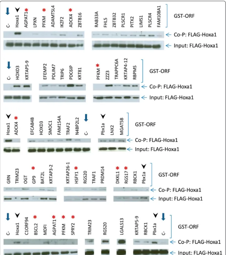
To validate the 59 interactions identified by the Y2H screen by an orthogonal assay we turned to affinity co-purification of a FLAG-Hoxa1 fusion protein co-expressed with glutathione S-transferase (GST)-tagged candidate interactors in transfected COS7 or HEK293T cells. In absence of GST-partners, there was no or very weak back-ground binding of FLAG-Hoxa1 onto the glutathione-agarose beads (Figure 1). As positive controls we measured Hoxa1 dimer formation [30,31] and the reproducible interaction between Hoxa1 and Pbx1a [32] (Figure 1). In total, affinity purification from co-transfected cells confirmed 45 out of the 59 Y2H inter-actors (Table 1 and 2), in the presence of which a detectable amount of FLAG-Hoxa1 remained associated to the GST-fusion/glutathione-agarose beads and could be detected on western blots. It should be noted however that some interactions could not be confirmed because the corresponding GST-ORF fusion was expressed at an undetectable level, if at all (data not shown).
Bioinformatics functional analysis
To determine if Hoxa1 preferentially targets parti-cular biological functions or pathways, we tested for stat-istical enrichment in regards to the Gene Ontology GO [33]), Kyoto Encyclopedia of Genes and Genomes KEGG; [34]) and Pathway Commons databases (www. pathwaycommons.org).
We observed that six GO terms were significantly overrepresented (Table 3). These enriched annotations are consistent with known functions of Hoxa1, linking our set of interactors to developmental and transcription factor function. There were several additional enriched, though not statistically so, GO terms linked to develop-ment and transcription factors (Table 3).
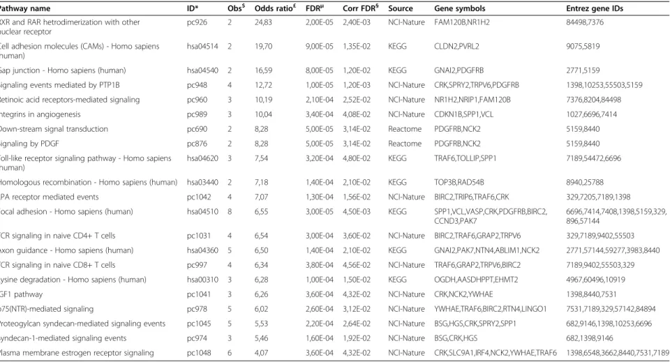
The immediate interactors of Hoxa1 were not enriched for annotated pathways, which could be due to incomplete coverage or relative sensitivity of the Y2H assay [35], or be intrinsic to the way Hoxa1 interacts with pathways, needing only one or few direct contacts. To account for the latter possibility, we also analyzed second-degree interactors, proteins that interact with Hoxa1 targets. Proteins associated with 21 pathways are overrepresented compared to random expectation (Table 4), showing that Hoxa1 could play a role in vari-ous processes other than gene regulation, such as focal adhesion, axon guidance or several signaling cascades.
Lambert et al. BMC Developmental Biology 2012, 12:29 Page 2 of 17
Table 1 Interaction partners for Hoxa1 revealed by yeast two-hybrid screening using DB-Hoxa1
Protein symbol Protein name Gene ID UniProtKB/
Swiss-Prot Protein function BiFC signal Confirmed byco-purification
ADAMTSL4 (TSRC1) ADAMTS-like 4 54507 Q6UY14 TNF-induced apopotosis Nuclear Y
BAT2L HLA-B associated transcript 2-like 84726 Q5JSZ5 Unknown Nuclear Y
C1orf94 chromosome 1 open reading frame 94 84970 Q6P1W5 Protein binding Cytoplasmic Y
CCDC33 coiled-coil domain containing 33 80125 Q8N5R6 Protein binding / N
EFCAB4B EF-hand calcium binding domain 4B 84766 Q9BSW2 Calcium binding Nuclear, and vesicular Y EFEMP2*§ EGF-containing fibulin-like extracellular
matrix protein 2 30008 O95967 Fibulin-like, unknown n.d. Y
FAM154A family with sequence similarity 154, member A
158297 Q8IYX7 Unknown Nuclear, vesicular, cytoplasmic Y FHL5 (ACT) four and a half LIM domains 5 9457 Q5TD97 Transcription factor (zinc finger),
and kinesin and actin-binding protein
Nuclear Y
GPRIN2 G protein regulated inducer of neurite
outgrowth 2 9721 O60269 G protein interaction / N
HOXA1§ homeobox A1 3198 P49639 Transcription factor (homeodomain) Nuclear Y
HOXD3 homeobox D3 3232 P31249 Transcription factor (homeodomain) Nuclear Y
HSFY1 heat shock transcription factor, Y-linked 1 86614 Q96LI6 Transcription factor (heatshock factor) / N
KRTAP26-1§ keratin associated protein 26-1 388818 Q6PEX3 Keratin associated Nuclear Y
KRTAP3-2§ keratin associated protein 3-2 83897 Q9BYR7 Keratin associated Nuclear Y
KRTAP3-3§ keratin associated protein 3-3 85293 Q9BYR6 Keratin associated / N
KRTAP4-12*§ keratin associated protein 4-12 83755 Q9BQ66 Keratin associated Cytoplasmic Y
LGALS13 lectin, galactoside-binding, soluble, 13 29124 Q9UHV8 Lipase activity, signaling (regulator of protein kinases)
Nuclear, vesicular, cytoplasmic Y LNX2 ligand of numb-protein X 2 222484 Q8N448 Molecular scaffold, E3 ubiquitin ligase,
signaling regulator (Notch), associated to cell adhesion molecules
Nuclear and cytoplasmic Y
LPXN* leupaxin 9404 O60711 Signaling (focal adhesion), Transcription
factor
Vesicular and cytoplasmic Y MGAT5B (GnT-VB) mannosyl (α-1,6-)-glycoprotein
β-1,6-N-acetyl-glucosaminyltransferase, isozyme B
146664 Q3V5L5 Glycosyltransferase, focal adhesion dynamics
Nuclear Y
N4BP2L2 (PFAAP5) NEDD4 binding protein 2-like 2 10443 Q92802 Transcription factor or co-regulator Nuclear Y NR4A1 (Nur77) nuclear receptor subfamily 4, group A,
member 1 3164 P22736 Transcription factor (nuclear hormonereceptor) / N
OGT O-linked N-acetylglucosamine (GlcNAc) transferase
8473 O15294 Glycosyltransferase, transcription co-regulator
Nuclear and cytoplasmic Y PCSK5§ proprotein convertase subtilisin/kexin
type 5 5125 Q92824 Pro-protein convertase / N
Lambert et al. BMC Development al Biology 2012, 12 :29 Page 3 of 17 http://ww w.biomedce ntral.com/1 471-213X/12 /29
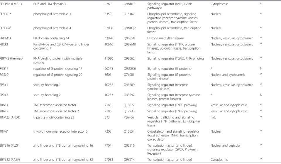
Table 1 Interaction partners for Hoxa1 revealed by yeast two-hybrid screening using DB-Hoxa1 (Continued)
PDLIM7 (LMP-1) PDZ and LIM domain 7 9260 Q9NR12 Signaling regulator (BMP, IGFBP pathways)
Cytoplasmic Y
PLSCR1* phospholipid scramblase 1 5359 O15162 Phospholipid scramblase, signaling regulatior (receptor tyrosine kinases, protein kinases), transcription factor
Nuclear Y
PLSCR4§ phospholipid scramblase 4 57088 Q9NRQ2 Phospholipid scramblase, transcription
factor Nuclear Y
PRDM14 PR domain containing 14 63978 Q9G2V8 Histone methyltransferase Nuclear, vesicular, cytoplasmic Y RBCK1 RanBP-type and C3HC4-type zinc finger
containing 1
10616 Q9BYM8 Signaling regulator (TNFR, protein kinases), ubiquitin ligase, transcription factor
Nuclear, vesicular, cytoplasmic Y
RBPMS (Hermes) RNA binding protein with multiple splicing
11030 Q93062 Signaling regulator (TGFβ), RNA binding Nuclear, vesicular, cytoplasmic Y RGS17 regulator of G-protein signaling 17 26575 Q9UGC6 Signaling regulator (G proteins) / N RGS20 regulator of G-protein signaling 20 8601 O76081 Signaling regulator (G proteins,
protein kinases) Nuclear and cytoplasmic Y SPRY1 sprouty homolog 1 10252 O43609 Signaling regulator (receptor
tyrosine kinases)
Nuclear, vesicular, cytoplasmic Y SPRY2 sprouty homolog 2 10253 O43597 Signaling regulator (receptor tyrosine
kinases, protein kinases) / N
TRAF1 TNF receptor-associated factor 1 7185 Q13077 Signaling regulator (TNFR pathway) Vesicular and cytoplasmic Y TRAF2 TNF receptor-associated factor 2 7186 Q12933 Signaling regulator (TNFR pathway) Vesicular and cytoplasmic Y TRIM23 (ARD1) tripartite motif-containing 23 373 P36406 Vesicular trafficking and signaling
regulator (TNF pathway), E3 ubiquitin ligase
n.d. Y
TRIP6* thyroid hormone receptor interactor 6 7205 Q15654 Cytoskeleton and signaling regulator (focal adhesion, TNFR), transcription co-regulator
Nuclear Y
ZBTB16 (PLZF) zinc finger and BTB domain containing 16 7704 Q05516 Transcription factor (zinc finger), signaling regulator (GPCR, ProRenin Receptor)
Nuclear and vesicular Y
ZBTB32 (FAZF) zinc finger and BTB domain containing 32 27033 Q9Y2Y4 Transcription factor (zinc finger) Cytoplasmic Y
* Previously reported by Rual et al., 2005. §Revealed by both AD-ORF and DB-ORF screening. aY = yes; N = no. Lambert et al. BMC Development al Biology 2012, 12 :29 Page 4 of 17 http://ww w.biomedce ntral.com/1 471-213X/12 /29
Table 2 Interaction partners for Hoxa1 revealed by Y2H screening using AD-Hoxa1
Protein symbol Protein name Gene ID UniProtKB/
Swiss-Prot Protein function BiFC signal Confirmed byco-purification
ADCK4 aarF domain containing kinase 4 79934 Q96D53 Ser/Thr kinase / N
AGPAT1 1-acylglycerol-3-phosphate
O-acyltransferase 1 10554 Q99943 Acetyltransferase / N
BSCL2 (Seipin) Berardinelli-Seip congenital lipodystrophy 2
26580 Q96G97 Unknown / N
DKKL1 (Soggy) dickkopf-like 1 27120 Q9UK85 Signaling modulator (Wnt pathway) / N
EFEMP2*§ EGF-containing fibulin-like
extracellular matrix protein 2 30008 O95967 Fibulin-like, unknown n.d. Y
FAM108A1 family with sequence similarity 108, member A1
81926 Q96GS6 unknown Nuclear Y
GP9 glycoprotein IX (platelet) 2815 P14770 Multifunctional receptor, cytoskeleton and signaling regulator (integrins, focal adhesion, PI3K)
/ N
GRN* granulin 2896 P28799 Growth factor, transcription factor
(in GRN precursor form) n.d. Y
HOXA1§ homeobox A1 3198 P49639 Transcription factor (homeodomain) Nuclear Y
HSD3B7 hydroxy-delta-5-steroid dehydrogenase,
3β- and steroid delta-isomerase 7 80270 Q9H2F3 Dehydrogenase / N
IKZF2 (Helios) IKAROS family zinc finger 2 22807 Q9UKS7 Transcription fator (zinc finger) Nuclear Y
KRT81 keratin 81 3887 Q14533 Intermediate filament Nuclear and cytoplasmic Y
KRTAP26-1§ keratin associated protein 26-1 388818 Q6PEX3 Keratin associated Nuclear Y
KRTAP3-2§ keratin associated protein 3-2 83897 Q9BYR7 Keratin associated Nuclear Y
KRTAP3-3§ keratin associated protein 3-3 85293 Q9BYR6 Keratin associated / N
KRTAP4-12*§ keratin associated protein 4-12 83755 Q9BQ66 Keratin associated Cytoplasmic Y
KRTAP5-9 keratin associated protein 5-9 3846 P26371 Keratin associated Vesicular and cytoplasmic Y LIMS1 (PINCH1) LIM and senescent cell antigen-like
domains 1 3987 P48059 Cytoskeleton and signaling regulator(focal adhesion, integrins, receptor tyrosine kinases)
Nuclear Y
MDFI* (I-mfa) MyoD family inhibitor 4188 Q99750 Signaling regulator (channels, Wnt, JNK pathways) - Transcription factor (I-mfa domain),
Nuclear, vesicular, cytoplasmic Y
PCSK5§ proprotein convertase subtilisin/kexin
type 5 5125 Q92824 Pro-protein convertase / N
PDCD6IP (Alix) programmed cell death 6 interacting protein
10015 Q8WUM4 Endosome formation and vesicular trafficking, cytoskeleton and signaling regulator (Focal adhesion, TNFR pathway, EGFR, PDGFR)
Vesicular and cytoplasmic Y
PFKM phosphofructokinase, muscle 5213 P08237 Glycolysis / N
Lambert et al. BMC Development al Biology 2012, 12 :29 Page 5 of 17 http://ww w.biomedce ntral.com/1 471-213X/12 /29
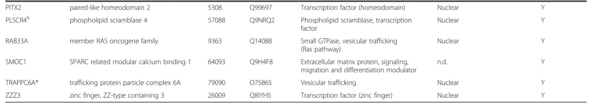
Table 2 Interaction partners for Hoxa1 revealed by Y2H screening using AD-Hoxa1 (Continued)
PITX2 paired-like homeodomain 2 5308 Q99697 Transcription factor (homeodomain) Nuclear Y
PLSCR4§ phospholipid scramblase 4 57088 Q9NRQ2 Phospholipid scramblase, transcription
factor Nuclear Y
RAB33A member RAS oncogene family 9363 Q14088 Small GTPase, vesicular trafficking (Ras pathway)
Nuclear Y
SMOC1 SPARC related modular calcium binding 1 64093 Q9H4F8 Extracellular matrix protein, signaling,
migration and differentiation modulator n.d. Y TRAPPC6A* trafficking protein particle complex 6A 79090 O75865 Vesicular trafficking Nuclear Y ZZZ3 zinc finger, ZZ-type containing 3 26009 Q8IYH5 Transcription factor (zinc finger) Nuclear Y
* Previously reported by Rual et al., 2005. §Revealed by both AD-ORF and DB-ORF screening. aY = yes; N = no. Lambert et al. BMC Development al Biology 2012, 12 :29 Page 6 of 17 http://ww w.biomedce ntral.com/1 471-213X/12 /29
Figure 1 Validation of 45 out of the 59 interactions revealed for Hoxa1 by affinity co-purification on glutathione-agarose beads. Candidate interactors were fused with a GST-tag and co-expressed in transfected cells with a FLAG-Hoxa1 fusion protein. Western blots were run to detect FLAG-Hoxa1 from cell extracts before (Input) or after (Co-P) purification. The Hoxa1-Hoxa1 or PBX1A-Hoxa1 interactions were used as positive controls (see lanes with arrowheads). Negative control corresponds to transfected cells with the only FLAG-Hoxa1 fusion protein (C-, lanes with blue arrows). Some interactors which could not be confirmed by co-purification are also shown (red asterisks).
Lambert et al. BMC Developmental Biology 2012, 12:29 Page 7 of 17
Hoxa1-mediated interactions take place in distinct cell compartments
We tested the 45 validated Hoxa1 interacting proteins by Bimolecular Fluorescence Complementation (BiFC) assay, which not only tests for protein interactions but can also visualize where the distinct interactions occur in live cells. For BiFC, the ORF corresponding to each interactor was fused C-terminally to the N-terminal 173 amino acids of the Venus fluorescent protein (VN173), while the Hoxa1 ORF was fused downstream of the C-terminal moiety of Venus (amino acids 155 to 243; VC155). Detectable fluorescence in cells transfected for the complementary VN173 and VC155 fusion proteins means that a functional Venus has been reconstituted, indicating that the partner proteins inter-act. As a preliminary control, BiFC was assayed for the well-established Hoxa1-PBX1A interaction (Figure 2). The VN173-PBX1A and VC155-Hoxa1 fusion proteins provided fluorescence complementation (Figure 2A), whereas the VN173-PBX1A/VC155 and VN173/VC155-Hoxa1 combinations did not (Figure 2B, C). This there-fore supported that the N- and C-terminal Venus fragments did not reassociate if not fused to interact-ing proteins. In addition, the immunocytolocalization of Venus consistently revealed that the VN173- and VC155-containing fusion proteins displayed a broad intracellular distribution that completely encompassed the narrower BiFC signal. In agreement with these con-trols, like the VN173-PBX1A fusion (Figure 2B), none of the VN173-interactor fusions provided fluorescence
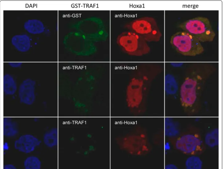
alone or in the presence of the VC155 Venus fragment alone (data not shown). For 41 out of the 45 interactors tested specific fluorescence was observed upon addition of the VC155-Hoxa1 fusion protein. Distinct patterns of intracellular interactions were observed (see Table 1 and 2, Figure 3). For 31 proteins, interactions took place in the nucleus (Figure 3A and C). Of these, 16 proteins appeared to contact Hoxa1 exclusively in the nucleus, while 15 also displayed other patterns of subcellular fluorescence complementation. Among the proteins found to bind Hoxa1 in the nucleus, some were known transcription factors (Table 5) or were known to have nuclear functions, but other were not (e.g. LGALS13, LIMS1, LNX2, MGAT5B, RBPMS, RAB33A, RGS20, TSCR1). A set of proteins shared a similar interaction pattern characterized by a diffuse, finely-punctuated cytoplasmic signal without nuclear staining (Figure 3B). This subcellular localization pattern was observed for different proteins reported to participate in a common signaling pathway. Examples are TRAF, TRIP or PDCD6IP (also known as Alix) which are found asso-ciated with the TNFR family of receptors [36-41], SPRY1 and PDCD6IP modulating RTK downstream signaling [42-46], PDLIM7 (alias LMP1) and RBPMS (also known as Hermes) which are involved in the BMP/TGFβ sig-naling regulation [47,48] and LPXN, PDCD6IP and TRIP6 known to associate with focal adhesion sites and related signal transduction [49-53]. As a control, in cells co-expressing GST-TRAF1 fusion and wildtype Hoxa1, proteins displayed an overlapping intracellular distribution
Table 3 Gene Ontology (GO) enrichment analysis
GO term Obs* Odds ratio$ P-value£ Corr P-valueμ
keratin filament 6 102,352 3,62292E-10 1,19194E-07
pattern specification process 6 12,3435 2,01548E-05 0,00331547
regionalization 5 14,3981 0,000048482 0,00531685
cranial nerve morphogenesis 2 116,248 0,000331888 0,0272978
kidney development 3 21,403 0,000559756 0,0306933
zinc ion binding 16 2,99882 0,000514454 0,0338511
embryonic development 6 5,58735 0,00120012 0,0564059
receptor signaling protein serine/threonine kinase activity 4 8,11769 0,00208634 0,0624006
developmental process 20 2,38882 0,00204634 0,0673246
negative regulation of MAP kinase activity 2 19,4953 0,00596027 0,0676182
regulation of transcription factor import into nucleus 2 31,8952 0,00251195 0,0688694
cation binding 19 2,14904 0,00649842 0,071266
anterior/posterior pattern formation 3 13,3725 0,00198155 0,0724365
cytoskeletal part 8 3,92527 0,00191605 0,0787977
inner ear morphogenesis 2 27,0115 0,00335617 0,0788701
* number of Hoxa1 interactors annotated with the corresponding GO term.
$the odds ratio represents the enrichment of the corresponding GO term in the set of Hoxa1 interaction partners, an odds ratio of 10 meaning that the considered GO term is observed 10 times more than expected at random.
£probability to see at least the number of proteins corresponding to the GO term at random. μP-value including a correction for multiple testing.
Lambert et al. BMC Developmental Biology 2012, 12:29 Page 8 of 17
Table 4 Pathways enriched in secondary Hoxa1 interactors
Pathway name ID* Obs$ Odds ratio£ FDRμ Corr FDR§ Source Gene symbols Entrez gene IDs
RXR and RAR hetrodimerization with other nuclear receptor
pc926 2 24,83 2,00E-05 2,40E-03 NCI-Nature FAM120B,NR1H2 84498,7376 Cell adhesion molecules (CAMs) - Homo sapiens
(human) hsa04514 2 19,70 9,00E-05 1,35E-02 KEGG CLDN2,PVRL2 9075,5819
Gap junction - Homo sapiens (human) hsa04540 2 16,59 8,00E-05 1,20E-02 KEGG GNAI2,PDGFRB 2771,5159
Signaling events mediated by PTP1B pc948 4 12,72 1,00E-05 1,20E-03 NCI-Nature CRK,SPRY2,TRPV6,PDGFRB 1398,10253,55503,5159 Retinoic acid receptors-mediated signaling pc960 3 10,19 2,10E-04 2,52E-02 NCI-Nature NR1H2,NRIP1,FAM120B 7376,8204,84498 Integrins in angiogenesis pc989 3 10,04 3,40E-04 4,08E-02 NCI-Nature CDKN1B,SPP1,VCL 1027,6696,7414 Down-stream signal transduction pc690 2 8,28 5,00E-05 3,14E-02 Reactome PDGFRB,NCK2 5159,8440
Signaling by PDGF pc876 2 8,28 5,00E-05 3,14E-02 Reactome PDGFRB,NCK2 5159,8440
Toll-like receptor signaling pathway - Homo sapiens (human)
hsa04620 3 7,54 3,20E-04 4,80E-02 KEGG TRAF6,TOLLIP,SPP1 7189,54472,6696 Homologous recombination - Homo sapiens (human) hsa03440 2 7,18 1,40E-04 2,10E-02 KEGG TOP3B,RAD54B 8940,25788 LPA receptor mediated events pc1042 4 7,07 1,30E-04 1,56E-02 NCI-Nature BIRC2,TRIP6,TRAF6,CRK 329,7205,7189,1398 Focal adhesion - Homo sapiens (human) hsa04510 8 6,55 3,00E-05 4,50E-03 KEGG SPP1,VCL,VASP,CRK,PDGFRB,BIRC2,
CCND3,PAK7 6696,7414,7408,1398,5159,329,896,57144 TCR signaling in naive CD4+ T cells pc1031 4 6,54 3,00E-04 3,60E-02 NCI-Nature BIRC2,TRAF6,GRAP2,TRPV6 329,7189,9402,55503 Axon guidance - Homo sapiens (human) hsa04360 5 6,50 1,40E-04 2,10E-02 KEGG GNAI2,PAK7,NTN4,ABLIM1,NCK2 2771,57144,59277,3983,8440 TCR signaling in naive CD8+ T cells pc997 4 6,34 3,80E-04 4,56E-02 NCI-Nature TRAF6,GRAP2,TRPV6,BIRC2 7189,9402,55503,329 Lysine degradation - Homo sapiens (human) hsa00310 3 6,28 1,00E-04 1,50E-02 KEGG OGDH,AASDHPPT,EHMT2 4967,60496,10919
IGF1 pathway pc1041 3 6,26 3,60E-04 4,32E-02 NCI-Nature CRK,NCK2,YWHAE 1398,8440,7531
p75(NTR)-mediated signaling pc978 5 6,02 2,60E-04 3,12E-02 NCI-Nature YWHAE,TRAF6,BIRC2,RTN4,LINGO1 7531,7189,329,57142,84894 Proteogylcan syndecan-mediated signaling events pc1045 5 5,53 2,20E-04 2,64E-02 NCI-Nature BSG,HGS,CRK,SPRY2,SPP1 682,9146,1398,10253,6696 Syndecan-1-mediated signaling events pc974 3 5,46 1,60E-04 1,92E-02 NCI-Nature BSG,CRK,HGS 682,1398,9146
Plasma membrane estrogen receptor signaling pc1048 6 4,07 3,60E-04 4,32E-02 NCI-Nature CRK,SLC9A1,IRF4,NCK2,YWHAE,TRAF6 1398,6548,3662,8440,7531,7189
* pathway identifier.
$ number of Hoxa1 interactors belonging to the corresponding pathway.
£the odds ratio represents the enrichment of the corresponding pathway, an odds ratio of 10 meaning that the considered pathway is observed 10 times more than expected at random. μfalse discovery rate as computed through the simulation.
§corrected false discovery rate accounting for multiple testing.
Lambert et al. BMC Development al Biology 2012, 12 :29 Page 9 of 17 http://ww w.biomedce ntral.com/1 471-213X/12 /29
consistent with the BiFC signal observed with VN173-TRAF1/VC155-Hoxa1 (Figure 4). Fourteen interactors tested displayed variable interaction patterns, showing mostly nuclear to nuclear and cytoplasmic or nuclear and vesicular BiFC signal (Figure 3A and C). This heteroge-neous distribution suggests a coordinated shuttling be-tween cell compartments for Hoxa1 and some partners (e.g. MDFI, OGT, PITX2, PRDM14, RBCK1, RBPMS, SPRY1, ZBTB16). The specific associations between Hoxa1 and 41 interactors detected by BiFC shows that Hoxa1 can associate dynamically with distinct categories of proteins in distinct intracellular domains.
Discussion
By a high-throughput Y2H screen we identified 59 Hoxa1 interacting proteins among which 45 were con-firmed by co-precipitation from animal cells. The intra-cellular localization of 41 interactions was further detected by a BiFC approach. This is the first exhaustive screen and analysis for interactors of a Hox protein. Our
data support the conclusion that Hox proteins, and Hoxa1 in particular, known as crucial transcription factors controlling developmental processes can fulfill unexplored roles in cell signaling, cell adhesion, or ves-icular trafficking.
Hoxa1 appears to interact with several proteins found to be part of molecular platforms associated with a few signaling pathways (TNFR superfamily, RTK, BMP/ TGFβ, Focal adhesion,. . .), membrane dynamics and ves-icular trafficking (Table 5). These platforms contact activated receptors at the plasma membrane and can positively or negatively modulate the downstream signal-ing or subsequent internalization in the endosomal com-partment. By interacting with these proteins Hoxa1 could either act as a modulator or an effector of these signaling pathways. The BiFC assay revealed that most of the interactors involved in signaling pathways display a similar pattern of Hoxa1 interaction in culture cells. LPXN, PDLIM7, PDCD6IP, RBPMS, SPRY1, TRAF1, TRAF2 and TRIP6, for example, showed a BiFC signal Figure 2 Bimolecular Fluorescence Complementation assay reveals the Hoxa1-PBX1A interaction in culture cells. COS7 cells were transfected with combinations of VN173, VN173-PBX1A, VC155 and VC155-Hoxa1 expression vectors. Upon interaction between PBX1A and Hoxa1, the VN173 and VC155 moieties of the Venus fluorescent protein brought together provide a fluorescent signal (A). Fluorescence complementation does not appear when the VN173 or VC155 fragments are expressed instead of the corresponding fusion proteins (B-C). As a control, expression of Venus fragments is detected by immunocytochemistry (anti-GFP). The BiFC signal shows colocalization with the anti-GFP immunofluorescence (D-F).
Lambert et al. BMC Developmental Biology 2012, 12:29 Page 10 of 17
Figure 3 (See legend on next page.)
Lambert et al. BMC Developmental Biology 2012, 12:29 Page 11 of 17
in the cytoplasm, with fine punctuated staining probably related to vesicular compartments (Figure 2B). Although further experiments are required to identify these com-partments, our data suggest that Hoxa1 interacts with distinct modulators of a given pathway at the level of shared molecular platforms. Finally, some interactors such as MDFI, OGT, RBCK1, RBPMS or SPRY1 display various patterns of Hoxa1 interaction from cell to cell, possibly indicating dynamic partnerships depending on cell physiological state (Figure 3A and C).
Some links might be drawn between the molecular, cellular and developmental processes involving Hoxa1 and its interactors. LIMS1 for example is expressed in neural crest cells and plays an important role in neural crest development through TGFβ signaling [54]; in mouse, a downregulation of SPRY1 inhibits the rhombomere4-derived neural crest cells to colonize the 2nd branchial arch [55]; RBPMS is expressed in the outflow tract of the developing heart [56], a territory colonized by Hoxa1 positive cells [57]. An important group of interactors consists in transcription factors. Some of them are known to be involved in embryonic patterning or cell fate decision (HOXD3, MDFI, PITX2 for example). In that regard, ZBTB16 (better known as PLZF) is a particularly relevant Hoxa1 interactor. It is expressed during hindbrain development at rhombomere boundaries and, like Hoxa1, has been pro-posed to control hindbrain segmentation [58]. Tran-scriptional coregulators, like the SET-domain histone methyl-transferase PRDM14 or the O-linked-N-acetyl-glucosamine (GlcNac) transferase OGT, have also been identified as Hoxa1 interactors which may contribute to Hoxa1-mediated gene regulation. Most significantly, OGT has recently been shown to be the homologue of the Drosophila Super sex combs (Sxc) protein. Sxc is associated to Polycomb complexes and is required for their ability to repress gene expression, including Hox genes [59].
Conclusions
We presented here the first large-scale Hox interac-tome characterized so far. Although only a handful of interactors are known for other Hox proteins, some interactors identified here for Hoxa1 are shared with other Hox proteins [28]. PLSCR1 has been shown to contact HOXA9 and HOXB6, and HOXA9 is also
contacted by TRIP6. RBPMS is able to interact with HOXA9 and HOXB9. These interactions, as well as other described here, underline that Hox proteins should be viewed not only as gene regulators, but also as compo-nents of signal transduction and modulation of cell-to-cell communication, cell adhesion and vesicular trafficking.
Methods
Yeast two-hybrid screening
The mouse Hoxa1 coding sequence was amplified from the pGIH327 expression plasmid[60] and cloned into pDONR-223 by Gateway BP recombinational reaction (attB1.1 primer: GGGGACAACTTTGTACAAAAAAGT TGGCATGAACTCCTTTCTGG; attB2.1 primer: GGG GACAACTTTGTACAAGAAAGTTGGGTAGTGGGAG GTAGTCAGAGTGTC; Invitrogen). By Gateway LR recombinational cloning, Hoxa1 was then transferred into pDEST-DB and pDEST-AD-CYH2 centromeric destination vectors [29] to code for Gal4 DNA binding domain (DB)-Hoxa1 and Gal4 activation domain (AD)-Hoxa1 fusion proteins, respectively.
MATα Y8930 and MATa Y8800 yeast strains (geno-type: trp1-901; leu2-3, 112; ura3-52; his3-200; gal4Δ; gal80Δ; GAL2-ADE2; LYS2::GAL1-HIS3; met2::GAL7-lacZ; cyh2R) were used for yeast two-hybrid (Y2H)
screens. The DB-Hoxa1 coding construct was first tested for auto-activation by transforming it into the MATα Y8930 yeast strain and testing for expression of the HIS3 reporter gene in the absence of any AD-hORF fusion protein, on a solid synthetic complete medium lacking leucine and histidine (Sc-L-H) and supplemented with 1mM 3-amino-triazol (3AT) [29]. The DB-Hoxa1 con-struct did not auto-activate.
High-throughput Y2H screens were essentially per-formed as described [29]. Briefly, DB-Hoxa1 and AD-Hoxa1 vectors were transformed into MATα Y8930 or MATa Y8800 yeast strains, respectively. The DB-Hoxa1 construct in MATα Y8930 was mated with MATa Y8800 containing the AD-hORF library [27], and for the other configuration DB-hORFs library in MATα Y8930 were mated with AD-Hoxa1 in MATa Y8800. After overnight growth at 30°C, diploid yeast cells were transferred to plates lacking histidine, leucine and tryptophan, supple-mented with 1mM 3AT (Sc-L-T-H+3AT), to select for those with elevated expression of the GAL1-HIS3 re-porter gene.
(See figure on previous page.)
Figure 3 Bimolecular Fluorescence Complementation assay reveals the Hoxa1-mediated interactions in culture cells. MCF10A cells were transfected with VN173-hORF and VC155-Hoxa1 fusion proteins. Upon interaction between the partner proteins, the VN173 and VC155 moieties of the Venus fluorescent protein brought together provide a fluorescent signal. The interactions between Hoxa1 and its interactors can be classified according to their intracellular pattern: (A) nuclear, (B) cytoplasmic or associated to vesicles, (C) nuclear and cytoplasmic and/or vesicular.
Lambert et al. BMC Developmental Biology 2012, 12:29 Page 12 of 17
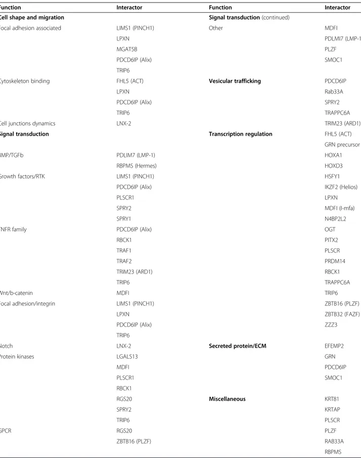
Table 5 Functional classification of Hoxa1 interactors
Function Interactor Function Interactor
Cell shape and migration Signal transduction (continued)
Focal adhesion associated LIMS1 (PINCH1) Other MDFI
LPXN PDLMI7 (LMP-1)
MGAT5B PLZF
PDCD6IP (Alix) SMOC1
TRIP6
Cytoskeleton binding FHL5 (ACT) Vesicular trafficking PDCD6IP
LPXN Rab33A
PDCD6IP (Alix) SPRY2
TRIP6 TRAPPC6A
Cell junctions dynamics LNX-2 TRIM23 (ARD1)
Signal transduction Transcription regulation FHL5 (ACT)
GRN precursor
BMP/TGFb PDLIM7 (LMP-1) HOXA1
RBPMS (Hermes) HOXD3
Growth factors/RTK LIMS1 (PINCH1) HSFY1
PDCD6IP (Alix) IKZF2 (Helios)
PLSCR1 LPXN
SPRY2 MDFI (I-mfa)
SPRY1 N4BP2L2
TNFR family PDCD6IP (Alix) OGT
RBCK1 PITX2
TRAF1 PLSCR
TRAF2 PRDM14
TRIM23 (ARD1) RBCK1
TRIP6 TRAPPC6A
Wnt/b-catenin MDFI TRIP6
Focal adhesion/integrin LIMS1 (PINCH1) ZBTB16 (PLZF)
LPXN ZBTB32 (FAZF)
PDCD6IP (Alix) ZZZ3
TRIP6
Notch LNX-2 Secreted protein/ECM EFEMP2
Protein kinases LGALS13 GRN
MDFI PDCD6IP PLSCR1 SMOC1 RBCK1 RGS20 Miscellaneous KRT81 SPRY2 KRTAP TRIP6 PLSCR GPCR RGS20 PLZF ZBTB16 (PLZF) RAB33A RBPMS
Lambert et al. BMC Developmental Biology 2012, 12:29 Page 13 of 17
Positive colonies were picked, grown on Sc-L-T plates, and retested on Sc-L-T-H, as well as on medium lacking Adenine (Sc-L-T-A) and Sc-L-T-H-A+3AT, to select for colonies with high GAL1-HIS3 and GAL2-ADE2 reporter gene activity. To detect any spontaneous auto-activators arising in the course of the screen, positive colonies were transferred in parallel onto cycloheximide containing media (Sc-H+CHX). Candidate colonies that grew on Sc-H+CHX were discarded.
The identities of candidate interacting pairs was deter-mined by sequencing PCR products amplified directly from yeast cells using primers specific to Gal4DB and Gal4AD (DB primers: GGCTTCAGTGGAGACTGATA TGCCTC, GGAGACTTGACCAAACCTC TGGCG; AD primers: CGCGTTTGGAATCACTACAGGG, GGAGAC TTGACCAAACC TCTGGCG). PCR products were puri-fied (Qiagen kit # 28104) and sequenced.
The protein interactions from this publication have been submitted to the IMEx (http://www.imexconsortion.org)
consortium through IntAct [pmid: 19850723] and assigned the identifier IM-15418.
Co-precipitation assays
The Hoxa1 coding sequence was transferred from the
pDONR-223 GatewayW vector to pDEST-FLAG
mam-malian expression vector by GatewayWLR recombination
reaction. Open reading frames coding for interactors from the hORFeome were cloned into a pDEST-GST mammalian expression vector by the same procedure.
COS7 and HEK293T cells were maintained in Dulbec-co’s modified Eagle’s medium (DMEM) low glucose or high glucose respectively (Gibco/Invitrogen) supple-mented with Glutamine, 10% fetal bovine serum (Gibco/ Invitrogen), 100 IU/ml penicillin, and 100 μg/ml strepto-mycin (Gibco/Invitrogen). Cell lines were maintained at 37°C in a humidified, 5% CO2atmosphere. For transient
transfection, 1.4 × 10⁵ (COS7) or 4 × 10⁵ (HEK293T)
cells were plated into six-well plates. Twenty-four hours Figure 4 Hoxa1 and TRAF1 intracellular distributions overlap. MCF10A cells were transfected with GST-TRAF1 and Hoxa1 expression vectors. The immunolocalization of GST-TRAF1 and Hoxa1 (anti-GST, anti-TRAF1 and anti-Hoxa1 immunocytofluorescence) reveals that the partner proteins display partially overlapping intracellular distribution.
Lambert et al. BMC Developmental Biology 2012, 12:29 Page 14 of 17
after plating, cells were transfected with TransFectin™ reagent (BioRad). One and a half μg of pDEST-FLAG-Hoxa1 expression vector and 3μg of pDEST-GST-hORF were mixed with 250μl of serum-free medium and added to a mix of 1 μl of TransFectin™ and 250μl of serum-free medium. Forty-eight hours after transfection, cells were lysed with Tris–HCl pH7.5 20mM, NaCl 120mM, EDTA 0.5mM, NP40 0.5%, glycerol 10% and Complete™ prote-ase inhibitor (Roche).
Cell lysates were cleared by centrifugation for 5 min-utes at 13,000 g. Cleared lysates were incubated over-night on gluthatione-agarose beads (Sigma # G4510). Beads were cleared 3 times with the lysis buffer. Beads and third wash samples were then loaded on SDS-PAGE, transferred on nitrocellulose membrane and processed for detection of FLAG tagged proteins with an anti-FLAG M2 antibody (Sigma # F1804).
Bimolecular Fluorescence Complementation assay (BiFC)
pDEST-VN173 and pDEST-VC155 plasmids were obtained by cloning sequences encoding N-terminal residues 1–173 and C-terminal residues 155–243 of the yellow fluorescent protein VENUS, respectively, within the pDEST-v1899-FLAG vector instead of the 5’ [KpnI|HindIII] 3xpDEST-v1899-FLAG- 3xFLAG-fragment (VN173F primers : GAGGTACCATGGTGAG CAAGGGCGAGGAGC, GGAGAAGCTTCTCGATGTTGT GGCGGATC; VC155 primers: AAGGTACCATGGCCGAC AAGCAGAAGAACGGC, GGAAAAGCTTCGTGGACCG GTGCTTGTACAGC).
The Hoxa1 coding sequence was transferred from
the pDONR-223 GatewayW vector to pDEST-VC155
mammalian expression vector by GatewayW LR
recom-bination reaction. Open reading frames coding for interactors from the hORFeome were cloned into the pDEST-VN173 mammalian expression vector by the same procedure.
MCF10A cells were maintained at 37°C in a
humidi-fied 5% CO2 atmosphere, in DMEM-F12+L-glutamine
medium (Gibco/Invitrogen) supplemented with 5% horse serum (Gibco/Invitrogen), 100 IU/ml penicillin (Gibco/Invitrogen), 100 μg/ml streptomycin (Gibco/Invi-trogen), 100 ng/ml of cholera toxin (Gentaur), 20 ng/ml of human Epidermal Growth Factor (hEGF; Sigma), 500 ng/ml hydroxycortisone (Sigma) and 10 μg/ml insulin (Sigma). For transfection, 3 × 10⁵ cells were seeded on
glass cover slips in 24-well plates. Twenty-four hours after plating, cells were transfected with TransFectin™ reagent (BioRad) or JetPRIME (Polyplus). For JetPRIME transfection, a total of 500 ng of plasmid DNA were transfected per well: 100 ng of pDEST-VN173-hORF, 20 ng of pDEST-VC155-Hoxa1 and 380 ng carrier DNA. DNA was mixed with 50 μl JetPRIME buffer and 1 μl of JetPRIME was added further. For TransFectin™-mediated transfection, 500 ng of pDEST-VN173-hORF and 500 ng
of pDEST-VC155-Hoxa1 were mixed with 50 μl of serum-free medium and added to a mix of 1 μl of TransFectin™ and 50 μl of serum-free medium. Twenty-four hours after transfection, cells were fixed with 4% formaldehyde for 30 minutes, rinsed three times in PBS and once in TBS-0,1% Triton X100. Glass
cover slips were mounted in VectashieldW-DAPI
medium (Vector laboratories). BiFC were then analysed by confocal microscopy (LSM710, Zeiss, Jena, Germany; Plan-Apochromat 63x/1.40 Oil DIC M27 objective; Oil refraction index 1.5 imaging medium; PMT camera). Images were acquired by using the ZEN 2010 software, and subsequently processed with ZEN 2008 Light Edition.
Immunocytolocalization
COS7 and MCF10A cells were maintained, seeded on coverslips and transfected as described here above. Twenty four hours after transfection, cells were fixed with 4% formaldehyde for 30 minutes. Cells were further blocked with 10% low-fat milk in TBS-0.1% Triton X100 solution for 45 min at room temperature, followed by overnight incubation in TBS-0.1% Triton X100 solution at 4°C, with a rabbit polyclonal anti-GFP (Invitrogen A11122, diluted 1/200), a mouse anti-GST (Sigma G1160, diluted 1/50), a mouse monoclonal anti-TRAF1 (Santa Cruz, sc-6253, diluted 1/50), or a rabbit poly-clonal anti-Hoxa1 (Abcam ab64941, diluted 1/50), as primary antibodies. Cells were rinsed three times for 30 min in TBS-0.1% Triton X100 solution and incubated for 45 min at room temperature with a goat anti-rabbit IgG-AF555 (Molecular Probes 4413, diluted 1/750), a goat anti-mouse IgG-FITC (SantaCruz sc-3699, diluted 1/100), or a bovine anti-rabbit IgG-TRITC (SantaCruz sc-2367, diluted 1/100), as secondary antibodies. Cells were rinsed three times and glass cover slips were mounted in VectashieldW-DAPI medium (Vector
labora-tories). Slides were then analysed by confocal micros-copy (LSM710, Zeiss, Jena, Germany; Plan-Apochromat 63x/1.40 Oil DIC M27 objective; Oil refraction index 1.5 imaging medium; PMT camera). Images were acquired by using the ZEN 2010 software, and subsequently pro-cessed with ZEN 2008 Light Edition.
Gene Ontology annotation and pathway analysis
Gene Ontology (GO) annotations were downloaded from Entrez Gene (September 2009), pathway data from KEGG (September 2008) and Pathway Commons (September 2008) databases. From Pathway Commons, we analyzed the pathways originally annotated in NCI-Nature[pid.nci.nih.gov] and Reactome [61].
Fisher’s Exact Test was used to determine GO annota-tion and pathway enrichment of Hoxa1 direct targets, using the space of human proteins that have been tested
Lambert et al. BMC Developmental Biology 2012, 12:29 Page 15 of 17
in our Y2H experiment, the human ORFeome v3.1 [27]. The corrected p-value was computed using the Benjamini-Hochberg multiple testing correction. We limited our results to GO annotations and pathways for which at least two Hoxa1 targets were annotated for.
To estimate the significance of indirect targets enrich-ment we ran 100,000 simulations for which the identity of the direct targets was randomized. The interactors of these targets were identified in an unbiased protein-protein interaction network [28], to avoid study bias inherent to literature curation. Interactors belonging to each pathway were counted, and the resulting distribu-tion compared to the observed counts. An empirical False Discovery Rate (FDR) determined the significance of the enrichment, with the FDR computed as the proportion of random trials giving at least the observed number of indirect targets in the analyzed pathway. The FDR was corrected for multiple testing using the Bonferroni correction. Pathways with a corrected FDR < 0.05 and at least two observed proteins were considered significant.
Abbreviations
TALE: Three Amino acid Loop Extension; TBP: TATA Binding Protein; HMG: High Mobility Group; CBP: CREB Binding Protein; Y2H: Yeast two-hybrid; BMP: Bone Morphogenetic Protein; TGF: Tumor Growth Factor; TNF: Tumor Necrosis Factor; RTK: Receptor Tyrosine Kinase; BiFC: Bimolecular Fluorescence Complementation; DB DNA: Binding domain; AD: Activation Domain; 3AT: 3-Amino-Triazol; GO: Gene Ontology; FDR: False Discovery Rate; ORF: Open Reading Frame; GST: Glutathione S-Transferase; NCoR: Nuclear receptor Co-Repressor; SMRT: Silencing Mediator of Retinoic acid and Thyroid hormone receptor; HDAC: Histone Deacetylase; KAP: Keratin Associated Protein.
Competing interests
The authors declare that they have no competing interests. Authors' contributions
BL carried out most of the molecular biology, yeast two-hybrid and cell biology experiments, made a substantial contribution to data analysis and drafted the manuscript. JV contributed to the co-precipitation experiments and substantially contributed to the BiFC assay and immunocytofluorescent detection of proteins. SR and IB set up the BiFC assay and the BiFC controls. NS carried out the bioinformatics analyses. JCT helped in the yeast two-hybrid screening and data interpretation. MV conceived and provided the materials required for the high-throughput yeast two-hybrid assay. RR conceived the study, significantly contributed to data interpretation and helped in drafting and revising the manuscript. All authors read and approved the final manuscript.
Acknowledgments
We are grateful to Michael Cusick and Matija Dreze for helpful comments on the manuscript. We are also grateful to Abdelmounaim Errachid (IMABIOL imaging platform) for technical assistance in confocal microscopy. We thank Samir Mérabet for providing us with plasmids coding for the VENUS VN173 and VC155 moieties and for helping us in setting up the BiFC assay. This work was supported by the Belgian Fund for Scientific Research (FNRS, FRSM grant 3.4.536.06F), the “Direction Générale des Technologies, de la Recherche et de l’Energie” of the Walloon Region (WALEOII grant n°516054), the Fonds Spéciaux de Recherche (FSR) of the Université catholique de Louvain (UCL), and by US National Human Genome Research Institute grant R01-HG001715 to MV. BL and IB held a FRIA fellowship from the Belgian Fund for Scientific Research and a FSR grant from UCL. NS is supported by a return grant from Belspo (Belgian Federal Government). MV is a Chercheur Qualifié Honoraire of the Belgian Fund for Scientific Research.
Author details
1Molecular and Cellular Animal Embryology group, Life Sciences Institute
(ISV), Université Catholique de Louvain, Louvain-la-Neuve 1348, Belgium.
2Bioinformatique des Génomes et des Réseaux (BiGRe), Université libre de
Bruxelles, Bruxelles, Belgium.3GIGA-R and Gembloux Agro Bio-Tech,
Université de Liège, Liège 4000, Belgium.4Center for Cancer Systems Biology
(CCSB) and Department of Cancer Biology, Dana-Farber Cancer Institute, Boston, MA 02215, USA.5Department of Genetics, Harvard Medical School,
Boston, MA 02115, USA.
Received: 16 October 2012 Accepted: 16 October 2012 Published: 22 October 2012
References
1. Alexander T, Nolte C, Krumlauf R: Hox genes and segmentation of the hindbrain and axial skeleton. Annu Rev Cell Dev Biol 2009, 25:431–456. 2. Iimura T, Denans N, Pourquie O: Establishment of Hox vertebral identities
in the embryonic spine precursors. Curr Top Dev Biol 2009, 88:201–234. 3. Narita Y, Rijli FM: Hox genes in neural patterning and circuit formation in
the mouse hindbrain. Curr Top Dev Biol 2009, 88:139–167.
4. Wellik DM: Hox patterning of the vertebrate axial skeleton. Dev Dyn 2007, 236(9):2454–2463.
5. Mann R, Lelli K, Joshi R: Hox specificity unique roles for cofactors and collaborators. Curr Top Dev Biol 2009, 88:63–101.
6. Gehring WJ, Kloter U, Suga H: Evolution of the Hox gene complex from an evolutionary ground state. Curr Top Dev Biol 2009, 88:35–61. 7. Cillo C, Faiella A, Cantile M, Boncinelli E: Homeobox genes and cancer.
Exp Cell Res 1999, 248(1):1–9.
8. Mark M, Rijli FM, Chambon P: Homeobox genes in embryogenesis and pathogenesis. Pediatr Res 1997, 42(4):421–429.
9. Hassan M, Saini S, Gordon J, van Wijnen A, Montecino M, Stein J, Stein G, Lian J: Molecular switches involving homeodomain proteins, HOXA10 and RUNX2 regulate osteoblastogenesis. Cells Tissues Organs 2009, 189(1–4):122–125.
10. Moens C, Selleri L: Hox cofactors in vertebrate development. Dev Biol 2006, 291(2):193–206.
11. Zhu A, Kuziora MA: Homeodomain interaction with the beta subunit of the general transcription factor TFIIE. J Biol Chem 1996, 271(35):20993–20996.
12. Um M, Li C, Manley JL: The transcriptional repressor even-skipped interacts directly with TATA-binding protein. Mol Cell Biol 1995, 15(9):5007–5016.
13. Chen Y, Knezevic V, Ervin V, Hutson R, Ward Y, Mackem S: Direct interaction with Hoxd proteins reverses Gli3-repressor function to promote digit formation downstream of Shh. Development 2004, 131(10):2339–2347. 14. Kataoka K, Yoshitomo-Nakagawa K, Shioda S, Nishizawa M: A set of Hox
proteins interact with the Maf oncoprotein to inhibit its DNA binding, transactivation, and transforming activities. J Biol Chem 2001, 276(1):819–826.
15. Bai S, Shi X, Yang X, Cao X: Smad6 as a transcriptional corepressor. J Biol Chem 2000, 275(12):8267–8270.
16. Li X, Nie S, Chang C, Qiu T, Cao X: Smads oppose Hox transcriptional activities. Exp Cell Res 2006, 312(6):854–864.
17. Zappavigna V, Falciola L, Helmer-Citterich M, Mavilio F, Bianchi ME: HMG1 interacts with HOX proteins and enhances their DNA binding and transcriptional activation. EMBO J 1996, 15(18):4981–4991.
18. Chariot A, van Lint C, Chapelier M, Gielen J, Merville MP, Bours V: CBP and histone deacetylase inhibition enhance the transactivation potential of the HOXB7 homeodomain-containing protein. Oncogene 1999, 18(27):4007–4014.
19. Shen W, Chrobak D, Krishnan K, Lawrence HJ, Largman C: HOXB6 protein is bound to CREB-binding protein and represses globin expression in a DNA binding-dependent, PBX interaction-independent process. J Biol Chem 2004, 279(38):39895–39904.
20. Saleh M, Rambaldi I, Yang X-J, Featherstone MS: Cell signaling switches HOX-PBX complexes from repressors to activators of transcription mediated by histone deacetylases and histone acetyltransferases. Mol Cell Biol 2000, 20(22):8623–8633.
21. Topisirovic I, Kentsis A, Perez JM, Guzman ML, Jordan CT, Borden KLB: Eukaryotic Translation Initiation Factor 4E Activity Is Modulated by HOXA9 at Multiple Levels. Mol Cell Biol 2005, 25(3):1100–1112.
Lambert et al. BMC Developmental Biology 2012, 12:29 Page 16 of 17
22. Derossi D, Joliot AH, Chassaing G, Prochiantz A: The third helix of the Antennapedia homeodomain translocates through biological membranes. J Biol Chem 1994, 269(14):10444–10450.
23. Brunet I, Di Nardo A, Sonnier L, Beurdeley M, Prochiantz A: The topological role of homeoproteins in the developing central nervous system. Trends Neurosci 2007, 30(6):206–207.
24. Chisaka O, Musci TS, Capecchi MR: Developmental defects of the ear, cranial nerves and hindbrain resulting from targeted disruption of the mouse homeobox gene Hox-1.6. Nature 1992, 355(6360):516–520.
25. Lufkin T, Dierich A, LeMeur M, Mark M, Chambon P: Disruption of the Hox-1.6 homeobox gene results in defects in a region corresponding to its rostral domain of expression. Cell 1991, 66(6):1105–1119. 26. Zhang X, Zhu T, Chen Y, Mertani HC, Lee KO, Lobie PE: Human growth
hormone-regulated HOXA1 is a human mammary epithelial oncogene. J Biol Chem 2003, 278(9):7580–7590.
27. Lamesch P, Li N, Milstein S, Fan C, Hao T, Szabo G, Hu Z, Venkatesan K, Bethel G, Martin P, Rogers J, Lawlor S, McLaren S, Dricot A, Borick H, Cusick ME, Vandenhaute J, Dunham I, Hill DE, Vidal M: hORFeome v3.1: a resource of human open reading frames representing over 10,000 human genes. Genomics 2007, 89(3):307–315.
28. Rual J-F, Venkatesan K, Hao T, Hirozane-Kishikawa T, Dricot A, Li N, Berriz GF, Gibbons FD, Dreze M, Ayivi-Guedehoussou N, et al: Towards a proteome-scale map of the human protein-protein interaction network. Nature 2005, 437(7062):1173–1178.
29. Dreze M, Monachello D, Lurin C, Cusick ME, Hill DE, Vidal M, Braun P: High-quality binary interactome mapping. Methods Enzymol 2010, 470:281–315.
30. Fernandez CC, Gudas L: The truncated Hoxa1 protein interacts with Hoxa1 and Pbx1 in stem cells. J Cell Biochem 2009, 106(3):427–443. 31. Phelan ML, Featherstone MS: Distinct HOX N-terminal arm residues are
responsible for specificity of DNA recognition by HOX monomers and HOX·PBX heterodimers. J Biol Chem 1997, 272(13):8635–8643. 32. Phelan ML, Rambaldi I, Featherstone MS: Cooperative interactions
between HOX and PBX proteins mediated by a conserved peptide motif. Mol Cell Biol 1995, 15(8):3989–3997.
33. Ashburner M, Ball CA, Blake JA, Botstein D, Butler H, Cherry JM, Davis AP, Dolinski K, Dwight SS, Eppig JT, et al: Gene ontology: tool for the unification of biology. The Gene Ontology Consortium. Nat Genet 2000, 25(1):25–29.
34. Kanehisa M, Araki M, Goto S, Hattori M, Hirakawa M, Itoh M, Katayama T, Kawashima S, Okuda S, Tokimatsu T, et al: KEGG for linking genomes to life and the environment. Nucleic Acids Res 2008, 36(Database):D480–D484. 35. Venkatesan K, Rual JF, Vazquez A, Stelzl U, Lemmens I, Hirozane-Kishikawa T, Hao T, Zenkner M, Xin X, Goh KI, et al: An empirical framework for binary interactome mapping. Nat Methods 2009, 6(1):83–90.
36. Lee NK, Lee SY: Modulation of life and death by the tumor necrosis factor receptor-associated factors (TRAFs). J Biochem Mol Biol 2002, 35(1):61–66.
37. Lee SY, Choi Y: TRAF1 and its biological functions. Adv Exp Med Biol 2007, 597:25–31.
38. Dempsey PW, Doyle SE, He JQ, Cheng G: The signaling adaptors and pathways activated by TNF superfamily. Cytokine Growth Factor Rev 2003, 14(3–4):193–209.
39. Li L, Bin L, Li F, Liu Y, Chen DC, Zhai Z, Shu H: TRIP6 is a RIP2-associated common signaling component of multiple NF-kappaB activation pathways. J Cell Sci 2005, 118(Pt3):555–563.
40. Mahul-Mellier A-L, Strappazzon F, Petiot A, Chatellard-Causse C, Torch S, Blot B, Freeman K, Kuhn L, Garin J, Verna J-M, et al: Alix and ALG-2 Are Involved in Tumor Necrosis Factor Receptor 1-induced Cell Death. J Biol Chem 2008, 283(50):34954–34965.
41. Odorizzi G: The multiple personalities of Alix. J Cell Sci 2006, 119(Pt 15):3025–3032.
42. Mason J, Morrison D, Basson M, Licht J: Sprouty proteins: multifaceted negative-feedback regulators of receptor tyrosine kinase signaling. Trends Cell Biol 2006, 16(1):45–54.
43. Cabrita MA, Christofori G: Sprouty proteins, masterminds of receptor tyrosine kinase signaling. Angiogenesis 2008, 11(1):53–62.
44. Schmidt MH, Hoeller D, Yu J, Furnari FB, Cavenee WK, Dikic I, Bogler O: Alix/ AIP1 antagonizes epidermal growth factor receptor downregulation by the Cbl-SETA/CIN85 complex. Mol Cell Biol 2004, 24(20):8981–8993.
45. Stasyk T, Schiefermeier N, Skvortsov S, Zwierzina H, Peranen J, Bonn GK, Huber LA: Identification of endosomal epidermal growth factor receptor signaling targets by functional organelle proteomics. Mol Cell Proteomics 2007, 6(5):908–922.
46. Lennartsson J, Wardega P, Engström U, Hellman U, Heldin C-H: Alix Facilitates the Interaction between c-Cbl and Platelet-derived Growth Factor Î2-Receptor and Thereby Modulates Receptor Down-regulation. J Biol Chem 2006, 281(51):39152–39158.
47. Sangadala S, Boden SD, Viggeswarapu M, Liu Y, Titus L: LIM Mineralization Protein-1 Potentiates Bone Morphogenetic Protein Responsiveness via a Novel Interaction with Smurf1 Resulting in Decreased Ubiquitination of Smads. J Biol Chem 2006, 281(25):17212–17219.
48. Sun Y, Ding L, Zhang H, Han J, Yang X, Yan J, Zhu Y, Li J, Song H, Ye Q: Potentiation of Smad-mediated transcriptional activation by the RNA-binding protein RBPMS. Nucl Acids Res 2006, 34(21):6314–6326. 49. Lipsky BP, Beals CR, Staunton DE: Leupaxin is a novel LIM domain protein
that forms a complex with PYK2. J Biol Chem 1998, 273(19):11709–11713. 50. Pan S, Wang R, Zhou X, Corvera J, Kloc M, Sifers R, Gallick GE, Lin SH,
Kuang J: Extracellular Alix regulates integrin-mediated cell adhesions and extracellular matrix assembly. EMBO J 2008, 27(15):2077–2090. 51. Pan S, Wang R, Zhou X, He G, Koomen J, Kobayashi R, Sun L, Corvera J,
Gallick GE, Kuang J: Involvement of the conserved adaptor protein Alix in actin cytoskeleton assembly. J Biol Chem 2006, 281(45):34640–34650. 52. Cabezas A, Bache KG, Brech A, Stenmark H: Alix regulates cortical
actin and the spatial distribution of endosomes. J Cell Sci 2005, 118(Pt 12):2625–2635.
53. Bai C-Y, Ohsugi M, Abe Y, Yamamoto T:ZRP-1 controls Rho GTPase-mediated actin reorganization by localizing at cell-matrix and cell-cell adhesions. J Cell Sci 2007, 120(16):2828–2837.
54. Liang X, Sun Y, Schneider J, Ding J-H, Cheng H, Ye M, Bhattacharya S, Rearden A, Evans S, Chen J: Pinch1 Is Required for Normal Development of Cranial and Cardiac Neural Crest-Derived Structures. Circ Res 2007, 100(4):527–535.
55. Trokovic N, Trokovic R, Partanen J: Fibroblast growth factor signalling and regional specification of the pharyngeal ectoderm. Int J Dev Biol 2005, 49(7):797–805.
56. Gerber WV, Yatskievych TA, Antin PB, Correia KM, Conlon RA, Krieg PA: The RNA-binding protein gene, hermes, is expressed at high levels in the developing heart. Mech Dev 1999, 80(1):77–86.
57. Makki N, Capecchi MR: Hoxa1 lineage tracing indicates a direct role for Hoxa1 in the development of the inner ear, the heart, and the third rhombomere. Dev Biol 2010, 341(2):499–509.
58. Ivins S, Pemberton K, Guidez F, Howell L, Krumlauf R, Zelent A: Regulation of Hoxb2 by APL-associated PLZF protein. Oncogene 2003, 22(24):3685–3697.
59. Gambetta MC, Oktaba K, Muller J: Essential Role of the Glycosyltransferase Sxc/Ogt in Polycomb Repression. Science 2009, 325(5936):93–96. 60. Remacle S, Shaw-Jackson C, Matis C, Lampe X, Picard J, Rezsohazy R:
Changing homeodomain residues 2 and 3 of Hoxa1 alters its activity in a cell-type and enhancer dependent manner. Nucleic Acids Res 2002, 30(12):2663–2668.
61. Matthews L, Gopinath G, Gillespie M, Caudy M, Croft D, de Bono B, Garapati P, Hemish J, Hermjakob H, Jassal B, et al: Reactome
knowledgebase of human biological pathways and processes. Nucleic Acids Res 2009, 37(Database):D619–D622.
doi:10.1186/1471-213X-12-29
Cite this article as: Lambert et al.: Protein interactions of the transcription factor Hoxa1. BMC Developmental Biology 2012 12:29.
Lambert et al. BMC Developmental Biology 2012, 12:29 Page 17 of 17