UNIVERSITÉ DE MONTRÉAL
THE EFFECT OF ZOOPLANKTON ON THE SURVIVAL OF ESCHERICHIA COLI IN MICROCOSMS (SDI)
TAREK A L FARAJ
DÉPARTEMENT DES GÉNIES CIVIL, GÉOLOGIQUE ET DES MINES ÉCOLE POLYTECHNIQUE DE MONTRÉAL
MÉMOIRE PRÉSENTÉ EN VUE DE L’OBTENTION DU DIPLÔME DE MAÎTRISE ÈS SCIENCES APPLIQUÉES
(GÉNIE CIVIL) NOVEMBRE 2017
UNIVERSITÉ DE MONTRÉAL
ÉCOLE POLYTECHNIQUE DE MONTRÉAL
Ce mémoire intitulé :
THE EFFECT OF ZOOPLANKTON ON THE SURVIVAL OF ESCHERICHIA COLI IN MICROCOSMS (SDI)
présenté par : FARAJ A L Tarek
en vue de l’obtention du diplôme de : Maîtrise ès sciences appliquées a été dûment accepté par le jury d’examen constitué de :
M. COMEAU Yves, Ph. D, président
Mme DORNER Sarah, Ph. D, directrice de recherche
M. BURNET Jean-Baptiste, Ph. D, membre et codirecteur de recherche Mme BICHAI Françoise, Ph. D, member
DEDICATION
ACKNOWLEDGEMENTS
My thanks are due, first and foremost, to my supervisor, Professor Sarah Dorner for accepting me into her research group, and providing me a highly stimulating opportunity. Thanks for your remarkable availability, scientific rigor, and constant support.
On the second place, I would also like to thank my co-supervisor M. Jean-Baptiste Burnet for all his support and technical advices. I consider myself very privileged to learn from him every day. Jean-Baptiste has been more than available for me anytime I needed him. It was an honour to share this experience with him.
Special thanks to Mme. Jacinthe Mailly, M. Yves Fontaine, Mme. Julie Philibert, Mme. Mireille Blais M. Denis Bouchard and Mme.Mélanie Bolduc; for the technical support and laboratory training, research would not work without you all. Also, many thanks for Laura Razafinjanahary and her assistant in different matters. I would like to thank all laboratory technicians and assistants in CHAIRE INDUSTRIELLE CRSNG EN EAU POTABLE and LABORATOIRES CREDEAU, polytechnique Montréal.
I have also to thank all colleagues and friends I met in this three-year experience of studies at Polytechnique Montréal, who made my experience unforgettable.
RÉSUMÉ
Escherichia coli, une des bactéries indicatrices fécales les plus couramment utilisées, joue un rôle clé dans le suivi et l'évaluation de la qualité de l'eau. Comprendre son devenir en tenant compte de divers facteurs environnementaux (ex. prédation par le zooplancton) est essentiel pour évaluer la qualité microbiologique de l'eau et de protéger le consommateur. De nombreuses études ont été menées sur les effets de la température, de l’irradiation solaire ou des nutriments. Quelques données limitées démontrent l’effet du broutage par le protozooplancton (nanoflagellés et ciliés hétérotrophes) sur le devenir d’E. coli dans l'eau. Cependant, la capacité du métazooplancton à influencer ou à contrôler l'indicateur fécal dans l'eau reste encore peu comprise, malgré l’omniprésence de cette communauté planctonique dans nos ressources aquatiques.
Dans cette étude, nous étudions l’impact du métazooplancton sur la survie d’E. coli dans l’eau. Une première série d’expériences a été menée en laboratoire afin d'étudier comment le métazooplancton, et en particulier le cladocère Daphnia, pouvait éliminer E. coli de l’eau dans diverses conditions en eau synthétique et en eau de lac. Ensuite, nos expériences visaient à comparer la pression de prédation de plusieurs représentants du métazooplancton sur E. coli dans de l’eau de surface. Enfin, les travaux se sont penchés sur l’étude de la variation saisonnière du taux de prédation (ou taux de broutage) sur E. coli par des communautés zooplanctoniques indigènes (à la fois méta- et protozooplancton) dans de l’eau de lac.
En microcosmes d’eau synthétique, l’espèce modèle D. pulex (32 ind.L-1) ingère et induit un effet significatif sur la survie d’E. coli. Cependant, en réduisant d’un facteur 1000 la concentration initiale en E. coli dans l’eau, les taux d’abbattement diminent de 1.65 j-1 à 0.62 j-1, témoignant d’une plus faible probabilité de rencontre entre Daphnia et E. coli. En eau de lac, l'influence de Daphnia sur les taux d’abattement d'E. coli. Augmente avec la densité de population du cladocère, et atteint près de 0.47 j-1 en présence de 65 ind.L-1. Il est intéressant de noter également qu’un part majeur de la diminution des concentrations en E. coli est attribuable à des facteurs liés à la matrice - probablement par la présence d’autres communautés bactérivores et/ou par des effets de compétition bactérienne.
Dans une matrice constituée d'un mélange d'eaux usées et d'eaux de surface pour simuler un évènement de contamination fécale en présence de métazooplancton en densités de population
représentatives pour les eaux de surface, D. magna (36 ind. L-1) semble exercer l’impact le plus important sur E. coli avec un taux d’abattement moyen de 2,33 j-1. À densités de populations égales (36 ind. L-1), D. pulex induit des taux d’abattement d’E. coli compris entre 0,99 et 0,62 j-1. Le rotifère Brachionus calyciflorus possède quant à lui la plus faible pression de prédation sur E. coli avec un taux moyen ne dépassant pas 0,21 j-1, cela malgré une densité de population plus importante (500 ind.L-1). La raison la plus probable de leur faibleimpact sur E. coli semble provenir de leur taille plus réduite et par conséquent de vitesses de filtration nettement plus faibles que celles de Daphnia.
Enfin, nous mettons en évidence une certaine variabilité saisonnière du taux de broutage des communautés zooplanctoniques d’eau douce sur E. coli. Cependant, les raisons de ces variations semblent complexes à appréhender et nécessiteraient davantage de données. Des facteurs associés non seulement à la composition et l’abondance relatives des groupes de zooplancton mais également la température semble influencer la prédation sur E. coli dans les eaux de surface. En conclusion, le rôle potentiellement épurateur de Daphnia se confirme pour E. coli dans l’eau. Cependant, il est important d’être conscient que les taux d’abattement d’E. coli dépendent de multiples facteurs, dont certains comme la ratio prédateur-proie, la taille du zooplancton (et leur taux de filtration) ainsi que le type de matrice et de ses composantes biotiques, ont été mis en évidence durant ce travail. Les résultats offrent une vision plus complète de l'effet du zooplancton sur E. coli dans l’eau et aident à expliquer les différences qui peuvent être observées à différents moments de l'année dans les milieux aquatiques naturels. Le type de données acquises au cours de ce travail devraient également permettre d’améliorer les modèles actuels sur le devenir et le transport d’E. coli dans l’eau.
ABSTRACT
Escherichia coli is one of the most commonly used fecal indicator bacteria (FIB), and thereby plays a key role in water quality monitoring and assessment. Understanding its fate under a variety of environmental factors (ex. predation by bacterivorous zooplankton) is essential for the assessment of water quality to ensure public health. Much research has been done on the effects of temperature, sunlight irradiation, or nutrient scarcity. Limited knowledge has been generated on the effect of protozooplankton grazing (heterotrophic nanoflagellates and ciliates) on the fate of E. coli in water. In contrast, the capability of metazooplankton communities - which are widespread in freshwater ecosystems - to impact or control the fecal indicator in water remains poorly understood.
In this study, we investigate the role of metazooplankton on the survival of E. coli. We first perform laboratory feeding experiments to investigate how metazooplankton, especially the filter-feeding Cladoceran Daphnia, could impact the fate of E. coli under different experimental conditions in synthetic and lake water microcosms. Then, we compare the grazing pressure of different metazooplankton species on E. coli in surface water microcosms. Finally, we describe the seasonal evolution of grazing on E. coli by natural metazooplankton and protozooplankton communities in lake water.
In synthetic water matrices, the model species D. pulex (32 ind.L-1) ingested E. coli and increased its loss rates. Following a 1,000-fold reduction in E. coli initial concentrations, decay rates decrease from 1.65 d-1 to 0.62 d-1, reflecting the lower probability of encounters between Daphnia and E.- coli. In lake water matrices with a D. pulex density ranging from 0 to 65 ind.L-1, we observed that E. coli loss rates increased with Daphnia densities, reaching 0.47 d-1 in the presence of D. pulex at 65 ind.L-1. Also, a significant portion of the E. coli population loss was associated with matrix-related factors - most likely due to predation by other bacterivorous biota and/or bacterial competition.
When simulating a fecal pollution event in water containing representative metazooplankton population densities, D. magna (36 ind. L-1) showed a significant impact on the E. coli loss rate and reached 2.33 d-1. At the same population density, D. pulex impacted the E. coli population with a loss rate between 0.99 and 0.62 d-1. With E. coli loss rates of 0.21 d-1,the small rotifer Brachionus
500 ind.L-1. Although additional investigations are warranted, the low impact of the rotifer most likely results from its smaller size and lower filtering rate.
Finally, we report a seasonal evolution of grazing pressures on E. coli in a freshwater bay. Variations in community grazing rates on E. coli appear to be complex to understand and may be linked to species composition and abundance but also to water temperature and the occurrence of cyanobacteria.
In conclusion, Daphnia appears to be an efficient filter-feeder for the removal of E. coli in water. However, E. coli loss rates depend on a variety of factors such as predator to prey ratio, size of the zooplankton (and their filtration rate) and the type of matrix and its components (biotic interactions). These results provide a more comprehensive view of the effect of zooplankton on E. coli bacteria within water bodies and help to explain differences that can be observed at various times of the year in natural aquatic environments. Data on grazing rates should prove helpful for the improvement of current fate and transport models.
TABLE OF CONTENTS
DEDICATION ………...iii ACKNOWLEDGEMENTS………iv RÉSUMÉ ...v ABSTRACT...vii TABLE OF CONTENTS………...………...ix LIST OF TABLES...xii LIST OF FIGURES………...xivLIST OF APPENDICES ...xviii
CHAPTER 1 INTRODUCTION ……….1
1.1 Fecal pollution of aquatic resources ... 1
1.2 Regulations of drinking and recreational water quality ... 3
1.3 Zooplankton (Daphnia spp) role in food web in water bodies ... 4
1.4 Thesis organization ... 5
CHAPTER 2 LITERATURE REVIEW………...6
2.1 Fate and transport of E. coli in water ... 6
2.1.1 Temperature ... 7
2.1.2 Advection and sedimentation ... 8
2.2Biotic interactions between water bacterial communities and zooplankton communities ... 9
2.2.1 The zooplankton including the filter-feeder Daphnia ... 10
2.2.2 Some interactions between zooplankton (Daphnia) and bacterial communities including E. coli ... 12
2.3 Hypotheses ... 16
CHAPTER 3 MATERIAL AND METHODS……….……….17
3.1 Maintenance of laboratory cultures ... 18
3.1.1 Algae cultures ... 18
3.1.2 Zooplankton cultures ... 21
3.2 Origin of E. coli ... 24
3.3 Impact of D. pulex on the decay rates of E. coli ... 25
3.3.1 Synthetic water ... 25
3.3.2 Lake water ... 30
3.4 Impact of various metazooplankton species on E. coli decay rates ... 32
3.4.1 Preparation of microcosms ... 34
3.4.2 Experimental procedure ... 35
3.4.3 Enumeration of settled and Daphnia-associated E. coli ... 38
3.5Impact of natural zooplankton communities on the decay rates of E. coli in lake water ... 39
3.5.1 Adaptation of the dilution method ... 39
3.5.2 Sample collection and in situ analyses ... 40
3.5.3 Microcosm setup ... 41
3.5.4 Laboratory analyses ... 42
3.6 Enumeration of E. coli USEPA method 1604 ... 43
3.7 Statistical analyses ... 43
CHAPTER 4 RESULTS………...44
4.1 Experiment 1 – Impact of D. pulex on E. coli. ... 44
4.1.1 Synthetic water ... 44
4.1.2 Lake water ... 46
4.2.1 First assay ... 49
4.2.2 Second assay ... 50
4.2.3 Enumeration of settled and Daphnia-associated E. coli ... 52
4.3Impact of natural zooplankton communities on the decay rates of E. coli in lake water ... 55
CHAPTER 5 DISCUSSION……….…58
5.1 Addressing research needs ... 58
5.2 Does Daphnia affect the fate of E. coli in water? ... 59
5.2.1 Synthetic water ... 59
5.2.2 Lake water ... 60
5.3 How do different metazooplankton species affect the fate of E. coli? ... 62
5.4 How do natural meta – and protozooplankton communities affect the fate of E. coli in water and how does their predation pressure evolve seasonally? ... 65
CHAPTER 6 CONCLUSION AND RECOMMENDATIONS……….68
BIBLIOGRAPHY……….……….70
LIST OF TABLES
Table 3. 1: The concentration of E. coli and algae spike with the Daphnia. ... 27
Table 3. 2: Experimental conditions for determining the fate of E. coli upon exposure to a gradient of D. pulex in lake water ... 31
Table 3. 3: Concentrations of E. coli and zooplankton population densities used in experiment #2 to assess the differential impact of zooplankton species on the fate of E. coli in surface water microcosms spiked with raw sewage. ... 36
Table 4. 1: First order kinetics of E. coli decay upon exposure to Daphnia pulex. ... 45
Table 4. 2: Characterisation of zooplankton biota in the lake water matrix sampled at Missisquoi Bay (QC), Canada on Sept. 1, 2015. HNF, heterotrophic nanoflagellates; NA, not applicable; * expressed in ind.L-1 and ind.mL-1 for metazooplankton and protozooplankton, respectively; ** if >10 ind.L-1. ... 46
Table 4. 3: First order kinetics of E. coli decay upon exposure to Daphnia pulex. ... 47
Table 4. 4: The sonicated Daphnia and their E. coli results CFU.ml-1 at the end of the experiment. ... 53
Table 5. 1: E. coli loss rate (d-1) in absence or presence of Daphnia spp and rotifer. ... 63
Table A. 1 : The quantities of sea salt and stock solutions to prepare the ADaM medium. ... 83
Table A. 2 : The added reagents and final water quality to prepare the EPA medium. ... 84
Table A. 3 : The components of stock solutions, trace elements and vitamins to prepare the BBM medium. ... 85
Table A. 4 : The components of Dehydrated MI agar and deionized or distilled water for EPA medium preparation. ... 87
Table A. 5 : Temporal evolution of E. coli upon exposure to D. pulex. Results are shown as average ± standard deviation (n=3) andare expressed in CFU.mL-1 (log CFU.mL-1). ... 90 Table A. 6 : Temporal evolution of E. coli upon exposure to D. pulex. Results are expressed as
average ± standard deviation (n=3). Results are expressed in CFU.mL-1 (log CFU.mL-1). .. 92 Table A. 7 : Temporal evolution of E. coli upon exposure to D. pulex. D. magna (clones 1 and 2).
Results are shown as average ± standard deviation (n=3) and expressed in CFU.mL-1 (log CFU.mL-1). ... 93 Table A. 8 : Temporal evolution of E. coli upon exposure to D. pulex. D. magna and Brachionus
calyciflorus. Results are expressed as average ± standard deviation (n=3). Results are expressed in ... 94 Table A. 9 : Physico-chemical measurements for all conditions, during the whole experiment
(Second assay), measurements of PH, Oxygen (mg/L), Temperature (°C), and Turbidity (UNT). ... 95 Table A. 10 : Zooplankton community composition of Missisquoi Bay water used for assessing the
grazing pressure on E. coli using the dilution method (*na = not available). ... 96 Table A. 11 : Physico-chemical and nutriment composition of Missisquoi bay water used for
LIST OF FIGURES
Figure 2. 1 : The life cycle of the cladoceran Daphnia pulex (asexual and sexual phases). ... 11
Figure 3. 1: experimental setup. ... 17 Figure 3. 2: A) algal cultures of Scenedesmus quadricauda, Pseudokirchneriella subcapitata, and
Ankistrodesmus falcatus. B) harvesting of algae culture in 250 mL bottles under laminar flow cabinet before centrifugation. C and D) 1L Erlenmeyer flasks inoculated with freshly harvested algae for re-culturing. ... 19 Figure 3. 3: Neubauer counting chamber, (www.slideshare.net). ... 20 Figure 3. 4: Model species used during the present work ... 21 Figure 3. 5: Zooplankton cultures for Daphnia pulex, Daphnia magna (clones 1 or “KLEINE” and
2 or “XINB3”) and Brachionus calyciflorus. ... 23 Figure 3. 6: The location of samples collection (Missisquoi Bay. Quebec, Canada). Sampling point
for experiment 1 is "Philipsburg" and for experiment 3 is "Venise-en-Québec". ... 24 Figure 3. 7: Impact of D. pulex on the decay rates of E. coli in synthetic water (ADaM). ... 25 Figure 3. 8: Zooplankton wheel used for incubation of bottle microcosms during experiment 1. 27 Figure 3. 9: USEPA method 1604 for enumeration of culturable E. coli. A and B) vacuum filtration
ramp. C and D) Blue E. coli colonies on MI agar plates after incubation at 35°C during 18 - 24 h). ... 28 Figure 3. 10: Overview of the second experimental setup using lake water. Blue: raw lake water;
light blue: 53 µm-filtered lake water. ... 30 Figure 3. 11: Overview of the second experimental setup. E. coli with D. pulex, D. magna and B.
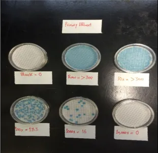
calyciflorus in a matrix of river water mixed with primary wastewater (no mixing of the microcosms). ... 33 Figure 3. 12: Enumeration of E. coli in the primary effluent sample on MI agar plates (USEPA
Figure 3. 13: Second experiment's microcosms, E. coli in presence or absence of D. pulex, D. magna (clone 1 and 2) and rotifers B. calyciflorus, in mixed river-raw sewage (A, B and C). Also, Physical and chemical measurements by HACH Probe (HQ40d) (D). ... 37 Figure 3. 14 : Grazing rates (g) correspond to the negative slope (a) of the line after linear-regression of the data (see figure above). ... 40
Figure 4. 1: Temporal evolution of E. coli concentrations in synthetic water (ADaM) in absence or presence of Daphnia pulex (32 ind. L-1) during 48 hours and for varying E. coli (103 or 106 CFU.mL-1) and algae (0.1 or 1.7 cells.mL-1) concentrations. (Daphnia dashed lines, controls, full lines) 3 colors only, 3 conditions. See appendix section 2.1, table 1 for more details. ... 44 Figure 4. 2: Effect of E. coli initial concentration (103 or 106 CFU.mL-1) and algae quantity (low
or high algal food) on E. coli loss rates (in d-1) following 48 hours incubation in absence or presence of D. pulex at densities of 32 ind.L-1. Results from figure 4.1. were converted into decay rates using the formula described in page 25 (equation 2). Letters indicate significantly (p<0.05) different E. coli decay rates among conditions in presence of Daphnia. * significant (p<0.05) difference between absence and presence of Daphnia for a given condition (Burnet et al., 2017). ... 45 Figure 4. 3: Temporal evolution of E. coli concentration in lake water in presence of increasing
Daphnia pulex population densities of 8, 32 and 65 ind. L-1. Two controls, composed of raw
and 53 µm-filtered lake water devoid of D. pulex were performed to assess the natural removal of E. coli in presence of local biota of all sizes and smaller than 53 µm, respectively. RLW, raw lake water; FLW, 53 µm-filtered lake water. ... 47 Figure 4. 4: Grazing of D. pulex on E. coli in a lake water matrix. Decay rates (in day-1) of E. coli
are measured following 48 hours incubation in presence of a D. pulex gradient (8, 32 and 65 ind.L-1) in 53 µm-filtered lake water. Filtered or raw lake water samples were used as controls to determine E. coli decay rates in the absence of D. pulex. Letters indicate significant differences between treatments in filtered lake water; asterisks report significant differences from raw lake water. LW, lake water. ... 48 Figure 4. 5: E. coli concentration (103 CFU.mL-1) versus time in water matrix (river water mixed
-1) during 48 hours of incubation. The decrease in E. coli concentrations is more apparent with
Daphnia. ... 49
Figure 4. 6: E. coli average decay rates (in d-1) following 48 hours incubation in the absence or presence of Daphnia pulex and Daphnia magna (clones c1 and c2) at densities of 36 ind.L-1. E. coli were added to the microcosms at an initial concentration of ~103 CFU.mL-1. ... 50 Figure 4. 7: Grazing of D. magna clone1, D. pulex and the rotifer B. calyciflorus on E. coli in a
matrix of river water and primary wastewater effluent. ... 51 Figure 4. 8: E. coli decay rates (in d-1) following 48 hours incubation in the absence or presence
of Daphnia pulex and Daphnia magna (clone 2) at densities of 36 ind.L-1 and the rotifer B. calyciflorus at densities of 500 ind.mL-1. E. coli were added to the microcosms at an initial concentration of ~103 CFU.mL-1. ... 51
Figure 4. 9: Zooplankton-mediated decay rates (after subtraction of decay rates observed in control microcosms) for D. pulex (n=6), D. magna clone 1 (n=3), D. magna clone 2 (n=6) and B. calyciflorus (n=3). ... 52
Figure 4. 10: Average concentrations of E. coli (CFU.ml-1) remaining in microcosm sediments at the end of the second assay. ... 53 Figure 4. 11 : Illustrate the removed E. coli, remained in sediments, remained in water and
associated with Daphnia at the end of experiment (second assay). ... 54 Figure 4. 12 : : In situ physical and chemical parameters for the 7 sampling occasions (June to
October 2015). ... 55 Figure 4. 13 : Metazooplankton relative densities (individuals.L-1) for the 7 sampling occasions
(June to October 2015). ... 56 Figure 4. 14: Protozooplankton densities for the 7 sampling occasions (June to October 2015). Left:
ciliates in individuals.L-1; right: HNF in individuals.mL-1. NA, not available. ... 56 Figure 4. 15: Zooplankton community grazing rates (d-1) as determined by the dilution method for
Figure A. 1 : Survival rate of Daphnia pulex in microcosms after 72h (Synthetic water experiment). ... 91 Figure A. 2 : Zooplankton community grazing rates (d-1) as determined by the dilution method for
the 7 sampling occasions (June to October 2015). ... 98 Figure A. 3 : Location of Saint-Hyacinthe wastewater treatment plant (google map), and collecting
LIST OF APPENDICES
CHAPTER 1
INTRODUCTION
1.1 Fecal pollution of aquatic resources
Surface water contaminated by fecal contamination such as Escherichia coli, and Enterococci (ENT) is a public health concern and a prevailing global environmental issue. This contamination occurs because of droppings from wildlife, inadequate sewage treatment (human fecal pollution), agricultural runoff, or faulty septic systems (Harwood et al., 2000; Johnson et al., 2004). The fecal indicator bacteria (FIB), which include fecal coliforms, Escherichia coli, and Enterococci, are an indicator of fecal contamination of bodies of fresh water. Consequently, the presence of fecal contamination is an indicator that a potential health risks exist for individuals exposed to the contaminated water. Therefore, they are used as a standard of microbial contamination of recreational water quality and as well as drinking water (US Environmental Protection Agency, 1986). Water polluted by fecal pollution is still one of the most critical origins of both epidemic and endemic worldwide diseases in both developing and developed countries. Releasing wastewater into water bodies without appropriate treatment is one of the high-risk factors which leads to disease and infections from fecal contamination. Many bacteria, viruses, and protozoa are transmitted via fecal pollution resuling in waterborne diseases including gastroenteritis, hepatitis, diarrhea, and respiratory and skin diseases. Exposure comes from inhalation or ingestion, via either direct consumption (drinking) or recreational activities (WHO, 2004; Stirling, 2001).
Waterborne diseases are prevalent not only in developing countries, but are also a serious challenge in developed countries. In the United States between the period of 1986 to 2000, 5,905 outbreaks were reported, with 95 outbreaks associated with recreational water. (Pandey et al., 2014). An outbreak in Milwaukee, U.S.A. in 1993 was responsible for the infection of around 403,000 people, having led to an estimated 50 deaths. In Milwaukee, Cryptosporidium contaminated the water supply due to inadequate filtration of water from Lake Michigan. In Walkerton, Ontario, Canada in 2000, the E. coli O157:H7 and Campylobacter contaminated the municipal well water and this contamination resulted 2,300 people falling ill, and seven dead (Craun et al., 2010).
It is important to monitor for fecal pollution in water for the protection of public health. When analyzing water samples for microorganisms (pathogens) of public health concern, it is difficult, expensive and time-consuming to test directly for the presence of the large variety of disease
causing pathogens. Therefore, water is tested for indicator species (coliforms and enterococci), which may be present when pathogens are present, and occur in high enough concentrations that they will be above detection limits (Glassmeyer et al., 2005).
Total coliforms have long been used as bacterial indicators. Total coliforms are common bacteria in the environment. They are Gram-negative, oxidase-negative, non-spore forming rods that ferment lactose with gas production at 35–37 °C after 24 to 48 hours in a medium with bile salts and detergents (Cabral, 2010). Total coliform counts are not necessarily a measure of fecal pollution, because total coliforms include bacteria that are naturally present in the environment and are not of fecal origin. However, their presence in water environment gives an indication of the possibility of existence of disease-causing pathogens, such as bacteria, viruses, and parasites (WHO, 2008; Medema, 2003; Payment, 2003). In addition, fecal coliforms are coliforms that ferment lactose at 44.5 °C, in a medium with bile salts (WHO, 2008; Grabow, 1996; Medema, 2003; Payment, 2003). Fecal coliforms are bacteria that exist in the intestines and feces (fecal origin) and include bacteria such as Escherichia coli (E. coli) (Young, 1992).
One of the potentially pathogenic bacteria in contaminated water from untreated wastewater is Escherichia coli, and it is also a fecal indicator. Escherichia coli has long been considered the most suitable bacterial indicator of fecal contamination in drinking water, and the testing methods are well developed and standardized (Edberg, 2000; Snozzi, 2001). E. coli are gram-negative, non-spore forming, rod-shaped, facultatively anaerobic bacteria and can ferment lactose with gas production after 24-84h at 37°C (Bitton, 1980). Also, they are defined as thermophilic coliforms that produce indole from tryptophan (feature used to confirm the presence of E. coli), but are also defined as a coliform able to produce β-glucuronidase or GLUC (although taxonomically up to 10% of environmental E. coli may not). All over the world, E. coli is used as an indicator organism to identify water and food samples that contain unacceptable levels of fecal contamination (for example, no E. coli is acceptable in distributed drinking water). The presence of E. coli in samples has been used as an indicator of fecal contamination since the turn of the 20th century (Environmental Protection Agency, U. S. 2000) and its presence indicates that there is a heightened risk of the presence of other fecal pathogenic bacteria and viruses (ex. Salmonella spp. or Hepatitis A) (Brüssow, 2004; Atlas, 1993). Recreational water guidelines from the USEPA revealed that in terms of prediction of occurrence of gastrointestinal illness in freshwater, the E. coli bacteria was a more reliable and consistent indicator than enterococci in terms of predicting illnesses (Wade et
al., 2003; U.S. EPA 2002). Compared to other coliforms, E. coli is shed in larger quantities, which facilitates its detection in water, soils, or food. In addition, E. coli O157:H7 and other strains of toxigenic E. coli are responsible for gastroenteritis that can cause effects and symptoms such as diarrhea, abdominal cramps, severe bloody diarrhea, and vomiting (Arnone and Walling 2007; WHO, 2003).
As the general test for coliforms includes bacteria that are not from fecal origins (e.g. Enterobacter, Klebsiella, Citrobacter), E. coli is considered a more specific indicator of fecal contamination. Therefore, to distinguish the E. coli from non-fecal coliforms, testing of the lack of an enzyme (selective for the E. coli organism) is required to confirm presumptive fecal coliforms or by using elevated temperature (44.5°C) (Francy et al., 1993; EPA). Furthermore, several studies (Abreu-Acosta and Vera, 2011; Wu, 2011 and Molleda 2008) have shown that the bacterial indicator organisms, including Escherichia coli, total coliforms, and fecal coliforms have limitations in their use to regulate and monitor pathogen levels in treated wastewater and the environment because of their lack of correlation with pathogens. As an example, Marino et al., 2005 studied the influence of the temperature and pH on E. coli concentrations. E. coli decreased with the increasing of pH and temperature, while Vibrio cholerae and Enterococcus faecalis increased. This difference showed why E. coli are sometimes not well correlated with V. cholerae. In drinking water distribution systems, the existence of a single positive sample for total coliforms is not enough to trigger public health action. Rather, if more than 5% of samples are positive, then actions and notifications are required (Edberg, 2000).
1.2 Regulations of drinking and recreational water quality
The bacteria in the coliform group (total coliforms, thermotolerant (faecal) coliforms, E. coli) and the enterococci have been used to monitor drinking and recreational waters for the presence of faecal contamination. However, testing for every possible waterborne disease causing microorganism would be prohibitive in terms of the financial resources necessary, the time required to perform the analyses, and the difficulties of isolating and quantifying. In addition, testing requires a proper laboratory, specialized equipment, and highly trained and experienced microbiologists. Therefore, authorities monitor for non-pathogenic faecal indicator bacteria that are present in high numbers in both human and animal faeces, which are indicative of faecal contamination and suggest the possible presence of enteric pathogens (Health Canada, Guidelines
for Canadian Recreational Water Quality – Third Edition). Outbreaks of illness associated with recreational water use result from exposure to infectious pathogens in recreational water venues that can be treated (pools and hot tubs or spas) or untreated (lakes and oceans). The outbreaks of illness associated with untreated recreational water can be caused by Cryptosporidium or by Escherichia coli (E. coli O157:H7 or E. coli O111) (Hlavsa et al., 2015). Also, drinking water contamination events can cause disruptions in water service, resulting in a large impact on public health. Waterborne disease outbreaks can be caused by Legionella or the parasite Cryptosporidium (Benedict, 2017).
In Canada, for recreational waters, the indicator organism used for primary contact activities is Escherichia coli (E. coli) for fresh waters and Enterococci for marine waters (Guidelines for Canadian Recreational Water Quality – Third Edition). For drinking water, E. coli is currently the best available indicator of recent faecal contamination in drinking water systems. E. coli can be used to provide an indication of the magnitude of a problem and thus inform the public health response (Health Canada, Guidelines for Canadian Drinking Water Quality). Also, in the province of Québec Canada the E. coli is the preferred bacterial indicator. The regulations for drinking water and recreational water (treatment and distribution in Québec) are based upon E. coli concentrations. (Ministère du Développement durable, de l'Environnement et de la Lutte contre les changements climatiques, Québec (MDDELCC, 2017); Regulation respecting the quality of drinking water (Q-2, r.40)).
1.3 Zooplankton (Daphnia spp) role in food web in water bodies
Daphnia spp has been well recognized as a keystone species in food webs and its important role as a primary consumer in aquatic food chains. The Cladocerans such as Daphnia species and protozoans are considered the major consumer of bacteria in freshwater lakes (JiJRGENS 1994; Pace 1990). Daphnia spp have shown to have the potential for reducing the microbial communities through several surveillance studies conducted. Therefore, this may lead to controlling the microbial population in natural waters (Siciliano et al., 2015).
The crustacean Daphnia has become a cornerstone of systems biology research for alternative testing and methodologies (Kurz and Ewbank, 2007). Also, in terms of measuring and screening toxicity, the use of Daphnia (D. magna and D. pulex) is considered a high sensitivity analytical
tool for screening toxicity of common environmental chemicals and monitoring of effluents and contaminated waters (Zhou et al., 2008 and Persoone et al., 2009). In addition, the ease of cultivating the crustaceans Daphnia, its well-studied biology, and its wonderful attributes of a short development time and large brood size make it suitable model for various environmental studies (Lambolez et al.,1994 and Seco et al., 2003).
The harmful effects of chemical treatment of wastewater makes ecological alternatives attractive; alternatives such as plants, invertebrate zooplankton, and fish that are able to remove algae and bacteria, and improve water quality (Shiny et al., 2005). In wastewater treatment, Daphnia magna filtration removed particles (diameters below 30 μm) efficiently removing E. coli bacteria. However, the removal depends on abiotic parameters such as water temperature and the hydraulic retention time (HRT) (Serra et al., 2014). Also, the filtration of Daphnia magna efficiently removed suspended sludge particles and was efficient at wastewater disinfection (Serra et al., 2016). Daphnia magna improved the water quality of wastewater, reducing the particle volume concentration of small particles. Experiments were performed in both the laboratory and in a mesocosm system (Pau et al., 2013).
From all material mentioned previously, we can illustrate the importance of zooplankton and their possible ability to impact the pathogenic bacteria such as E. coli in natural waters.
1.4 Thesis organization
This thesis is separated in the following manner:
Chapter 1 introduces the concepts of water fecal pollution and regulations of drinking and recreational water quality in Canada, as well as some basic information about the role of Daphnia spp in water bodies.
Chapter 2 provides the literature review concerning the fate and transport of E. coli in water, and biotic interactions between E. coli and zooplankton communities.
Chapter 3 contains the results.
Chapter 4 presents the experimental techniques and methods used in this work. Chapter 5 presents the discussion.
CHAPTER 2
LITERATURE REVIEW
A large body of knowledge exists on the grazing of protozooplankton on planktonic or non-pathogenic microbial communities in water (Sheer and Sheer, 2002). Also, it is well known that protozooplankton species are reservoirs and vectors of pathogenic microorganisms in the environment (Barker and Brown, 1994; Greub and Raoult, 2004). In contrast, similar knowledge on metazooplankton is rather limited (Bichai et al., 2008; Nowosad et al., 2007). Rotifers and cladocerans have a cosmopolitan geographical distribution, are among the most common biota of freshwater ecosystems, and they are both capable of significant clearance of their planktonic preys in natural environments. Among the rare studies on metazooplankton-pathogen interactions, rotifers have been shown to ingest Cryptosporidium oocysts and Gairdia cysts (Fayer et al., 2000; Trout et al., 2002; Stott et al., 2003). Connelly et al. (2007) provide evidence that the cladoceran D. publicaria has the potential to substantially decrease the number of infectious Cryptosporidium and Giardia in freshwater. In laboratory conditions, Schallenberg et al., 2005 also shows that D. publicaria can remove Campylobacter from water. Ingestion of microorganisms is also suspected to modify their settling rates upon enclosure into fecal pellets (Brookes et al., 2004). However, the number of field studies reporting zooplankton predation on fecal pathogens and indicators is limited (Nowosad et al., 2007; Bichai et al., 2010).
2.1 Fate and transport of E. coli in water
E. coli is the most widespread indicator of fecal pollution, and it is very important to understand its survival and transport in water. In order to assess the severity of contamination for making appropriate management decisions, it is essential to understand E. coli survival rates in the aquatic environment (Blaustein et al., 2013). Additionally, to assess the risk of pathogens, it is recommended to understand critical variables influencing their fate and distribution in water (lakes and reservoirs). Sampling for E coli should be paired with other biological, physical, and chemical parameters, because several factors control the transport, distribution, and inactivation of pathogens in natural water environments (Brookes et al., 2004).
Several biotic and abiotic environmental factors influence the survival (fate) of allochthonous bacteria. Biotic factors such as grazing by protozoa is one of the main biological processes able to control allochthonous bacteria and their density (Van Limbergen et al.,1998). The control of
bacteria then depends on the digestion capacity of the grazer, and the bacterial concentration (Barcina et al.,1997). With regards to abiotic factors, physicochemical stress can lead bacteria to enter to a viable but non-culturable state or dormancy. Also, temperature, nutrient scarcity, visible light, and osmotic stress all have negative influences on the survival of bacteria. For instance, in aquatic systems the UV light has a negative effect by increasing the loss of culturability and the formation of active but non-culturable cells (e.g. Barcina et al.,1997). Menon et al. (2003) reported that in the North Sea, mortality rates of both autochthonous and fecal bacteria increased with the increase of temperature. Part of this mortality may be due to the grazing of a larger community of protozoa (heterotrophic nanoflagellates), which also rose with temperature increases. Wcislo and Chrost (2000) reported that in the aquatic environment, the disappearance of E. coli was related to the grazing of microflagellates. Also, the grazing of flagellates on bacteria was "size-selective" and E. coli with sizes of (0.5-2 μm) were more susceptible to predation than other small autochthonic bacteria. They also reported the effect of bacteriophages on the survival time of E. coli and found it to be insignificant. Their results also confirmed the influence of other factors on the survival of E. coli in aquatic ecosystems including other heterotrophic bacteria and the impact of temperature.
Sampling and measuring E. coli must be representative of environmental conditions. Therefore, it is necessary to understand detailed processes governing E. coli survival, including predation, which has been identified as a dominant process.
2.1.1 Temperature
Activity and abundance of the free-living bacteria is strongly influenced by temperature, one of the most important environmental factors affecting the transport and survival of bacteria (Peierls and Paerl, 2010). Bacteria in fresh waters survive longer at low temperatures than at high temperatures (Barcina et al.,1986). Despite other environmental factors controlling E. coli survival rates, such as sunlight intensity, pH, predation, and salinity, temperature is considered as the major factor influencing E. coli survival in water (Blaustein et al., 2013). At various temperatures in filtered and unfiltered river water microcosms, the mortality of E. coli increased with temperature (Flint, 1987). Likewise, E. coli and Enterococcus spp had significantly higher inactivation rates at 20°C than at 14°C (Noble, 2004).
On the other hand, The E. coli population structure in Dunes Creek was more varied in summer than in winter although the reasons remained unclear (Whitman et al., 2008). Temperature also
affects the survival of bacteria through its influence on other environmental factors, and by controlling the metabolic rate of bacterial cells (Jones, 1971; EPA, 1985). For instance, the influence of temperature on E. coli survival can be enhanced through the predatory activity of protozoa (Barcina et al., 1986a, b), but is also directly linked to temperature in the absence of eukaryotes (Anderson et al, 1983). Temperature and light penetration in natural environments are related. For example, a temperature gradient often occurs in natural aquatic environments. In the absence of light (for example, in darker, turbid waters) enteric bacterial survival is enhanced (Gould and Munro, 1980; Evison and Morgan, 1982), and the mortality of coliform bacteria is more likely to increase as temperature increases (Gameson and Gould,1985). Blaustein et al. (2013) reported that the E. coli in different water sources had highly variable survival rates at the same given temperature. This is potentially due to variations in biological and physical survival factors. Also, they demonstrated that the inactivation of E. coli in rivers and wastewater was faster than in lakes and other cleaner water sources.
2.1.2 Advection and sedimentation
Horizontal and vertical processes in water are important in determining the distribution of particles, fecal indicator bacteria, and pathogenic microorganisms. Horizontal transport in lakes and reservoirs is driven by wind and internal waves, which also generate vertical movements (Justin et al., 2004). In rivers, horizontal transport is primarily through advection. With precipitation, pathogens can be transported into rivers, lakes, and reservoirs, from runoff and inflow. In lakes, inflows may sink to lower depths, depending on temperature and salinity gradients, and flow along the bathymetry towards the deepest point. If pathogens are present in the inflow, they are diluted, and if they reach the deepest points where it is cold and dark, some pathogens such as Cryptosporidium oocysts can survive for extended periods (Justin et al., 2004; Deen et al., 2000).
Vertical transport of fecal indicator bacteria or pathogens occurs through settling processes. The aggregation of pathogens with particles containing organic matter plays an important role in the distribution and transportation of pathogens and survival in water environments. (Belle and Gerba, 1979). Aggregation may allow a consortia of bacteria cells to be more resistant to the effects of surrounding environmental stressors, then to transport and colonize in the host (Gilbert et al., 1993). Surface charges play a key role in pathogen–particle interactions in aqueous system (Ongerth and Pecoraro, 1996). For instance, in surface waters, Cryptosporidium oocysts may have negative
surface charges, the same as gatherings of natural particles, and in the same way as responding metal salt coagulation (Ongerth et al., 1996). Moreover, the sedimentation occurs when microorganisms attach to particles and settle in the sediments. Indicator organisms can persist in sediments, and may use the sediment to provide protection from environmental stressors (Anderson, 1983; Fish, 1995). In the mesocosms experiment by (Fish, 1995), the density of E. coli increased over the sample time when the mesocosms contained sterile water and sediment, probably due to the lack of environmental stressors. While, E. coli concentration decreased when the mesocosms contained only sterile water.
The size and density of particles are important factors in estimating the settling rate and the vertical distribution of pathogens. Particulate material may assist with the aggregation of pathogens, thereby influencing the rate of pathogen settling, but could also influence the predation of pathogens (Reynolds, 1984). It is important to consider particle size ranges when determining the settling and resuspension characteristics of pathogens (Brookes et al., 2004). The settling speed of a single oocyst in water was about 0.03 m day-1, and it increased when it was associated with
biologically treated sewage effluent particles (Medema et al., 1998). In addition, in their sedimentation kinetics, G. lamblia cysts and C. parvum oocysts could be qualified by oocysts size and density, as well as the density and viscosity of the sedimentation medium. The movements of oocysts in water occur more easily because of water flow, wind, temperature, and movement of aquatic organisms than because of gravitational settling (Medema et al., 1998). In the Lake of Burragorang in Sydney, Australia, field measurements show that the estimation of sedimentation rates of oocysts is around 5–10 m/day-1, meanwhile, the free-floating oocysts settle very slowly. Consequently, oocysts need to be attached to particles to have high settling velocities, which are necessary for modelling pathogen transport (Hawkins et al.,2000).
2.2 Biotic interactions between water bacterial communities and
zooplankton communities
Although much has been described about the abiotic stressors and factors are controlling the fate of pathogens and indicator organisms (viability and/or infectivity) in water, such as temperature, solar radiation (UV and visible), nutrient scarcity, salinity etc., quite limited information exists on the biotic effects (Blaustein et al., 2013; Wanjugi and Harwood, 2013).
2.2.1 The zooplankton including the filter-feeder Daphnia
What are zooplankton? Zooplankton are small animals that float freely in the water column of lakes and oceans, depending on water movement. The sizes of zooplankton community of most lakes ranges from a few tens of microns (Protozoa) to up to 2mm (macrozooplankton). The dominant groups of zooplankton in most lakes are protozooplankton, which is the protozoa, and metazooplankton, which are rotifers and crustaceans (Cladocerans Daphnia and Copepods). Zooplankton play a important role in water-based food webs, they can be food for fish or predators, grazing on algae, bacteria, protozoa, and other invertebrates (Vanni,1988).
One important zooplankton is the Daphnia. Daphnia - better known as water fleas - are characterized by flattened leaf-like legs that are used to produce a water current to act as a filtering apparatus, and are members of the order Cladocera. They have an enclosed body in an uncalcified shell or carapace (1-5 mm long), and are ubiquitous in freshwater aquatic environments (Ebert, 2005). In addition, Daphnia can feed on algal blooms (for proteins and carbohydrates), bacteria, and yeast. The Daphnia life cycle starts with an egg, then progresses to juvenility, adolescence, and adult life, with an average lifespan of between 40-56 days (Pennak, 1989). In normal environmental conditions, Daphnia have the ability to rapidly clone themselves asexually. However, when environmental conditions deteriorate, such as a lack of food availability or poor temperatures, Daphnia can procreate sexually and produce resting cysts (eggs), which are able to hatch when conditions improve (Figure 2.1).
Figure 2. 1 : The life cycle of the cladoceran Daphnia pulex (asexual and sexual phases).
Daphnia are important because they have an important role as a model for ecological studies and are an essential part of the food web in water bodies. They have been well recognized as a keystone species in food webs, and for their important role as a primary consumer in aquatic food chains. The Cladocerans such as the Daphnia species and protozoans are considered to be the major consumer of bacteria in freshwater lakes (Jijrgens 1994; Pace 1990). Also, evidence based on grazing phenomenon suggests it’s one of the major forces shaping the bacterial community structure in waters (Güde, 1989). In addition, Daphnia are often used in the environmental monitoring of the aquatic environment and in bioassays. For instance, the Environmental Protection Agency (EPA) recommended Daphnia magna and Daphnia pulex as standard aquatic test species for toxicity tests. Moreover, compared to other zooplankton, the Daphnia organisms are more intensive grazers (filter feeders) and they are widely available in many surface waters like ponds. Daphnia have the potential as a community to filter a large volume of water (Cyr et al., 1992). Also, Daphnia can be cloned naturally and thus, the culturing of Daphnia is easy and cost-efficient (Weber, 1991 and EPA, 2002). In general, Daphnia are among the best-studied subjects in ecology literature (Ebert, 2005).
2.2.2 Some interactions between zooplankton (Daphnia) and bacterial
communities including E. coli
Generally, the bacteria that have been ingested by zooplankton might be either digested (although yet unproven) or excreted out back into the water environment. Regarding the predation by indigenous biota and their effect on E. coli survival in water, a lot of information has been gathered for protozooplankton (heterotrophic nanoflagellates (HNF) and ciliates). However, much less is known for the role of metazooplankton grazing (Bichai et al., 2008).
Regarding the association of bacteria with zooplankton in aquatic systems, bacteria in general may attach to zooplankton either directly to the exoskeleton (Nagasawa and Nemoto, 1988) or to other particles such as food particles or aggregates (Simon et al. 2002, Carrias and Sime-Ngando 2009). In addition, bacteria may be found inside the gut of zooplankton due to ingestion (Tang, 2005). While there is a lack of information on zooplankton being physically associated with pathogenic microorganisms in water, bacterial settlement of the exoskeleton surface of zooplankton organisms and attachment to planktonic animals is more validated as a mode for pathogens to be transferred from the water phase in natural systems. Certain bacteria can attach themselves to the surface of zooplankton organisms where they discover a microhabitat that enables them to persist longer in the environment. So far, the most widely known example is the case of Vibrio cholerae, the bacterium responsible for cholera (Cottingham et al. (2003). Tang et al. (2010) reported that bacteria interact with zooplankton in several different ways, with implications for microbial production, evolution, biogeochemical fluxes, diversity maintenance, and dispersal.
Knowledge of the grazing effects of metazooplankton on E. coli is still limited as compared to protozooplankton, which is one of the main biological processes controlling allochthonous bacteria and their concentrations in water (Barcina et al.,1997). Menon et al. (2003), reported that in river and in the coastal areas, protozooplankton grazing was responsible for more than 90% of the overall mortality rate of fecal and autochthonous bacteria. In a constructed reservoir, microflagellate grazing was found to be the main factor responsible for E. coli mortality, and the microflagellates were more size-selective of E. coli cells (Wcislo and Chrost, 2000). In addition, in lake ecosystems, protozoans or metazoans have been shown to play a key role as bacterial consumers depending on the structure of the food web. Heterotrophic nanoflagellates (HNF) can change the bacterial community such that smaller cells dominate because they selectively consume the large-sized
bacterial cells (Jürgens at al., 1994). Grazing information is still limited to a few zooplankton groups and environments, for example, copepods in coastal and estuarine waters (Menon et al., 2003). There are few studies that have examined microorganism survival during ingestion by zooplankton species. These studies generally do not provide any information on water-borne pathogens that zooplankton grazing in natural conditions with realistic concentrations (Menon et al., 2003; Wcislo and Chrost., 2000; Barcina et al., 1997 and Jürgens et al.,1994).
However, a few studies show the effect of metazooplankton on E. coli. A feeding rate experiment of Daphnia magna was done by McMahon and Rigler (1965). Daphnia magna in different concentrations (50, 100 and 20 ind/mL) fed on four different foods (E. coli, Saccharomyces cerevisiae, Chlorella vulgaris, and Tetrahymena pyriformis). The feeding behavior of Daphnia magna was approximately similar for all conditions. Although maximum rates expressed as cells/hr-1 varied tremendously, the maximum volume of the different foods that were consumed were similar. The filtering rates for E. coli per Daphnia magna (2.8-3.3-mm size) was 5.6 million cells/hr. However, filtering efficiency was independent of the size of food (ranging from 0.9 µ3 to
1.8 X 10⁴ µ3).
McMahon and Rigler (1963) studied the crustacean Daphnia magna incubated with two foods (Chlorella vulgaris and Saccharomyces cerevisiae). The feeding rate was proportional to the concentration of food, although there were critical thresholds above and below which little effect was observed on feeding rates. There exists an effect on the feeding rate of Daphnia by the food present in their gut before starting an experiment. Daphnia pre-fed at a very low rate do, for a short time, feed more rapidly than those prefed at a higher rate. However, this study did not observe a decrease in feeding rate with time (McMahon and Rigler, 1963). However, Hadas et al. (1983) reported that Daphnia magna is able to ingest bacteria and algae, also a cell-free extract (CFE) of Daphnia magna was able to lyse E. coli cells by causing damage to E. coli cells as represented in the release of the enzymes of the bacterial cell.
Cladocerans, especially the Daphnia genus, are important in terms of filtration capacity. They are able to ingest pelagic food particles (including bacteria) over a wide range of sizes by collecting them with their thoracic appendages (Brendelberger 1991; Riemann 1985). Also, Cladocerans like Daphnia are widespread and found in many lakes and ponds over the world. Their presence can
change the structure and performance of microbial food webs in freshwater ecosystems. Daphnia can apply significant grazing pressure on all components of microbial food webs (picoplankton, nanoplankton and microplankton) because they are able to filter a large spectrum of particle sizes (Jürgens 1994). In terms of ingestible particle sizes by Daphnia, the planktonic bacteria are at the lower level, while Nanoplanktonic protozoans (flagellates and small ciliates) are in the optimum size range. Thus, Daphnia could become the main bacterivores and consume the major portion of bacterial production. Also, Daphnia grazing can have a positive influence effect on bacterioplankton (Jürgens 1994). In terms of Cladocerans filler mesh size and its capability of retention of very small particles, four Cladocera species (Daphnia magna, Daphnia pulex, Simocephalus vetulus, and Diaphanosoma brachyurum) had retention efficiencies of between 33 and 154% of food particles (consisting of green algae, free-living planktonic bacteria and cyanobacteria). Although, the Cladoceran species with coarser filtering meshes showed lower retention efficiencies for bacteria and cyanobacteria, the Cladocerans can utilize free-living, single-celled, planktonic bacteria as a resource of food (Brendelberger. 1991). In addition, Degans et al., (2002) reported that there was a quick change in structure and biomass bacterioplankton community upon exposure to Daphnia grazing and heterotrophic nanoflagellates (HNF). He concluded that Daphnia have a clear impact on the lake bacterioplankton, either by direct grazing on bacteria or grazing on protozoan bacterivores. Daphnia magna lived on diets containing small proportions of heterotrophic bacteria (E. coli 20% and Flavobacterium ≤ 50%) and Daphnia grew significantly in these diets compared to pure algal diets, suggesting the occurrence of nutritional upgrading by these bacteria. Thus, the nutrition of Daphnia may have improved with the presence of the more diverse bacterial composition (Freese and Martin-Creuzburg., 2013). Also, high biomass of Daphnia pulex can sufficiently keep the bacterioplankton below the carrying capacity, since the bacteria growth was about one doubling per day and the Daphnia was able to balance it by the grazing activity (Jürgens at al., 1994).
In freshwater habitats, there is a complex interaction between protozooplankton communities and metazooplankton communities because Daphnia can feed upon protozooplankton (ciliates and flagellates) (Jürgens at al., 1994). In the study of Jürgens at al. (1994), they reported that the Daphnia pulex predation and grazing on the protozoans (nanoflagellates) was much higher than on bacteria. When the Daphnia pulex were removed, the heterotrophic nanoflagellates developed and
became the main bacterivores. This means that when large Daphnia populations are present, the heterotrophic nanoflagellates become unimportant.
Daphnia spp, have the potential for reducing some bacterial communities, as demonstrated through surveillance studies. The presence of Daphnia can lead to the control of the abundance of microbial communities in natural waters (Siciliano et al., 2015). Under artificial experimental conditions, Daphnia pulicaria could naturally control Giardia lamblia cysts (zoonotic parasites) and Cryptosporidium parvum oocysts by controlling their density, viability, and infectivity (Connelly et al., 2007). In addition, Daphnia carinata and Campylobacter jejuni were incubated for 72 hours in simulated and natural aqueous conditions. The predation of Daphnia carinata caused death and reduced the Campylobacter jejuni concentrations, as compared to the control without Daphnia carinata. The reduction in the Campylobacter jejuni concentration was 2 logs (Schallenberg et al., 2005). Moreover, an experimental study on controlling pathogens in nature demonstrated that the Cladocerans Daphnia pulex and Moina macrocopa reduced Vibrio cholerae densities more efficiently than rotifers (Ramirez et al., 2012). On the other hand, some authors reported that the filter-feeders (Cladocerans) are not able to digest all kinds of bacteria. Unlike rod shaped bacteria, general coccoidal bacteria survived the digestion of Daphnia ambigua. Therefore, perhaps "viable gut-passage" is one of the mechanism of bacteria to survive cladoceran grazing (King et al., 1991). However, even though there is no direct evidence of bacterial ingestion, digestion, or death by zooplankton, grazing of zooplankton on bacteria is expected to be an important factor in the reduction of bacteria in natural environments (Pace 1988).
2.3 Hypotheses
- In view of the filtration capacity of metazooplankton communities and the experimental evidence that they ingest FIB and pathogens with the potential to reduce their numbers (Schallenberg et al., 2005; Connelly et al., 2007), it is expected that grazing by metazooplankton, especially Daphnia, significantly contributes to the removal and potential inactivation of fecal bacteria in surface waters (Boehm et al., 2005).
- It is expected that the grazing pressure varies over the year since zooplankton population dynamics follow seasonal trends.
2.4 Objectives
This research has been organised into three specific objectives that have been adressed in this master’s thesis:
1. Using the model species D. pulex, determine the impact of Daphnia grazing on E. coli under various experimental conditions
- Under different effect of matrix (presence of algal food, type of water). - Under different Daphnia densities.
2. Assess the grazing impact of various metazooplankton species (Daphnia, Moina and Brachionus) on E. coli.
3. Quantify the grazing impact of natural zooplankton communities on E. coli and determine how this impact evolves seasonally.
CHAPTER 3
MATERIAL AND METHODS
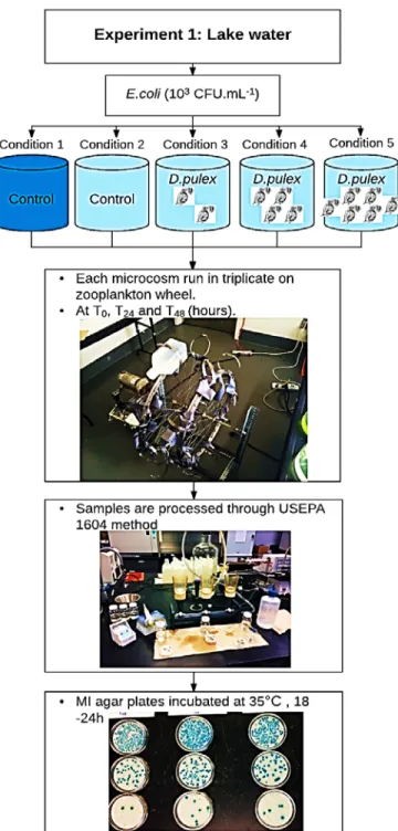
Three different experiments with different designs and conditions, and with different zooplankton species were performed. The first experiment was done with only Daphnia pulex, either in synthetic water medium (ADaM), or lake water, to study the interactions Daphnia- E. coli upon exposure of the FIB to the grazer. The second experiment was done by Daphnia pulex, Daphnia magna, and rotifers Brachionus calyciflorus. Finally, the third experiment was done by using natural metazooplankton and protozooplankton communities (Figure 3.1).
3.1 Maintenance of laboratory cultures
Before starting our experiments two cultures were maintained in our lab: Algae cultures and Zooplankton cultures. The algae cultures were used as food for the zooplankton cultures’ growth, then the zooplankton cultures were used in our experiments to assess their impact on E. coil bacteria.
3.1.1 Algae cultures
Cultures from three algal species (Scenedesmus quadricauda, Pseudokirchneriella subcapitata, and Ankistrodesmus falcatus) were kindly provided by Prof. Melania Cristescu from the Department of Biology, McGill University and were maintained in modified BBM (Bold's Basal Medium, see appendix A seection 1.3) in 1L Erlenmeyer flasks with continuous stirring (magnetic stir bar) and air bubbling. The cultures were grown during a photoperiod of 18 hours of light and 6 hours of dark using triphosphorus fluorescent lights at an ambient temperature of 20 ± 1°C. The volume of culture medium was changed on a weekly basis. The harvest of algae cultures was done during the log phase after 7 to 8 days growth. Liquid algae cultures were distributed into 250mL centrifuge flasks and centrifuged at 4000rpm (3350g) during 10 minutes. The supernatant was discarded to remove the waste products generated by the algal metabolism during growth (Figure 3.2).
Figure 3. 2: A) algal cultures of Scenedesmus quadricauda, Pseudokirchneriella subcapitata, and Ankistrodesmus falcatus. B) harvesting of algae culture in 250 mL bottles under laminar flow cabinet before centrifugation. C and D) 1L Erlenmeyer flasks inoculated with freshly harvested algae for re-culturing.
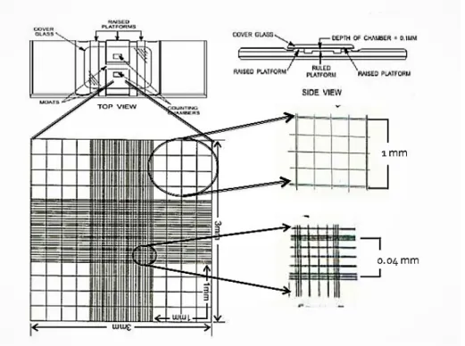
The algal pellet was resuspended in BBM medium and the algae concentration was determined using a Neubauer counting chamber (Figure 3.3). A 1000-fold dilution of the resuspended pellets was prepared by successive Log10 dilutions. Then, approximately ~200 µL of the 1000x dilution
was transferred to the counting chamber, covered with a coverslip and placed under an epifluorescence microscope (Olympus, 10x or 20x magnification) to count algal cells. The average of at least two counts was taken and calculated using the following formula to estimate the algae concentration.
𝐶𝐶 = (𝑁𝑁 / 𝑉𝑉) x 𝑑𝑑 (1) where C is the stock concentration (expressed in cells. mL-1), N is the average number of cells counted from min. two “16 squares-areas”, V is the volume of one square and d is the dilution factor.
Figure 3. 3: Neubauer counting chamber, (www.slideshare.net).
Finally, 1 mL of each algae species was used to re-culture them by inoculating them in new BBM medium (500 to 1000 ml), before pooling the freshly harvested algae and storing the stock at 4°C for daily feeding of Daphnia cultures.
3.1.2 Zooplankton cultures
3.1.2.1 Pictures of the model species
In this work, the following species were used to assess their impact on the decay rates of E. coli (Daphnia pulex, Daphnia magna, Brachionus calyciflorus and Moina) (Figure 3.4).
Figure 3. 4: Model species used during the present work
3.1.2.2 Daphnia
Daphnia pulex Leydig, 1860 were purchased from Carolina Biology Supply (Burlington, CA) and two species of Daphnia magna (clone1 “KLEINE” from Germany and clone 2 “XINB3” from Finland) were kindly provided by Prof. Melania Cristescu from the Department of Biology at McGill University. Daphnia was grown in Artificial Daphnia Medium (ADaM) (Kluttgen et al.,
1994), (see appendix A section 1.1). The cultures (approximately 6 to 7 L) (Figure 3.5) were maintained in the laboratory at 20°C and fed with the algae mixture (Scenedesmus quadricauda, Pseudokirchneriella subcapitata, and Ankistrodesmus falcatus). The daily amount of algae food varied from 1 to 2 ml per microcosm, which is about 108.mL–1 or 105.mL–1. Dead Daphnia bodies, molts, and settled algae were removed regularly with small pipettes. The culture medium was changed every 7 to 10 days.
3.1.2.3 Brachionus calyciflorus
Resting eggs of Brachionus calyciflorus were purchased (www.brineshrimpdirect.com) for cultures of the model rotifer. Resting eggs were resuspended in tap water and the vial was shaken vigorously to hydrate the eggs, which were then poured into 20 mL freshwater (no chlorine) in a shallow wide petri dish that provided a high surface area to volume ratio allowing sufficient oxygen exchange. The dish was covered with a clear lid to reduce evaporation for 24 to 72 hours. After 24 hours, the eggs hatched and the rotifer juveniles were fed with a few drops of algae (Scenedesmus quadricauda) until the water produced a faint green color. At 48 hours, the culture was transferred into a 500mL beaker of EPA medium (see appendix A section 1.2) and fed with enough algae to produce a light green color. After 4 days, the culture was transferred to a 2L beaker of EPA medium and fed daily with Scenedesmus quadricauda following the instructions of the provider. Also, in the initial stage of rotifer culture, it was important to provide the food routinely but not in excess because dark green water could impede reproduction due to high pH. As for Daphnia cultures, rotifer cultures were maintained at 18 hours of light / 6 hours of dark periods at 20 ± 1°C.
Figure 3. 5: Zooplankton cultures for Daphnia pulex, Daphnia magna (clones 1 or “KLEINE” and 2 or “XINB3”) and Brachionus calyciflorus.
3.1.2.4 Moina sp.
The crustacean Moina sp. was isolated from the Saint-Hyacinthe wastewater treatment plant (WWTP) from a secondary treatment basin (appendix A section 3). Moina individuals were collected with a zooplankton net (mesh size of 53 µm), and brought back to the laboratory where they were transferred to EPA medium and kept under the same conditions as Daphnia and Brachionus (20°C, 18:6 light-dark cycles). The diet for Moina consisted of the same algae mixture as for Daphnia supplemented with yeast (1 teaspoon of yeast dissolved in 500 mL of 100 degree tap water). The amount of yeast feeding was adjusted with the color of the culture and Moina was fed 3 to 4 times a week. (NFC: Moina - Intensive Culturing Method. GHemsath at alascom_att.com. Tue, 14 Sep 1999).
Unfortunately, due to the difficulties that were encountered in the maintenance of the Moina sp. culture the experiments were ended because of the high mortality of Moina sp.
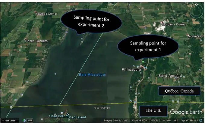
3.2 Origin of E. coli
A water sample from Missisquoi Bay, a shallow transboundary bay of Lake Champlain (Québec, Canada) (Figure 3.6), was cultured on MI agar and single blue colonies were isolated, sub-cultured on Tryptic Soy Agar (TSA) during 24h at 37°C, and confirmed for indole production using Kovac’s reagent. Confirmed colonies were then inoculated in Tryptic Soy Broth (TSB) during 24h at 37°C. Cells were washed by two centrifugal steps. The cell pellets then re-suspended in new TSB tube containing 0.22 µm-filtered glycerol (10-15% final v/v). Several 1mL aliquots of this suspension were then aseptically transferred to cryotubes and stored at -80°C before further use. A new aliquot of E. coli sister culture was used for every new experiment. To obtain a new E. coli concentration for new experiment, inoculating of new E. coli cells from the cryotubes on Tryptone Soy Agar (TSA) and incubated at 35°C during 18-20 hours. Then, some colonies were removed by the loop and re-suspended in sterile phosphate buffer. The suspension was adjusted to an OD600 of 1.0
(corresponding to ~109 CFU.mL-1) by using a spectrophotometer (Unico SpectroQuest Model SQ2800 Single Beam UV/Visible Scanning Spectrophotometer).
Figure 3. 6: The location of samples collection (Missisquoi Bay. Quebec, Canada). Sampling point for experiment 1 is "Philipsburg" and for experiment 3 is "Venise-en-Québec".