HAL Id: tel-02180596
https://tel.archives-ouvertes.fr/tel-02180596
Submitted on 11 Jul 2019HAL is a multi-disciplinary open access archive for the deposit and dissemination of sci-entific research documents, whether they are pub-lished or not. The documents may come from teaching and research institutions in France or abroad, or from public or private research centers.
L’archive ouverte pluridisciplinaire HAL, est destinée au dépôt et à la diffusion de documents scientifiques de niveau recherche, publiés ou non, émanant des établissements d’enseignement et de recherche français ou étrangers, des laboratoires publics ou privés.
cells
Aikaterini Papaefthymiou
To cite this version:
Aikaterini Papaefthymiou. Role of the Srf transcription factor in adult muscle stem cells. Human health and pathology. Université Sorbonne Paris Cité, 2016. English. �NNT : 2016USPCB120�. �tel-02180596�
Université Paris Descartes
Ecole doctorale Bio Sorbonne Paris Cité
Laboratory of Neuromuscular development, Genetics and Physiopathology
Institut Cochin
Role of the Srf transcription factor in adult muscle
stem cells
By Aikaterini Papaefthymiou
PhD thesis in Development Discipline
Supervised by Dr. Athanassia Sotiropoulos
Defended on 30th of November 2016
In front of a jury composed of:
Montarras Didier: Research Director - Institut Pasteur, Rapporteur
Duprez Delphine: Research Director - CNRS, Rapporteur
Hénon Sylvie: Professor - University of Paris Diderot, Examinateur
Coletti Dario: Associate Professor - University of Sapienza (Rome), Examinateur
Pichon Sabrina: Lecturer - University of Paris Diderot, Examinateur
Résumé: Le muscle squelettique adulte est un tissu avec une grande plasticité étant donné qu’il
adapte sa taille suite à la surcharge fonctionnelle et il régénère suite à une lésion. La base de cette plasticité est la myofibre et les cellules souches associées, les cellules satellites (CS). Suite aux stimuli, les CS sortent de la quiescence, elles s’activent, proliférent, s’engagent dans la voie myogénique et fusionnent entre elles ou bien avec la fibre pre-éxistante. Une partie des CS retourne à la quiescence afin de maintenir le « pool » de progéniteurs. Ce projet a pour objectif de mieux caractériser des voies de signalisation responsables des adaptations des CS au cours de la régénération et le l’hypertrophie compensatoire.
Srf est un facteur de transcription, particulièrement exprimé dans les muscles. Les gènes cibles de Srf sont des gènes qui participent à la régulation de la prolifération cellulaire et des gènes codant des protéines sarcomériques du muscle ou bien des gènes ayant un rôle dans l’adhésion cellulaire, la migration et l’organisation du cytosquelette. Il a été montré que la perte de fonction de Srf dans la lignée de cellules musculaire C2C12 inhibe leur prolifération et leur différenciation et que Srf contrôle l’expression de MyoD qui est un gène de détermination myogénique. Aucune donnée n’est disponible à ce jour concernant la fonction de Srf dans les CS in vivo. Nous avons généré des souris dépourvues de Srf spécifiquement dans les CS adultes. Les CS ont été recruitées par l’hypertrophie et la régénération musculaire. En parallèle des études ex vivo ont été menées afin de préciser si les phénotypes observés sont cellule-autonomes et afin de disséquer les mécanismes sous-jacents. Nous montrons que la perte de Srf dans les CS affecte fortement les processus de régénération et d’hypertrophie suggérant un rôle de Srf dans le contrôle du destin cellulaire de CS. Nos études montrent que la perte le Srf dans les SC n’affecte pas leur prolifération et leur engagement dans la différenciation myogénique. Par contre, leur motilité et leur capacité de fusion sont fortement réduites. Afin d’identifier les effecteurs de Srf impliqués dans la motilité et le défaut de fusion des CS mutantes, nous avons réalisé des études transcriptomiques et identifié le set de gènes dont l’expression est altérée par la perte de Srf dans des conditions de prolifération et de différenciation. L’analyse des fonctions altérées nous a indiqué que la voie de signalisation du cytosquelette d’actine était perturbée. En effet les CS dépourvues de Srf expriment moins d’actine et présentent une organisation du cytosquelette d’actine perturbée. Des expériences de sauvetage utilisant un modèle de souris permettant la surexpression inductible d’actine alpha dans les CS dépourvues de Srf ont montré que la surexpression d’actine chez les mutants Srf était suffisante pour rétablir partiellement l’organisation du cytosquelette et améliorer les capacités de fusion des CS. De manière intéressante, seule la fusion hétérotypique (entre une cellule contrôle et une cellule mutante), et pas la fusion homotypique (entre deux cellules mutantes), est rétablie par l’expression de l’actine. In vivo, le rétablissement de la fusion hétérotypique restaure la croissance hypertrophique des muscles alors que l’altération de la régénération chez les mutants Srf n’est que faiblement améliorée par la surexpression de l’actine. Cette étude nous a permis d’avoir une vision d’ensemble et mécanistique de la contribution du facteur de transcription Srf dans la biologie des CS et de mettre en évidence l’importance structurale du maintien du cytosquelette d’actine pour la fusion des cellules musculaires.
Mots clés : cellules souches musculaires, serum response factor, hypertrophie musculaire,
Title: Role of the Serum Response Factor (SRF) transcription factor in adult muscle stem cells
Abstract: The adult skeletal muscle is a high plastic tissue as it adapts its size upon overload and it is
capable of regeneration upon muscle lesion. The skeletal muscle is composed of a specialized syncytium, the myofiber, which is the functional unit of the muscle and a small population of myogenic progenitors, residing adjacent to the myofibers, termed as satellite cells (SCs). SCs are the muscle-specific stem cells which endow the skeletal muscle with its remarkable capacity to repair and to maintain homeostasis during muscle turnover. In resting adult muscles, SCs are quiescent but they activate upon exposure to stimuli. The activated SCs (myoblasts) proliferate extensively and subsequently differentiate and fuse between them or pre-existing myofibers, a series of cellular events called myogenesis. In parallel to the myogenesis, a reserve population of SCs escapes the myogenic program and self-renews to replenish the SC pool. The current project aims to further characterize the signalling pathways involved in SC functions during muscle regeneration and compensatory hypertrophy (CH).
Srf is a muscle-enriched transcription factor with Srf-target genes implicated in cell proliferation, differentiation (sarcomeric proteins), adhesion, migration and cellular cytoskeleton. Studies in C2C12 mouse myogenic cell line showed that Srf loss prevent the myoblast proliferation and differentiation by down-regulating the expression of the myogenic determinant MyoD gene.
We used a genetic murine model for adult SC-specific Srf-loss in order to conduct in vivo and ex vivo studies for the Srf role in SCs. Compensatory hypertrophy and regeneration are the two means by which SCs were recruited. We show that loss of Srf in SCs affects the regeneration process and the CH suggesting the Srf role in the SC fate. Srf-depleted SCs display probably no defect in their proliferation and differentiation but reduced capacity in motility and fusion. Transcriptomic analysis revealed altered actin cytoskeleton and signalling. Srf-depleted SCs show reduced actin expression and altered actin cytoskeleton. Rescue of actin expression in Srf-depleted SCs partially restored the cytoskeleton organization and the fusion process. Interestingly by actin overexpression only the heterotypic/asymmetric fusion was established but not the homotypic/symmetric fusion. Therefore actin overexpression restored the hypertrophic growth in the CH (in vivo model of heterotypic fusion) but failed to do so in the regeneration (in vivo model of homotypic fusion).
This study contributed to the in vivo investigation of the Srf mechanistic role in adult SCs and underlined the importance of actin cytoskeleton maintenance in the fusion of myogenic cells.
Keywords: muscle satellite cells, serum response factor, skeletal muscle hypertrophy, skeletal
L’enfer, c’est les autres….
Jean-Paul Sartre
Acknowledgements
Pour l’achevement de ce travail, il y a beaucoup de gens qui sont impliqués directement ou indirectement et qu’ils méritent plus qu’un merci. Tout d’abord mes rapporteurs qui ont montré toute leur volonté, flexibilité, gentillesse, compréhension d’accepter d’être mes rapporteurs qui veut dire : rendre mon manuscript en retard. Monsieur Didier Montarras et Madame Delphine Duprez, je vous remercie très chaleureusement et j’éspère que mon manuscript ne vous fatiguera pas beaucoup. Ensuite, je voudrais dire un grand merci aux autres membres de jury pour l’honneur qu’ils m’ont fait de consacrer leur temps pour évaluer mon travail : Merci Madame Sylvie Hénon, Madame Sabrina Pichon et Monsieur Dario Coletti.
La personne grace à elle j’ai pu faire ma thèse malgré que je me plainds pour les comptages éternelles est ma chef, Madame Athanassia Sotiropoulos à qui je dois beaucoup plus qu’elle peut imaginer. Athanassia, ton prénom en grec signifie « immortalité », et qui étaient immortels en Antiquité ? Είσαι θεά, γιατί μου έδωσες την ευκαιρία να κάνω αυτό το διδακτορικό κ το οποίο ήταν το στήριγμα μου για να έχω να πιαστώ σε όλα αυτά τα δύσκολα χρόνια. Πήρες το ρίσκο να με δεχτείς στο εργαστήριο χωρίς να με γνωρίζεις, το ξέρω ότι το μετάνιωσες, το ξέρω ότι σε κούρασα...δεν ξέρω εάν πρέπει να σου πω ευχαριστώ ή συγνώμη. Κυρίως σε ευχαριστώ όχι για την επιστημονική επίβλεψη που αναμφισβήτα με δίδαξες πολλά αλλά γιατί είσαι καλός άνθρωπος κ είχες πολύ υπομονή μαζί μου. Θέλω να ξέρεις ότι εκτιμώ σε όλα τα σημεία αυτά που μου πρόσφερες και να ξέρεις ότι με βοήθησες πολύ να σταθώ όρθια και να ανταπεξέλθω σε όλα τα εμπόδια. Νιώθω ένοχη που δε μπόρεσα να είμαι καλύτερη για σένα!
Ma chère Voahangy, un grand merci suffit? Non, ça ne suffit pas…Pour moi tu étais une mère à Paris, je n’oublierai jamais ce samedi dans le L2…et tout le temps, toute l’énergie, tout le courage et les conseilles que tu m’as offerts avec toute ta générosité. Heuresement que tu existes !
Pascal, le chef male du labo : vous êtes très romantique, pour moi c’est ça qui compte…et vous avez une équipe très romantique ! Quel plaisir d’avoir un chef comme vous ! Vous m’avez beaucoup encouragé pendant cette thèse là ! Je vous remercie très profondement !
Philli, my philli, le seul homme sur terre qui peut me comprendre et partager mes soucis pour la vie. Mon cher Philippe un merci ne suffit pas, combien de petits cœurs je dois dessiner pour t’accorder mon amour ? Tu es exceptionnel, j’avais la chance de partager le bureau romantique avec toi, je sais que les autres sont jaloux !!!
Alessandra, la vie est dure, tu le sais bien ! Nous sommes brilliantes, tu le sais bien ! Quel soulagement de partager la routine et la misère de comptages avec toi ! Sache que je t’apprecie beaucoup ! Milles merci !!
Ulduz, you ‘re fantastique my dear ! Ton énergie, ta positivité, l’envie pour vivre, exactement comme moi !! Baby thanks you so much for your hot heart and not only….Thanks to you, I found a treasure you know what I mean !!!
Chiara, courage pour le comptage : les premières 10.000 fibres sont difficiles après la main va automatiquement !! Bella I am just joking, enjoy your PhD and keep laughing! Je te remercie beaucoup ma petite pour ton bon cœur ! Tu vas réussir !
Maud, je n’ai jamais rencontré une personne si motivéé que toi. Tu prends quoi ? Je voudrais te remercier non seulement pour les bons moments voire Grèce et shopping aux Etats Unis mais que chaque fois tu étais présente à mon écoute, à me motiver (ça c’est difficile quandmême), stimuler et toujours positive et optimiste. Je t’embrasse fort !
Gaelle, la vie est dure et pas que…Merci beaucoup pour ton humour et de rigoler et surtout merci pour ton offre à la communauté : Jecco etc…qui aide à l’orientation des doctorants. Bon encore je ne sais pas quoi faire dans cette vie ! Continue !
Franscesc, la vie est aussi dure ! Combien de fois je m’amuserais avec toi et Emilie ? Combien d’heure de microscopie fluorescente dans le noir ? C’est toujours un plaisir d’avoir des colleques comme toi. Bonne continuation pour ton post-doc !
Nathalie je te remercie pour l’encouragement et tes compliments. Toujours gentille et souriante ! Matthieu,….le dernier et le meilleur…le destin, c’était la ville !!!
Merci beaucoup encore aux autres membres du laboratoire Evelyne et Stephanie et aux anciens membres du laboratoire, Josiane la chic, Christophe le drôle, Jean-François (très chic et drôle) et Aude dotée du talent en dessin !
Merci encore à tous les gens de l’institut (secrétaires, gardiens, femmes de menage) qui m’ont aidé avec l’administartion française et avec d’autres choses….et les gens de la plateforme de la microscopie pour le soutien technique pour l’acquisition et le traitement des images.
Antigone, une existence « tragique », qui approuve comme moi que « la vie est une punition » sinon elle ne serait pas mon amie cordiale ! « D’où moment qu’elle a été designée ouvreuse du chaos, elle n’a pas le temps de s’occuper de la poésie… Il est temps que vous, vous écriviez les poèmes que personne ne lira et qu’ils réstont dans des livres fermés…Dans l’éventualite d’un fin définitif, devenez les prophètes les plus vains, puisque que personne ne sera pas là de vous défendre…»
Merci aux gens qui pensent comme elle !!!
Ce doctorat a été financé par l’état Grec (I.K.Y.) et l’Association Française de Myologie (AFM) que je remercie très profondement ! Grace à eux et au financement de ma famille j’ai eu la possibilité d’effectuer ma thèse en France.
La ville
Tu t'es dit "J'irai ailleurs, un autre pays, un nouveau rivage doivent exister, une ville autre.
Tous mes efforts ici sont condamnés; et mon cœur n'est que mort, enterré.
Jusqu'à quand ce marasme? Où que je tourne mes yeux, où mon regard se pose, je ne vois que ruines celles de ma vie gâchée, depuis toutes ces années
ici, où je ne suis que l'épave de moi-même. Il n'y aura pas d'autres pays, tu chercheras en vain d'autres rivages, la ville te poursuivra. Dans ces mêmes
rues tu iras roder. Et tu vieilliras dans ces mêmes quartiers; tes cheveux
blanchiront dans ces mêmes maisons. Toutes les routes te ramèneront ici,
dans cette même ville.
Pour ce qui est d'ailleurs - n'espère pas - pour toi point de navire, point de chemin.
De la façon dont ici, dans ce petit coin tu as raté ta vie, tu l'as ruinée partout, sur toute la terre.
Cavafis
Table of Contents
ACKNOWLEDGEMENTS ... 5
ABBREVIATIONS ... 10
INTRODUCTION ... 12
1.SKELETAL MUSCLE: AN ORGAN FOR LOCOMOTION AND ENERGY METABOLISM ... 12
PART 1: ORIGINS OF SKELETAL MUSCLE AND SATELLITE CELLS ... 13
CHAPTER 1:TRUNK AND LIMB MYOGENESIS ... 14
CHAPTER 2:UPSTREAM REGULATORS OF MYOGENESIS ... 16
CHAPTER 3:MYOGENIC REGULATORY FACTORS ... 19
PART 2: DESCRIPTION OF MYOFIBERS ... 25
PART 3: EXTRACELLULAR MATRIX ... 27
PART 4: SATELLITE CELLS ... 29
CHAPTER 1:TRANSCRIPTIONAL CONTROL OF ADULT MYOGENESIS ... 30
CHAPTER 2:QUIESCENCE ... 32
CHAPTER 3:ACTIVATION ... 33
CHAPTER 4:SELF-RENEWAL ... 36
CHAPTER 5:FUSION ... 37
CHAPTER 6:HETEROGENEITY IN SATELLITE CELL POPULATION ... 41
CHAPTER 6:SATELLITE CELL FATE IN AGEING ... 43
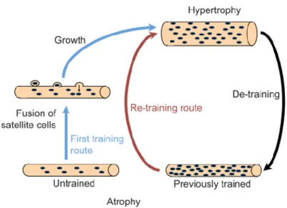
PART 5: SKELETAL MUSCLE MASS – HYPERTROPHY AND ATROPHY ... 45
CHAPTER 1:HYPERTROPHY ... 47
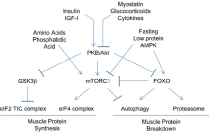
CHAPTER 2:SIGNALLING ... 48
PART 6: SKELETAL MUSCLE REGENERATION ... 54
CHAPTER 1:INFLAMMATION RESPONSES ... 55
CHAPTER 2:ECM,FIBROBLAST AND SCCROSS-TALK ... 55
CHAPTER 3:SCMIGRATION ... 57
2.SERUM RESPONSE FACTOR ... 58
PART 2: ACTIN: A KNOWN SRF-TARGET GENE ... 64
CHAPTER 1:LINKING ACTIN DYNAMICS AND SRF TRANSCRIPTIONAL ACTIVITY ... 66
CHAPTER 2:MRTFS ... 67
PART 3: SRF ACTIVITY IN MUSCLE TISSUE ... 70
CHAPTER 1:SRF-EXPRESSION PATTERN IN MOUSE DEVELOPMENT ... 70
CHAPTER 2:SRF-LOSS IN CARDIAC MUSCLE ... 71
CHAPTER 3:SRF-LOSS IN SMOOTH MUSCLE ... 71
CHAPTER 3:SRF-LOSS IN SKELETAL MUSCLE ... 72
OBJECTIF OF THE STUDY ... 75
RESULTS ... 77
PART 1:SRF CONTROLS SATELLITE CELL FUSION DURING SKELETAL MUSCLE HYPERTROPHY THROUGH THE MAINTENANCE OF ACTIN ARCHITECTURE ... 77
PART 2:SRF ROLE IN SATELLITE CELLS DURING SKELETAL MUSCLE REGENERATION ... 123
DISCUSSION AND PERSPECTIVES ... 135
Abbreviations
ABPs: Actin-Binding ProteinsbHLH: basic Helix-Loop-Helix BMP: Bone Morphogenetic Protein BrdU: Bromo-deoxy-Uridine
CH: Compensatory Hypertrophy CSA: Cross-Sectional Area CTX: Cardiotoxin
E: Embryonic Day ECs: Endothelial Cells ECM: Extra Cellular Matrix EdU: Ethylo-deoxy-Uridine
FCM: Fusion-Competent Myoblast FGF: Fibroblast Growth Factor FC: Founder Cell
GI: Gastro Intestinal tract
HGF: Hepatocyte Growth Factor IEGs: Immediate-Early Genes IGF-1: Insulin-like Growth factor-1 IP: Intraperitoneal
KO: Knock-out
MADS: MCM1-Agamous-Deficiens-SRF MAPKs: Mitogen-Activated Protein Kinases MEF2: Myocyte Enhancer Factor 2
MND: Myo-Nuclear Domain
MRFs: Myogenic Regulatory Factors
MRTFs: Myocardin Related Transcription Factors Mstn: Myostatin
Mammalian Target of Rapamycin: mTOR MyHC: Myosin Heavy Chain
Pl: Plantaris muscle
QSCs: Quiescent Satellite Cells SCs: Satellite Cells
SMCs: Smooth Muscle Cells SRE: Serum Response Element SRF: Serum Response Factor TA: Tibialis Anterior
TCFs: Ternary Complex Factors TF: Transcription Factor
Tg: Transgene
TGF-β: Transforming Growth Factor-β TMX: Tamoxifen
TNF-α: Tumor Necrosis Factor-α
Introduction
1. Skeletal Muscle: an organ for locomotion and
energy metabolism
Movement is a defining feature of all animals and the evolutionary advantages of efficient locomotion led to several solutions for the construction of the motile organs (the muscles) in all animal phyla. Skeletal muscle, which constitutes about 40% of the mass of the human body, is the organ with the specificity to transform the chemical energy to mechanical energy (potential and kinetic energy) as well as heat energy. Its metabolism affects the metabolic balance of the entire organism and is the major body protein reservoir. Skeletal muscle is the most flexible structure in vertebrate organisms as it exerts diverse functions, enabling both crushing with great force and movement with exquisite precision (Braun and Gautel, 2011). The main muscle activity is the contraction of muscle cells, which is under the voluntary control and it is initiated by the nerves impulses.
The functional cellular units responsible for skeletal muscle contraction are cylindrical, multinucleated muscle fibers (myofibers). The skeletal myofiber is a specialized syncytium1,
which contains thousands of myonuclei within a common, undivided cytoplasm, forming an elongated cell under the plasma membrane called sarcolemma.
The myofiber nuclei are postmitotic and under normal conditions cannot re-enter a proliferative state to contribute to additional nuclei. They lose this ability of mitosis once the myogenic progenitors’ nuclei have been incorporated into myotubes during the embryonic and postnatal development. In adult life, somatic stem cell populations participate in the homeostasis of their host tissues. During postnatal life, myofiber growth and repair of skeletal muscle depend on muscle stem cells, otherwise named satellite cells (Mauro, 1961; Moss and Leblond, 1971).
Part 1: Origins of Skeletal Muscle and Satellite cells
The three germ layers (endoderm, mesoderm, and ectoderm) are formed during gastrulation. The mesoderm is at the origin of muscles, gonads, secretory organs, and connective tissue. Mesoderm is anatomically separated into paraxial, intermediate, and lateral mesoderm, with respect to position from the midline (Bentzinger et al., 2012). Skeletal muscle cells of higher vertebrates arise during midgestation (in mice between embryonic day 9 (E9) and E12) from three different locations within the middle layer of cells in the primitive embryo: the segmented somitic paraxial mesoderm, the unsegmented cranial paraxial mesoderm and the prechordal mesoderm; these represent different parts of the mesoderm along the rostrocaudal axis (Braun and Gautel, 2011).
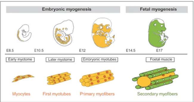
In mouse, skeletal muscle development occurs in several phases (Figure 1). First, in the E8.5 of gestation, primary myofibers develop displaying the earliest expression of myogenin and muscle-specific genes (desmin, titin and a-actin genes). Around E13 of gestation, secondary myofibers form in trunk and limbs, and they develop parallel to the primary myofibers, that serves as a scaffold for the orientation of differentiating myocytes of a second wave of myoblasts (Buckingham and Mayeuf, 2012).
Satellite cells (SCs), the stem cells of adult skeletal muscle, arise around E17 of development as a unique myogenic cell lineage. They constitute the principal proliferative cell population of developing skeletal muscle. Late in fetal development at around E16.5-18.5, these cells occupy a satellite cell position adjacent to the myofibers, a characteristic of progenitor cells in postnatal muscle (Kassar-Duchossoy et al., 2005; Relaix et al., 2005).
Further skeletal muscle maturation occurs during the postnatal period for about 2-3 weeks with the SC nuclear accretion to contribute to multinucleated myofibers (Tajbakhsh, 2009). The adult number of myonuclei and satellite cells is established by 3 weeks of postnatal development. Subsequently, the muscle mass undergo extensive hypertrophic growth with increased cellular protein content to dominate in order to achieve growth of the musculature to adult size (White et al., 2010).
Figure 1 : Waves of skeletal muscle formation. There is a first wave of skeletal muscle formation, termed embryonic or primary myogenesis. This is followed by a second wave of fetal or secondary myogenesis. The timing of this transition depends on the onset of innervation which varies at different sites in the embryo. E: Embryonic day of mouse development. From (Buckingham and Mayeuf, 2012)
Chapter 1: Trunk and Limb Myogenesis
Skeletal muscle in the trunk and limbs derives from somites that progressively form by segmentation of paraxial mesoderm on either side of the neural tube and notochord, following an anterior-posterior developmental gradient. Somites are the first metameric structures and a characteristic paradigm of segmentation in mammalian embryos (Bentzinger et al., 2012). Segmentation starts from embryonic day 8 (E8.0). The somite is initially an epithelial ball of cells that subsequently distribute into a ventral mesenchymal sclerotome, giving rise to the bones of the vertebral column and ribs and an adjacent (dorsal-most part) syndetome, a source of muscle tendons in the trunk. The dorsal part of the somite, the dermomyotome, retains an epithelial structure for longer and gives rise to dorsal dermis and all the skeletal muscles of the trunk and limbs, as well as endothelial and smooth muscle cells of blood vessels, and brown fat (Buckingham and Rigby, 2014).
Myotome
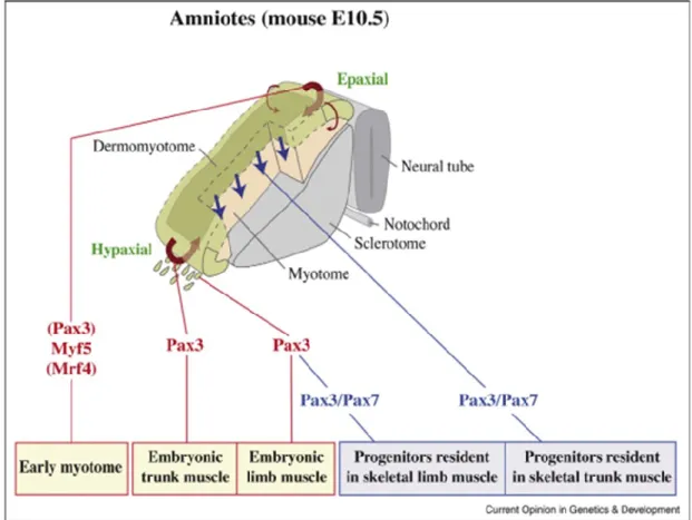
The first muscle mass to form, under the dermomyotome, is the myotome, which has an epaxial and a hypaxial component (Figure 2). Dorsal muscles are formed from the epaxial part of the dermomyotome and myotome, whereas lateral trunk and limb muscles are
derived from the hypaxial domains (Buckingham et al., 2003). Myogenic precursor cells delaminate from the edges of the dermomyotome and they differentiate into myocytes to form the underlying differentiated muscle of the myotome. Subsequently, these myocytes undergo cell fusion to form multinucleated muscle fibers followed by cleavage and reorganization of the growing and splitting trunk muscle mass. At the level of the fore- and hindlimbs, cells delaminate from the hypaxial dermomyotome and migrate into the early limb bud where they subsequently differentiate into skeletal muscle (Buckingham and Mayeuf, 2012).
Figure 2 : Trunk and limb skeletal muscles in amniotes originate from the somites. The dermomyotome contains multipotent progenitor cells of different cell types, including the skeletal muscle progenitors. Even if Pax3 does not directly control early epaxial myogenesis (i.e. formation of the early myotome), all muscle cells derived from the somite have expressed Pax3 in their history. The early myotome and embryonic myofibres originate from cells that have expressed only Pax3 (indicated in red). In the central portion of the dermomyotome (darker green region) and in muscle progenitors once they have migrated to the limb, Pax7 expression is initiated in Pax3 positive cells. Pax3 expression, contrary to Pax7, is downregulated in cells that will contribute to fetal myogenesis (indicated in blue). From (Buckingham and Vincent, 2009)
Both delamination and migration depend on the presence of c-met, a tyrosine kinase receptor, which interacts with its ligand hepatocyte growth factor HGF, produced by
non-somitic mesodermal cells that thus delineate the migratory route. In mutant mouse embryos, which lack functional c-met or HGF, skeletal muscle is absent from the limbs. Transcription of the c-met gene depends on Pax3 transcription factor (Epstein et al., 1996).
Cranial Myogenesis
Craniofacial muscles are associated with head (including extra-ocular muscles, jaw muscles and facial muscles) and neck structures. The formation of head muscles differs significantly (evolutionarily, morphologically, molecularly) from the formation of their counterparts in the trunk and limbs. In the embryo, these structures derive from distinct mesoderm populations. The head musculature originates from the unsegmented cranial paraxial mesoderm, which is positioned along both sides of the neural tube and notochord, and it is located anterior to the somites (Sambasivan et al., 2011a).
Chapter 2: Upstream Regulators of Myogenesis
2.1. Paired-Homeobox Transcription Factors
Pax genes encode evolutionarily conserved (paired box: a DNA-binding sequence) transcription factors that play critical roles in organ development and tissue specification. Pax genes are divided into four subfamilies based on sequence similarities depending on the presence of an additional DNA-binding homeodomain and/or an octapeptide region, which serves as a binding motif for protein co-factors (Blake and Ziman, 2014).
Pax3 and Pax7 constitute one of the four Pax subfamilies. Pax7 is unique in the family for the presence of a C-terminal 14 amino acids sequence, the OAR (Otp/aristaless/Rax) (Mayran et al., 2015). Similarities in their protein sequence and expression pattern reflect a common origin, as they arose by duplication from a unique ancestral Pax3/7 gene at the onset of vertebrate evolution (Paixão-Côrtes et al., 2015). Pax3 and Pax7 function upstream of the myogenic regulatory factors (see the corresponding paragraph) in the trunk and limbs and thus control the entry of cells into the myogenic program (Relaix et al., 2005). Unlike the myogenic regulatory factors, Pax3 and Pax7 are not tissue specific, being also expressed in neuroectoderm, in subdomains of the brain, in the dorsal neural tube, and in neural crest (Buckingham and Rigby, 2014).
Pax3
In the mouse, Pax3 is expressed in presomitic mesoderm and then throughout the somite, before becoming restricted to the dermomyotome as somites mature (Buckingham
and Rigby, 2014). The mutation of Pax3 in Splotch mice leads to defects in neural tube closure (spina bifida and exencephaly), in the migration of the neural crest cells and in limb muscle formation. Pax3-mutant embryos show somite defects and abnormalities in segmentation. In consequence, the myotome fails to form correctly leading to trunk muscle defects and perinatal death (Tremblay et al., 1998).
Cells in the central dermomyotome are marked by the expression of both Pax3 and Pax7 proteins however, only Pax3 is expressed in long-range migrating cells (see Figure 2). Pax3-mutant mice have no limb muscles that depend on the delamination and migration of muscle progenitor cells from the hypaxial dermomyotome (Tremblay et al., 1998). Despite Pax3 and Pax7 have partially overlapping functions during muscle development; Pax7 can substitute for Pax3 function in somite and trunk muscle development, but only partially in the formation of limb muscles involving long-range migration of muscle progenitor cells (Relaix et al., 2004).
Pax3 Expression in Postnatal Muscles
Analysis of adult mice in which the Pax3 gene is targeted with nLacZ reporters revealed the presence of β-galactosidase–positive satellite cells in adult skeletal muscle. The number of such cells varies between muscles. They are particularly abundant in the diaphragm but are much less frequent in hindlimb muscles, with the exception of the gracilis muscle. In contrast, 50% of forelimb muscles express Pax3. As in the embryo, expression of Pax3 is not detectable in head muscles. Most ventral trunk muscles are positive, with a striking juxtaposition in the rib area, where intercostal muscles are mainly negative, whereas body wall muscles such as the serratus caudalis dorsalis are positive (Relaix et al., 2006).
Pax7
Although the Pax3 function is revealed by the Splotch mutation, no spontaneous mutant is available which indicates the role of Pax7. Pax7 has been originally described as a member of the paired-homeobox gene family that is specifically expressed during somitogenesis and neurogenesis in the embryo. Pax7 transcripts are present in the myotome of the somites and persist during differentiation into skeletal muscles of the trunk and limbs (Jostes et al., 1990). Its expression pattern both in ectoderm- and mesoderm-derived tissues, suggests a function in central nervous system development and skeletal muscle.
In parallel to myofiber formation, a subpopulation of myogenic precursor cells that do not express the myogenic regulatory factors and maintain Pax3/Pax7 expression is observed in the myotome at E10.5 (see Figure 2). In the absence of both Pax3 and Pax7, further
muscle development is arrested and only the early embryonic muscle of the myotome forms. Cells not expressing Pax3 or Pax7 die or assume a non-myogenic fate (Kassar-Duchossoy et al., 2005; Relaix et al., 2005).
Pax7 Germline Mutant Phenotype
Pax7 germline mutants are born but they have increased lethality (die within 2–3 weeks) and growth retardation with reduced body weight although the muscle formation is not affected. The cause of death is probably because of dysgenesis of neural crest derivatives (Mansouri et al., 1996).
The markedly decreased muscle mass, the reduced fiber caliber and the reduced number of myonuclei of Pax7 mutant muscle suggested that the postnatal growth phase of skeletal muscle, normally mediated by satellite cells, was deficient in the absence of Pax7 and is attributable to an absence of functional satellite cells (Kuang, 2006). Deletion of Pax7 in mice leads to normal numbers of stem cells at birth followed by excessive wasting of stem cells during the first weeks of postnatal development (Oustanina et al., 2004; Relaix et al., 2006).
Satellite cells (SCs), the skeletal muscle stem cells, also express the paired box transcription factor Pax7 (Seale et al., 2000). Pax7 is an essential transcriptional factor (TF) for the survival (Relaix et al., 2006), proliferation (Oustanina et al., 2004) and self renewal (Olguin and Olwin, 2004) of SCs and for an efficient regeneration mediated by Pax7-expressing cells (Günther et al., 2013; Lepper et al., 2011; Sambasivan et al., 2011b).
2.2. Sine Oculis–Related Homeobox Transcription Factors
In addition to Pax3 and Pax7, the homeodomain proteins Six1 and Six4 are important upstream regulators of myogenesis that directs dermomyotomal multipotent progenitors toward the myogenic lineage (Figure 3). Unlike Pax3 and Pax7, these factors are also present in differentiated skeletal muscle (Buckingham and Rigby, 2014). Six family proteins are transcription factors characterized by the presence of two conserved domains, a Six-type homeodomain that binds to DNA and an amino-terminal Six domain that interacts with coactivators or corepressors of transcription (Bentzinger et al., 2012). Six proteins bind to and translocate the eyes-absent homologs Eya1 and Eya2 to the nucleus, where they act as cofactors to activate Six target genes, such as Pax3, MyoD, MRF4 and myogenin (Grifone, 2005).
Six4 germline mutant mice do not present major developmental defects (Ozaki et al., 2001), while Six1 null mice do not survive and have defects in many organs, including rib, craniofacial and muscle deficiencies (Laclef et al., 2003). Six1 and Six4 double germline
mutant mice develop a more severe phenotype than in the single Six1 mutant with loss of all muscles derived from the hypaxial dermomyotome (limb and many trunk muscles). At the limb bud level, Six1 and Six4 homeogenes control early steps of myogenic cell delamination and migration from the somite through the control of Pax3 gene expression (Grifone, 2005). Myf5 expression within the epaxial dermomyotome, which is independent of Pax3, is unaffected in Six1 and Six4 double mutants, and dorsal muscles arising from this structure are the only remaining axial muscles in these mice (Giordani et al., 2007). Six orchestration together with Pax, at the onset of myogenesis is illustrated by regulation of the Myf5 and MyoD gene (Relaix et al., 2013).
In adult muscle Six1 acts as a main determinant of fast-fiber type acquisition and maintenance (Sakakibara et al., 2014, 2016) and as a critical regulator of SC self-renewal to ensure a proper muscle regeneration (Le Grand et al., 2012).
Chapter 3: Myogenic Regulatory Factors
The process of skeletal myogenesis during embryonic development is regulated by a family of muscle-specific transcription factors that are expressed in a spatially and temporally ordered manner (Figure 3). The family of basic helix-loop-helix (bHLH) transcription factors consisted of Myf5, MyoD, Myogenin (MyoG), and MRF4, form the myogenic regulatory factors (MRFs). The ancestral MRF gene is assumed to have given rise to the four family members in vertebrates, shown by evolutionary analyses of amino acid sequences of this family members (Atchley et al., 1994).
The MRFs cooperatively activate transcription and myogenesis through protein-protein interactions with members of the myocyte enhancer factor 2 (MEF2) family of MADS domain transcription factors (Molkentin et al., 1995) and the ubiquitously-expressed E proteins (Londhe and Davie, 2011). They have potential target genes with E boxes (cis-acting DNA control elements which are present in the promoters and enhancers of muscle-specific genes) (Arnold and Braun, 1996).
The MRFs have differential patterns of expression in the developing somites and are exclusively expressed in skeletal muscle2. In the mouse embryo, Myf5 is the earliest MRF to
be expressed in the dorsomedial lip of dermomyotome at E8.0. Myogenin is expressed after Myf5 at E8.5 and MRF4 at E9.0. MyoD is the last to be expressed in the somite at E10.5. Cells migrating toward the limb do not express MRFs until they have reached the limb bud (Francetic and Li, 2011). The MRFs null mutations analyses also suggest overlapping functions between them.
The existence of dominant-acting regulators in muscle cell differentiation has been predicted from early cell fusion experiments which demonstrated that muscle-specific genes can be transactivated in non-muscle nuclei of heterocaryons (the parental nuclei remain intact) between differentiated skeletal muscle cells and various non-muscle cell types (Blau et al., 1983). When they are overexpressed in nonmuscle cells, they will activate the myogenic program, with suppression of other cell fates and formation of differentiated muscle (Weintraub et al., 1991). Since then the possibility of converting one cell type to another by transdifferentiation has become a major issue in the stem cell field.
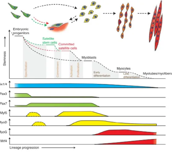
Figure 3 : Hierarchy of transcription factors regulating progression through the myogenic lineage. Muscle progenitors that are involved in embryonic muscle differentiation skip the quiescent satellite cell stage and directly become myoblasts. Some progenitors remain as satellite cells in postnatal muscle and form a heterogeneous population of stem and committed cells. Activated committed satellite cells (myoblasts) can eventually return to the quiescent state. Six1/43 and Pax3/7 are master regulators of early lineage specification,
whereas Myf5 and MyoD commit cells to the myogenic program. Expression of the terminal differentiation genes, required for the fusion of myocytes and the formation of myotubes, are
performed by both myogenin (MyoG) and MRF44. From (Bentzinger et al., 2012). This figure
refers the requirement of each TF on myogenic cellular level and not in developmental time.
MyoD
Lineage-specific markers are expressed in proliferating myoblasts prior to terminal differentiation and the myoblast determination gene number 1 or MyoD is one of them. MyoD was isolated as the cDNA clone hybridized to a myoblast/myotube cDNA library, as the key regulator that can change the cell fate in transfected fibroblast-like 10T1/2 cells by inducing the myogenic phenotype (Davis et al., 1987). The phenomenon of myogenic conversion remains remarkable in that a single transcription factor can exert this overriding effect and this set of experiments establish the conceptual framework for the discovery of induced pluripotent stem cells from somatic cells (Hochedlinger, 2010).
MyoD transcripts are detectable at E10.5 in the somites and myotomal muscles that had already expressed MyoG two days earlier. MyoD expression pattern in somites, after the MyoG expression, suggest that early myogenesis in the myotome is not dependent on this TF (Sassoon et al., 1989). MyoD is the last of the four myogenic HLH transcription factors to be activated in development and thus, may be essential for the SC function in postnatal growth and/or regeneration of skeletal muscle rather than in the regulation of de novo differentiation of skeletal myocytes (Megeney et al., 1996).
Mice carrying a null MyoD mutation are viable and fertile, and they do not exhibit any morphological or physiological abnormalities in skeletal muscle (Rudnicki et al., 1992). Mutant animals reveal an abundant expression of Myf5 in their muscle indicating that functional redundancy is a feature of the MRF regulatory network and that Myf5 may partially substitute for the function of MyoD in the control of the skeletal myogenic developmental program.
MyoD is dispensable for the development of skeletal muscle however, in its absence the postnatal growth is attenuated and muscle regeneration is less efficient confirming its important role in SC function. mutant animals show increased number of MyoD-deficient satellite cells but they exhibit decreased rates of proliferation (Megeney et al., 1996). Thus, the lack of MyoD promotes the satellite cell self-renewal rather than progression through the developmental program as the MyoD mutant myoblasts are differentiation defective (Cornelison et al., 2000; Sabourin et al., 1999). In this case, MyoD plays a cell autonomous and non-redundant role in regulating the dynamic balance between proliferation, differentiation and renewal that normally establishes an appropriate satellite cell pool size.
In addition to its importance in the myogenic determination and differentiation, MyoD has a role in the proliferation and growth of myoblasts as it was shown by the ex vivo culture of MyoD-null myoblasts and their sequential passages. Primary cultures of MyoD –/– mice
were poorly growing and displayed signs of senescence a phenotype more accentuated over the four subsequential passages accompanied with the progressive loss of Myf5 expression (Montarras et al., 2000). Further study confirmed the MyoD capacity to interact with and to affect components of the cell cycle machinery by initiating the expression of Cdc6 gene after myoblasts transition out of quiescence in order to progress through the cell cycle (Zhang et al., 2010). This could justify the early requirement for MyoD expression within hours after the SC activation and before SC duplication (Jones et al., 2005).
In the adult, MyoD is not expressed by quiescent satellite cells but in activated, proliferating myoblasts (Yablonka-Reuveni and Rivera, 1994). Nevertheless, adult satellite cells have passed through a developmental stage where the MyoD locus was active (Kanisicak et al., 2009).
Myf5
Among the four MRFs, Myf5 is the earliest to be expressed at E8 in the somites, in the epaxial domain, adjacent to the neural tube, subsequently is activated in the opposing hypaxial domain and is markedly reduced after E14 (Francetic and Li, 2011). Newborn Myf5-deficient animals were viable and fertile (Kaul et al., 2000). In addition, Myf5 mutants lack epaxial (dorsal) muscles (Kassar-Duchossoy et al., 2004).
Myf5-mutant mice show a delay in the myotome development although they expressed the normal set of MRFs (Braun et al., 1992). Similarly, inactivation of the Myf5 gene also allows apparently normal muscle development in the newborn mice, supporting the notion of an extraordinary plasticity in the formation of the myogenic lineage, with the myogenic factors Mrf4 and MyoD expressed in the somites at later developmental stages substituting for the absence of Myf5 (Kassar-Duchossoy et al., 2004).
Skeletal muscles of adult Myf5 null mice exhibit a subtle progressive myopathy. Adult Myf5 null mice exhibit perturbed muscle regeneration with a significant increase in muscle fiber hypertrophy, delayed differentiation, adipocyte accumulation, and fibrosis after freeze-injury. Satellite cell numbers are not significantly altered in Myf5 null animals and they show a modest impaired proliferation under some conditions in vitro (Gayraud-Morel et al., 2007).
Mice deficient for both MyoD and Myf5 were immobile and died soon after birth. Newborn mice are totally devoid of skeletal muscle fibers and myoblasts (Kassar-Duchossoy et al., 2004; Rudnicki et al., 1993). Fetal myogenesis was severely compromised whereas Mrf4 partially rescues embryonic myogenesis in Myf5/MyoD double-knockout mice (Kassar-Duchossoy et al., 2004). Mice triple mutant for Myf5, Myod and Mrf4 totally lack skeletal muscle and myoblasts (Kassar-Duchossoy et al., 2004). These experiments suggest that formation of skeletal muscle depends on determination factors Myf5, MyoD and Mrf4 which direct multipotent progenitors into the myogenic lineage (Sambasivan and Tajbakhsh, 2007).
MyoG
Myogenin (MyoG) is another member of the MRF which was isolated for inducing the myogenic fate of non myogenic cell line and it shares a very high homology with MyoD in its amino-terminal region (Wright et al., 1989). MyoG binds to the regulatory regions of muscle-specific genes and activates their regulation. Thus, whereas MyoD and Myf5 are active in the lineage specification of muscle cells, myogenin appears to mediate muscle cell differentiation. During the embryonic myogenesis, the MyoG is expressed earlier that the MyoD in the myotomal cells of the first somites whereas they are expressed synchronously in the limb buds (Sassoon et al., 1989).
Mice lacking myogenin are born immobile and die at birth because of a virtual absence of myofibers. MyoG is indispensable for the skeletal muscle formation during embryogenesis contrary to the MyoD and Myf5-mutants. MyoG-mutant mice show severe reduction of skeletal muscle mass, drastically reduced fiber density with mononucleated cells replacing most of the mature muscle cells (Hasty et al., 1993; Nabeshima et al., 1993).
The initial phase of myotomal differentiation occurred normally in the myogenin-mutant embryos. Primary myogenesis was delayed whereas secondary myogenesis was dramatically affected indicating that myogenin is not required for the initial aspects of myogenesis, including myotome formation and the appearance of MyoD-expressing myoblasts (Venuti et al., 1995). Myogenin is not essential for commitment of cells to the myogenic lineage, but is important for terminal differentiation. Lack of myosin heavy chain and actin (markers for differentiated muscle cells) expression was diminished in MyoG-mutant mice demonstrating its importance for biochemical and morphological differentiation of skeletal muscle cells (Venuti et al., 1995).
Postnatal deletion of MyoG leads to 50% lethality before P10. The survived mice were observably smaller in size, not accompanied however with reduced myofiber caliber or compensatory upregulation of any of the myogenic bHLH TF expression. The absence of MyoG did not alter postnatal skeletal muscle growth or function and reveal “an unsuspected
non-cell autonomous role for myogenin in the regulation of tissue growth” (Knapp, 2006). Deletion of MyoG in adult mice renders the skeletal muscle resistant to neurogenic atrophy by diminishing the expression of MuRF1 and Atrogin1 (E3 ubiquitin ligases) and thus preventing the proteasome-mediated protein degradation. Denervation-induced upregulation of MyoG triggers MyoG-dependent transcriptional cascade, involving MuRF1 and Atrogin1, leading to muscle atrophy (Moresi et al., 2010).
Mrf4
Mrf4 or Myf6 represents the fourth member of MRFs that is capable of converting fibroblasts to stable determined myogenic cells at a very high frequency (Rhodes and Konieczny, 1989). Mrf4 is expressed transiently in the myotome at the same time as Myf5 at the onset of myogenesis and then becomes up-regulated during late fetal development to eventually become the predominant myogenic bHLH factor expressed in adult skeletal muscle (Hinterberger et al., 1991). Based on its expression pattern, it has been proposed that Mrf4 regulate skeletal muscle maturation and aspects of adult myogenesis (Le Grand and Rudnicki, 2007).
A recent study confirmed the function of Mrf4 in adult muscle fibers as Mrf4 knockdown in adult skeletal muscle induces hypertrophy and prevents denervation-induced atrophy. This effect is dependent on an increase in Mef25 transcriptional activity and the
consequent upregulation of Mef2 target genes, suggesting that these two TFs act together to regulate growth in adult muscle. Mrf4 is a negative regulator of muscle growth by repressing Mef2 activity (Moretti et al., 2016).
Three Mrf4 knock-out mice have a range of phenotype from viable with no muscle defects, to lethal phenotype with some muscle defects. Since Myf5 and Mrf4 are genetically linked on the same chromosome, knock-out in one gene results in a cis effect by which the expression of the other is also decreased or lost (Olson et al., 1996). In the viable knock-out, Mrf4-mutant mice showed only a slight reduction in expression of a subset of muscle-specific genes but showed a dramatic increase in expression of myogenin, suggesting that it may compensate for the absence of Mrf4 and demonstrating that Mrf4 is required for the down-regulation of myogenin expression that normally occurs in postnatal skeletal muscle (Zhang et al., 1995).
In the Mrf4 and MyoD double mutant the myogenin null phenotype is phenocopied suggesting therefore that Mrf4 or MyoD is necessary to activate the myogenin gene (Kassar-Duchossoy et al., 2004)
Part 2: Description of Myofibers
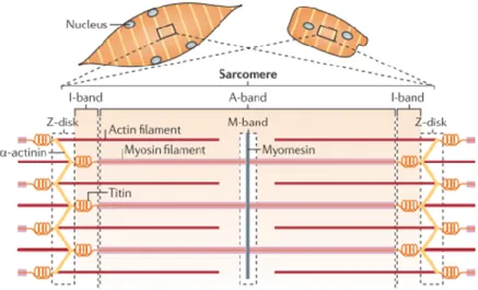
Myofibers are composed of myosin II motor proteins and actin filaments that generate force and movement. In the striated muscles that are used for locomotion, actomyosin contraction is amplified in serial and parallel arrangements of numerous contractile units, called sarcomeres, which is the basic functional unit of skeletal muscle (Figure 4). These are made up of actin and myosin filaments arranged in highly ordered, almost crystalline arrangements, as well as hundreds of regulatory proteins such as the troponin–tropomyosin complex, scaffolding and cytoskeletal crosslinking proteins such as αactinin, myomesin and the kinase titin (Braun and Gautel, 2011).
This group of proteins, which is essential for the ordered assembly of actin and myosin filaments into sarcomeres, combines architectural, mechanical and signaling functions in muscle and summarized as the sarcomeric cytoskeleton (Gautel and Djinović-Carugo, 2016). Such signaling functions that are interpreted in gene expression, protein synthesis and protein degradation are the translation of mechanical stimuli to biochemical signals (muscle mechanotransduction).
Responding to the signals from motor neurons, myofibers depolarize and release calcium from the sarcoplasmic reticulum. This drives the movement of actin and myosin filaments relative to one another and leads to sarcomere shortening and muscle contraction (Yin et al., 2013a).
Each myofiber is anchored at its extremities to tendons at the myotendinous junctions. Myotendinous junctions are anatomical regions which connects the skeletal muscle to the bone for transmitting the contractile force between the two tissues.
Figure 4 : Striated muscle structure. The contractile machinery of skeletal muscle syncytial myotubes (left) and single cardiomyocytes (right) is formed from long arrays of sarcomere units, which are joined into myofibrils. The sarcomere (bottom) is constructed from interdigitating, antiparallel filaments of actin and myosin, the elastic titin filaments and the crosslinker proteins for actin — α-actinin, myosin and myomesin. Sarcomeres contain many other accessory components, including proteins involved in transcriptional regulation and turnover control. Adapted from (Braun and Gautel, 2011)
Skeletal muscle comprises different fiber types based on their physiological properties. Skeletal muscle fibers can be grouped into a slow-contracting/fatigue-resistant type and a fast-contracting/fatigue-susceptible type. Slow and fast muscles differ in the metabolism type (oxidative or glycolytic). Myofibers also vary in terms of their myosin heavy chain (MyHC) isoforms (I, IIA, IIB, IIX). The fiber type profile can change, in response to hormonal, neural influences and mechanical load, rendering the muscle a plastic tissue (Schiaffino and Reggiani, 2011).
Part 3: Extracellular Matrix
Skeletal muscles are composed primarily of contractile material. However, muscle is a composite tissue of connective tissue, blood vessels and nerves, these “minor” tissues (in terms of relative mass) may strongly influence muscle function. Normal muscle function is also influenced by the skeletal muscle extracellular matrix (ECM) or basement membrane that coats the skeletal myofibers. The primary function of extracellular matrix is to endow tissues with their specific mechanical and biochemical properties. ECM bears the majority of muscle passive load as shown by biomechanical studies and alterations in ECM properties are associated to muscle pathology with ECM fibrosis (Gillies and Lieber, 2011). Also, acellular basement membrane provide a scaffold to orient and constrain cells during regeneration (Vracko and Benditt, 1972).
Each myofiber is surrounded by the endomysium (also called the basement membrane or basal lamina). Bundles of myofibers are surrounded by the perimysium, while the entire muscle is contained within the epimysium (Figure 2). The epimysium layer is continuous with the tendons that attach the muscles to the bones (Lund and Cornelison, 2013). Additionally, ECM in skeletal muscle is critical for both longitudinal and lateral force transmission from muscle fibers to tendons (Purslow, 2010).
The basal lamina of skeletal muscle is composed of type IV collagen, proteoglycans, glycoproteins and matrix remodeling enzymes. These ECM molecules are mainly synthesized and excreted by interstitial muscular fibroblasts but can also be produced and remodeled by myoblasts during muscle development and regeneration (Chapman et al., 2016).
Figure 5 : Skeletal muscle extracellular matrix structure. Skeletal muscles are composite and hierarchical tissues with three layers of ECM, the epi-, peri- and endomysium. Skeletal muscle fibroblasts reside in the extracellular space between muscle fibers and fascicles, where they secrete ECM proteins to be incorporated into skeletal muscle ECM. From (Chapman et al., 2016)
Part 4: Satellite Cells
Satellite cells are skeletal muscle-specific adult stem cells and their presence seems to be a common feature of all adult skeletal muscles. They account for 2-7% of the nuclei associated with a particular myofiber and the proportion varies with age and muscle group (Gibson and Schultz, 1983). Satellite cells contribute to increase the diameter and length of the existing fibers, as they are the main source of myogenic cells after birth and during regeneration. The satellite cells were initially described in the course of an electron microscopic study in the peripheral region of the skeletal muscle fiber of the frog and were named based on their anatomic location on the surface of muscle fibers, between the myofiber plasmalemma and the basal lamina (Mauro, 1961).
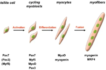
In resting muscle, satellite cells remain in a non-proliferative, quiescent state. Upon injury or growth stimulus, satellite cell become activated, enter the cell-cycle and turn into proliferating myoblasts, which differentiate and eventually fuse (Figure 6) to pre-existing myotubes or to each other to form new myotubes (Le Grand and Rudnicki, 2007). Terminal differentiation leads to the activation of many muscle-specific genes, including those encoding the sarcomeric proteins (such as myosin light and heavy chains, a-actin, troponin I, and troponin T) muscle enzymes, structural proteins of the contractile apparatus and specialized membrane receptors.
Figure 6 : Schematic representation of adult myogenesis. Quiescent skeletal muscle satellite cell can become activated following stimuli originating from their associated fiber or from the micro-environment. Their proliferating progeny, the skeletal myoblasts, express the paired-box transcriptions factors Pax7 and Pax3, as well as the myogenic regulatory factors Myf5 and MyoD. Once committed to differentiation, myoblasts stop cycling and loose expression of
Pax7, Pax3 and Myf5. Differentiating myogenin+ve myocytes will then align and fuse to form multinucleated myofibers. MRF4 is further required for hypertrophy of the new fibers. From (Le Grand and Rudnicki, 2007)
The descendents of activated satellite cells called myoblasts or myogenic precursor cells: they are the transient amplifying population, undergoing multiple rounds of division and express MyoD. Satellite cells appear to form a population of stem cells that are distinct from their daughter myoblasts as defined by biological and biochemical criteria (Yablonka-Reuveni et al., 2008). A reserve population escapes from the differentiation fate and return back to quiescence that assures the replenishment of satellite cell population.
The replenishment of the satellite cell population is a critical aspect of muscle tissue regeneration. Repeated injury experiments have shown that satellite cell numbers remain constant even after multiple traumas. The satellite cell pool is constantly replenished during lifetime, although there is a decline in satellite cell numbers and a reduced proliferative capacity in aged individuals (Dumont et al., 2015a). Thus, satellite cells display two hallmarks of stem cells: lineage-specific differentiation and self-renewal (Buckingham and Montarras, 2008).
Chapter 1: Transcriptional Control of Adult Myogenesis
Adult myogenesis takes place during muscle regeneration and SCs undergoing myogenesis to form de novo muscle fibers. During adult adaptation of muscle, aspects of early developmental programs can be reactivated and can cooperate with specific factors to determine fibertype characteristics. This process, in many but not all respects, recapitulates embryonic myogenesis (Yin et al., 2013a). The myogenic potential of satellite cells is under the molecular control of specific Paired-box and bHLH transcription factors which tightly orchestrate the balance of myogenic progression during muscle regeneration (Le Grand and Rudnicki, 2007).
SCs are identified by the expression of Pax7 and Pax7-expressing SCs are required for muscle regeneration as it has been shown by genetic ablation of such cells (Lepper et al., 2011; Sambasivan et al., 2011b). Pax7 is able to drive transcription in quiescent satellite cells, activated and proliferating satellite cells. Expression is then downregulated in cells that initiate terminal differentiation, but is maintained (and transcriptionally active) in those that withdraw from immediate differentiation (Zammit, 2006).
Satellite cells and primary myoblasts lacking Pax7 undergo cell cycle arrest and precocious differentiation. Ex vivo Pax7 deletion in wild type myoblasts resulted in a striking reduction in the levels of Myf5 mRNA, a reduction in MyoD expression and an
increase in the numbers of satellite cells expressing myogenin, suggesting cell cycle arrest and precocious differentiation (von Maltzahn et al., 2013). In contrast, overexpression of Pax7 caused an increase in the number of EdU-incorporating Pax7-positive SCs and a reduction of nonproliferating cells, as well as an impairment of SC differentiation (Günther et al., 2013). Conditional inactivation of the Pax7 expression in adult satellite cells leads to progressive loss of SCs and to differential changes of the SC signature. SCs after loss of Pax7 expression maintain some SC features for several weeks, but not indefinitely. Inactivation of Pax7 gives rise to rare atypical SCs with reduced heterochromatin condensation. Pax7-deficient, hypoproliferative SCs are mainly removed by differentiation into myocytes (Günther et al., 2013). Pax7 induces chromatin modifications that stimulate transcriptional activation of target genes to regulate entry into the myogenic developmental program (McKinnell et al., 2008).
The myogenic identity is established by the presence and function of specific transcription factors, the MRFs. The Myf5 locus is active in 90% of quiescent satellite cells, which suggests most satellite cells are committed to the myogenic lineage (Kuang et al., 2007). MyoD and Myf5 are crucial to drive the gene expression program of activated SCs. MyoD and Myf5 are induced in satellite cells in vivo within 3 hours of cardiotoxin-mediated muscle injury, implicating them in the early stages of satellite cell activation (Cooper et al., 1999). The differentiation factors myogenin and Mrf4 are involved in later phases of myogenesis. Induction of myogenin is necessary and sufficient for the formation of myofibers (Le Grand and Rudnicki, 2007). However the specific role of MyoG in adult myogenesis has not been described since the MyoG inactivation in adult satellite cells has not been yet studied. Mrf4 is further required to prevent hypertrophy of the myofibers (Moretti et al., 2016). MyoD expression is restricted to cells exhibiting myogenic capacity and is a marker of myogenic commitment that directs the upregulation of differentiation-linked genes (Tapscott, 2005). Myogenin acts downstream of MyoD and Myf5 since most myoblasts in culture express MyoD or Myf5, but turn on myogenin only when induced to differentiate. During muscle regeneration myogenin mRNA sequences in mononuclear cells were detected as early as 6 h after injury, peaked between 24 and 48 h, and thereafter declined to pre-injury levels at about 8 days. The presence of myogenin mRNA at 6 to 48 h indicates that transcription of this gene is occurring at the same time as replication of muscle precursor cells in vivo (Grounds et al., 1992).
Translational control of MRF expression also accounts for the transition through the sequential myogenic stages. In quiescent SCs the expression of the Myf5 protein is avoided by sequestration of the Myf5 mRNA in messenger ribonucleoprotein granules and by the action of the microRNA-31, which blocks Myf5 translation (Crist et al., 2012). MyoD protein
expression is also prevented in quiescent SCs by the action of tristetraprolin, a protein that promotes the decay of MyoD mRNA (Hausburg et al., 2015). Moreover, a general repression of translation, mediated by the phosphorylation of the eukaryotic initiation factor eIF2α, preserves the quiescent state of satellite cells, as cells unable to phosphorylate eIF2α exit quiescence and activate the myogenic program (Zismanov et al., 2016).
Myogenic differentiation is blocked by the inhibitor of differentiation (Id), a bHLH protein that lacks the basic DNA-binding domain and interacts with either MyoD or E proteins. When myoblasts exit the cell cycle Id expression is downregulated, allowing functional heterodimers to be formed and promoting muscle differentiation-specific gene expression (Puri and Sartorelli, 2000).
Chapter 2: Quiescence
The stable quiescence of adult stem cells in their niche is best demonstrated by measuring the frequency at which they undergo DNA synthesis (Fuchs et al., 2004). In resting adult muscles, satellite cells exist in a non-cycling, quiescent state. In 2014, it was shown that the quiescence is not really a dormant state but is composed of two distinct functional phases, G0 and an 'alert' phase termed G(Alert). Stem cells actively and reversibly shift between these phases in response to injury-induced systemic signals (Rodgers et al., 2014). Thereby, the quiescent state is distinct from the cell cycle exit observed prior to differentiation, the most notable difference being its reversibility, which allows cells to return to a proliferative state in response to injury (Dumont et al., 2015a). The rapid cell cycle re-entry of satellite cells after injury suggests that the quiescent state is highly regulated and represents a “ready” state that is primed for activation.
Microarray analyses revealed that quiescent SCs (QSCs) possess a unique transcriptional profile that distinguishes them from their more activated progeny. QSCs express approximately 500 uniquely enriched genes (Fukada et al., 2007; Liu et al., 2013; Pallafacchina et al., 2010). Although the functional importance of many of these genes is yet to be understood, it seems likely that this transcriptional program inhibits QSC proliferation, anchors satellite cells in their anatomical niche and provides mechanisms for the efficient transport and processing of lipids that are required for metabolic reactions characteristic of quiescent cells (Almada and Wagers, 2016).
QSCs have a different epigenetic signature that has to be preserved through self renewing divisions to assure the original transcriptional state (epigenetic memory). The histone code has been shown to act as a source of inheritable epigenetic information as it can be transmitted from one cell generation to the next. The chromatin of QSCs is maintained at a permissive state in which few genes are epigenetically repressed