Année 2015
Thèse N° 73
Management of childhood asthma
in the emergency department
THESE
PRESENTEE ET SOUTENUE PUBLIQUEMENT LE 26/05/2015
PAR
Mme.
Chaimaa KOUDRI
Née Le 15 Juillet 1987 à Casablanca
POUR L’OBTENTION DU DOCTORAT EN MEDECINE
MOTS-CLES:
Asthma – Children – Emergency – Management
JURY
M
R.Professeur
d’Anesthésie-réanimation
S. YOUNOUS
M
RProfesseur Agrégé de Pédiatrie
.
M. BOURROUS
M
meProfesseur Agrégée de Pédiatrie
. G. DRAISS
M
meProfesseur Agrégée de Pneumo-phtisiologie
.
L. AMRO
PRESIDENT
RAPPORTEUR
JUGES
FACULTE DE MEDECINE ET DE PHARMACIE
MARRAKECH
Au moment d’être admis à devenir membre de la profession médicale, je
m’engage solennellement à consacrer ma vie au service de l’humanité.
Je traiterai mes maîtres avec le respect et la reconnaissance qui leur sont dus.
Je pratiquerai ma profession avec conscience et dignité. La santé de mes
malades sera mon premier but.
Je ne trahirai pas les secrets qui me seront confiés.
Je maintiendrai par tous les moyens en mon pouvoir l’honneur et les nobles
traditions de la profession médicale.
Les médecins seront mes frères.
Aucune considération de religion, de nationalité, de race, aucune considération
politique et sociale, ne s’interposera entre mon devoir et mon patient.
Je maintiendrai strictement le respect de la vie humaine dés sa conception.
Même sous la menace, je n’userai pas mes connaissances médicales d’une façon
contraire aux lois de l’humanité.
Je m’y engage librement et sur mon honneur.
LIST OF
FACULTE DE MEDECINE ET DE PHARMACIE
MARRAKECH
Doyen Honoraire: Pr Badie Azzaman MEHADJI
ADMINISTRATION
Doyen : Pr Mohammed BOUSKRAOUI
Vice doyen à la recherche et la coopération : Pr.Ag. Mohamed AMINE
Secretaire Générale : Mr Azzeddine EL HOUDAIGUI
Professeurs de l’enseignement supérieur
Nom et Prénom Spécialité Nom et Prénom Spécialité
ABOULFALAH Abderrahim Gynécologie-
obstétrique FINECH Benasser Chirurgie – générale AIT BENALI Said Neurochirurgie GHANNANE Houssine Neurochirurgie AIT-SAB Imane Pédiatrie KISSANI Najib Neurologie
AKHDARI Nadia Dermatologie KRATI Khadija Gastro- entérologie AMAL Said Dermatologie LMEJJATI Mohamed Neurochirurgie ASMOUKI Hamid Gynécologie
-obstétrique B LOUZI Abdelouahed Chirurgie – générale ASRI Fatima Psychiatrie MAHMAL Lahoucine Hématologie - clinique BENELKHAIAT BENOMAR
Ridouan Chirurgie - générale MANSOURI Nadia Stomatologie et chiru maxillo faciale BOUMZEBRA Drissi Chirurgie Cardio
-Vasculaire MOUDOUNI Said Mohammed Urologie BOUSKRAOUI Mohammed Pédiatrie A MOUTAOUAKIL
Abdeljalil Ophtalmologie
CHABAA Laila Biochimie NAJEB Youssef Traumato- orthopédie
CHELLAK Saliha (
Militaire) Biochimie- chimie OULAD SAIAD Mohamed Chirurgie pédiatrique CHOULLI Mohamed Khaled Neuro pharmacologie RAJI Abdelaziz Oto-rhino-laryngologie
EL FEZZAZI Redouane Chirurgie pédiatrique SAMKAOUI
Mohamed Abdenasser Anesthésie- réanimation EL HATTAOUI Mustapha Cardiologie SARF Ismail Urologie ELFIKRI Abdelghani
( Militaire ) Radiologie SBIHI Mohamed Pédiatrie B ESSAADOUNI Lamiaa Médecine interne SOUMMANI
Abderraouf Gynécologie- obstétrique A/B ETTALBI Saloua Chirurgie réparatrice et
plastique YOUNOUS Said Anesthésie- réanimation FIKRY Tarik Traumato- orthopédie
A
Professeurs Agrégés
Nom et Prénom Spécialité Nom et Prénom Spécialité
ABKARI Imad Traumato
-orthopédie B EL OMRANI Abdelhamid Radiothérapie ABOU EL HASSAN Taoufik Anésthésie
-réanimation FADILI Wafaa Néphrologie ABOUCHADI Abdeljalil (
Militaire ) Stomatologie et chir maxillo faciale FAKHIR Bouchra GynécologieA - obstétrique ABOUSSAIR Nisrine Génétique FOURAIJI Karima Chirurgie pédiatrique B ADALI Imane Psychiatrie HACHIMI Abdelhamid Réanimation médicale ADERDOUR Lahcen Oto- rhino
-laryngologie HAJJI Ibtissam Ophtalmologie
ADMOU Brahim Immunologie HAOUACH Khalil Hématologie biologique AGHOUTANE El Mouhtadi Chirurgie
pédiatrique A HAROU Karam GynécologieB - obstétrique AIT AMEUR Mustapha (
Militaire ) Hématologie Biologique HOCAR Ouafa Dermatologie AIT BENKADDOUR Yassir Gynécologie-
obstétrique A JALAL Hicham Radiologie AIT ESSI Fouad Traumato
-orthopédie B KAMILI El Ouafi El Aouni Chirurgie pédiatrique B ALAOUI Mustapha ( Militaire
) Chirurgiepéripherique - vasculaire KHALLOUKI Mohammed Anesthésie- réanimation AMINE Mohamed Epidémiologie
-clinique KHOUCHANI Mouna Radiothérapie AMRO Lamyae Pneumo- phtisiologie KOULALI IDRISSI
Khalid ( Militaire ) Traumato- orthopédie ANIBA Khalid Neurochirurgie KRIET Mohamed (
Militaire ) Ophtalmologie ARSALANE Lamiae (Militaire
BASSIR Ahlam Gynécologie
-obstétrique A LOUHAB Nisrine Neurologie
BELKHOU Ahlam Rhumatologie MADHAR Si Mohamed Traumato- orthopédie A BEN DRISS Laila ( Militaire ) Cardiologie MANOUDI Fatiha Psychiatrie
BENCHAMKHA Yassine Chirurgie réparatrice
et plastique MAOULAININE Fadl mrabih rabou Pédiatrie BENHIMA Mohamed Amine Traumatologie -
orthopédie B MATRANE Aboubakr Médecine nucléaire BENJILALI Laila Médecine interne MEJDANE Abdelhadi (
Militaire ) Chirurgie Générale BENZAROUEL Dounia Cardiologie MOUAFFAK Youssef Anesthésie - réanimation BOUCHENTOUF Rachid (
Militaire ) Pneumo- phtisiologie MOUFID Kamal( Militaire ) Urologie BOUKHANNI Lahcen Gynécologie
-obstétrique B MSOUGGAR Yassine Chirurgie thoracique BOUKHIRA Abderrahman Toxicologie NARJISS Youssef Chirurgie générale BOURRAHOUAT Aicha Pédiatrie B NEJMI Hicham Anesthésie- réanimation BOURROUS Monir Pédiatrie A NOURI Hassan Oto rhino laryngologie BSISS Mohamed Aziz Biophysique OUALI IDRISSI
Mariem Radiologie
CHAFIK Rachid Traumato
-orthopédie A QACIF Hassan ( Militaire ) Médecine interne CHAFIK Aziz ( Militaire ) Chirurgie
thoracique QAMOUSS Youssef ( Militaire ) Anésthésie- réanimation CHERIF IDRISSI EL
GANOUNI Najat Radiologie RABBANI Khalid Chirurgie générale DRAISS Ghizlane Pédiatrie RADA Noureddine Pédiatrie A
EL BOUCHTI Imane Rhumatologie RAIS Hanane Anatomie pathologique EL HAOURY Hanane Traumato
-orthopédie A ROCHDI Youssef Oto-rhino- laryngologie EL MGHARI TABIB Ghizlane Endocrinologie et
maladies métaboliques
SAMLANI Zouhour Gastro- entérologie EL ADIB Ahmed Rhassane Anesthésie
-réanimation SORAA Nabila Microbiologie - virologie EL ANSARI Nawal Endocrinologie et
maladies métaboliques
TASSI Noura Maladies infectieuses EL BARNI Rachid ( Chirurgie- générale TAZI Mohamed Illias Hématologie- clinique
EL HOUDZI Jamila Pédiatrie B ZAHLANE Mouna Médecine interne EL IDRISSI SLITINE Nadia Pédiatrie ZAOUI Sanaa Pharmacologie EL KARIMI Saloua Cardiologie ZIADI Amra Anesthésie -
réanimation EL KHAYARI Mina Réanimation
médicale
Professeurs Assistants
Nom et Prénom Spécialité Nom et Prénom Spécialité
ABIR Badreddine (Militaire) Stomatologie et Chirurgie maxillo faciale
FAKHRI Anass Histologie- embyologie cytogénétique
ADALI Nawal Neurologie FADIL Naima Chimie de Coordination
Bioorganique ADARMOUCH Latifa Médecine
Communautaire (médecine préventive, santé publique et hygiène) GHAZI Mirieme (Militaire) Rhumatologie AISSAOUI Younes (
Militaire ) Anesthésie réanimation - HAZMIRI Fatima Ezzahra Histologie – Embryologie - Cytogénéque
AIT BATAHAR Salma Pneumo- phtisiologie IHBIBANE fatima Maladies Infectieuses
ALJ Soumaya Radiologie KADDOURI Said (
Militaire ) Médecine interne ARABI Hafid (Militaire) Médecine physique
et réadaptation fonctionnelle
LAFFINTI Mahmoud
Amine ( Militaire ) Psychiatrie ATMANE El Mehdi ( Militaire
) Radiologie LAHKIM Mohammed (Militaire) Chirurgie générale BAIZRI Hicham (
Militaire ) Endocrinologie et maladies métaboliques LAKOUICHMI Mohammed ( Militaire ) Stomatologie et Chirurgie maxillo faciale
BELBACHIR Anass Anatomie-
pathologique LOQMAN Souad Microbiologie et toxicologie environnementale BELBARAKA Rhizlane Oncologie médicale MARGAD Omar (
Militaire ) Traumatologie orthopédie -BELHADJ Ayoub (Militaire) Anesthésie
-Réanimation MLIHA TOUATI Mohammed (Militaire) Oto-Rhino Laryngologie -BENHADDOU Rajaa Ophtalmologie MOUHSINE Abdelilah
(Militaire) Radiologie BENLAI Abdeslam (
Militaire ) Psychiatrie NADOUR Karim(Militaire) Oto-Rhino Laryngologie
-CHRAA Mohamed Physiologie OUBAHA Sofia Physiologie
DAROUASSI Youssef
vasculaire (Militaire) Réanimation EL HARRECH Youness
(Militaire) Urologie SERHANE Hind Pneumo- phtisiologie
EL KAMOUNI Youssef
(Militaire) Microbiologie Virologie TOURABI Khalid (Militaire) Chirurgie réparatrice et plastique EL KHADER Ahmed (Militaire) Chirurgie générale ZARROUKI Youssef Anesthésie -
Réanimation EL MEZOUARI El Moustafa
All letters cannot find the right words...
All words cannot express the appreciation,
Love, Respect, recognition...
Also, it is simply that
To my dear mother Lhajja Amina
Affable, honorable, amiable you represent for me the symbol of ultimate goodness, tenderness
source and example of dedication that has not stopped encouraging me and praying for me.
No dedication is enough to express what you deserve for all the sacrifices you have stopped
giving me since my birth, during childhood and even in adulthood.
I dedicate this work to you as a token of my deep love. May Allah, the Almighty, you preserve
and grant you health, long life and happiness.
To my dear father Lhaj Mustapha
This modest work, which is primarily yours, is that the consecration of your great efforts and
your immense sacrifices. Without you I cannot get where I am.
I hope to stay still worthy of your esteem.
Your kindness and generosity are endless. Your prayers have been for me a great moral support
all through my studies.
May Allah Almighty protect you from harm, fill you with health, happiness and grant you a
long and happy life so I can make you a minimum of what I owe you.
To my dear husband Doctor Abdelilah
When I met you, I found the man of my life, my soul mate and the light of my way.
My life by your side is full of surprises.
Your sacrifices, your support, your unequaled kindness, your deep commitment have allowed me
to succeed my studies.
Without your help, your advice and your encouragement this work could not exist.
May God meet our paths for a serene common long and that this work is testimony of my
gratitude and my sincere and faithful love.
To my dear little girl Chahd
My little darling, the icing on the cake that has given meaning to my life and illuminates by all
its splendor my life, I thank you for making our sweet lives, our best days and our lives as filled.
No cannot express my pride and my love for you.
May God Almighty protect you and gives you a life full of happiness and success.
To my dear brothers, Anas, Hamza and Saad
In recognition of the commitment, love and affection I have for you.
You have supported me and filled all through my career. Hoping this work to be testimony of my
deepest feelings and gratitude.
Thank you again for your encouragement that have never been lacking.
May God provide you happiness, health and prosperity.
To my dear stepfather Lhaj Abdellatif and my stepmother Lhajja
Keltoum
You have welcomed me with open arms in your family.
In recognition of the commitment, love and affection I have for you.
I dedicate this work with you my best wishes for happiness, success, health and long life.
IN MEMORY OF MY GRANDPARENTS
I wish you to be present that day to share with me the best moments of my life, but alas ... God
wanted otherwise.
Hoping this work is a prayer for the repose of your souls.
May Allah the almighty, the merciful, reward yourself and your souls rest in peace.
To all my uncles and aunts
Please accept the expression of my deep gratitude for your support, encouragement, and
affection.
I hope you find in the dedication of this work, the evidence of my sincere feelings and my wishes
for health and happiness.
To all my family members, big and small
Please find in this modest work the expression of my affection.
To all my friends and colleagues,
To all the times we had together, all our memories!
I wish you long life full of happiness and prosperity.
I dedicate this work to you as a token of my gratitude and my respect.
To all my teachers of primary, secondary, and faculty of medicine of Marrakech.
To all who are dear to me and I inadvertently failed to mention.
To our Master and thesis president, Professor Said YOUNOUS,
Professor of Anesthesia and reanimation.
Thank you for the honor you have done us by accepting to chair this jury.
Your seriousness, your competence and sense of duty have greatly impressed us.
Please find here the expression of our respectful consideration and deep admiration for all of
your scientific and human qualities.
This work is an opportunity for us to express our deep gratitude.
To our Master and thesis judge, Professor Ghizlane DRAISS,
Professor of Pediatrics.
Thank you for your valuable participation in the development of this work.
Allow us to express our admiration for your human and professional qualities.
Please find here the expression of our esteem and consideration.
To our Master and thesis judge, Professor Lamyae AMRO,
Professor of Pneumophtisiology
You have honored us with great sympathy to accept to sit among our thesis jury.
Please find here the expression of our respect and our acknowledgments.
To our Master and thesis supervisor, Professor Mounir
BOURROUS, Professor of Pediatrics.
You have entrusted us this rich work of interest and guided us every step of its implementation.
You always reserved for us the best reception, despite your professional obligations.
Your tireless encouragement, your kindness, your kindness deserve all of admiration.
We take this opportunity to express our deep gratitude while witnessing you our respect.
AD : Atopic Dermatitis
BHR : bronchial hyperreactivity BMI : body mass index
CBC : complete blood count CRP : C-reactive protein ED : emergency department EIA : exercice induced asthma FEV : Forced Expiratory Volume GERD : Gastro-oesophageal reflux GINA : the Global Initiative for Asthma GOAL : Gaining Optimal Asthma Control GST : Glutathione S-transferase HDM : house dust mite
HRV : human rhinovirus
ICU :
ICS : inhaled corticosteroids intensive care unit
IM : intramuscular
ISAAC : International study of asthma and allergy in childhood IV : intravenous
LABA : long-acting beta-2 agonist MDI : metered-dose inhaler NAC : National Asthma Council
NAEPP : National Asthma and Education and Prevention Program NHLBI : National Heart, Lung, and Blood Institute
OCS : oral corticosteroids
PEFR : The peak expiratory flow rate PSA : Pediatric status asthmaticus RCT : randomized controlled trials RSV : respiratory syncytial virus SABA : short-acting beta-2 agonists SHS : second hand tobacco smoke
PATIENTS AND METHODS 3 I. Patients: 4 1. Inclusion criteria: 4 2. Exclusion criteria: 4 II. Methods: 4 1. Type of work: 4 2. Collecting data: 5 3. Statistical analysis: 5 RESULTS 6 I. Anamnestic Profile: 7 1. Age : 7 2. Sex 7 3. Socioeconomic level: 7 4. Type of residence 8 5. Personal history: 9
6. Family history of asthma and / or atopy: 10
II. Pecipitating factors: 10
III. Natural history of disease 11
1. Reason for consultation 11
2. functional signs: 11
3. Duration of the crisis 12
4. Seasonal predominance 13
IV. Clinical examination: 13
V. Paraclinical investigation 14
VI. Therapeutic profile 15
VII. Classification 15
VIII. Follow up and Evolution 16
DISCUSSION 18
I. Definition of asthma 19
II. Pathophysiology of asthma: 19
1. Inflammation and Airway Remodeling 19
2. Bronchoconstriction and airway hyperresponsiveness. 21
III. EPIDEMIOLOGY: 21
1. Prevalence 21
2. Mortality 22
3. Morbidity 22
IV. Risk factors: 23
5. Allergens: 27
6. Gastro-esophageal reflux: 28
V. Environmental factors: 29
1. Passive smoking: 29
2. Socio-economic factors: 29
VI. Associated Allergic reactions: 30
VII. Clinical diagnosis: 31
1. Paroxysmal asthma attack 31
2. Status asthmaticus or acute severe asthma 32
3. Exercise induced asthma: 33
VIII. Classification of asthma 33
IX. Complications: 34
1. Infection: 34
2. Mechanical complications: Pneumo-mediastinum, pneumothorax
and subcutaneous emphysema: 35
X. Differential diagnoses: 36
XI. Para clinical investigations: 37
1. Chest X-ray : 37
2. Skin tests: 38
3. Respiratory function tests: 38
4. Peak Expiratory Flow Rate: 39
5. Blood gases: 40
XII. Therapeutic management: 40
1. Treatment of the usual acute asthma attack: 40
2. Treatment of status asthmaticus: 46
3. Proposed treatment to return home: 48
4. Maintenance treatment of asthma: 49
XIII. Control and follow of asthma: 50
XIV. Education: 53
1. Self-monitoring and Periodic Assessment: 53
2. Environmental Control and Avoidance: 54
XV. Evolution: 55 XVI. Prevention 55 XVII. Summary: 57 CONCLUSION 58 APPENDICE 60 RESUMES 63
Asthma is the most common chronic lower respiratory disease in childhood throughout the world which affects more than 6.6 million children in the United States. It is estimated that in economically developed countries, approximately 10% of the pediatric population is affected by this disease.
Asthma most often starts early in life and has variable courses and unstable phenotypes which may progress or remit over time (1).
It is among the leading causes of school absentees, hospitalizations and frequent visits to hospital emergency, but the most worrying still increasing constant of the prevalence in the last decades. The Pediatric asthma exacerbations account for more than 1.8 million emergency department (ED) visits annually (2).
The management of an asthma exacerbation is complex, involving a temporal and multi-disciplinary evaluation and reevaluation to adjust asthma medications and make a disposition decision. It is challenging to provide standardized care in a fast-paced and overcrowded environment like the ED (3).
The evolution of asthma is relatively unknown, 40%-50% of children followed for asthma will not be bothered by this disease as adults and nearly 50% of children follow for asthma will not disappear this disease after puberty. Morbidity and asthma mortality in children have become a problem in recent years, the most often due to inadequate therapeutic management (2,4).
The assessment of asthma control has become pivotal in the management of asthma. Uncontrolled asthma can lead to exacerbations requiring the patient to seek immediate care, frequently in an ED setting.
Our study will focus on asthma in children between 2-15 years, based on the analysis of anamnesis, clinical and para-clinical of children's cases presented to the pediatric emergency department (ED) of University Hospital Mohamed VI Marrakech.
The goal of this study is to assess the management of asthma and make an asthma care protocol in the pediatric ED to help standardize care and reduce time to disposition decision.
PATIENTS
AND
I.
Patients:
1.
Inclusion criteria:
The criteria for inclusion of subjects in our study were: An age range from 2 to 15 years
The admission of patients to pediatric emergency department of university hospital Med VI.
A diagnosis of asthma retained on anamnestic, clinical, and para-clinical data. The occurring of an asthma attack, whatever its gravity.
2.
Exclusion criteria:
Age less than 2 years and more than 15 years. Other respiratory diseases than asthma. Improperly filled records.
II.
Methods:
1.
Type of work:
Our work is a prospective study of 216 of asthmatic children, which were presented to the pediatric emergency department of university hospital Med VI Marrakech for 1 year from 1st January 2014 to 31 December 2014.
2. collecting data:
This study included data collected through the exploitation records (see appendice) and filled out by duty doctors with the following information:
Epidemiological data. Precipitating factors. Natural history of disease. Clinical data.
Paraclinical data. Therapeutic data. Evolutive data.
3. Statistical analysis:
We had simple and univariate data, processed by Microsoft Excel in duration of two weeks after consulting the Epidemiological Laboratory of the Faculty of Medicine and Pharmacy of Marrakech.
I. Anamnestic Profile:
1. Age :
In our population studied, the age of our patients was between 2 and 15 years. Average age was 6.32 years.
Figure 1: Distribution of patients according to age ranges
2. Sex
The sex ratio was 1.5 with male predominance (60%).
3. Socioeconomic level:
The majority of patients (55%) in our study were of medium socioeconomic level.
60%
34%
6%
Figure 2 : Distribution of patients according to socioeconomic level
4. Type of residence
The residents were unhealthy in 23.1% (50) of cases.
Figure 3: Distribution of patients according to their residence 15% 55% 30% 0% 10% 20% 30% 40% 50% 60%
Hight Medium Low
sunny
77%
non sunny 23%
5. Personal history:
5.1. History of Asthma
The majority of children (83.8%) included in our study had already developed an asthma attack and only 63.8% of them had received previous treatment.
Figure 4: Distribution of patients according to their history of asthma
5.2. Personal atopy:
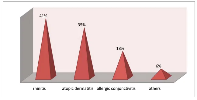
The personal atopy was associated with asthma in 95 children (44%), these types were represented in the following figure:
Figure 5: Distribution of patients according to their personal atopy. followed for asthma previous asthma attack Previous treatment
52%
83.8%
63.8%
rhinitis atopic dermatitis allergic conjonctivitis others 41%
35%
18%
In addition to the asthma and atopic history, children also presented others. The main one was respiratory infection in 141 children (65.27%) and the chronic vomiting in only 19 (8.7%).
6. Family history of asthma and / or atopy:
More than half of the patients (57%) had a family history of asthma and /or atopy, most frequently in mothers (29%).
Figure 6: Distribution of patients according to their family history
II. Pecipitating factors:
Table I: Distribution of patients according to precipitating factors.
Precipitating factors Number of cases Percentage
passive smoking 112 51.85% Allergens 203 94% physical exercise 177 81.9% Respiratory infection 123 57% Chimney smoke 3 1.38% air pollutant 37 17.1% father 16% mother 29% siblings 8% Grandparents 12% Aunts and uncles 24% Cousins 11%
In our study, we found that the allergens were the main precipitating factors (94%). This percentage includ: Dust mites (64%) Mildew (24.4%) Indoor plant (47.8%) Domestic animals (5.3%)
III. Natural history of disease
1.
Reason for consultation
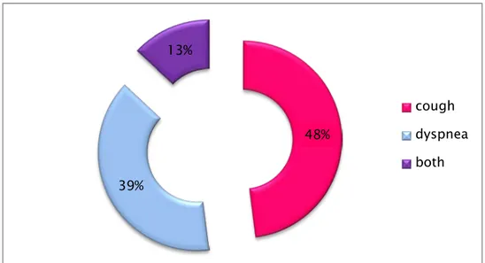
In our study, the reason for consultation was: cough (104 children), dyspnea (84 children) or both (28 patients).
Figure 7: Distribution of patients according to their reason for consultation
2.
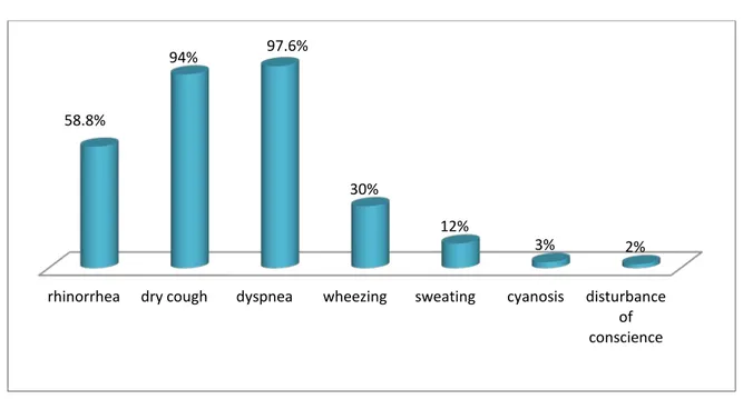
Functional signs:
Dyspnea and cough were found, respectively, in 211 and 203 patients. However, other signs like wheezing (65 patients) and rhinorrhea (127 patients) were also presents.
48% 39% 13% cough dyspnea both
Figure 8: Distribution of patients according to their functional signs
3.
Duration of the crisis
The duration of the crisis was varied between 2 and 14 hours, the average was 5.39 hours.
Figure 9: Distribution of patients according to the duration of their crisis rhinorrhea dry cough dyspnea wheezing sweating cyanosis disturbance
of conscience 58.8% 94% 97.6% 30% 12% 3% 2% 0 20 40 60 80 100 120 140 160 2-6 Hours 7-10 Hours 11-14 Hours 153 45 3,5
4.
Seasonal predominance
Children included in our study were admitted frequently in autumn and winter period with a pick in November when 35 children (16.2%) were admitted.
Figure 10: Distribution of patients according to their seasonal predominance
IV. Clinical examination:
All patients had wheezes (100%). Indeed, clinical signs are shown in the table below: Table II: Distribution of patients according to clinical signs
CLINICAL SIGNS Number of cases Percentage (%)
polypnea 137 63.4% Fever 71 32.9% distended chest 82 38% wheezes 216 100% tachycardia 31 14.3% subcutaneous emphysema 3 1.4%
decreased breath sounds 13 6%
0% 2% 4% 6% 8% 10% 12% 14% 16% 18%
V. Paraclinical investigation
C-reactive protein (CRP) and complete blood count (CBC) were requested respectively in 52 (24%) and 65 (30.1%).
Figure 11: Paraclinical investigation requested
The chest X-ray was performed in 151 cases (69.9%) with results below:
Figure 12: The result of the chest x-ray 30.1% 24% 69.9% CBC CRP Chest x- ray 24% 53% 43% 2% 0% 10% 20% 30% 40% 50% 60% Normal Thoracic distension Inectious focus Complications
VI. Therapeutic profile
We found in our study that the treatment of choice was nebulization of salbutamol used for 208 (96.3%) children.
Table III: Distribution of patients according to their Therapeutic profile
Treatment Number of cases Percentage
Oxygen Therapy 119 55.1% Salbutamol nebulization 208 96.3% injection 8 3.7% Corticotherapy oral 109 50.5% injection 121 56% Hydration 72 33.33% Antibiotic therapy 127 58.8% Other treatment 5 2.3%
Antibiotics were prescribed for 127 children (58.8%), the most used drug was Amoxicillin-clavulanic acid (57: 26.4%), followed by Amoxicillin (39: 18%), and macrolides (31: 14.4%).
VII. classification
The crisis severity was classified according only to clinical signs. Indeed, the crisis was mild in the majority of cases (80%).
Figure13: Distribution of patients according to their crisis’s severity
VIII. Follow up and Evolution
− We found that 62 children had regular follow of their asthma (28.7%). The rest of the patients were distributed among poor compliance (58: 26.8%), not followed (37: 17%) and disregarded (59: 27.5%).
Figure14: Distribution of patients according to their following 80% 18% 2% mild moderate sever 28,50% 27% 17% 27,50% 0,00% 5,00% 10,00% 15,00% 20,00% 25,00% 30,00%
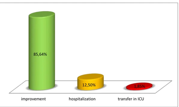
− We found that 185 children (85.6%) presented an improvement and received ambulatory treatment, 27 (12.5%) had been hospitalized on pediatric service A and only 4 (1.9%) was admitted in Intensive Care Unit (ICU).
Figure 15: Distribution of patients according to their evolution. improvement hospitalization transfer in ICU
85,64%
I. Definition of asthma: (5)
The Global Initiative for Asthma (GINA) definition is:
Asthma is a heterogeneous disease, usually characterized by chronic airway inflammation. It is defined by history of respiratory symptoms such as wheeze, shortness of breath, chest tightness and cough that vary over time and in intensity, together with variable expiratory airflow limitation.
II. Pathophysiology of asthma:
1. Inflammation and Airway Remodeling
The inflammation in asthma is mediated by multiple cell types including mast cells, eosinophils, T lymphocytes, macrophages, neutrophils, and epithelial cells (6). Asthma has allergic and non allergic presentations, based on the presence or absence of IgE antibodies to common environmental allergens. Both variants are characterized by airway infiltration by T-helper (Th) cells, which secrete a predominantly Th2 milieu (cytokines IL-4, IL-5, and IL-13) (6), (7).These cytokines stimulate mast cells, cause eosinophilia, promote leukocytosis, and enhance B-cell IgE production.
Although mild asthma symptoms are episodic and reversible, with progression, severity long-term and permanent airway changes can be present. Long term changes can include airway smooth muscle hypertrophy and hyperplasia; increased mucus production (and associated risk of mucus plugs); and edema (6). In the subepithelial layer, thickening can range from 7 to 23 mm, versus 4 to 5 mm in normal subjects, and more commonly affects the smaller airways (2–6 mm) (7-9). Permanent changes can include thickening of subbasement membrane, subepithelial edema and fibrosis, airway smooth muscle hypertrophy and hyperplasia, blood vessel proliferation and dilation, and mucus gland hyperplasia and hypersecretion (6, 9, 10).
There is likely an occult process of bronchial inflammation that precedes clinical symptoms of asthma. Bronchial biopsies of children with early respiratory symptoms who progressed to asthma had higher concentrations of eosinophils in the bronchial mucosa and thicker subepithelial lamina reticularis than those who did not, and these findings were present before the clinical presentation of disease (11). This suggests that an inflammatory milieu may precede clinical symptoms in people with asthma. Furthermore, in patients who had clinical symptoms of asthma that then seemed to go into remission, evidence of inflammation and remodeling persist on follow-up biopsies (12, 13).
Clinical symptoms are a late manifestation of lower airway inflammation. There are many treatments for asthma symptoms, but asthma is not a curable disease, and there
is evidence that inflammation is life-long and occurs even when no symptoms are present.
2. Bronchoconstriction and airway hyperresponsiveness.
The bronchoconstriction that occurs in asthma exacerbations is the main cause of obstructive symptoms. Airway hyperresponsiveness, or twitchy airways, occurs secondary to inflammation and airway remodeling. There is a distinct correlation between airway hyperresponsiveness and the degree of inflammation present. Bronchoconstriction can be induced by several pathways. Allergen-induced bronchoconstriction is caused by IgE-dependent mast cell degranulation, with resultant release of histamine, tryptase, leukotrienes, and prostaglandins (14-16). In addition to these mechanisms, bronchoconstriction by mast cell degranulation can also occur secondary to osmotic stimuli, which is likely the cause of exercise induced bronchoconstriction (6).
III. EPIDEMIOLOGY:
Known since antiquity, asthma was long considered a disease relatively benign. It became a concern after the Second World War, particularly in the early 1960s and was recorded deaths epidemic in Anglo-Saxon countries (17).
1. Prevalence
According to the global burden of asthma in 2003 the average prevalence of symptoms in Middle East children was 10.7%. The reported range for the prevalence of symptoms starts from 7.5% in Morocco to 17% in Kuwait (18). Based on the mentioned report; prevalence of asthma in Iran was about 5.5% in total population and 10% in childhood (18).
The International Study of Asthma and Allergy in Childhood (ISAAC) examined the epidemiology of asthma in Morocco. It included three Moroccans centers (Casablanca, Rabat, and Marrakech). The first surveys in Morocco in 1996 estimated the prevalence of asthma between 2 and 5.5%. Ten years later, as part of the same study, the prevalence of asthma was 6.6% among
children of Rabat and 12.1% in the same population in Casablanca. in contrast in Marrakech, less polluted city, the study found a prevalence of 17.9% (19). The prevalence of asthma in children in the Safi region was 3.4% in 2010 (20).
2. Mortality
Mortality from asthma has become a concern in recent years, due to the increase in the prevalence of asthma, increased severity of crisis, low patient compliance as well as poor management. Mortality among asthmatic children varies according to the authors from 0.4% to 0.7% (21). Asthma was recently reported to remain the sixth leading cause of death of children between 5 and 14 years of age in the United States. In Sweden, a study by Bergströma et al during 10 years period from 1994 to 2003, found 75 deaths suspected to be due to asthma (22).
In our study, we had no case of death.
3. Morbidity
Asthma is considered as an affection of the lifetime which 50% begins before the age of 2 years. Webb et al noted the persistence of crisis in 59% of children aged 3 years and a half after bronchiolitis (22). Similarly, Buffum still find 60% at 5 years with asthma among children who had asthma before the age of 2 years (23). In contrast, Park et al found that 20% of asthmatics at age 10 years among those who had presented dyspnea events were infants (24).
The frequency of hospitalization for asthma has also increased. This data is found in different countries: she has in fifteen years multiply by 3 in the USA and by 4 in Canada (25). In Morocco, the number of hospitalizations of children with asthma varies according to the months of the year, peaked in November and decreased in June and July (20).
In France, half of hospital admissions for children relate to asthma and hospitalization rates remained stable among children. A study of more than 1 year was conducted in 14 pediatric units in France, including children aged 3 years and older hospitalized for asthma
exacerbation. Data from 727 hospitalizations were collected. In 48% of hospitalizations, the children were 3-5 years. Among children with asthma, 57% had been admitted to the hospital for asthma exacerbation, 37% were admitted to the hospital or in the emergency department, and control of the asthma in the previous month was unacceptable in 46% of cases (26).
IV. Risk factors:
Numerous potential risk factors have been studied in relation to the development of asthma. Atopy is frequently identified as a strong risk factor for the development of asthma, yet there is no direct correlation between the two (27, 28). Some studies have demonstrated that early dust mite sensitization and maternal asthma are very significant predictors of asthma (29,30). Parental smoking is a significant risk factor for acute lower respiratory tract infections in infants, and the development of wheezing and asthma in children. Air pollution and viral infections are well-established triggers for asthma exacerbations, but there is conflicting data whether these factors contribute to developing asthma (33-36). Microbial exposure is inversely correlated with the development of asthma and atopy, and may account for the disparate prevalence of asthma in urban versus rural (specifically farming) environments (37-39). Ultimately, it is likely that asthma develops in genetically susceptible individuals through a combination of complex environmental exposures.
Figure 17: Effect of the interaction of various types of exposures on the asthma
Allergen-induced
•
House dust
mite
•
Pollen
•
Ragweed
•
Cockroach
•
mold
Non- allergen-induced
•
Ozone
•
Cigarette
•
Infection
•
Aspirin
•
Exercice
•
Cold air
Intrinsic
Asthma
1. Age of onset:
Most epidemiological studies report that the vast majority of children with asthma have their asthma during childhood.
Some authors have studied the influence of this factor on the asthma prognosis. The Barr et al studies found no dependence between the age of early onset and evolution of asthma. Gerritsen's study confirms this same observation (40).
Finally, a study by Sears in 2003 showed that the age of early onset is a risk factor for persistence and severity of asthma (41).
In our study, the age of the majority of our patients (60%) was between 2 and 6 years.
2. Atopy
The main personal risk factor for bronchial asthma is atopy in the individual or his/her family. The most important risk factor is atopy in the family. Some genes related to concurrent atopy, Ig E response and asthma have been found. These genes are located on chromosomes 5, 11 and 14. Children whose parents do not have extrinsic asthma have an asthma prevalence of 8 %, which increases to 15 % with asthma in one parent and 28.6 % with asthma in both parents. The incidence of asthma in first-degree relatives is 3-6 times of normal (42). The study by Martin et al found that eczema has a bad influence on the prognosis of asthma. Against, Wittis study showed that there is no relationship between eczema and the prognosis of asthma (42).
The study of Kocevar published in 2002, which estimated the number of hospital days and readmission in asthmatic Norwegian children with and without allergic rhinitis, has demonstrated that the allergic rhinitis is associated with an increase of the duration of hospitalization in these children (43).
Our study objectified that 44% had a personal atopy and/or family atopy, allergic rhinitis occupied the predominant place in 41% of children.
3. Genetic predisposition:
Asthma is a complex disease that results from the interaction between genetic predisposition and environmental factors
It seems that the inheritance of asthma and atopy does not follow the classical Mendelian patterns (43). In 1909, Drinkwar by studying an example of three generations suggested that asthma was transmitted in a Mendelian dominant mode (44).
Martinez et al studies in USA found the influence of several genes including one major gene transmitted respectively by an autosomal co-dominant and autosomal recessive. The role of HLA class 2 in the specific immune response to allergens has historically been initiated in the early years by the discovery of an association between IgE Specific Ra of 5 allergens and HLA-DW2 located almost exclusively in asthmatic (45).
A study in Italy in 2011 of 127 asthmatic children showed that imbalance between the oxidation forces and antioxidant defense systems has been implicated in the pathogenesis of asthma. Glutathione S-transferase (GST) plays an important role in cell protection against inflammation. The results suggest that the GSTA1 and GSTO2 are asthma genes involved in the increased risk of developing asthma in the Italian population (45).
In our study, the family history of asthma and/or atopy was recorded in more than half of cases (57%), most frequently in mothers (29%).
4. Infection-Related Asthma
4.1. Viruses:
The role of infection in asthma is varied in that it may exacerbate established asthma or be a contributing factor to the initial development of the clinical onset of asthma. Mounting evidence implicates both roles, with particular viral pathogens, namely human rhinovirus (HRV) and respiratory syncytial virus (RSV), among the most likely culprits in asthma inception (46,47).
In addition, outpatient wheezing illnesses caused by RSV and HRV early in life may increase the risk for subsequent wheezing episodes and the development of asthma (46). Recent studies have found that over 80% of wheezing episodes were associated with viral respiratory infections (47). In more than 60% of these children, the synsicial respiratory virus (RSV) was detected. The close link between bronchiolitis induced by viruses and the development of asthma has been demonstrated in several studies (48).
In our study, more than half of the children followed for asthma (57%) had a history of recurrent respiratory viral infections.
4.2. Bacterial pathogens:
Bacteria such as Lactobacillus and Helicobacter pylori are reported to be protective against asthma; whereas other bacteria are associated with an increased risk of asthma (49). One study demonstrates associations between neonatal hypopharyngeal colonization of Haemophilus influenzae, Streptococcus pneumoniae, and Moraxella catarrhalis, with the subsequent increased risk of developing recurrent wheezing and childhood asthma (50).
It is unclear from these findings whether early colonization with these organisms in some way influences the development of asthma or if the presence of these organisms is a reflection of an altered immune system that predisposes to altered host airway responses to respiratory pathogens. Acute wheezing episodes of preschool children are associated with these bacterial pathogens, with a frequency similar to that seen with viruses (51).
5. Allergens:
High allergen exposure in the home and allergic sensitisation is a cause of acute exacerbations of asthma in children (52). Allergen exposure in schools might also be important. Low-dose exposure to cat allergen on the clothes of classmates at school is sufficient to cause deterioration of asthma (53).
The aeroallergens likely susceptible to intervention are pets, cockroaches, moulds, and house dust mites. House dust mites are a controversial area, although there is little evidence for routine use of avoidance measures for most children sensitised to these aeroallergens.
People with asthma of all levels of severity are commonly exposed to allergens in the home (54). Pet and cockroach sensitisation might be a marker for high morbidity, although whether cockroach sensitisation can be separated from the effects of low socioeconomic status is arguable (56). An interaction between passive smoking and pet sensitisation might exist. The use of synthetic bedding might be associated with severe wheeze (55).
The study of Platts-Mills et al (56) showed, in patients allergic to dust mites, improved clinical signs and decreased bronchial hyperresponsiveness (BHR), after a sojourn of two months in a hospital without mites. Recent studies have revealed a recurrence of symptoms and worsening non specific BHR when children were staying again at home, full of mites, reflecting the likely role of the latter in asthma symptoms (57, 58).
In our study, we found that the dust mite was the main precipitation factor, present in 203 patients (94%).
6. Gastro-esophageal reflux:
Gastro-esophageal reflux disease (GERD), which is the passive regurgitation of gastric contents retrograde into the esophagus may be associated with asthma (59). However, exact causal relationship has not been confirmed between asthma and GERD (60). The relationship between asthma and GERD has been detected in many studies by 24 h pH monitoring (61, 62).
V. Environmental factors:
1. Passive smoking:
Children are likely to be exposed to second hand tobacco smoke (SHS) at home (63). A study in Japan by Kaneita et al has shown that 64.8% of 6 month old children live with smoking parent(s), and of those, 57.9% of parents smoke indoors at home. Although, many previous studies have revealed the risk of SHS for childhood asthma (64).
There has been no prospective study of the risk of paternal smoking for asthma in children aged 2 years or less and only one study for children aged 3–4 years. Further, a wide range of estimated effect size of postnatal maternal smoking on incidence of childhood asthma was observed, indicating a need to confirm the results. A previous study by Kanoh et al. using data from the Longitudinal Survey of Newborns in the 21st Century, reported a positive hazard risk between parental smoking and childhood asthma incidence (65). Furthermore, although parents are encouraged to smoke outdoors or not to smoke (66), the difference in the contribution of these parental smoking behaviors to the risk reduction of asthma among their children has not been sufficiently evaluated (67).
In our population, 51.8% of asthmatic children were exposed to parental smoking.
2. Socio-economic factors:
Factors related to lifestyle and socioeconomic status appears to be involved in the expression of the manifestations of asthma (68).
The prevalence of asthma may vary according to life styles, it is higher in children living in rural areas, and especially if they live in the country during the first two years of life (69). The prevalence of asthma also varies according to the socio-economic level. Asthma is more common in people of higher social class as shown by the English and Swiss studies with prevalence of 13 and 9% in the highest class against 8 and 5% in the lowest (70). The social class
difference involves multiple environmental factors that may be more directly involved in the determinism of asthma.
In addition to the different lifestyles (heating, types of bedding, carpets ...), other factors may explain this predominance: better medical knowledge of diseases, excessive medical consumption, the use of higher hygiene products and finally the highest maternal age.
The majority of patients (85%) in our study had a low or medium socioeconomic level.
VI. Associated Allergic reactions:
Seasonal allergic rhinitis is one of the most common allergic diseases and its prevalence is steadily increasing. Asthma and allergic rhinitis are two manifestations of one common allergic respiratory syndrome. The relationship between asthma and allergic rhinitis is complex and upper and lower airways interact with each other. They often occur together and allergic rhinitis increases the risk of asthma development (71).
Although the relationship between allergic rhinitis and asthma has been well established, direct links between nasal and bronchial inflammation in seasonal allergic rhinitis without asthma remain to be exactly investigated. Several authors have focused on the effect of natural allergen exposure or nasal challenge on lower airway, but the dynamics of bronchial inflammation in patients with pollen allergic rhinitis during and out of the pollen season are not completely clear yet (71).
One study (72) was made in four cities of Mexico, about prevalence allergy (rhinitis) in children aged 6 to 7 years and adolescents aged 13 to 14 years of primary and secondary schools. In children aged 6 to 7 years, the results were in this order: current rhinitis (27.9%), rhinoconjunctivitis (24.2%), and associated allergies (9.2%). The corresponding frequencies in adolescents 13 to 14 years were respectively 33.3%, 34.1% and 18.4%. All children with rhinitis also had asthma symptoms, and symptoms of atopic dermatitis (AD).
AD is a common condition in infancy but disappears around age 3 years in a significant proportion of children. The prognosis is mostly determined by the severity and the presence of atopic sensitization. Early AD is associated with asthma at school age, but in many of these asthmatic children, wheezing manifests before or with the onset of AD. Children with AD and wheeze have a marked loss in lung function, suggesting a distinct phenotype rather than a progressive development from AD to asthma. (73).
In our study, the allergic rhinitis was associated with asthma in 41% of cases followed by atopic dermatitis in 35%.
VII. Clinical diagnosis:
1. Paroxysmal asthma attack: (5)
The classic triad of asthma attack includes cough, wheeze, and dyspnea. However, patients often present with only 1 of these symptoms, which can make diagnosis challenging. In studies of patients presenting solely with wheeze, cough, or dyspnea, only 24 to 35% were eventually diagnosed with asthma. Symptoms of asthma are typically worse at night or early in the morning. A personal and family history of asthma and atopy favor the diagnosis.
Clinical examination revealed chest distension, increased the sound and especially wheezing predominate in expiration
The exacerbation of asthma in children is defined by GINA 2014 as an acute or sub-acute deterioration in Symptom control that is sufficient to cause distress or risk to health, and necessitates a visit to a health care provider or requires treatment with systemic corticosteroids.
Early symptoms of an exacerbation may include any of the following: • An acute or sub-acute increase in wheeze and shortness of breath. • An increase in coughing, sneezing, nasal itching.
• Impairment of daily activities, including feeding. • A poor response to reliever medication.
In our study, 14.3% of children had tachycardia and only 3% had cyanosis.
2. Status asthmaticus or acute severe asthma:
(74)Pediatric status asthmaticus (PSA) is a medical emergency warranting prompt recognition and intervention. A status asthmaticus or severe asthma exacerbation is defined as an acute episode that does not respond to standard treatment with short acting b2-agonists and corticosteroids. Although, a large variation exists in this definition between authors. In other definitions, need for hospitalization, emergency room visit or decline in peak expiratory flow (PEF) is also taken into account. PSA can result in respiratory insufficiency as well as circulatory failure and is potentially life-threatening.
Clinically, it is a dramatic asphyxia, acute respiratory failure made of:
Respiratory syndrome with dyspnea, cyanosis, inability to cough and, Sweating, pallor, chest distended, tympanic, disappearance of wheezing
Tachycardia with presence paradoxical pulse and cardiovascular collapse
Neuropsychiatric syndrome with anxiety, disturbance of consciousness, confusion and stupor.
The Radiography is essential to ensure the absence of complications (pneumothorax, pneumomediastinum and infection).
The arterial blood gas shows severe hypoxia, but especially hypercapnia indicator of alveolar hypoventilation, importance of bronchial obstruction and depletion of the patient.
Finally, the onset of acidosis is a poor prognostic factor. In case of hypercapnia, the gasometric response to treatment should be evaluated within two hours after startup.
Data on the incidence or prevalence of PSA are scarce. In a USA cohort, admission for status asthmaticus between 1992 and 2006 approximately halved from 1.92 to 0.93 per 1000 children. In contrast, ICU care related to asthma increased from 0.09 to 0.31 per 1000 patients (75).
In our study, 4 patients presented a Status asthmaticus and were admitted to ICU.
3. Exercise induced asthma:
The clinical expression of Exercise induced asthma (EIA): is that of an asthma attack whose only characteristic is to follow an exercise. Typically, it occurs in the exercise, in a subject with normal respiratory function, and reaches its maximum intensity 5 to 10 minutes after cessation of exercise (76). In some cases, the EIA may be the only manifestation of asthmatic disease (77).
A retrospective study was conducted in four high schools in Rabat, with 1179 students and 16 teachers of physical education. The study found 70 asthmatic children (6%), 62.5% teachers felt that children with asthma cannot continue the effort and only 18% of students use asthma prophylaxis before the sports sessions (78).
The EIA is not a cons-indication to the sport in asthmatic children and does not justify any exemption in physical education. It remains accessible to administered therapeutic few minutes before exercise (79).
In our study, the physical exercise induced asthma attacks was found in 81.9% of children.
VIII. Classification of asthma:
(80)To address diversity and guide management, several factors have been used to classify pediatric asthma.
According to the National Asthma Education and Prevention Program's (NAEPP) Guidelines for the Diagnosis and Management of Asthma , asthma can be divided into four levels of asthma severity: mild intermittent, mild persistent, moderate persistent, and severe persistent.
In children older than 5 years, three major features are recommended in determining level of severity: frequency of asthma symptoms during the day, frequency of nighttime asthma symptoms, and measures of pulmonary function. These Guidelines divided the Forced Expiratory Volume (FEV) predicted into three levels: above 80% predicted typical of mild asthma (both intermittent and persistent), from 60 to 80% predicted typical of moderate asthma, and below 60% predicted typical of severe asthma.
Table IV: Classification of asthma severity (80)
Symptoms Nocturnal symptoms FEV or PEF Severe persistent Continuous limited physical activity frequent < 60% predicted Variability›30% Moderate Daily attacks physical activity >1 Time a week 60 to 80 % predicted Variability >30% Mild persistent >1Time a week
But<1Time a day > 2 Time a month
>80% predicted Variability 20 to 30%
Intermittent
<1 Time a week Asymptomatic
And Normal PEF between attacks
<2 Time a month >80% predicted Variability <20%
FEV: Forced Expiratory Volume
;
PEF: Peak Expiratory FlowIn our study, the crisis severity was classified according only to clinical signs. Indeed, the crisis was mild in the majority of cases (80%).
IX. Complications:
1. Infection:
It’s a classic but rare complication, antibiotic therapy is justified if the patient is febrile, if there is purulent secretion and radiological home. The frequency of respiratory infections associated with bacteria during asthma attacks varies according to the authors from 10 to 17% (81).
2. Mechanical complications : Pneumo-mediastinum, pneumothorax
and subcutaneous emphysema:
The frequency of pneumomediastinum in children's asthma attacks is 0.3-5%. The diagnosis should be suspected in a sudden deterioration of respiratory status, retro-sternal pain radiating to the arms and neck, aggravated by breathing movements and sometimes swallowing. The essential clinical sign is the perception of a snowy crepitation of the upper cervical and thoracic region painful on palpation. The pneumomediastinum results on chest x ray deal by Hyper-V linear lights along the mediastinum, lifting the two pleural layers (82).
In our patients, the subcutaneous emphysema was found in 3 children (1.9%), with improvement under treatment.
The vast majority of cases, pneumomediastinum and subcutaneous emphysema are benign complication of asthma. Treatment coincides with that of the crisis itself, and their disappearance is done in a few days. The hospitalization of the child is indispensable because of the risk of pneumothorax. Pneumothorax, it is the consequence of a pneumo-mediastinum or pleural bubble rupture; it is a rare event (83).
X. Differential diagnoses:
Table V: Important differential diagnoses in pediatric asthma (84).
Condition Characteristics
Transient infant wheezing (eg, recurrent bronchiolitis
Onset in infancy No associated atopy
Associated with maternal smoking Cystic fibrosis Recurrent wheeze and failure to thrive Primary ciliary dyskinesia
Associated recurrent otitis media and sinusitis Initial oxygen requirement postnatally
Situs inversus in 50%
Primary ciliary dyskinesia Persistent moist cough in combination with wheeze Purulent sputum suggests bacterial cause Structural abnormality
(eg, tracheomalacia, bronchomalacia
Onset usually from or shortly after birth but occasionally later
Vocal cord dysfunction
High-pitched inspiratory stridor and dyspnea May be spontaneous or exercise induced
Blunting of inspiratory volume loop on spirometry Inhaled foreign body Sudden onset Differential air entry or wheeze on examination
Cardiac failure
Associated with congenital or acquired heart disease (eg, dilated cardiomyopathy after viral infection)
Eosinophilic lung disorders including allergic bronchopulmonary
aspergillosis
Skin prick test positivity to Aspergillus fumigatus Raised serum IgE
Infiltrates on chest radiograph Anxiety causing hyperventilation
No wheeze audible
Spirometry at the time of symptoms may help distinguish
Exercise induced asthma No respiratory symptoms other than with exercise Exercise testing may distinguish Milk aspiration/cough during feeds Symptomatic particularly with liquids
XI. Para clinical investigations:
1. Chest X-ray :
Chest radiographs can be used to help exclude other diagnoses of wheezing or cough, especially in patients with first-time wheezing (85). Acute-onset, unilateral wheezing suggests foreign body aspiration, and patients may show hyperinflation on chest radiographs. Chest radiographs may show a structural abnormality in a patient with chronic wheezing that fails to respond to bronchodilator therapy (86-88).
Chest radiograph in patients with respiratory failure may help rule out complications from asthma such as pneumothorax or pneumomediastinum as well as other contributing causes for respiratory distress such as infection or cardiac disease. Although no clear guidelines exist, chest radiographs should be considered in the following instances: asymmetric wheezing, wheezing that fails to respond to bronchodilator therapy, or patients with respiratory failure (88).
Two patients in our study, which was initially treated as asthma had presented a foreign body in the chest X-ray.
Figure 19: Chest X-ray objectified a foreign body in a 5 year old girl who was treated initially as asthma
2. Skin tests:
The skin prick test is a commonly used procedure in secondary care for assessing a specific sensitization in allergic respiratory diseases. Up to 85% of asthma patients show positive reactions to skin tests for common aeroallergens (89). Research has demonstrated the reliability and predictive validity of allergy skin testing.
Failure to use this combined approach could result in deficiencies in diagnosis and asthma management. Tschopp et al. recently concluded that the skin prick test should be used as a primary tool by clinicians to assess respiratory allergic diseases, as the test is comparably economical and provides immediate educational information for patient and physician (89).
3. Respiratory function tests:
Evaluation of lung function is important for both diagnosis and monitoring. Nevertheless, normal lung function tests do not exclude a diagnosis of asthma, especially for intermittent or mild cases (89). Therefore, these tests are considered supportive. Performing the tests when the child is symptomatic may increase sensitivity.
In children younger than 5 years, newer lung function tests that require less cooperation have been used (such as oscillometry or specific airway resistance). However, these are not generally available outside specialized centers (90).
Respiratory function tests are recommended for children old enough to perform it properly; the proposed range of minimum age is between 5 and 7 years.
Assess the disease by the only clinic, causes an under appreciation and under treatment which can be harmful to the respiratory future of the child. Only lung function test confirms a sense of well being breathing. It is used to classify the severity of the disease and adapt treatment; it must be repeated at regular intervals to adjust the therapeutic stage of the disease (91).
4. Peak Expiratory Flow Rate:
The peak expiratory flow rate (PEFR) is the measure of airflow during a brief, forceful exhalation. The measurement of peak flow rates can be taught to the patient and routinely used at home to monitor disease severity (92).
Reduced peak flow measurements do not differentiate between obstructive and restrictive diseases; spirometry and sometimes measurement of lung volumes are necessary to distinguish the two. Peak flow measurements are not sufficient to distinguish upper airway obstruction (eg, vocal cord dysfunction) from asthma. The validity of PEFR measurements depends on patient effort and technique. There is also no standardization among peak flow measurements. Despite the shortcomings, many patients use PEFR to successfully follow the progression of their asthmatic disease (93).
Fonceca et al followed 75 children aged 5–16 years with persistent asthma for a 12-week period. Their study showed little or no evidence of correlation between clinical severity scores and the values of FEV and PEF but showed a positive correlation between PEF and FEV obtained by spirometry and they establish PEF as a reasonable measure for use in the home when spirometry cannot be obtained. The researchers state that their findings reinforce the use of PEF for the management of children with asthma but state that PEF should not be the only objective parameter used in monitoring asthma control (94,95).
Figure 20: Peak Expiratory Flow (PEF)
5. Blood gases:
It is required in severe asthma attacks to assess respiratory failure. Respiratory alkalosis is common at a moderate asthma attack, marked by hypoxia with hypocapnia. The transition to normocapnia and especially to hypercapnia indicates decompensation and respiratory acidosis.
In our study, the blood gas was used only in ICU, and the oxygen saturation was less than 92% in 32.5% of our patients.
XII. Therapeutic management:
1. Treatment of the usual acute asthma attack:
Early recognition and treatment of the asthma exacerbation is essential for success in overall management. Patients presenting to the ED with acute asthma should be quickly evaluated
for the adequacy of airway, breathing, and circulation. This evaluation should include a complete set of vital signs, pulse, oximetry, respiratory rate, and an assessment of respiratory effort.
Treatment should be started immediately. Current recommendations are outlined in the 2007 National Asthma and Education and Prevention Program (NAEPP), Expert Panel Report (EPR-3) coordinated by the National Heart, Lung, and Blood Institute of the National Institutes of Health and the 2005 Canadian Asthma Consensus Guidelines (96). Goals of therapy include correction of hypoxemia, rapid reversal of airway obstruction, and treatment of inflammation.
1.1. B2- Agonists:
B2-Agonists are potent bronchodilators that act on b receptors to quickly and effectively relax bronchial smooth muscle. Short-acting b-agonists are the recommended first-line therapy for the acute asthma exacerbation (96). Albuterol is the most commonly used b2-agonist for acute asthma. B2-Agonists can be administered in multiple forms, including metered-dose inhaler
For acute asthma, 2 to 6 inhalations of albuterol are given with MDI and spacer device. Therapy is repeated every 20 minutes for up to 4 hours until there is maximal improvement in respiratory symptoms. Alternatively, if MDI is unavailable or if the patient is unable to use proper techniques, 2.5 to 5 mg of nebulized albuterol is given every 20 minutes to a total of 3 doses (maximum of 15 mg in one hour). Albuterol can also be given as a continuous nebulization at a rate of 10 to 15 mg over 1 hour (96). Therapy should be titrated to an objective measure of airflow obstruction (eg, FEV1 or PEF) and clinical response. B-Agonists should only be used for relieve of acute symptoms. Adverse effects associated with b-agonists include tremor,
(MDI), nebulizer, subcutaneous injection, and intravenous injection. Traditionally, aerosolized bronchodilators have been administered by continuous-flow nebulization. However, multiple studies have found little difference in efficacy between MDI and nebulizer therapy (96).
Hospital admission rates are equivalent between the 2 modalities, MDI is more cost-effective and time-effective than nebulizer therapy (96). An additional concern with aerosolized or nebulized medication is its potential to spread infectious agents.
tachycardia, palpitations, hyperglycemia, and hypokalemia. Current evidence does not support the use of intravenous b2-agonists for the treatments of acute severe asthma (96). A meta-analysis including 21 pediatric studies and more than 2000 children demonstrated that in acute asthma spacers were as effective as nebulizers in limiting hospitalization rates and reducing the time spent in emergency department (96).
In our study, the majority of children (96%) used nebulized salbutamol.
Figure 21: the metered-dose inhaler with spacer
1.2. Anticholinergics:
Anticholinergics block the action of acetylcholine on the parasympathetic autonomic system. They decrease vagally mediated smooth muscle contraction in the airways leading to bronchodilation. Anticholinergics are recommended for the treatment of severe asthma, in combination with short-acting b-agonists (97). The synergistic effects of these 2 agents decrease hospitalization rates and improve lung function. Ipratropium bromide is the most commonly used anticholinergic agent for the treatment of asthma.
For the acute exacerbation, 0.5 mg of ipratropium is given by nebulization every 20 minutes for 3 doses. Alternatively, 8 puffs with MDI and spacer can be given every 20 minutes for up to 3 hours. A recent study (97) showed the benefit of a multiple-dose protocol of ipratropium combined with albuterol in patients with severe asthma exacerbations (FEV <50%).