Task-Induced Functional Connectivity of Picture
Naming in Healthy Aging: The Impacts of Age
and Task Complexity
Perrine Ferré1, Julien Jarret1 , Simona Maria Brambati1,2, Pierre Bellec1,2 , and Yves Joanette1
1Centre de recherche de l’Institut universitaire de gériatrie de Montréal, Montréal, Québec, Canada 2
Université de Montréal, Québec, Canada
Keywords: fMRI, functional connectivity, healthy aging, picture naming, language, task-induced functional connectivity
ABSTRACT
The topological organization of the brain, governed by the capacity of brain regions to synchronize their activity, allows for cost-effective performance during everyday cognitive activity. Functional connectivity is an fMRI method deemed task-specific and demand-dependent. Although the brain undergoes significant changes during healthy aging, conceptual knowledge and word-production accuracy are generally preserved. The
exploration of task-induced functional connectivity patterns during active picture naming may thus provide additional information about healthy functional cerebral mechanisms that are specifically adapted to the cognitive activity at hand. The goal of this study is to assess and describe age-related differences in functional connectivity during an overt picture-naming task, as well as to compare age-related differences under complex task demand, defined by lexical frequency. Results suggest both age-specific and task-specific mechanisms. In the context of preserved behavioral performance in a picture-naming task, older adults show a complex array of differences in functional connectivity architecture, including both increases and decreases. In brief, there is increased segregation and specialization of regions that are classically assigned to naming processes. Results also expand on previous word-production studies and suggest that motor regions are particularly subject to age-related differences. This study also provides the first indication that intrinsic task demand, as manipulated by lexical frequency, interacts little with the relationship between age and functional connectivity. Together, these findings confirm the value of task-induced functional connectivity analysis in revealing the brain organization that subserves task performance during healthy aging.
INTRODUCTION
The aging of the population worldwide creates both immense opportunities and numerous challenges regarding health and wellness. This critical demographic change is compelling neuroscientists to engage in studies of cognition in aging, so they can better understand the constituents of cognitive health, which is central to quality of life (WHO, 2018). Although many cognitive abilities typically decline with healthy aging, conceptual knowledge is long preserved (Ben-David, Erel, Goy, & Schneider, 2015;Goulet, Ska, & Kahn, 1994;Salthouse, 2014). Its impairment may be an early marker of major or mild neurocognitive disorders (Blackwell et al., 2004;Miller, Rogers, Siddarth, & Small, 2005;Reilly, Peelle, Antonucci, & Grossman, 2011). The assessment of conceptual knowledge classically includes picture
a n o p e n a c c e s s j o u r n a l
Citation: Ferré, P., Jarret, J., Brambati, S. M., Bellec, P., & Joanette, Y. (2020). Task-induced functional connectivity of picture naming in healthy aging: The impacts of age and task complexity. Neurobiology of Language, 1(2) 161–184.https://doi.org/10.1162/ nol_a_00007 DOI: https://doi.org/10.1162/nol_a_00007 Supporting Information: https://doi.org/10.1162/nol_a_00007 Received: 30 September 2019 Accepted: 04 March 2020
Competing Interests: The authors have declared that no competing interests exist. Corresponding Author: Perrine Ferré perrine.ferre@umontreal.ca Handling Editor: Jonathan Peelle Copyright: © 2020 Massachusetts Institute of Technology. Published under a Creative Commons Attribution 4.0 International (CC BY 4.0) license.
naming, a sensitive word-production test at the core of many clinical assessment tools (e.g., intraoperative language mapping, differential diagnosis;Moritz-Gasser, Herbet, & Duffau, 2013). Although older individuals often complain about proper name retrieval (Condret-Santi et al., 2013), they often obtain accuracy scores similar to those of younger adults when naming pictures of common names. Yet, this typically occurs at the expense of longer response times (Baciu et al., 2016;Hoyau et al., 2017;Wierenga et al., 2008) in possible relation with a general slowing in information processing (Feyereisen, Demaeght, & Samson, 1998). The dissociation between time and accuracy makes this task particularly interesting, as it suggests that older adults engage in adaptive mechanisms to perform it adequately. This study thus aims to explore the specific neuro-functional processes that underlie picture naming, a preserved cognitive ability.
The field of cognitive neuroscience of aging (Cabeza, Nyberg, & Park, 2016) has revealed a dynamic interplay between declines in structural and functional resources and plasticity phe-nomena that support cognitive performance. Consistent patterns of enhanced recruitment with older age, including greater prefrontal bilateral activation, have been found across many cog-nitive tasks using task-induced blood oxygen level–dependent (BOLD) amplitude activation dif-ferences (Cabeza, 2002;Davis, Dennis, Daselaar, Fleck, & Cabeza, 2008). Such manifestations of functional brain adaptation potentially relate to greater demands for cognitive-control pro-cesses in older adults (Park & Reuter-Lorenz, 2009;Schneider-Garces et al., 2010). When age-related differences such as decreased specificity and more diffuse brain activity are not as-sociated with performance, a dedifferentiation phenomenon is invoked (Cabeza et al., 2018). Although such phenomena have generally been explored using abilities known to decline with age, less is known about the functional organization that sustains a well-preserved ability.
Older individuals may exhibit longer latencies and phonological imprecision (Feyereisen, 1997;Goulet et al., 1994;Shafto, James, Abrams, & Tyler, 2017). Some complain about word-finding difficulties, but mostly so for proper names and with no relation to objective cog-nitive difficulty (Condret-Santi et al., 2013). However, on average, their accuracy is preserved, sometimes even improved, when a simple common names task is used, where accuracy and not response time is scored, and when there are few time constraints (LaBarge, Edwards, & Knesevich, 1986; Salthouse, 2014;Schmitter-Edgecombe, Vesneski, & Jones, 2000;
Verhaegen & Poncelet, 2013;Wierenga et al., 2008). Overall, naming abilities for common names appear behaviorally preserved when task demand is controlled, at least until 65 years of age (Salthouse, 2014). Picture naming typically involves an extensive neural network reflect-ing every cognitive step required to produce a known word. Although many interindividual var-iations exist, the core areas have been described in young adults (Duffau, Moritz-Gasser, & Mandonnet, 2014;Friederici & Gierhan, 2013;Price, 2012;Sarubbo et al., 2016). The occipital cortex and the middle and inferior posterior temporal cortex are involved in the semantic cessing of visual attributes, along with the orbital inferior frontal gyrus (IFG) for multimodal pro-cessing. Together, the opercular IFG, premotor cortex, insula, and inferior parietal and superior temporal cortex are in charge of phonological processing and articulation. These core areas, although segregated, do not operate in isolation. In particular, age-related neurofunctional re-organization will often happen at the scale of networks, if not the whole brain, by changing ac-tivity interactions between regions (Perry et al., 2015;Tomasi & Volkow, 2012;Tsvetanov et al., 2016). Thus, functional connectivity (FC) is a tool of choice to study neurofunctional mecha-nisms associated with aging.
FC in healthy aging has been studied mainly using resting-state and associated networks, such as the default mode network (DMN) (Sala-Llonch, Bartrés-Faz, & Junqué, 2015). The in-tegrity of DMN connectivity appears to be particularly affected by age and related to perfor-mance (Mak et al., 2017). Generally speaking, a decrease in within-network FC is reported,
along with increased FC between functionally related regions (Mak et al., 2017;Sala-Llonch et al., 2015). This pattern appears in line with previous activation studies that demonstrated decreased deactivation of the DMN during task performance in older individuals (Persson, Pudas, Nilsson, & Nyberg, 2014). Examples of increased brain FC with regions belonging to the same functional network but typically not recruited by younger adults have also been detected using lexicose-mantic tasks (Agarwal, Stamatakis, Geva, & Warburton, 2016;Hoyau et al., 2018;La et al., 2016;Marsolais, Perlbarg, Benali, & Joanette, 2014;Muller, Mérillat, & Jäncke, 2016). Some studies of word production have reported BOLD signal increases in parietal, frontal, or temporal regions with age (e.g.,Hoyau et al., 2017;Meunier, Stamatakis, & Tyler, 2014), as well as contra-lateral recruitment (e.g.,La et al., 2016;Meinzer et al., 2012). Along with an impact of age, effects of behavioral performance and task demand are also expected: A recent meta-analysis focusing on semantic cognition and aging (Hoffman & Morcom, 2018) concluded that augmented activation was principally observed when older adults performed worse than younger adults, for example, during tasks drawing on executive functions.
However, some authors have recently argued in favor of state-dependent differences in re-gional functional connectivity with age (Campbell & Schacter, 2016;Greene, Gao, Scheinost, & Constable, 2018;Samu et al., 2017). In regard to lexical knowledge, recent evidence points toward distinct FC patterns for different tasks (e.g., synonyms, antonyms, picture naming) in older adults (Ferré et al., 2019;Varangis, Razlighi, Habeck, Fisher, & Stern, 2019).
It is important to note that a task paradigm allows for direct manipulation of the task require-ments to characterize neural mechanisms that underlie brain-behavior associations and poten-tial strategic mechanisms (Crowell et al., 2019;Finn et al., 2017;Greene et al., 2018;Persson, Lustig, Nelson, & Reuter-Lorenz, 2007;Steffener et al., 2014). Prior studies of aging and cog-nition have concluded that domain-general control processes become more involved when general cognitive load increases, to which older adults appear particularly sensitive (Campbell et al., 2016;Park & Reuter-Lorenz, 2009; Peelle, 2019;Wang, Dew, & Cabeza, 2015). Yet, little is known about the impact of manipulating task requirements through psy-chometric criteria (Cole, Smith, & Beckmann, 2010). For example, lexical frequency influ-ences brain activity and performance during naming (e.g., Basso et al., 2013; Burke, MacKay, Worthley, & Wade, 1991). Indeed, low-frequency words trigger more tip-of-the-tongue states than high-frequency words (Burke et al., 1991), and older adults have more dif-ficulty producing the correct name for low-frequency items (Au et al., 1995;LaGrone & Spieler, 2006;Rogalski, Peelle, & Reilly, 2011). Lexical frequency thus offers another way to manipulate the requirements of a picture-naming task, using intrinsic task characteristics.
The general aim of this study is to characterize possible age-related differences in task-induced FC, using a picture-naming task, while considering the impact of lexical frequency as an indicator of task difficulty.
The first goal was to assess and describe age-related differences in FC of the regions acti-vated by the picture-naming task as well as in the DMN during word production. On the basis of previous reports of increased activity during task performance, older adults were expected to show higher FC in the regions activated by the task, in relation to greater reliance on lifelong accumulated semantic knowledge (Agarwal et al., 2016;Hoyau et al., 2018;Marsolais et al., 2014;Tran et al., 2018) or domain-general control mechanisms (Cabeza et al., 2018;Geerligs, Maurits, Renken, & Lorist, 2014;La et al., 2016). Decreases in FC within DMN regions, along with increases in the rest of the brain, are also expected during task performance, in line with most previous FC studies of aging (Andrews-Hanna et al., 2007;Damoiseaux et al., 2008;
Geerligs et al., 2014).
The second goal of the study was to describe the differential impact of lexical frequency on FC as a function of age. Differences between conditions were expected to be larger for older adults and to indicate enhanced connectivity, as a result of more demanding psycholinguistic process-ing and as observed in the context of greater task demand (Avelar-Pereira, Bäckman, Wåhlin, Nyberg, & Salami, 2017;Campbell & Schacter, 2016;Dixon et al., 2017;Dubois & Adolphs, 2016;Geerligs, Rubinov, & Henson, 2015;Grady, Sarraf, Saverino, & Campbell, 2016).
By gathering such a task-specific set of information, this study offers a baseline for further exploration of adaptive mechanisms subserving the preservation of cognition throughout life. METHOD
Population
Seventy-two participants (38 young adults, 34 older adults) gave their informed consent to participate in this study, in accordance with local ethics committee guidelines. Participants were native French speakers and right-handed, and were free of neurological disorders and a history of drug or alcohol dependency, major depression, or moderate to severe auditory or visual disorders. Adults 65-years-old or older underwent a cognitive and hearing screening. The Montreal Cognitive Assessment (Nasreddine et al., 2005) was used to test for mild cogni-tive impairment, with a standard cutoff score of 26. A pure-tone test was used to ensure that all older participants had hearing acuity within ISO standard 7029:2000. In addition, extensive screening questionnaires were used to exclude participants with MRI contraindications. The final sample, after quality control of fMRI preprocessing, included 37 young adults and 31 older adults (Table 1).
Task Design
Participants were asked to complete an overt object naming task during fMRI data acquisition: The Boston Naming Test (BNT) (Goodglass, Kaplan, & Weintraub, 1983). The BNT is among the most widely used picture-naming tasks in both clinical and experimental settings. It was therefore considered to provide a basis for knowledge development. Participants were asked
Table 1. Demographic and behavioral characteristics
Age group 18–35 (N = 37) 61–80 (N = 31)
Mean age (SD) 26 (5.2) 70 (5.5)
Sex (male/female) 21/16 12/19
Mean years of education (SD)
[min–max] 14 (3.3) [9–20] 14 (3.4) [11–21] Mean accuracy (SD) [min–max]
BNT 45 (7.7) [24–57] 48 (5.5) [34–55]
BNT– easy condition 27 (2.9) [23–30] 28 (2.2) [26–30] BNT– hard condition 18 (5.1) [7–27] 17 (3.9) [9–25] BNT– reaction time (seconds) 1284 (189.3) [959–1815] 1423 (208.1) [1001–1906]
MoCA – 28 (1.2) [26–30]
BNT, Boston Naming Test; MoCA, Montreal Cognitive Assessment.
to name the pictures they saw on the screen aloud, as fast as possible. Word-production pro-cesses occur rapidly, typically within 600 ms of seeing an object (Indefrey & Levelt, 2004). Pilot acquisitions with both young and healthy older adults occurred before settling on the final task paradigm.
Each of the 60 BNT stimuli was presented for 1,500 ms, and participants had an additional 1,500 ms to give their answer before the next trial. A long allowable response time was pre-ferred because it is only after 700 ms that neurofunctional response to the tip-of-the-tongue phenomenon is typically observed (Shafto & Tyler, 2014); moreover, the pilot acquisitions sug-gested the same thing. Many previous FC studies used resting-state and offline performance measures (Sala-Llonch et al., 2015), but those methods are suspected of reflecting the actual cognitive processes during the task less closely (Campbell & Schacter, 2016;Tran et al., 2018). Thus, an overt naming task was preferred in this study. An interstimulus interval of 350 ms separated the stimuli. A fixation cross indicated the end of the trial. The task was composed of 12 blocks lasting 17.5 s, with five images each to be named. The blocks were separated by rest epochs (fixation cross) lasting 17.5 s, for a total of 145 volumes. Half of the blocks had a low frequency (high requirement) level. Linguistic complexity was defined by the lexical fre-quency obtained from the French lexical database lexique3 (http://lexique.org/). To validate the assumption that a frequent item should generally be named successfully, each item’s cat-egorization was compared with the success rate of the test item from the French norms: A word designated as frequent systematically triggered 80% success (Roberts & Doucet, 2011).
Behavioral Analysis
Accuracy (number of correct responses given in the maximum time window) and response times (RTs) for all naming trials were first analyzed to determine whether task performance was similar across age groups. Three trained raters independently scored the accuracy results and reached a consensus. Age groups were compared for their total score as well as each frequency level using two independent sample t tests. An analysis of variance (ANOVA) tested the relationship between age group and accuracy or RT for each frequency level. No devia-tions from homoscedasticity or normality were observed. Three individuals were excluded from the behavioral analysis because of technical issues resulting in missing data (e.g., un-plugged microphone, inaudible answer).Table 1presents all the behavioral results.
fMRI Scanning and Data Processing
MRI images were acquired with a 32-channel head coil and a 3T SIEMENS TrioTim magnetic resonance imaging system. Participants were instructed to limit their movement to the extent possible, and a practice session was held before the scanning session to ensure they knew how to limit their movement. Foam rubber pads within the head coil also restricted head movement. Earplugs reduced scanner noise. A microphone was oriented toward the mouth to allow vocal recording, using MR Confon ( https://www.crsltd.com/tools-for-functional-imaging/audio-for-fmri/mr-confon/). When necessary, vision was corrected using MRI-175 compatible lenses that matched the distance prescription used by the participant. The task stimuli were presented using DMDX presentation software (Forster & Forster, 2003).
Anatomical images (T1) were acquired with a Multi echo multi planar rapid gradient echo pulse sequence and a Generalized autocalibrating partial parallel acquisition (GRAPPA) ac-celeration factor using the following parameters: field of view (FoV) = 256.0 mm2, matrix size = 256 × 256, 176 slices covering whole brain, 1 mm isotropic voxel size, echo time/repetition time (TE/TR) = 1.64/253 ms, flip angle = 7.0°.
Functional images (T2*) were acquired with an echo-planar imaging pulse sequence and a GRAPPA acceleration factor using the following parameters: FoV = 220 × 220 mm, matrix size = 74 × 74, 50 ascending slices covering the whole brain, 3 mm isotropic voxel size, TR/TE = 3,000/20 ms, flip angle = 90°. T2* image acquisition was oriented−30° from the anterior commissure–posterior commissure line (to reduce the signal loss from the anterior temporal lobes). The first five volumes of each run were automatically discarded during acquisition.
Preprocessing
Preprocessing of structural and BOLD functional metrics was done using SPM12 (http://www. fil.ion.ucl.ac.uk/spm/) and CONN toolboxes (v.18.b;www.nitrc.org/projects/conn) imple-mented in Matlab 2015b (https://www.mathworks.com/). Functional images were first coregis-tered and realigned to account for minor head motion using the CONN preprocessing pipeline.
Using the VBM12 toolbox in SPM12 (http://dbm.neuro.uni-jena.de/wordpress/vbm/), core-gistered structural (T1) images were segmented into gray matter, white matter, and cerebrospi-nal fluid, with a sampling of 1.5 mm × 1.5 mm × 1.5 mm using trilinear interpolation. After segmentation, we performed image normalization using Diffeomorphic Anatomical Registration through Exponentiated Lie (DARTEL;Ashburner, 2007) to create a custom tem-plate. The flow field images obtained during the DARTEL template creation were used to warp all realigned functional images and the coregistered structural images into the Montreal Neurological Institute (MNI) space. Images were modulated by multiplying the Jacobian de-formation parameters defined during normalization to preserve the total amount of original gray matter before normalization (Ashburner & Friston, 2005;Zhu et al., 2013). Next, the mod-ulated/warped images were smoothed with a 6 mm full-width at half-maximum (FWHM) iso-tropic Gaussian kernel and normalized into the MNI space.
Functional images were coregistered to the structural T1 images obtained during the pre-vious realignment step using CONN options. Smoothed normalized images were entered in the CONN toolbox.
A motion-censoring procedure was applied to remove unwanted motion, physiological, and other artifactual effects from the BOLD signal. An ART-based functional outlier detection method was used, as implemented in the CONN toolbox (Mazaika, Whitfield, & Cooper, 2005). The threshold was established using the maximum voxel displacement with a scrub-bing criterion established at 0.9 mm scan-to-scan head motion or global signal intensity 5 SD above the mean signal for the session (Mazaika, Hoeft, Glover, & Reiss, 2009; Whitfield-Gabrieli & Nieto-Castanon, 2012). A dummy variable represented each outlier in the first-level denoising step. The mean number of invalid scans was 4.84, or 3.34% of the total number of volumes. Three participants who presented more than 25% outliers out of their total volumes were excluded from further FC analysis.
An anatomical component-based noise correction method was also applied (Behzadi, Restom, Liau, & Liu, 2007), regressing the white matter and cerebrospinal fluid from the BOLD signal. This method has proven useful to improve the specificity of connectivity mea-surements (Muschelli et al., 2014), and it partially reduced the effect of vascular health on FC measures (Geerligs, Tsvetanov, & Henson, 2017).
The six realignment parameters (with their first temporal derivatives) and the task effect (BOLD time series orthogonalization to task effects) were also included as regressors.
Finally, a high-pass filter (<0.01 Hz) was applied after nuisance regression, to filter signals related to physiological or motion artifacts (Muschelli et al., 2014;Satterthwaite et al., 2013). A high-pass filter has been demonstrated to produce stronger, more reliable age effects than a bandpass filter (Geerligs et al., 2017).
Quality assessment was performed before and after denoising, by visually inspecting the overlay of functional and structural individual realigned images, the overlay of the functional images and the MNI template, as well as the BOLD functional time points movie and distri-bution of the scrubbed volumes across time. One participant was excluded from further FC analysis because of an isolated but massive amplitude movement. No other anomalies were noted.
In total, 4 participants were excluded of the initial 72. The remaining sample was com-posed of 68 individuals: 37 young adults and 31 older adults.
Functional connectivity fMRI analysis
Regions of interest (ROIs) were defined based on the BOLD signal characterizing the execution of the naming task (the main effect of naming contrast). This method reduces the number of observations and guides the FC data analysis. Reduction allows for easier and clearer interpre-tation and is deemed a simple yet powerful method (Fjell & Walhovd, 2016).
Task-induced ROI definition. A data-driven approach using a task- and sample-specific template was favored over canonical resting-state networks because the latter may be less sensitive to task-related changes in network connectivity (Crowell et al., 2019;Davis, Stanley, Moscovitch, & Cabeza, 2017). For example, several of the networks found in the left perisylvian language regions during active language tasks (e.g., left inferior frontal, posterior temporal, and inferior parietal cortices) may appear as a single network of correlated regions using a resting state (Jackson, Hoffman, Pobric, & Lambon Ralph, 2015; Liljeström, Stevenson, Kujala, & Salmelin, 2015;Tran et al., 2018). Thus, seed ROIs were based on the whole brain statistical parametric map of the task main effect for the whole sample. However, to reduce the number of observations and limit redundancy, we selected the activation peaks that best summarized the whole brain activation maps in the whole brain for the whole sample.
First, voxel-wise T-maps were constructed for each subject using a task > rest contrast (task main effect). Second-level group analyses were then performed to test for significant differ-ences across groups. Activation peaks of the union of the task main effect maps (t tests) in both age groups were determined. A sphere was centered on each peak with a radius of 7 mm using MARSBAR (http://marsbar.sourceforge.net). The Euclidean distance between the centroid’s
co-ordinates in the standard MNI space was also calculated, to verify that the distance between peaks was at least twice the width of the smoothing kernel (i.e., 12 mm).
Second, we grouped the activation peaks according to responses during the task and se-lected the most relevant from each grouping. A one-sample t tests was computed for all partici-pants using all activation peaks during the task condition. An ROI-to-ROI hierarchical clustering algorithm sorted all regions and grouped ROIs showing the most similar time series, using a network-based statistic (NBS;Zalesky, Fornito, & Bullmore, 2010) with a false discov-ery rate connection-level threshold of p < .0001 at the analysis level. The ROI with the largest size (i.e., the number of suprathreshold connections between this seed and all other ROIs) in each cluster was selected for further seed-to-voxel analysis.
To better compare these results with the literature, one additional seed was selected to rep-resent the DMN. Age-related differences in DMN connectivity have indeed been consistently
reported, especially in the posterior cingulate cortex (PCC;Andrews-Hanna et al., 2007;Chan, Alhazmi, Park, Savalia, & Wig, 2017;Geerligs et al., 2014; Kong et al., 2018;Tomasi & Volkow, 2010). In sum, a total of six ROIs were selected for further seed-to-voxel FC analysis.
Functional connectivity statistics. The spatial topography of seed-to-voxel FC was examined using the selected ROI spheres defined as seeds in previous steps.
Seed-to-voxel connectivity was measured at the first level using an haemodynamic response function-weighted general linear model (which deweights the initial scans within each block). The Fisher transform of the Pearson correlation between each seed time series and all other voxels was calculated.
Group-level analysis in CONN implemented repeated-measures analyses using the ReML estimation of covariance components and evaluated through F-statistical parameter maps. Correction for multiple comparisons was done using a combined voxel-level height threshold ( p < .001 uncorrected) and a cluster extent threshold ( p < .05 family-wise error [FWE] corrected).
Connectivity maps were automatically labeled by CONN using the anatomy toolbox v2.0 (Eickhoff et al., 2005) and manually checked using the AAL, Tzourio-Mazoyer, and Brodmann atlases.
The main contrasts of interest were the differences between age groups in absolute connec-tivity during the task condition—ignoring the fixation epochs—as tested by a two-sample t test for each ROI. Results from a simple linear association in FC studies can lead to misleading interpretations (Ferreira et al., 2016;Geerligs & Henson, 2016;Song et al., 2012). For exam-ple,“increases” in FC may reflect both an increase in the magnitude of a positive correlation or a loss of anticorrelation. To distinguish between processes and support interpretation, within-group mean connectivity maps during the task were thus extracted.
The second aim of the study was to investigate how the age effect on FC varies as a function of task demand. The condition (low vs. high frequency) by group (older vs. younger adults) interaction was tested. A 2 × 2 mixed ANOVA between age group and frequency level was computed for each ROI.
We were interested in the main effect of condition. The simple main effect of complex-ity in each group (paired t test) and mean within-group FC at each frequency level were explored for every significant interaction to support description and interpretation of the interaction.
RESULTS
Behavioral Results
Mean behavioral performance metrics (accuracy and RTs) were first compared between groups using an ANOVA. Interaction effects were tested by comparing accuracy scores and RTs in each lexical frequency condition for younger and older individuals. The hypothesis regarding both accuracy and reaction times were verified, and although frequency did interact with RT, no main effect of frequency on accuracy scores was observed.
As expected, younger (mean = 44.67, SD = 7.69) and older (mean = 43.50, SD = 7.93) adults did not differ significantly in their total BNT accuracy scores (F[1,63] = 0.005, p = 0.946), neither in the high-frequency (F[1,63] = 1.13, p = 0.292) nor in the low-frequency (F[1,63] = 0.89, p = 0.347) condition.
There was a significant difference in the overall mean RT of younger (mean = 1284.24, SD = 189.37) and older (mean = 1417.34, SD = 208.41) adults (F[1,63] = 5.74, p = 0.005). The inter-action between age group and word frequency was significant on RT (F[1,63]= 5.74, p=0.005), and when tested individually, interaction between age and RT is significant for the low frequency (F[1,63]= 10.16, p=0.002) but not the high frequency (F[1,63]= 3.037 p=0.086) condition, show-ing that older adults were significantly slower to answer in the more difficult condition.
Identification of the Task-Induced Regions of Interest
The thresholded map of the union of both age groups in the naming versus fixation contrast was used to determine peaks of activation in the task-related network. As expected, the nam-ing contrast activated an extensive bilateral occipital-temporal-parietal-frontal circuit in both younger and older adults. The maps of the two age groups were, for the most part, similar (Figure 1). The only significant difference was found in the older > younger contrast in the right inferior cerebellum lobule VIII (MNI: 27−54 −46). This cluster was encompassed in a more massive occipital cluster in the union activation maps, so it was not selected as a distinct seed in FC analysis.
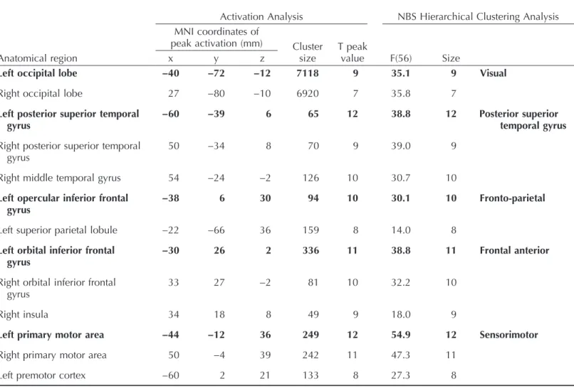
The task-induced statistical parametric map was composed of 13 clusters. The ROI-to-ROI analysis revealed that these clusters belonged to five distinct functional clusters: A visual occip-ital cluster, a sensorimotor cluster, a left frontoparietal cluster, a frontal anterior cluster, and a posterior superior temporal gyrus (STG) cluster. The seeds that presented the best spatial overlap and the most similar functional response to each canonical network were the left occipital, left premotor cortex, left orbital IFG, left opercular IFG, and left posterior superior temporal gyrus. MNI coordinates for the task-induced activation peaks, along with the NBS functional hi-erarchical clustering statistics, are presented inTable 2.
One additional ROI was selected in the PCC from among the CONN network ROI list to represent the DMN (MNI coordinates: x = 1 y =−61 z = 38).Figure 1illustrates the method and results of selecting the seed ROIs.
Age-Related Differences in Functional Connectivity
The main goal of this study was to assess and describe age-related differences in task-induced FC in the regions activated during the naming task.
The effect of age on FC topography during task completion was investigated using a seed-to-voxel FC for each selected ROI for the task and the PCC (t test between two independent samples). The mean within-group FC was also computed to better characterize the differences in topological FC organization. Results are reported with a voxel-level height threshold (p < .001 uncorrected) and a cluster extent threshold (p < .05 FWE corrected) and illustrated in
Figure 2. The mean and group difference maps for all other activation peaks are available inSupporting Information 1.
Based on previous reports of enhanced differences in FC architecture during a task, older adults were expected to show significant differences in the integration of both the task and default mode networks: decreased FC within the DMN, but increased FC within regions out-side the DMN, as well as increased FC in the task-activated regions with semantic and/or ex-ecutive control regions. The results only partially validated the initial expectations.
The task-induced FC architecture presented many similarities between age groups. Although small, significant clusters of age-related differences were nonetheless present for each ROI of the task and the DMN. Both increases and decreases were observed with aging.
Sensorimotor regions within the frontal lobe also exhibited FC increases. In older adults, the left primary motor cortex showed an age-related enlargement of ipsilateral connectivity sur-rounding frontoparietal motor areas (dorsolateral prefrontal cortex, middle frontal gyrus, and caudate nucleus).
Anterior frontal regions (i.e., the orbital IFG) mainly exhibited age-related FC increases in portions of the left dorsal striatum (caudate and putamen).
In contrast, there were decreases in the coupling between canonical anterior and posterior language regions: the left posterior STG ( Wernicke’s area) with the temporal-frontal junction (temporal pole, inferior frontal orbital cortex, frontal pole, and pars triangularis); as well as the Figure 1. Task-induced seed definition: Task-induced activation maps of younger and older adults, ROI functional hierarchical clustering, and selected seeds. Above: activation clusters in the main contrast of interest (task > rest) for both younger (in yellow) and older (in cyan) adults. Overlaps (in green) are observed between younger and older adults in the task contrast. Slice numbers are indicated on the sagittal axis. Below: Functional hierarchical clustering of the ROI spheres for the task-induced activation peaks. The 13 activation peaks belonged to five functional clusters. The most relevant activation peaks are indicated in bright yellow. For example, the left STG shows the largest number of connections with all other ROIs. Each colored connection (magenta, blue, green, orange) represent the distinct clusters, whereas the width and opacity are proportional to stats, with the most opaque and greatest width representing the highest connection between regions in each group-ing. ROI = region of interest; STG = superior temporal gyrus; premotor = premotor cortex; PMA = primary motor area; occ = occipital lobe; orIFG = orbital inferior frontal gyrus; opIFG = opercular IFG; MTG = middle temporal gyrus; SPL = superior parietal lobule. L = left; R = right.
left opIFG (Broca’s area) with the posterior inferior temporal regions (fusiform gyrus). The left visual occipital regions also showed decreased interaction with the occipital pole and superior frontal regions.
As anticipated, the DMN showed marked age-related differences in FC. There was de-creased coupling between the PCC and frontal portions of the DMN (medial prefrontal cortex, bilateral paracingulate cortex, bilateral middle and superior frontal gyrus, bilateral superior parietal lobule). Conversely, connectivity increased with nearby posterior regions, most of which do not correspond to the traditional description of the DMN (angular gyrus, supramar-ginal gyrus, superior lateral occipital cortex, superior parietal lobule).
Figure 2andSupplementary Material 1, illustrate a general trend, across both DMN and task-induced ROIs, for reduced long-range and increased short-range connectivity in older adults. For example, the left superior parietal lobule seed mainly exhibited age-related in-creases in FC with surrounding parietal and medial regions (dorsal striatum) but dein-creases with distant frontal and temporal areas. Similarly, the PCC showed increased coupling with posterior parietal regions but reduced coupling with frontal (medial and lateral) regions in older adults.
Table 2. Task-induced seed definition
Anatomical region
Activation Analysis NBS Hierarchical Clustering Analysis MNI coordinates of
peak activation (mm) Cluster size
T peak
value F(56) Size
x y z
Left occipital lobe −40 −72 −12 7118 9 35.1 9 Visual
Right occipital lobe 27 −80 −10 6920 7 35.8 7
Left posterior superior temporal gyrus
−60 −39 6 65 12 38.8 12 Posterior superior
temporal gyrus Right posterior superior temporal
gyrus
50 −34 8 70 9 39.0 9
Right middle temporal gyrus 54 −24 −2 126 10 30.7 10 Left opercular inferior frontal
gyrus
−38 6 30 94 10 30.1 10 Fronto-parietal
Left superior parietal lobule −22 −66 36 159 8 14.0 8 Left orbital inferior frontal
gyrus
−30 26 2 336 11 38.8 11 Frontal anterior
Right orbital inferior frontal gyrus
33 27 −2 81 10 32.2 10
Right insula 34 18 8 49 9 18.0 9
Left primary motor area −44 −12 36 249 12 54.9 12 Sensorimotor
Right primary motor area 50 −4 39 242 11 47.3 11
Left premotor cortex −60 2 21 133 8 27.3 8
Bold characters show the activation peaks selected as seeds, the most functionally relevant to the corresponding canonical functional network.
These patterns of differences in the regions activated by the task tend to confirm that young and older adults rely on different neurofunctional and cognitive processes to perform the word-production task.
The Impact of Lexical Frequency on Age-Related Differences in FC
The final goal of this study was to test whether there was a group difference when manipulating lexical frequency as an example of a task-specific requirement in a naming task (a condition-by-group interaction). Within-group statistical maps of between-condition contrasts and mean FC within age group for each lexical frequency level were computed to support the description and interpretation of the interaction.
Figure 2. Age-related differences in functional connectivity. Spatial maps show the seed-based con-nectivity of six most representative ROIs activated by the task, in addition to the default mode net-work. For each ROI (yellow sphere) the mean functional connectivity maps are presented for the older group, the younger group, and the difference between age groups (OA > YA). For example, older adults showed decreased coupling between the left posterior STG and the anterior IFG-insula region, compared with younger adults. ROI = region of interest; OA = older adult; YA = young adult; IFG = inferior frontal gyrus; opIFG = opercular inferior frontal gyrus; orIFG = orbital inferior frontal gyrus; occ = occipital lobe; STG = superior temporal gyrus; PCC = posterior cingulate cortex. L = left.
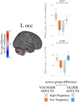
The manipulation of lexical frequency yielded a significant age-by-condition interaction for the left occipital ROI only. Contradicting the initial expectation, there was no systematic ev-idence of greater differences between conditions in FC in older adults than in young adults.
A simple main-effects analysis indicated double dissociations in each significant effect. As expected, older adults showed increased FC with lower lexical frequency (the more challeng-ing condition). Young adults, on the contrary, showed anti-correlated FC between each pair of regions in the same condition (Figure 3).
The left occipital ROI showed a significant interaction in coupling with the right posterior inferior temporal gyrus/fusiform gyrus and the right superior cerebellum. As can be seen in
Supplementary Figure 1, the right insula, which was not selected as one of the most represen-tative seeds, also exhibited an increase in FC with the right inferior cerebellum (I and VI). For the latter, although the age-by-condition interaction was significant, it did not reach statistical significance at the within-group level (main effect of condition in each group).
DISCUSSION
This study proposed a structured exploration of the FC characteristics revealed in healthy older adults during picture naming, a cognitive ability that is central to human functioning and widely used in clinical cognitive assessments. Most previous FC studies used the resting state to exam-ine cognitive activity. However, it was felt that task-induced FC is better adapted to describe possible changes in FC that are specific to the cognitive activity. By using seed-based FC during picture naming, while regressing the task effect and carefully controlling for major confounds,
Figure 3. Interaction between age and lexical frequency: Spatial maps and mean FC values of interaction between age and lexical frequency on task-induced FC. Spatial maps (on the left) show the significant interaction effect, FWE thresholded. Bars and whiskers on the right denote FC effect size for each group (younger and older adults) in each condition (high and low lexical frequency). Across-group differences in FC effect sizes are given above the bars. FC = functional connectivity; FWE = family-wise error; occ = occipital lobe; ITG = ; L = left; R = right.
this study confirms some of the initial hypotheses but also expands upon prior knowledge sur-rounding word production in aging.
In brief, the results show that, although the brain topography of picture naming is very similar in younger and older adults, the FC architecture underlying naming exhibits significant differ-ences. In older adults, there is less coupling of the traditionally described core structures of language acquisition (e.g., Broca’s and Wernicke’s areas), along with enhanced FC in regions involved in both semantic retrieval and motor control. This study also provides the first evi-dence that intrinsic task psychometric characteristics (i.e., lexical frequency) do not interact much with the age-FC relationship. Together, the findings confirm the mutual interest of task-induced FC over a resting-state paradigm to study brain organization in healthy aging.
A Common Naming Activation Network
By choosing a sample- and task-specific template, this study proposed a neurofunctional ex-ploration as similar as possible to the actual cognitive activity (Dickie et al., 2017;Geerligs et al., 2017;Salehi et al., 2018;Salehi, Karbasi, Scheinost, & Constable, 2017). As anticipated, the brain activation analysis yielded a large fronto-temporo-occipital network of higher activity during naming. This statistical parametric map is in line with the most prominent model of word processing (Hickok & Poeppel, 2007). Typical ventral semantic and dorsal phonological streams were revealed by brain activation studies (Baciu et al., 2016;Price, 2012) and brain stimulation mapping (Duffau et al., 2014) in younger adults, and these findings were replicated in studies of FC in aging (Hoyau et al., 2018;Indefrey, 2011). Moreover, the activation peaks were spatially and functionally coherent with the canonical functional networks that could be expected to be active during a naming task, such as the visual, sensorimotor, salience, and anterior and posterior language networks.
The task-related network involved the same regions across age groups, except for the right inferior cerebellum, which was more active in older than in younger adults. It is now recog-nized that the cerebellum is engaged in the processing of complex cognitive material (Bernard & Seidler, 2014; Keren-Happuch, Chen, Ho, & Desmond, 2014; Stoodley, Valera, & Schmahmann, 2012). In particular, the right inferior cerebellum lobule VIII has been described as the “sensorimotor cerebellum” because it is involved in overt motor processing (articula-tion) and phonological storage (Chen & Desmond, 2005). This may suggest increased phono-logical activity in older adults.
Some studies of word production previously reported increases in BOLD signals in parietal, frontal, or temporal regions with age (e.g.,Hoyau et al., 2017;Meunier et al., 2014), as well as contralateral recruitment (e.g.,La et al., 2016;Meinzer et al., 2012), to support performance. A recent meta-analysis focusing on semantic cognition and aging (Hoffman & Morcom, 2018) concluded that increased activation was principally observed when older adults performed worse than younger ones, for example, during tasks drawing on executive functions. Although examples of difficulties during naming are common in the literature (Feyereisen, 1997;Goulet et al., 1994;Shafto et al., 2017), mean accuracy is generally preserved in our study, in line with previous reports that examined accuracy with untimed tasks (LaBarge et al., 1986;Salthouse, 2014;Schmitter-Edgecombe et al., 2000;Verhaegen & Poncelet, 2013;Wierenga et al., 2008). In the context of preserved cognitive performance, then, the absence of such“compensatory” ac-tivity is not surprising. It should be mentioned that word production is not systematically preserved with age (Burke & Shafto, 2004) and, although the causes are still under study (Facal, Juncos-Rabadán, Rodríguez, & Pereiro, 2012;Schwartz & Frazier, 2005;Shafto, Burke, Stamatakis, Tam, & Tyler, 2007), the impact of task demand will require further investigation.
Age-Related Functional Connectivity Differences
The main question raised in this study was whether there were differences in the FC architec-ture during word production between younger and older healthy adults. Overall, a common FC architecture is observed across age groups during the task. Small yet significant, and po-tentially functionally meaningful, differences were observed: Both increases and decreases were seen in task-activated regions as well as in the DMN.
First, there is a general tendency for older adults to reduce long-range and increase short-range connectivity. This pattern may play a functional role and reflect the need for more local processing of neuronal information with age, at the expense of long-distance connectivity (see
Sala-Llonch et al., 2015, for a review), in line with the idea that functional organization be-comes more segregated with age (Cao et al., 2014;Ferreira & Busatto, 2013; Tomasi & Volkow, 2012). Alternatively, this pattern may be an unfortunate consequence of head move-ment (Power, Schlaggar, & Petersen, 2015;Van Dijk, Sabuncu, & Buckner, 2012). Although overt naming is deemed to be similar to natural/clinical conditions, it does induce consider-able task-related motion. Even though many precautionary and corrective measures were taken during the acquisition and preprocessing steps, some task-related movement remains. No stan-dard procedure has yet been shown to unequivocally eliminate the confounding factor of movement (Buckner, Krienen, & Yeo, 2013); however, scrubbing—as used in this study—is
an efficient method of reducing related artifacts (Power, Barnes, Snyder, Schlaggar, & Petersen, 2012;Yan et al., 2013). Further investigation in various task contexts and a better comprehension of the many structural, molecular, or physiological changes that occur in healthy aging will be required to determine which interpretation is most valid.
Functional Connectivity Differences in Task-Activated Regions
Earlier studies of FC in aging generally reported expansion of the system involved in word processing at a younger age (Agarwal et al., 2016; Hoyau et al., 2018; La et al., 2016;
Marsolais et al., 2014). Our results suggest that older adults use similar circuits but also rely on multiple different mechanisms during task performance.
First, the connectivity of the task-activated regions decreases along a fronto-temporo-occipital pathway (IFG and posterior STG, posterior fronto-temporo-occipital, and superior frontal gyrus), rem-iniscent of the semantic ventral stream traditionally described in younger adults (Duffau, 2015;
Hickok & Poeppel, 2007). Although frontotemporal connections have long been considered crucial for word production, findings emerging from brain surgery, stimulation, and multi-modal imagery in young adults suggest that their role is multimulti-modal rather than specific (Binder, Desai, Graves, & Conant, 2009;Chao, Haxby, & Martin, 1999;Duffau, 2015;Etard et al., 2000;Simons, Koutstaal, Prince, Wagner, & Schacter, 2003;Tyler, Cheung, Devereux, & Clarke, 2013). Inferior-frontal to posterior-temporal connections—which show less
coacti-vation in older adults—are, for example, involved during tasks that require high semantic con-trol (Duffau, 2015). This finding is in line with accumulated evidence that older adults rely less on executive control—and more on semantic retrieval—when performing a word production task, as demonstrated using univariate activation analysis (Ansado, Marsolais, Methqal, Alary, & Joanette, 2013;Baciu et al., 2016; Diaz, Rizio, & Zhuang, 2016; Hoyau et al., 2017;
Lacombe, Jolicoeur, Grimault, Pineault, & Joubert, 2015;Marsolais et al., 2014;Methqal, Marsolais, Wilson, Monchi, & Joanette, 2019) and dynamic FC (Hoyau et al., 2018). Together, these findings suggest that older adults depend less than younger adults on seman-tic control abilities to perform the picture-naming task and possibly rely on automatized pro-cesses instead.
A decrease in frontoparietal coupling (right insula, supramarginal gyrus, and parietal operculum) is in line with earlier assertions that increased FC in domain-general systems is not required for everyday language functioning (Campbell & Tyler, 2018) and contrasts with the general expectation that decreased resource allocation is a characteristic of aging (Park & Reuter-Lorenz, 2009). The insula and the supramarginal gyrus are structurally con-nected (Ghaziri et al., 2017) and are part of control networks that guide directed attention (Dosenbach et al., 2007;Menon & Uddin, 2010;Seeley et al., 2007;Zabelina & Andrews-Hanna, 2016). Their deactivation during speech was previously associated with top-down regulation of auditory attention, like that induced in a noisy environment (Elmer, Meyer, Marrama, & Jäncke, 2011), possibly to maximize the somatosensory feedback when speech production becomes error-prone (Golfinopoulos et al., 2011; Seghier et al., 2015).
Finally, another striking and unanticipated age-related feature is the tendency for in-creased coupling of functional brain activity within and between motor regions responsible for higher-level phonological processing—for example, significant increases in surrounding ipsilateral activity in the frontal motor regions in charge of articulation control and initia-tion, such as the left dorsolateral frontal cortex and middle frontal gyrus (Duffau, 2015;
Guenther, 2016;Wise, Greene, Büchel, & Scott, 1999). The pattern is coherent with pre-viously described indirect connectivity along the dorsal phonological stream between the primary motor and the opercula IFG (Broca’s area) via the premotor cortex (Margulies, Böttger, Watanabe, & Gorgolewski, 2013). The primary motor cortex also shows increased coupling with the caudate nucleus, which plays a role in the control of articulation (Argyropoulos, Tremblay, & Small, 2013; Duffau et al., 2014; Guenther, 2016;Margulies et al., 2013;Tremblay, Deschamps, & Gracco, 2016). In summation, more functional re-organization and less segregation can be seen along the dorsal phonological stream with increased age (Agarwal et al., 2016;Diaz et al., 2016;Martins, Simard, & Monchi, 2014;
Muller et al., 2016;Sörös, Bose, Sokoloff, Graham, & Stuss, 2011), in line with the hypoth-esized weakening of the links between phonological and lexical representations (Burke et al., 1991).
Functional Connectivity Differences in the DMN
In accordance with previous reports, the DMN showed an age-related increase with other pa-rietal regions during the task (Chan et al., 2017;Geerligs et al., 2014;Grady et al., 2016;Mak et al., 2017;Spreng, Stevens, Viviano, & Schacter, 2017). This kind of mechanism has been suggested to indicate a broad dedifferentiation of neural activity in later life (e.g.,Campbell, Grady, Ng, & Hasher, 2012). Alternatively, the interaction between the DMN and task com-ponents may indicate active strategic cognitive processes. According to the default-executive coupling hypothesis of aging (DECHA) model (Turner & Spreng, 2015), aging is characterized by a semanticization process: As cognitive control resources decline, cognitive behavior be-comes more and more influenced by past experiences and knowledge. The DMN therefore becomes increasingly engaged with task components to support task performance. Although Turner and Spreng specifically described increases in DMN-prefrontal lateral coupling, they used cognitive tasks that rely partly on executive processes. In fact, the increase in DMN ac-tivity has also been demonstrated with other regions (Damoiseaux et al., 2008;Sambataro et al., 2010) and the DECHA might therefore be extended to any region involved in the task in relation to the posterior DMN. The posterior DMN has been specifically described as a key actor in semantic retrieval processes, and in fact, the DMN and the semantic system share an overlapping functional network (Binder et al., 2009;Bonnelle et al., 2012;Krieger-Redwood et al., 2016).
The Impact of Lexical Frequency Manipulation
Behavioral analysis showed that there is an interaction between age, frequency, and RT, which suggests that lower frequency does make the cognitive activity more demanding for older adults, even though they still manage to accomplish the task. Lexical frequency did not influ-ence accuracy scores in either age group. More segregated frequency levels may have allowed for a stronger task-demand effect (Moberg, Ferraro, & Petros, 2000), and more robust conclu-sions will require the manipulation of task demands through more than two levels.
Although exploratory, our results show a few regions that are significantly mediated by in-trinsic task requirement according to age. With greater task demand, integration increases in older adults, whereas it decreases in young adults. Previous studies with other types of task load interpreted this pattern as an adaptive form of compensation, beneficial to performance (e.g.,Crowell et al., 2019;Nagels et al., 2012;Turner & Spreng, 2015). Those studies, and others that presented age-related compensation patterns (Cabeza, 2002; Campbell et al., 2012;Grady et al., 2016; Morcom & Henson, 2018;Park & Reuter-Lorenz, 2009;Rajah & D’Esposito, 2005;Turner & Spreng, 2015), advocated for the notion that there are domain-general mechanisms, expressed mostly through increased activity in prefrontal regions. This hypothesis builds upon the underlying assumption that older adults find just about any task more difficult than young adults, because of a general cognitive decline (Craik & Byrd, 1982). However, domain-general upregulation was mostly reported using tasks that represent a strong cognitive challenge for older adults (Hoffman & Morcom, 2018). To our knowledge, only one other study previously explored the impact of linguistic criteria on verbal fluency (Marsolais et al., 2014); it showed an interaction between age and increased FC in posterior regions. Using an intrinsic—linguistic—task demand, rather than general cognitive load, our findings are also in line with the concept of task-specific mechanisms (Campbell & Schacter, 2016;Campbell & Tyler, 2018;Hearne, Cocchi, Zalesky, & Mattingley, 2017;Peelle, 2019;
Samu et al., 2017).Campbell and Tyler (2018)suggested that domain-general networks should not be considered as stable, singular mechanisms. Thus, neurofunctional patterns invoked during a task may instead reflect processes that are specific to that task.
In this study, age differences between conditions are small in both size and number. Furthermore, the FC of the DMN was not significantly affected by lower lexical frequency, contrary to what had been reported previously in studies that manipulated executive task de-mands (Persson et al., 2007;Steffener, Habeck, & Stern, 2012). Instead, the interaction was significant in regions that are functionally meaningful and specialized for the task: the left oc-cipital cortex and fusiform gyrus, which together encode visual representations of objects (Mahon et al., 2007;Martin & Chao, 2001); the fusiform gyrus, the cerebellum, and the insula, which were previously reported to be active during the processing of low-frequency words (Basso et al., 2013); and, as mentioned earlier, the right inferior cerebellum lobule VIII, which is also related to phonological production (Chen & Desmond, 2005).
Greater insight into domain-general and task-specific mechanisms will be gained by large data set studies that manipulate various cognitive states, such as those initiated by projects such as Reference Ability Neural Network study (Stern et al., 2014) and Cambridge Centre for Ageing and Neuroscience, or CamCAN (Taylor et al., 2015).
Finally, some general methodological considerations should be raised regarding the diffi-culty of interpreting the neurobiological meaning of linear FC changes in group comparisons. Increased FC could either indicate decreased network adaptability to task demands ( Avelar-Pereira et al., 2017;Campbell & Schacter, 2016;Dixon et al., 2017;Dubois & Adolphs, 2016;
Geerligs et al., 2015;Grady et al., 2016) or reflect optimization for efficient (i.e., small)
network updates, reducing processing demands and supporting behavioral performance (Schultz & Cole, 2016;Shine et al., 2016). Although this study was not designed to rule on these options, the latter fits the current findings better: Considering the preservation of perfor-mance, and the tendency for the core naming network to shrink and for the dorsal phonolog-ical stream to suffer more from the impact of age, FC topography differences could indicate an efficient strategy relying on a more experienced, automatized, and specialized circuit in older adults. Alternatively, interindividual heterogeneity should not be underestimated: The intricate patterns emerging from averaged differences between groups could reflect more diverse brain physiology and functioning among older people, as has been reported frequently (e.g.,Amiri et al., 2014;Dong et al., 2012;Raz et al., 2005;Zuo et al., 2010). In addition, the task effect was regressed in this study, because this technique had previously been demonstrated to in-crease the reliability of task FC estimates (Cao et al., 2014;Cole, Bassett, Power, Braver, & Petersen, 2014). This choice may have reduced some of the true task-related effects on FC but was felt to be preferable considering the overt nature of the naming task and knowing that participant movement can have large effects on FC estimates (Mowinckel, Espeseth, & Westlye, 2012).
Conclusion
This study was designed to characterize the possible age-related reorganization of functional connectivity associated with a word-naming task, while manipulating task demand using the lexical frequency of the stimuli.
Word production is a basic yet essential cognitive activity central to human everyday function-ing. This study, like others before it, emphasizes that vocabulary knowledge increases throughout the life span, into the later stages of life, even though word-production processes slow down.
In this context, the task-induced functional architecture of older adults, compared with younger adults, is characterized by greater reliance on regions in charge of motor control areas, along with lesser inclusion of regions classically associated with word production, such as the IFG and the posterior STG. Earlier descriptions of the core word-production network were based mostly on samples of young adults and used resting-state paradigms. This study, using a sample- and task-specific design, suggests that older adults have a more efficient strat-egy, relying on a more experienced, specialized, and segregated, naming pathway.
This study also reinforces the notion that task-specific rather than domain-general mecha-nisms apply in older adults. Although additional involvement of frontal and bilateral executive regions was previously suggested to support the cognitive performance of older adults in every domain, it appears that those findings cannot be generalized to all cognitive states or types of task demand. Instead, older adults rely on highly specialized regions to perform more complex linguistic processes.
Considering the current limitations affecting the interpretation of FC changes with age, fur-ther exploration is definitely required to disentangle the complex interplay between preserved performance and neurofunctional behavior, using various tasks and stimuli.
FUNDING INFORMATION
Yves Joanette, Institute of Aging (http://dx.doi.org/10.13039/501100000026), Award ID: 191272. Simona Maria Brambati, Canadian Network for Research and Innovation in Machining Technology, Natural Sciences and Engineering Research Council of Canada (http://dx.doi. org/10.13039/501100002790).
AUTHOR CONTRIBUTIONS
Julien Jarret: Data curation: Equal. Simona Maria Brambati: Conceptualization: Supporting; Methodology: Supporting; Writing — Review & Editing: Supporting. Pierre Bellec: Methodology: Supporting; Supervision: Supporting; Writing— Review & Editing: Supporting. Yves Joanette: Conceptualization: Supporting; Funding acquisition: Lead; Project administration: Equal; Resources: Lead; Supervision: Supporting; Writing— Review & Editing: Supporting.
REFERENCES
Agarwal, S., Stamatakis, E. A., Geva, S., & Warburton, E. A. (2016). Dominant hemisphere functional networks compensate for struc-tural connectivity loss to preserve phonological retrieval with ag-ing. Brain and Behavior, 6, e00495. https://doi.org/10.1002/ brb3.495
Amiri, M., Pouliot, P., Bonnéry, C., Leclerc, P.-O., Desjardins, M., Lesage, F., & Joanette, Y. (2014). An exploration of the effect of hemodynamic changes due to normal aging on the fNIRS re-sponse to semantic processing of words. Frontiers in Neurology, 5, 249.https://doi.org/10.3389/fneur.2014.00249
Andrews-Hanna, J. R., Snyder, A. Z., Vincent, J. L., Lustig, C., Head, D., Raichle, M. E., & Buckner, R. L. (2007). Disruption of large-scale brain systems in advanced aging. Neuron, 56, 924–935.
https://doi.org/10.1016/j.neuron.2007.10.038
Ansado, J., Marsolais, Y., Methqal, I., Alary, F., & Joanette, Y. (2013). The adaptive aging brain: Evidence from the preservation of com-munication abilities with age. European Journal of Neuroscience, 37, 1887–1895.https://doi.org/10.1111/ejn.12252
Argyropoulos, G. P., Tremblay, P., & Small, S. L. (2013). The neo-striatum and response selection in overt sentence production: An fMRI study. NeuroImage, 82, 53–60.
Ashburner, J. (2007). A fast diffeomorphic image registration algo-rithm. NeuroImage, 38, 95–113.
Ashburner, J., & Friston, K. J. (2005). Unified segmentation. NeuroImage, 26, 839–851.
Au, R., Joung, P., Nicholas, M., Obler, L. K., Kass, R., & Albert, M. L. (1995). Naming ability across the adult life span. Aging, Neuropsychology, and Cognition, 2, 300–311. https://doi.org/ 10.1080/13825589508256605
Avelar-Pereira, B., Bäckman, L., Wåhlin, A., Nyberg, L., & Salami, A. (2017). Age-related differences in dynamic interactions among default mode, frontoparietal control, and dorsal attention networks during resting-state and interference resolution. Frontiers in Aging Neuroscience, 9, 152.https://doi.org/10.3389/fnagi.2017.00152
Baciu, M., Boudiaf, N., Cousin, E., Perrone-Bertolotti, M., Pichat, C., Fournet, N.,… Krainik, A. (2016). Functional MRI evidence for the decline of word retrieval and generation during normal aging. Age (Dordrecht, Netherlands), 38, 3. https://doi.org/ 10.1007/s11357-015-9857-y
Basso, G., Magon, S., Reggiani, F., Capasso, R., Monittola, G., Yang, F. J., & Miceli, G. (2013). Distinguishable neurofunctional effects of task practice and item practice in picture naming: A BOLD fMRI study in healthy subjects. Brain and Language, 126(3), 302–313.https://doi.org/10.1016/j.bandl.2013.07.002
Behzadi, Y., Restom, K., Liau, J., & Liu, T. T. (2007). A component based noise correction method (CompCor) for BOLD and perfu-sion based fMRI. NeuroImage, 37, 90–101.
Ben-David, B. M., Erel, H., Goy, H., & Schneider, B. A. (2015). “Older is always better”: Age-related differences in vocabulary scores across 16 years. Psychology and Aging, 30, 856–862.
https://doi.org/10.1037/pag0000051
Bernard, J. A., & Seidler, R. D. (2014). Moving forward: Age effects on the cerebellum underlie cognitive and motor declines. Neuroscience and Biobehavioral Reviews, 42, 193–207.https:// doi.org/10.1016/j.neubiorev.2014.02.011
Binder, J. R., Desai, R. H., Graves, W. W., & Conant, L. L. (2009). Where is the semantic system? A critical review and meta-analysis of 120 functional neuroimaging studies. Cerebral Cortex, 19, 2767–2796.https://doi.org/10.1093/cercor/bhp055
Blackwell, A. D., Sahakian, B. J., Vesey, R., Semple, J. M., Robbins, T. W., & Hodges, J. R. (2004). Detecting dementia: Novel neuro-psychological markers of preclinical Alzheimer’s disease. Dementia and Geriatric Cognitive Disorders, 17, 42–48. Bonnelle, V., Ham, T., Leech, R., Kinnunen, K., Mehta, M.,
Greenwood, R., & Sharp, D. (2012). Salience network integrity predicts default mode network function after traumatic brain in-jury. Proceedings of the National Academy of Sciences of the United States of America, 109, 4690–4695. https://doi.org/ 10.1073/pnas.1113455109
Buckner, R. L., Krienen, F. M., & Yeo, B. T. T. (2013). Opportunities and limitations of intrinsic functional connectivity MRI. Nature Neuroscience, 16, 832–837.
Burke, D. M., MacKay, D. G., Worthley, J. S., & Wade, E. (1991). On the tip of the tongue: What causes word finding failures in young and older adults? Journal of Memory and Language, 30, 542–579. Burke, D. M., & Shafto, M. A. (2004). Aging and language
produc-tion. Current Directions in Psychological Science, 13, 21–24. Cabeza, R. (2002). Hemispheric asymmetry reduction in older
adults: The HAROLD model. Psychology and Aging, 17, 85–100. Cabeza, R., Albert, M., Belleville, S., Craik, F. I. M., Duarte, A.,
Grady, C. L., … Rajah, M. N. (2018). Maintenance, reserve and compensation: The cognitive neuroscience of healthy age-ing. Nature Reviews Neuroscience, 19, 701–710.https://doi. org/10.1038/s41583-018-0068-2
Cabeza, R., Nyberg, L., & Park, D. C. (2016). Cognitive neurosci-ence of aging: Linking cognitive and cerebral aging. New York: Oxford University Press. https://doi.org/10.1093/acprof:oso/ 9780195156744.001.0001
Campbell, K. L., Grady, C. L., Ng, C., & Hasher, L. (2012). Age differ-ences in the frontoparietal cognitive control network: Implications for distractibility. Neuropsychologia, 50, 2212–2223.https://doi. org/10.1016/J.NEUROPSYCHOLOGIA.2012.05.025
Campbell, K. L., Samu, D., Davis, S. W., Geerligs, L., Mustafa, A., Tyler, L. K., & Cambridge Centre for Aging and Neuroscience. (2016). Robust resilience of the frontotemporal syntax system to aging. Journal of Neuroscience, 36, 5214–5227.https://doi. org/10.1523/JNEUROSCI.4561-15.2016
Campbell, K. L., & Schacter, D. L. (2016). Aging and the resting state: Is cognition obsolete? Language, Cognition and Neuroscience, 32, 661–668.https://doi.org/10.1016/j.bbi.2017.04.008
Campbell, K. L., & Tyler, L. K. (2018). Language-related domain-specific and domain-general systems in the human brain.